Micro-RNA Binding Site Polymorphisms in the WFS1 Gene Are Risk Factors of Diabetes
Mellitus
Zsuzsanna Elek1, Nóra Németh1, Géza Nagy2, Helga Németh1, Anikó Somogyi2, Nóra Hosszufalusi3, Mária Sasvári-Székely1, Zsolt Rónai1*
1Department of Medical Chemistry, Molecular Biology and Pathobiochemistry, Semmelweis University, Budapest, Hungary,22nd Department of Internal Medicine, Semmelweis University, Budapest, Hungary, 3Research Laboratory, 3rd Department of Internal Medicine, Semmelweis University, Budapest, Hungary
*ronai.zsolt@med.semmelweis-univ.hu
Abstract
The absolute or relative lack of insulin is the key factor in the pathogenesis of diabetes melli- tus. Although the connection between loss of function mutations of theWFS1gene and DID- MOAD-syndrome including diabetes mellitus underpins the significance of wolframin in the pathogenesis, exact role ofWFS1polymorphic variants in the development of type 1 and type 2 diabetes has not been discovered yet. In this analysis, 787 patients with diabetes and 900 healthy people participated. Genotyping of the 7WFS1SNPs was carried out by TaqMan assays. Association study was performed byχ2-test in combination with correction for multiple testing. For functional analysis, the entire 3’UTR of theWFS1gene was sub- cloned in a pMIR-Report plasmid and relative luciferase activities were determined. Linkage disequilibrium analysis showed a generally high LD within the investigated region, however the rs1046322 locus was not in LD with the other SNPs. The two miR-SNPs, rs1046322 and rs9457 showed significant association with T1DM and T2DM, respectively. Haplotype analysis also confirmed the association between the 3’UTR loci and both disease types.In vitroexperiments showed that miR-185 reduces the amount of the resulting protein, and rs9457 miRSNP significantly influences the rate of reduction in a luciferase reporter assay.
Genetic variants of theWFS1gene might contribute to the genetic risk of T1DM and T2DM.
Furthermore demonstrating the effect of rs9457 in binding of miR-185, we suggest that the optimal level of wolframin protein, potentially influenced by miR-regulation, is crucial in nor- mal beta cell function.
Introduction
Diabetes mellitus is a group of pathogenically heterogeneous diseases sharing the trait of abso- lute or relative insufficiency of insulin effect. Common forms of the disease are type 1 (T1DM) and type 2 diabetes mellitus (T2DM). T1DM results from autoimmuneβ-cell destruction lead- ing to insulin deficiency, whereas type 2 diabetes (T2DM) is the end point of a progressive
OPEN ACCESS
Citation:Elek Z, Németh N, Nagy G, Németh H, Somogyi A, Hosszufalusi N, et al. (2015) Micro-RNA Binding Site Polymorphisms in the WFS1 Gene Are Risk Factors of Diabetes Mellitus. PLoS ONE 10(10):
e0139519. doi:10.1371/journal.pone.0139519 Editor:Chiaho Shih, Academia Sinica, TAIWAN Received:June 2, 2015
Accepted:September 12, 2015 Published:October 1, 2015
Copyright:© 2015 Elek et al. This is an open access article distributed under the terms of theCreative Commons Attribution License, which permits unrestricted use, distribution, and reproduction in any medium, provided the original author and source are credited.
Data Availability Statement:All relevant data are within the paper and its Supporting Information files.
Funding:This work was supported by the Hungarian Scientific Research Fund "OTKA - Országos Tudományos Kutatási Alapprogramok", Grant Number: K83766. This grant only covered the costs of the research (lab costs etc.) and provided no personal costs for any author. The funder had no role in study design, data collection and analysis, decision to publish, or preparation of the manuscript.
Competing Interests:The authors have declared that no competing interests exist.
insulin secretion defect on the background of insulin resistance. Both T1DM and T2DM are considered to be complex diseases caused by multiple environmental and genetic risk factors.
Polymorphisms and mutations of genes associated with other, much rarer monogenic forms of diabetes have been in the focus of research for many years. One of these illnesses is the Wol- fram- or DIDMOAD-syndrome, characterized by diabetes insipidus, non-autoimmune and non-HLA-linked insulin dependent diabetes mellitus together with progressive degeneration of beta cells[1], optic atrophy and deafness[2]. The mutated gene causing the disease was denominatedWFS1and is located on chromosome 4 region p16[2]. It was suggested that poly- morphisms of theWFS1resulting in minor modulation of the gene function instead of com- plete loss might be in the genetic background of the common, polygenic forms of diabetes (T1DM and T2DM). Meta-analysis of association studies revealed that the rs1046320 and rs10010131 SNPs were significant risk factors of T2DM[3]. The two SNPs were in strong link- age disequilibrium with each other, and although no biological effect could be demonstrated, it is notable that the rs1046320 polymorphism is located in the 3’UTR of theWFS1gene.
Significance of the miRNA system in the fine regulation of protein synthesis has recently been discovered. Although the principle of miRNA-action is their binding to the 3’UTR of tar- get genes, prediction of this interaction based on solely sequence alignment is doubtful. 355 miRNAs were suggested to have a binding site in theWFS13’UTRin silicoby the miRWalk database[4], however this interaction could be confirmed by molecular methods only for miR- 21 and the members of the hsa-let-7 family. Similarly 11439 genes (includingWFS1) were pre- dicted to be the putative target of miR-185, whereas only 33 (excludingWFS1) have been con- firmedin vitroso far. Considerable data suggest that miRNAs have a direct role in insulin secretion and production, pancreatic islet development, insulin action and indirectly control glucose and lipid metabolism[5]. The miRWalk database suggested a connection between dia- betes mellitus and 140 miRNAs. Although miR-185 was not among these hits, it was demon- strated by a recent study that both miR-185 and miR-668 were expressed in pancreatic islets [6]. Moreover, analyzing numerous tissues it was observed that miR-185 was abundant, high expression level was detected in the brain, kidney, lung, placenta, prostate, spleen and thyroid glands[7]. On the other hand miR-668 showed an islet-specific expression[6]. Our earlier study [8] revealed that miR-668 not only bound to the 3’UTR of theWFS1gene, but this connection was influenced by the rs1046322 SNP, which showed a significant association with diabetes mellitus according to our current findings (Tables1,2,3and4).
In this study we demonstrate an association between both types (T1DM and T2DM) of diabetes mellitus and SNPs in theWFS1gene. Three of the investigated polymorphisms (rs1046320 A/G, rs1046322 A/G and rs9457 C/G) are located in the 3’UTR of the gene, rs1801208 A/G is in exon 8, whereas rs6824720 A/G, rs10010131 A/G and rs13147655 A/G are in intron 1, 4 and 5 respectively. We show that besides the rs1046322 locus, described earlier [8], the rs9457 SNP also influences miRNA binding in anin vitrosystem. These data might help in elucidating the molecular background of the disease and might contribute to the elabo- ration of novel, alternative therapeutic approaches.
Materials and Methods Participants, DNA-purification
407 patients diagnosed with T1DM (from the 3rdDepartment of Internal Medicine, Szentá- gothai Knowledge Center, Semmelweis University, 46.9% female, 53.1% male, disease onset at the age of 32.1 ± 10.5 years) and 380 patients with T2DM (from the 2ndDepartment of Internal Medicine at the Semmelweis University, 57.9% female, 42.1% male, disease onset at the age of 48.0 ± 12.7 years) participated in the study. The 900 healthy volunteers (59.0% female, 41.0%
Table1.Allele-wiseassociationanalysisoftheinvestigatedWFS1SNPsanddiabetesmellitus. T1DMT2DMDM SNP,Risk alleleFreq. T1DMFreq. ControlpORLower CIUpper CIFreq. T2DMFreq. ControlpORLower CIUpper CIFreq. DMFreq. ControlpORLower CIUpper CI rs6824720,G0.8410.8120.1451.2210.9331.5970.8750.8240.0041.5051.1371.9900.8580.8240.0271.2931.0291.624 rs10010131, G0.6390.6050.1341.1540.9571.3930.6890.6092.510−41.4251.1781.7220.6640.6080.0021.2701.0901.480 rs13147655, G0.6490.5990.0521.2330.9981.5230.6890.6064.510−41.4441.1761.7740.6690.6060.0021.3171.1081.566 rs1801208,G0.9590.9580.8831.0390.6271.7200.9560.9500.5501.1510.7251.8280.9580.9500.3521.2040.8141.782 rs1046320,A0.5980.5890.6751.0380.8711.2370.6740.5921.110−41.4291.1911.7140.6350.5910.0111.2051.0441.390 rs1046322,A0.1870.0903.910−11 2.3271.8023.0040.1390.0940.0011.5551.1912.0320.1630.0945.910−9 1.8871.5202.343 rs9457,C0.6010.5440.0081.2641.0621.5040.6310.5468.110−51.4211.1931.6980.6160.5465.110−51.3341.1601.533 Testswerecarriedoutusinganallele-wiseapproach.p:pvalueofstatisticalsignificanceoftheχ2-probe.Boldnumbersshownominallysignificantresults,underlinedpvaluesare significantusingFDRapproach,whereasdoubleunderlinednumbersmeansignificantresultsafterBonferronicorrectionformultipletesting.Freq.:allelefrequencies,OR,LowerCI, UpperCI:Odds-ratiowith95%upperandlowerconfidenceintervals.Analyseswerecarriedoutinbothdiseasetypes(T1DM:2N=814,control:2N=1634;T2DM:2N=760, control:2N=1784)aswellasinthecombinedpatientgroup(DM:2N=1574,control:2N=1800). doi:10.1371/journal.pone.0139519.t001
male, mean age: 39.0 ± 13.1 years) were recruited by the Institute of Psychology, Eötvös Lor- ánd University. The study protocol was approved by the Local Ethical Committee (ETT-TU- KEB ad.328/KO/2005, ad.323-86/2005-1018EKU from the Scientific and Research Ethics Committee of the Medical Research Council). To address the issue of population stratifica- tion, the participation was restricted to subjects of Hungarian origin, thus creating an ethni- cally homogenous population. All participants signed written informed consent before providing DNA sample for genetic testing. Buccal epithelial cells were collected by swabs in duplicates (two swabs per sample and two samples per person). DNA purification was initi- ated by the incubation of the buccal samples at 56°C overnight in 0.2 mg/ml Proteinase K cell lysis buffer. It was followed by denaturing the proteins with saturated NaCl solution. Finally, DNA was precipitated using isopropanol and subsequently by 70% ethanol. DNA pellet was resuspended in 100μL 0.5× TE (1× TE: 10 mM Tris pH = 8.0; 1 mM EDTA) buffer. Concen- tration of the DNA-samples was measured by NanoDrop1000 spectrophotometer.
SNP Genotyping
Seven SNPs in theWFS1gene were genotyped by using pre-designed TaqMan assays from Life Technologies (rs6824720: C__30903802_10; rs10010131: C__30473796_10; rs13147655:
C__22272143_10; rs1801208: C___2401737_10; rs1046320: C___2873369_10; rs1046322:
C___8841086_1_; rs9457: C___2873371_10). These assays contained the two primers and allele-specific fluorescent probes labeled by FAM and VIC, respectively. Reaction mixtures contained these primer–probe premixes, the TaqMan1Genotyping Master Mix (including AmpliTaq Gold1DNA-polymerase, deoxyribonucloside-triphosphates and buffer) according to manufacturer’s instructions as well as approximately 4 ng genomic DNA in a final volume of 6μL. A 7300 Real Time PCR instrument was used for amplification. Thermocycle was started by activating the hot start DNA-polymerase and denaturing genomic DNA at 95°C for 10 minutes. This was followed by 40 cycles of denaturation at 95°C for 15 sec, and combined annealing and extension at 60°C for 1 minute. Real-time detection was carried out during the latter step for higher sensitivity and reliability to verify the results of the subsequent post-PCR plate read and automatic genotype call.
Genotype data in“ped”format are available asS1–S4Tables.
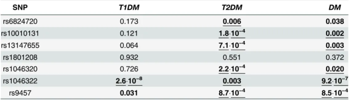
Table 2. Association analysis of the investigatedWFS1SNPs and diabetes mellitus by Cochran– Armitage trend test.
SNP T1DM T2DM DM
rs6824720 0.173 0.006 0.038
rs10010131 0.121 1.810−4 0.002
rs13147655 0.064 7.110−4 0.003
rs1801208 0.932 0.551 0.372
rs1046320 0.726 2.210−4 0.020
rs1046322 2.610−8 0.003 9.210−7
rs9457 0.031 8.710−4 8.510−4
The table depicts thepvalues of statistical significance. Bold numbers show nominally significant results, underlinedpvalues are significant using FDR approach, whereas double underlined numbers mean significant results after Bonferroni correction for multiple testing. Analyses were carried out in both disease types (T1DM:N= 407, control:N= 817; T2DM:N= 380, control:N= 892) as well as in the combined patient group (DM:N= 787, control:N= 900).
doi:10.1371/journal.pone.0139519.t002
Table3.Genotype-wiseassociationanalysisoftheinvestigatedWFS1SNPsanddiabetesmellitususingrecessivemodel. T1DMT2DMDM SNP,Risk genotypeFreq. T1DMFreq. ControlpORLower CIUpper CIFreq. T2DMFreq. ControlpORLower CIUpper CIFreq. DMFreq. ControlpORLower CIUpper CI rs6824720, GG0.7260.6710.1041.2970.9471.7750.7770.6870.0041.5911.1572.1890.7510.6870.0171.3761.0581.790 rs10010131, GG0.4300.3660.0461.3051.0041.6960.4880.3671.410−41.6451.2722.1260.4590.3663.510−41.4721.1901.820 rs13147655, GG0.4500.3770.0431.3541.0091.8170.4840.3730.0011.5751.1902.0840.4670.3730.0021.4731.1581.875 rs1801208, GG0.9190.9190.9881.0040.5971.6880.9120.9040.6921.1020.6831.7770.9150.9040.4891.1550.7681.735 rs1046320,AA0.3880.3520.2241.1690.9091.5030.4650.3511.810−41.6091.2542.0640.4260.3500.0021.3761.1241.685 rs1046322,AA0.0860.0127.010−10 7.6413.60816.1860.0300.0110.0232.6991.1096.5710.0580.0113.110−7 5.4652.64511.290 rs9457,CC0.3750.3030.0131.3771.0681.7760.4200.3047.510−51.6541.2882.1250.3970.3048.510−51.5051.2271.846 Tableshowsthefrequencyvaluesofhomozygotesfortheriskallele(recessivemodel).p:pvaluesofstatisticalsignificanceoftheχ2-probe.Boldnumbersshownominally significantresults,underlinedpvaluesaresignificantusingFDRapproach,whereasdoubleunderlinednumbersmeansignificantresultsafterBonferronicorrectionformultiple testing.Freq.:allelefrequencies,OR,LowerCI,UpperCI:Odds-ratiowith95%upperandlowerconfidenceintervals.Analyseswerecarriedoutinbothdiseasetypes(T1DM: N=407,control:N=817;T2DM:N=380,control:N=892)aswellasinthecombinedpatientgroup(DM:N=787,control:N=900). doi:10.1371/journal.pone.0139519.t003
Table4.Genotype-wiseassociationanalysisoftheinvestigatedWFS1SNPsanddiabetesmellitususingdominantmodel. T1DMT2DMDM SNP,Risk genotypesFreq. T1DMFreq. ControlpORLower CIUpper CIFreq. T2DMFreq. ControlpORLower CIUpper CIFreq. DMFreq. ControlpORLower CIUpper CI rs6824720,GG +AG0.9560.9530.8661.0610.5332.1110.9730.9600.3041.5130.6843.3450.9640.9600.7091.1250.6062.089 rs10010131, GG+AG0.8470.8430.8751.0290.7211.4680.8910.8510.0731.4250.9662.1010.8690.8510.3471.1550.8561.558 rs13147655, GG+AG0.8470.8220.3621.1990.8111.7700.8950.8380.0191.6381.0802.4860.8710.8380.1151.3060.9371.821 rs1801208,GG +AG1.0000.9970.3011.0030.9971.0081.0000.9950.1901.0050.9981.0111.0000.9950.0601.0050.9981.005 rs1046320,AA +AG0.8070.8250.4451.1290.8261.5440.8830.8320.0231.5251.0592.1960.8440.8310.4901.0990.8411.435 rs1046322,AA +AG0.2890.1682.710−6 2.0111.4972.7000.2480.1770.0041.5371.1422.0690.2680.1761.110−5 1.7171.3472.188 rs9457,CC +CG0.8280.7850.0811.3190.9661.8020.8420.7870.0261.4371.0431.9790.8350.7870.0151.3641.0621.751 Tableshowsthefrequencyvaluesofhomo-andheterozygouscarriersfortheriskallele(dominantmodel).p:pvaluesofstatisticalsignificanceoftheχ2-probe.Boldnumbersshow nominallysignificantresults,underlinedpvaluesaresignificantusingFDRapproach,whereasdoubledunderlinednumbersmeansignificantresultsafterBonferronicorrectionfor multipletesting.Freq.:allelefrequencies,OR,LowerCI,UpperCI:Odds-ratiowith95%upperandlowerconfidenceintervals.Analyseswerecarriedoutinbothdiseasetypes (T1DM:N=407,control:N=817;T2DM:N=380,control:N=892)aswellasinthecombinedpatientgroup(DM:N=787,control:N=900). doi:10.1371/journal.pone.0139519.t004
Plasmid construction
The full length 3’untranslated region (3’UTR) of theWFS1gene was amplified by PCR using a DNA-sample of an individual with known rs9457 (homozygous CC) genotype. The following primers were used: forward primer: 5’TCG GCG GAG CTC GGA TGG TCC GCC ACG AGG AGC 3’(where theSacI site is underlined) and reverse primer: 5’AAA GGA AAG CTT GCG CTG CAG GTT CCA CCA GAG G 3’(where theHind III site is underlined). The PCR prod- ucts were digested withSacI andHind III and were cloned downstream of the firefly luciferase gene in pMIR-Report plasmid (pMIR-REPORT miRNA Expression Reporter Vector System;
ABI) by T4 DNA ligase (New England BioLabs) using standard protocols. The generated reporter construct (denoted as C) carried the C (risk) allele, the other one (denoted as G) carry- ing the protective G allele was generated by Quick Change Lightning (Stratagene) site directed mutagenesis. A reference construct (referred to as Seed) contained a completely modified seed region: site-directed mutagenesis was employed to modify the appropriate segment of this con- struct. A control construct contained a different DNA-insert with the same length lacking any sequence complementary to miR-185 (denoted as Control). All constructs were confirmed by DNA sequencing.
MiRNA binding assay
Human Embryonic Kidney cell line (HEK293T) was cultured in DMEM medium (Invitrogen
—Gibco), supplemented with 10% bovine fetal serum (Lonza). The cells were maintained at 37°C in an atmosphere of 5% CO2. For miRNA assays the cells were seeded into 24-well plates and incubated for 24 hours before transfection. Subsequently, 0.05μg of the luciferase reporter construct were cotransfected with 0.2μgβ-galactosidase plasmid (Ambion) and 5 pmol of miR185 and/or miR668 (Sigma-Aldrich) using Lipofectamine 2000 (Invitrogen) according to the manufacturer’s instruction. When investigating the effect of the presence or absence of miR185 and miR668, miR20b was used as supplement to use the same amount of total miRNA in each sample to avoid any spurious effect. Cells were collected 48 hours after transfection, washed with PBS, then extracted by three consecutive freeze–thaw cycles and subsequent cen- trifugation. Supernatants were used for enzyme activity measurements. Luciferase activity was measured by adding 60μl Luciferin reagent (0.16 mg/ml Luciferin K, 20 nM Tricine, 2.6 nM MgSO4, 0.1 nM Na2EDTA, 33.3 nM DTT, 0.27 nM Li3CoA and 0.53 nM Na2ATP) to 12μl of each cell extract. Luminescence was measured using a Varioskan multi-well plate reader (Thermo Fisher Scientific, Inc.). Values for luciferase activity were normalized toβ-galactosi- dase activity (measured by standard protocol using the same Varioskan plate reader in pho- tometry mode). Each experiment was independently repeated three times, and each sample was studied in triplicate.
Statistical analysis
The Hardy–Weinberg equilibrium (HWE) for genotype distributions was assessed byχ2-test.
No significant difference (p>0.1) could be observed between the measured and expected genotype frequencies in the control group for any of the investigated SNPs. The frequency of both T1DM and T2DM is similar in females and males, and both disease types can develop at any age, although T1DM is more characteristic in juveniles, whereas symptoms of T2DM usu- ally appear in patients older than 40. Despite, age- and sex-matched study design was used in each analysis to rule out any spurious association. Allele-wise association analysis and geno- type-wise tests using dominant and recessive models were carried out by comparing the allele or genotype frequency values of each polymorphism in the patient and control group using SPSS v17.0 and HaploView v4.2[9]. GWASpi[10] was applied to perform Cochran–Armitage
trend test[11]. Correction for multiple testing was carried out to avoid false positive results using two different approaches. False Discovery Rate (FDR) was calculated by Microsoft Excel, results were verified using a web-based tool (http://www.sdmproject.com/utilities/?show=
FDR) [12]. The more stringent Bonferroni correction was also applied, the accepted level of statistical significance was 0.05 / 84 = 5.9510−4, as 7 loci and 3 patient categories (T1DM, T2DM and combined) were analyzed in 4 different approaches. Haplotype analysis was carried out by HaploView v4.2[9]. The same software package was used to linkage disequilibrium anal- ysis. ANOVA of the luciferase assays was carried out using GraphPad InStat (version 3.05).
Results
Marker selection
Seven SNPs of theWFS1gene were recruited in our study. Three (rs1046320, rs1046322 and rs9457) of them were located in the 3’UTR of the gene. The rs1046322 and rs9457 polymor- phisms were suggested to be miR-SNPs altering the binding site of the miR-668 and miR-185 respectively by the Patrocles[13] and PolymiRTS[14] databases. Moreover, putative regulatory effect of the rs1046322 site has previously been demonstrated byin vitroluciferase reporter sys- tem[8]. The rs1046320 SNP was shown to be in association with type 2 diabetes mellitus, how- ever no biological function could be detected[3]. The rs687420, rs10010131 and rs13147655 polymorphisms are intronic variants, whereas rs1801208 is located in the last exon of the gene.
These sites have been selected based on literature data, and our earlier thorough SNP analysis of theWFS1gene including 17 polymorphic loci[8] recruiting at least one polymorphism from each haplotype block.
Linkage disequilibrium analysis
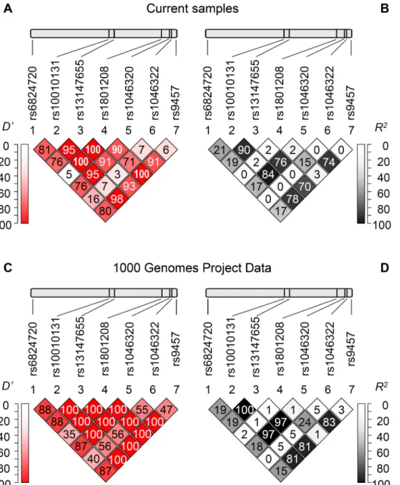
Fig 1Panel A and B show the pairwise linkage disequilibrium (LD) analysis based on our popu- lation including healthy control subjects and patients demonstrating Lewontin’sD’andR2val- ues, respectively. These results were in good agreement with the LD analysis using the data set of the 1000 Genomes Project (Fig 1Panel C and D). Generally a high level of LD can be observed in the gene region, although it is notable that the rs1046322 polymorphism was not in linkage disequilibrium with the other sites. Moreover the highD’values in combination with the lowR2values regarding the rs1801208 polymorphism show a partial linkage disequilibrium of this locus with other SNPs, demonstrating the occurrence of three and the lack of one haplo- type out of the four theoretical pairwise allele combinations.
Association studies
Table 1summarizes the results of the case-control study in an allele-wise setting. Analyses were carried out for both diabetes types as well as for the combined patient group using age- and sex-matched controls. Interestingly the risk allele was the same for the type 1 and the type 2 forms of the disease, and it is also remarkable, that it was only the rs1046322 SNP, at which this variant was the minor allele. It is also notable that polymorphisms in rather different loca- tions of the gene associated with diabetes mellitus, although no complete linkage disequilib- rium could be shown in this region. However the lowestpvalues of statistical significance could be detected in case of two 3’UTR SNPs. The rs1046322 and rs9457 polymorphisms showed nominally significant (p<0.05) association in all three (type 1, type 2 and combined) analyses, which remained significant in all cases after FDR correction for multiple testing. The association between the rs1046322 polymorphism and T1DM as well as that between rs9457 SNP and T2DM could be declared even using the more stringent Bonferroni approach
(accepted level of significance:p5.9510−4). Moreover, the rs10010131, the rs13147655 and the rs1046320 SNPs were also found to significantly associate with type 2 diabetes even using FDR or Bonferroni correction.
The Cochran–Armitage trend test was employed to extend the association study in a geno- type-wise manner (Table 2). In agreement with the allele-wise test, no association could be detected between the rs1046320 locus and T1DM (p= 0.726). The major effect of the rs1046322 SNP in T1DM was also confirmed (p= 2.63910−8), whereas a weak nominal association
Fig 1. Linkage disequilibrium analysis of the investigatedWFS1SNPs.Upper panels indicate the chromosomal positions of the polymorphisms, below the pairwise linkage disequilibrium data of the SNPs are demonstrated. Lewontin’sD’(Panels A and C) andR2values (Panels B and D) calculated based on our genotype data (Panels A and B) as well as based on the data obtained from the 1000 Genomes Project (Panels C and D) are shown. Dark background andD’orR2= 100 values mean strong linkage disequilibrium between the two appropriate loci. (Colored version of the figure is available online.)
doi:10.1371/journal.pone.0139519.g001
(p= 0.031) could be observed between rs9457 locus and the type 1 disease form. The Cochran– Armitage trend test detected association in the T2DM group in case of 6 SNPs, all of them remained significant using FDR correction. The allele- and genotype-wise analyses were in good agreement in the combined diabetes group as well. Strong association could be detected in case of two 3’UTR polymorphisms (rs1046322 and rs9457), the former one remained significant even using Bonferroni correction.
To further analyze association between the 7 WFS1 SNPs and diabetes mellitus in a more detailed and systematic way, genotype-wise tests were carried out applying two different approaches (Tables3and4). Frequency values of the genotype consisting of both risk alleles were computed for all loci in each patient group using the recessive model. In this setup a remarkably high odds ratio (7.641) was observed in case of the rs1046322 SNP in the T1DM dis- ease group, whereas the significant association between rs10010131, rs1046320, rs9457 polymor- phisms and T2DM was also confirmed. When using the dominant model, i.e. comparing the frequency of risk allele carriers and noncarriers, the effects detected previously could be observed.
Association was found between the rs1046322 locus and T1DM, as well as between all three 3’ UTR SNPs and the rs13147655 polymorphism and type 2 disease form using FDR correction, however none of these results remained significant by the stringent Bonferroni method. This observation shows that the effect of the risk allele is more pronounced in homozygous form sug- gesting that the recessive model is more appropriate in this case, however it can also be remarked that pure recessive or dominant inheritance can only be expected in case of monogenic diseases.
Based on the putative synergistic effect of the three 3’UTR SNPs (rs1046320, rs1046322 and rs9457) in translational regulation, these polymorphisms were included in a haplotype-wise association study. Investigation of the patient group with type 1 diabetes mellitus confirmed the major effect of the rs1046322 SNP. Frequency of the haplotypes containing the risk (“A”) allele of this site in combination with the risk variant of one or both SNPs (Table 5) was significantly higher in the patient group. This resulted in an outstanding odds ratio value, which was above 3 in all cases. Similar leader effect of a single polymorphism could not be observed in case of type 2 diabetes. The“AC”haplotype of the rs1046320 and rs9457 SNPs showed a highly significant association with the disease, on the other hand odds ratio was the lowest in case of this combina- tion, and it is also remarkable that these two polymorphisms are in linkage disequilibrium with each other, which can also contribute to the observed low statisticalpvalue of theχ2-probe.
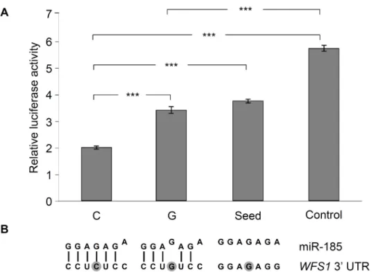
In vitromiRNA binding assay
Putative role of the rs9457 locus on miRNA binding was analyzed byin vitroluciferase assay, as according to the prediction of the PolymiRTS[14] database, this SNP is located in the bind- ing site of the seed sequence of microRNA-185 (miR-185). The 3’UTR region of theWFS1 gene was subcloned in a pMIR-Report luciferase reporter vector, construct with the“G”allele at the rs9457 locus was created by site directed mutagenesis. The same approach was used to generate the“seed mutant”construct, which lacked the entire binding site of the seed sequence of miR-185. Moreover a construct without any binding site of miR-185 (referred to as“Con- trol”) was also used. Luciferase enzyme levels were normalized toβ-galactosidase activities.
The lowest relative luciferase activity (35% of the“Control”) could be measured with the con- struct containing a“C”allele of the rs9457 polymorphism (Fig 2). Interestingly a single mis- match caused by the SNP resulted in a more, than 1.7 times increase (p<0.05) in luciferase level, and this value was practically identical with the activity of the“seed”construct. It might be related to the finding of thein silicosequence alignment, which showed only 6 complemen- tary bases out of the 7 nucletotides of the seed sequence in case of the“C”allele, and the“G” variant adds a further mismatch into the region (Fig 2, Panel B), resulting in a hypothetically
Table5.Haplotypeanalysisofthe3’UTRSNPsoftheWFS1gene. T1DMT2DMDM Haplotype,risk allelFreq. T1DMFreq. ControlpORLower CIUpper CIFreq. T2DMFreq. ControlpORLower CIUpper CIFreq. DMFreq. ControlpORLower CIUpper CI rs1046322– rs9457,AC0.1240.0423.410−14 3.2622.3694.4920.0670.0380.0011.8431.2672.6810.0980.0432.910−10 2.4121.8213.194 rs1046320– rs9457,AC0.5480.5420.7901.0230.8641.2110.6260.5550.0011.3411.1271.5960.5830.5420.0171.1811.0301.354 rs1046320– rs1046322,AA0.1240.0431.110−13 3.1652.3044.3470.0700.0400.0011.8091.2542.6090.1000.0454.010−10 2.3681.7953.124 rs1046320– rs1046322– rs9457,AAC
0.1200.0422.810−13 3.1522.2854.3480.0660.0380.0021.8051.2382.6300.0930.0436.110−9 2.2741.7133.019 p:pvalueofstatisticalsignificanceoftheassociationbetweentheinvestigatedhaplotypeandT1DM(2N=814,control:2N=1634)orT2DM(2N=760,control:2N=1784)orthe combinedpatientgroup(DM:2N=1574,control:2N=1800)assessedbyχ2-analysis.Boldnumbersmeanstatisticallysignificantassociation.Freq.:allelefrequencies,OR,Lower CI,UpperCI:Odds-ratiowith95%upperandlowerconfidenceintervals. doi:10.1371/journal.pone.0139519.t005
even weaker interaction between miRNA and mRNA. The putative role of miRNA-based regu- lation was further analyzed by investigating the haplotype containing the rs1046320“A”–
rs1046322“A”–rs9457“C”variants, which are the risk alleles of diabetes (Table 5) according to our association study. Earlier we demonstrated that the rs1046322 locus altered the binding efficiency of miR-668[8], now the combined effect of miR-185 and miR-668 was assessed. Rela- tive luciferase activities were measured in the presence or absence of any or both of miR-185 and miR-668, respectively. Presence of both miRNAs resulted in a 0.71 relative luciferase activ- ity (p= 0.013) compared to the reference sample (without miR-668 and miR-185). Application of only miR-185 caused a somewhat smaller effect (relative activity: 0.78,p= 0.036). On the other hand miR-668 did not result in a significant decrease (relative activity: 0.89,p= 0.251) in relative luciferase level. This observation might be explained by the fact that the rs1046320“A” allele leads to a mismatch in the binding site of the seed region of miR-668, thus its weaker binding and milder effect can be expected.
Discussion
Role of wolframin in the development of diabetes mellitus
Relationship between wolframin and diabetes mellitus has been suggested at numerous levels.
Although theWFS1gene is abundant, high expression level was detected in pancreatic islets[2]
Fig 2. Analysis of the effect ofWFS1rs9457 SNP on miR-185 binding by luciferase reporter assay.The entire 3’UTR region of theWFS1gene was subcloned into the pMIR luciferase riporter vector. Transient transfection were performed in HEK293 cells, for details, seeMethodsection. Luciferase activity values normalized toβ-galactosidase activity were measured,Panel Arepresents average±SD values of three independent experiments. (***p<0.001). The“C”luciferase construct harbored the rs9457 C allele, whereas the“G”contained the rs9457 G variant.“Seed”construct was generated by changing all seven nucleotides in the core binding site of WFS1 3’UTR region.“Control”construct contained a different DNA- insert with the same length lacking any sequence complementary to miR-185.Panel Bdemonstrates the sequence alignment of miR-185 and its binding site in theWFS13’UTR. Mismatches are shown by the shifted nucleotides of the miRNA, the rs9457 locus is shown by dark background.
doi:10.1371/journal.pone.0139519.g002
and in insulinomaβ-cell lines[15]. The 100 kDa wolframin protein possesses 9 transmembrane polypeptide regions and is located in the membrane of the endoplasmic reticulum (ER), the N-terminal of the protein is oriented in the cytoplasm, whereas the C-terminal is in the lumen of the ER. Its structure and localization suggest that the protein might play a role in membrane transport and might contribute to the Ca2+homeostasis of the ER[16]. This was confirmed by the observation of[17] that wolframin expression increases the Ca2+concentration of the cyto- sol in Xenopus oocytes. Connection between ER homeostasis and diabetes mellitus is well known. The increased insulin production caused by insulin resistance in T2DM leads to chronic ER stress and this contributes to the death ofβ-cells by apoptosis. It was demonstrated that glucose induced insulin secretion leads to increased WFS1 expression in wild type mice, whereas ER stress andβ-cell dysfunction can be observed inWFS1knock-out animals[18].
Our data further support the connection between wolframin and diabetes mellitus at a genetic level. Loss of function mutations are well known causes of the monogenic DIDMOAD syndrome[2], as well as some SNPs of the gene have been shown to be in association with T2DM[3]. Our data confirm the association between the gene and the disease, moreover extend this knowledge in some aspects. First, it is important to note that our study included a large group of both T1DM and T2DM patients and we detected association with both disease types. The rs1046322 SNP was found to be a strong risk factor of T1DM and showed a weaker association with T2DM as well. The rs9457 SNP was demonstrated to be in stronger associa- tion with the type 2 form of the disease, and the association between rs10010131 and T2DM[3]
was confirmed. Haplotype analysis revealed the significance of the 3’UTR polymorphisms in both forms of the illness. Although the symptoms and clinical appearance of T1DM and T2DM might be rather different, this observation is in agreement with the accelerator hypothe- sis of Wilkin[19] suggesting that patomechanism and some molecular dysfunctions overlap in the two disease types. On the other hand our results suggested and supported molecular and functional connection between the identified SNPs and the disease. Ourin vitromolecular assays demonstrated that the rs1046322[8] and the rs9457 SNPs are located in miRNA binding sites and might alter the binding efficiency of the corresponding miRNA. This effect might play a rolein vivoresulting in slightly different wolframin levels in case of the presence of dif- ferent allele and haplotype patterns.
MiRNA regulation
Numerous evidences suggest, that the impaired regulation of wolframin level can be one of the molecular mechanisms leading to diabetes. MiRNAs are important components of this regula- tory network, and the multiple connection between miRNAs and the disease has recently been demonstrated[20]. MiRNAs were shown to influence ADP: ATP ratio[21,22], exocytosis of insulin granules[23] and insulin synthesis[24]. MiRNAs were also suggested to contribute to the development of insulin resistance through the protein kinase B / GLUT 4 translocation pathway[25–27]. Altered expression of miRNAs was observed in both T1DM[28] and T2DM [29] including miR-185, which was shown to influenceβ-cell activity by targeting the mRNA of suppressor of cytokine signaling 3 (SOCS3)[30]. This is observation underlines the fact that the miRNA regulation network is rather complex. There are several miRNAs that influence the translation of the same target. It is notable that we earlier described anin vitrointeraction betweenWFS13’UTR and miR-668[8], and demonstrated in the current project that the rs1046322 SNP influencing the affinity of miR-668 toWFS1mRNA showed a strong associa- tion with diabetes mellitus. It was also observed that the WFS1 3’UTR with the risk haplotype (rs1046320“A”–rs1046322“A”–rs9457“C”) is sensitive to both miR-185 and miR-668, although the effect of miR-185 seemed to be stronger in thein vitroluciferase assay. Moreover,
one miRNA can be the regulator of numerous mRNAs. The data of Bao[30] and our results show, that these different pathways can be in the background of the same phenotype, as miR- 185 is suggested to be related to diabetes mellitus via two different (SOCS3andWFS1) targets.
Taking these data together, interaction of miRNAs andWFS1could be of great importance in the molecular background of diabetes mellitus. This is further supported by a recent com- prehensive study focusing on the miRNA profile of pancreaticβ-cells[6]. Comparison of the mRNA targets of islet specific miRNAs and candidate genes of T2DM based on genome-wide association studies resulted in six hits includingWFS1underlining the putativein vivorele- vance of the findings of our results obtained byin vitroluciferase assays.
Linkage disequilibrium study
Linkage disequilibrium analysis is an important supplement of association studies in several aspects. On one hand, loci in LD might help to identify candidate genes or chromosome regions associating with a phenotype, on the other hand although tag SNPs show statistical association, despite they are often not the polymorphisms that possess biological effect. Our earlier study analyzed 17 SNPs of theWFS1gene in another sample set[8], these results were used for SNP selection in this study. LD analysis of the 7 polymorphisms in our current work was in good agreement not only with the results of our earlier findings, but also with the LD analysis of the genotype data available from 1000 Genomes Project. These results suggest two observations. First, pairwiseD’andR2values of the rs1046322 SNP with other loci are low suggesting that this SNP is not a genetic marker only, but presumably possesses direct biolog- ical effect. This is in good agreement with our earlier observations[8] demonstrating the effect of rs1046322 SNP on the alteration on binding efficiency of miR-668in vitro, and it is notable that this miR was shown to be specific for pancreatic islets[6]. It is also in concor- dance with our data suggesting the functional role of rs9457 polymorphism in miR-185 bind- ing, which miR shows a high expression level in islet cells, and these observations support the importance of miR-based regulation of wolframin level in the molecular background of dia- betes. On the other hand, rs9457 SNP is in LD with several other loci, such as rs1046320.
This could clarify the results of an earlier study[3] demonstrating an association of the rs1046320 SNP with diabetes, however failing to show any functional effect of this locus.
It might be explained by our findings: as the two sites are in linkage disequilibrium, the rs1046320 polymorphism can be considered to be a genetic marker of the rs9457 site, which possesses a molecular function.
In conclusion, our study not only confirms that polymorphic variants of theWFS1gene are the genetic components of T2DM but also suggests loci that might play a molecular role in the development of the illness. Moreover, we demonstrated that these SNPs can also contribute to the development of T1DM form of the disease. Besides, we showed association between novel 3’polymorphisms and diabetes mellitus, these loci were demonstrated to be miR-SNPs altering the binding efficiency of miR-185 and miR-668. These results might contribute to the under- standing of molecular processes in the background of diabetes mellitus, which could be of clini- cal significance providing the bases of novel approaches of therapy or secondary prevention.
Supporting Information
S1 Table. SNP information file:“s1_dm.info”.This file contains the required information about SNPs necessary for the analysis of“.ped”files.
(INFO)