Compressive Properties of Commercially Available PVC Foams Intended for Use as
Mechanical Models for Human Cancellous Bone
Ákos Oroszlány, Péter Nagy, József Gábor Kovács
Department of Polymer Engineering, Faculty of Mechanical engineering, Budapest University of Technology and Economics
Műegyetem rkp. 3, H-1111 Budapest, Hungary
akos.oroszlany@audi.hu, nagy@pt.bme.hu, kovacs@pt.bme.hu
Abstract: Compressive properties of three commercially available rigid polyvinyl chloride (PVC) foams intended for use as possible model material for human cancellous bone were investigated. Quasi-static compression tests were performed on PVC foam blocks of different densities (0.10, 0.13 and 0.20 g/cm3) with a crosshead speed of 0.15 mm/sec to determine the compressive Young’s modulus, the yield strength and the energy absorbed until yield. The results were compared with data obtained on human cancellous bone and polyurethane (PUR) foams. Results showed that according to their Young’s modulus and yield strength the investigated 0.1 and 0.13 g/cm3 PVC foams are suitable as mechanical model material for Osteoporotic cancellous bone, while 0.20 g/cm3 PVC foam is suitable as model material for normal bone. According to the energy absorbed until yield the 0.10 g/cm3 PVC foams are suitable as mechanical model material for Osteoporotic(OP) cancellous bone. For the modeling of normal bone both the 0.13 and 0.20 g/cm3 PVC foams are suitable. Based on these results, it can be concluded that the examined PVC foams may prove suitable as a model material for OP and normal cancellous bone.
Keywords: PVC foam; compressive properties; synthetic bone; human cancellous bone;
Osteoporotic bone; cross-linking
1 Introduction
Cadaveric bone tissue is widely used for biomechanical tests and in medical implant-related research. Biological samples have a high variance in mechanical properties, caused by age, weight, sex and physical characteristics. This complicates the comparative testing of medical implants, and requires a higher number of test specimens. To reduce the high variance of properties between test specimens, the samples must be carefully selected. For these reasons the comparability and reproducibility of the results achieved often demands a larger number of test specimens. Comparative testing of orthopedic implants in human
cadavers is very limited not only because of biological but of ethical, and economical reasons, too [1-4].
Animal models provide a possible solution for biomedical research, since the healing of fractures and bone defect repair can be investigated. The use of living animal models have been disputed over the last 150 years. Animal models are simplified representations of the actual system of interest; they possess the same or similar functions and structures as the system under study. Biomedical studies using animal models may offer advantages over human cadavers, since the models are often simpler to control and manipulate, and ethical concerns may be less troublesome to address. The number of test specimens must still be large for reasons of comparability and reproducibility. When selecting an animal model bone phenotype, cross-species biomechanical properties should also be considered. Since there are no established standards, and there is a wide variety of bone shapes and sizes, a large number of variables must be considered when establishing mechanical testing procedures. The need for control groups, the care of animals etc. make animal models expensive, and should only be used when studying biological processes in the body. Because of these considerations, it is desirable to refine and reduce the use of animals and to find alternative models [5- 8].
Synthetic materials offer a wide range of possibilities as bone model materials.
The major advantage of synthetic materials is that they can be engineered to meet certain requirements, and will have constant material properties. The cellular structure of foams resembles that of cancellous bone and their mechanical properties like strength and stiffness are also similar to those of cancellous bone.
They provide an uncontaminated, clean test environment, which makes them the ideal choice when biological processes in the body are not part of the research. For this reason they can be used effectively in the development of bone fixating implants, since the measurement results are not affected by the biological diversity of even one species. Biomechanical measurements performed with the help of synthetic materials are easy to control, repeat and compare [6, 9].
Synthetic whole bones are widely used for the testing of fracture reconstruction systems in cases when anatomic correspondence is of concern. Such systems are used for example in the reconstruction of human long bones, spine and vertebra.
Heier et al. compared replicate femurs and tibias made of short-glass-fiber- reinforced (SGFR) epoxy, and fiberglass-fabric-reinforced (FFR) epoxy to model human femurs under bending, axial, and torsional loads [10]. Sommers et al.
studied an osteoporotic long-bone model and validated surrogate models for normal bone. They found that validated surrogate models for osteoporotic bone were also needed because of the differences between the two models [11].
Johnson et al. prepared synthetic thoracic vertebrae from open-cell rigid foam to study their morphological and mechanical (static, dynamic) properties. Their research showed that synthetic open-cell foam vertebrae with a fiberglass resin cortex offers an alternative to human vertebral bone in static and dynamic
biomechanical experiments [12]. Wähnert et al. prepared a distal femur model from 0.15 g/cm3 density PUR foam to model osteoporotic bone. Their results showed that their method for customizing artificial bones could provide suitable results, although it disregarded cortical bone [13].
The whole bone model is not always in in vitro biomechanical tests, in most cases it is enough to use a part of the bone (eg. only the femoral head instead of the whole femur). In practice, several materials have been used as model material for bones. Filled epoxy resins, glass or carbon fiber composites, or solid polyurethane (PUR) are all used to substitute cortical bone [9]. For the modeling of cancellous bone mostly rigid or semi-rigid porous polymeric foams are used. Szivek et al.
studied several PUR mixtures for the substitution of cellular bone. They measured the elastic modulus, yield and compressive strength of different, closed cell PUR foams, and concluded that with appropriate ratios of isocyanate to polyol a porous bone-like foam structure can be achieved. The mechanical properties of such foams are reproducible and could resemble those of human bone [14-15].
Thompson et al. studied under torsion and axial loading the shear and compressive properties of four types of rigid polyurethane foams, which are sold for biomechanical tests. They concluded that these foamsmay be used to simulate the elastic but not the failure properties of cancellous bone [16]. Shepherd et al.
compared the compressive properties of three kinds of PUR foam to that of normal and osteoporotic human bone. In their study they measured the Young's modulus (E), yield strength (σy) and energy absorbed utill yield (ΔEy) of the studied foams. They concluded that the 0.16 g/cm3 foam could substitute osteoporotic human bones during in vitro testing of biomedical implants in cases when fracture stress is of concern. The 0.32 g/cm3 foam could substitute normal human bones in the same situation. Furthermore, neither of the studied PUR foams should be used when energy dissipation or fatigue is of concern [17].
Although PUR foams are widely used as bone model material, Shikimani et al.
used polycarbonate (PC) plates and compared their results with measurements made on the mandibular bone of dogs [18-19]. Palissery et al. studied cross- linked, closed-cell PVC foam as the potential model material for cancellous bone for in vitro biomechanical tests of orthopedic devices. In their work, they studied the cyclic tension and compression behavior of the PVC foam applying 1/3 of the ultimate strength in each cycle. In their work they concluded that the performance of PVC foam during tension and compression testing is qualitatively similar to that of cancellous bone. Furthermore, bone model material should be selected based not only on similar static behavior, but also on similar compression/tension strength ratio and similar fatigue properties as well, particularly with respect to material property degradation [20].
The aim of this paper was to determine whether PVC foams could also be suitable for the mechanical modeling of normal and OP bone. Suitability was determined by the method developed by Li and Aspden [21] for the comparison of cancellous bone of patients with osteoporosis or osteoarthritis. Li and Aspden compared the Young's modulus, yield strength, and energy absorbed until yield as a function of
the density of the studied bone samples. Shepherd et al. [17] used the same method in their study on commercially available PUR foams used as a mechanical model for OP human bones. We used this method since both Shepherd and Apsden used the same method during their research, which enables us to compare our result not only to human bone, but also to PUR. Determining such mechanical properties may help us to select the relevant PVC foams as appropriate models in studies about the mechanical evaluation of implant performance.
2 Methods and Materials
2.1 Materials
Closed-cell, cross-linked PVC foams with a density of 0.10, 0.13 and 0.20 g/cm3 were examined in this study. The results were compared with those of PUR foams of a density of 0.09, 0.16 and 0.20 g/cm3 studied by Shepherd et al [17] and human bones studied by Li and Aspden [21]. The mechanical properties of the PVC foams used for this study and those of the PUR foams used by Shepherd are listed in Table 1, as provided by the manufacturer. Shepherd used the 0.16 and 0.20 g/cm3 PUR foams to model low and medium density cancellous bone, and the open-cell rigid 0.09 g/cm3 PUR foam to model very low density cancellous bone. To facilitate comparison, the PVC foams were selected to have similar density, compressive and tensile strength to the PUR foams studied by Shepherd.
All PVC foams were delivered by Alcan Airex AG free of charge for research purposes in block form, with dimensions of 400×800×30 mm.
Table 1
Material properties as provided by the manufacturers
Foam Material Density [g/cm³]
Compressive strength
[MPa]
Young’s modulus [MPa]
Tensile strength
[MPa]
Sawbones® PUR 0.09 0.6 16 1.0 Sawbones® PUR 0.16 2.2 58 2.1 Sawbones® PUR 0.32 8.4 210 5.6 AIREX® C70.90 PVC 0.10 1.9 125 2.7 AIREX® C70.130 PVC 0.13 2.8 170 3.8 AIREX® C70.200 PVC 0.20 5.2 280 6.0 Using a fine saw blade on a jigsaw machine, cube-shaped specimens were machined with nominal dimensions of 10×10×10 mm. The size of each specimen was measured and recorded with a Mitutuyo Digimatic digital caliper and the actual dimensions of the specimens were used for calculation. Six blocks were prepared from each PVC foam.
2.1 Method
The quasi-static unconstrained compression tests were conducted on a Zwick Z005 materials testing machine fitted with a load cell of 1000 N, and a self- aligning compression plate (Fig. 1). The use of a self-aligning compression plate was necessary so that the compression would be uniaxial and no buckling of the specimens would occur because of shape inaccuracies.
Figure 1
Measurement setup: a) Parallel plate, b) Self-aligning compression plate
Engineering stress was calculated with formula 1.
0
) ) (
( A
x x F
, (1)where σ(x) is the engineering stress [MPa] as a function of the crosshead displacement (x) of the machine, F(x) is the force [N] recorded by the load cell as a function of the crosshead displacement of the machine and A0 is the original cross-sectional area [mm2] of the PVC foam block. In order to simplify the evaluation process, a fifth grade polynomial was fitted to the results of the stress- strain curve. This polynomial was used for the calculation of the absorbed energy and the first derivative needed for the calculation of the Young modulus. formula 2 is the typical form of a fifth grade polynomial.
0 1 2 2 3 3 4 4 5
)
5( x C x C x C x C x C x C
, (2)where x is the crosshead displacement in [mm]. Ci-s are the constants specific to each measured foam block, where i={0, 1 , 2 , 3 , 4 , 5}.
The engineering strain was calculated by dividing the displacement of the machine crosshead (at each data point) by the initial height of the PVC foam block (formula 3.)
h
0 h
(3)where ε is the engineering strain [%], Δh is the displacement [mm] of the machine crosshead and h0 is the initial height [mm] of the PVC foam block.
The yield strength was calculated according to the method described by Li and Apsden [21] and used by Shephard et al. [17] for the study of commercially available PUR foams as mechanical model material for OP human bones. Li and Apsden defined the yield strength as the stress at which Young’s modulus is reduced to 97% of its original value. In this work, the yield strength was calculated according to formula 4.
)
(
yy
x
(4)where σy is the yield strength [MPa], εy is the strain at yield.
Young’s modulus was calculated with formula 5.
) ( )
( x x
E
(5)where E(x) is Young’s modulus [MPa] as a function of the crosshead displacement, σ’(x) is the first derivative of the stress-strain curve as a function of the crosshead displacement of the machine.
The energy absorbed until yield was calculated by integrating the polynomial equation of the engineering stress-strain curve between the limit of zero and the strain point at which the yield strength was determined (Eq. 6).
y
dx x E
y
0
)
(
(6)where ΔEy is the energy absorbed until yield [kJ/m3], εy is the strain at yield, and σ(x) is the fifth grade polynomial fitted to the stress-strain curve.
3 Results and Discussion
Six cube-shaped specimens were machined with nominal dimensions of 10×10×10 mm from each studied PVC foam. The size of each specimen was measured and recorded and in the evaluation of the measurements the actual size of the specimens were used for calculation. Statistical comparisons were made using a two-sample t-test with a significance level set at 0.05.
Results
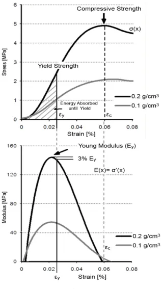
Figure 2 shows two characteristic stress-strain and modulus-strain curves from the compression testing of two PVC foam blocks. Figure 2a shows the characteristic stress-strain curves of a 0.20 g/cm3 and 0.10 g/cm3 density foam blocks. The expression for Young’s modulus of the material is given by the gradient of the curve. Figure 2b shows Young’s modulus as a function of the strain. Young’s modulus was determined as the maximum value of the curve in Figure 2b. The
yield point was defined as the stress at the end of the peak region, when Young’s modulus is reduced by 3%.
Figure 2a and 2b have the same strain axes to allow easy comparison. The curves on the figure are typical of those obtained in this study. The energy absorbed until yield is the area under the stress-strain curve up to the yield point (hatched area on Figure 2a for the 0.20 g/cm3 density PVC foam).
Figure 2
Characteristic Stress-strain and Young’s modulus curves of high and low density PVC foams. (a) Stress-strain curves of low and high density PVC foams, (b) Young's modulus determined as the gradient of the curve. The yield point is defined as the point at which Young's modulus decreases to 97% of its maximum value. The area under the stress-strain curve up to the yield point is defined as the
energy absorbed until yield.
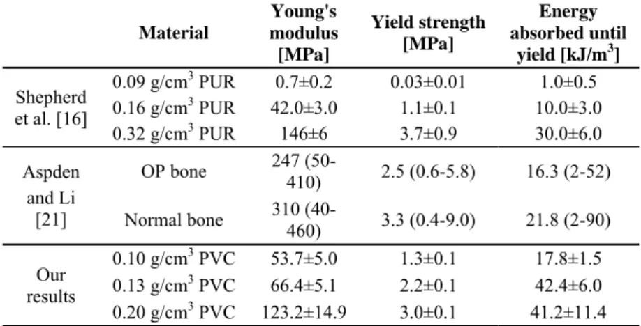
Table 2 summarizes the results for the three studied PVC foams and compares their values with those obtained by Shepherd et al. and Li and Apsden. The values compared are Young’s modulus, yield strength and energy absorbed until yield.
Table 2
Compression testing results of human bone, PUR and PVC foams.
Results of foams are given as mean± standard deviation. The results for bones are given as mean and min-max of measurement range.
Material
Young's modulus [MPa]
Yield strength [MPa]
Energy absorbed until
yield [kJ/m3] Shepherd
et al. [16]
0.09 g/cm3 PUR 0.7±0.2 0.03±0.01 1.0±0.5 0.16 g/cm3 PUR 42.0±3.0 1.1±0.1 10.0±3.0 0.32 g/cm3 PUR 146±6 3.7±0.9 30.0±6.0 Aspden
and Li [21]
OP bone 247 (50-
410) 2.5 (0.6-5.8) 16.3 (2-52) Normal bone 310 (40-
460) 3.3 (0.4-9.0) 21.8 (2-90) Our
results
0.10 g/cm3 PVC 53.7±5.0 1.3±0.1 17.8±1.5 0.13 g/cm3 PVC 66.4±5.1 2.2±0.1 42.4±6.0 0.20 g/cm3 PVC 123.2±14.9 3.0±0.1 41.2±11.4 Discussion
The purpose of the study was to determine whether closed-cell, cross-linked PVC foams could be suitable for the mechanical modeling of normal and OP bone. The method used was first published by Li and Aspden for the comparison of cancellous bone of patients with osteoporosis or osteoarthritis [21]. Shepherd et al.
used the same method in their study on commercially available PUR foams as mechanical model materials for OP human bones [17]. Using exactly the same method as Li and Aspden, and Shepherd et al. provides a unique opportunity to compare the studied mechanical properties of human bone, PUR and PVC foams.
Such comparative studies for different bone model materials are extremely rare, since different research groups use different methods for the validation of their material.
The 0.10 g/cm3 PVC foam was used to model very low density cancellous bone the same way as the 0.09 g/cm3 PUR foam studied by Shepherd et al. The PVC foam is much stronger than the PUR foam, and the results suggest that it could model severely osteoporotic bones. The Young’s modulus of this material is close to the lower limit of the modulus of the OP bone. The yield strength is one-half of the average value for osteoporotic bone, but it is within the limits measured by Aspden and Li. The lightest foam’s absorbed energy until yield is practically equal to that of the OP bone. According to these results the lowest density closed-cell, cross-linked, PVC foam studied in this paper is suitable as a mechanical model material for severely osteoporotic cancellous bone.
In terms of yield strength and Young’s modulus, the 0.13 g/cm3 PVC foam is a more suitable model material for osteoporotic cancellous bone than the 0.10 g/cm3 PVC foam. In contrast to the 0.10 g/cm3 density PVC foam and the 0.16 g/cm3 PUR material studied by Shepherd et al., the yield strength of this PVC foam matches that of the osteoporotic human cancellous bone, although its Young’s modulus is still at the lower end of the range of human osteoporotic bone’s Young’s modulus.
The results for the 0.20 g/cm3 PVC foam quantitatively match those for the 0.32 g/cm3 PUR material, and those for normal human bone. This makes it a suitable material for the modeling of normal human cancellous bone during in vitro biomechanical tests.
Previous papers used different methods to study possible substitute materials for human cancellous bone during in vitro biomechanical tests. Fatigue tests on rigid PUR foams showed that these materials do not model the behavior of cancellous bone precisely under dynamic and cyclic loading [9, 22]. Fresh animal and human bone (both cortical and cancelous) have viscoelastic properties that make their properties and behavior unique [23-25]. PVC foams have also displayed such properties, which could make them a better model material for trabecular bone during in vitro biomechanical tests [20, 26, 27].
The results of this paper indicate that cross-linked, closed-cell PVC foams could be more suitable for the modeling of human cancellous bone than currently used PUR foams. Based on Young’s modulus, yield strength and energy absorbed until yield, they represent human cancellous bone better than PUR foams. Their viscoelastic, and energy absorbing properties also make them a more suitable material for the modeling of cancellous bone.
The different test specimen geometry used in our study does not affect the properties measured since these were calculated from the original cross section of the specimens. Furthermore, our test specimens and those of Aspden et al. and Shepherd et al. have comparable dimensions and cross section/height aspect ratios.
Keaveny et al., and Pilkey et al. showed in their works that much higher cross section/height aspect ratio is needed for a significant difference in measurement results [28, 29].
In this paper we compared the Young’s modulus, yield strength and energy absorbed until yield of three cross-linked, solid, closed-cell PVC foams, with those of rigid closed-cell and open-cell PUR foams, and osteoporotic and normal density bones. Our results indicate that further comparative research would be needed on these materials. Not only compressive but bending and tensile tests would be needed, and fatigue behavior should also be compared under different loads. Any similarities found between the mechanical properties of PVC foam and cancellous bone would strengthen the case that PVC foams are suitable as a human cancellous bone model.
Conclusions
From the results of the above-mentioned and discussed measurements, the following conclusions can be drawn:
The investigated cross-linked, rigid cellular PVC foams are adequate model materials for osteoporotic and normal cancellous human bone.
Based on Young’s modulus and yield strength, the investigated 0.1 and 0.13 g/cm3 PVC foams are suitable as mechanical model materials for osteoporotic cancellous bone.
Based on Young’s modulus and yield strength, the investigated 0.20 g/cm3 PVC foam is suitable as a model material for normal bone.
Based on energy absorbed until yield, the 0.10 g/cm3 PVC foam is a suitable mechanical model material for osteoporotic cancellous bone. For the modeling of normal bone, both the 0.13 and 0.20 g/cm3 PVC foams are suitable.
In summary, it can be concluded that according to the results of this research, cross-linked closed-cell rigid PVC foams are suitable as mechanical model materials for human cancellous bone.
Acknowledgement
This paper was supported by the János Bolyai Research Scholarship of the Hungarian Academy of Sciences. The authors would like to thank Arburg Hungaria Ltd. for the injection molding machine.
This work is connected to the scientific program of the "Development of quality- oriented and harmonized R+D+I strategy and functional model at BME" project.
This project is supported by the New Széchenyi Plan (Project ID: TÁMOP- 4.2.1/B-09/1/KMR-2010-0002).
The work reported in this paper has been performed in the framework of the project "Talent care and cultivation in the scientific workshops of BME" project.
This project is supported by the TÁMOP - 4.2.2.B-10/1-2010-0009 grant.
This work was supported by the Hungarian Scientific Research Fund (OTKA PD 105995)
References
[1] E. M. Meslin, K. A. Quaid: Ethical Issues in the Collection, Storage, and Research Use of Human Biological Materials; Journal of Laboratory and Clinical Medicine, 144, 5, 2004, 229-234
[2] C. E. Dunham, S. E. Takaki, J. A. Johnson, C. E. Dunning: Mechanical Properties of Cancellous Bone of the Distal Humerus; Clinical Biomechanics, 20, 8, 2005, 834-838
[3] E. Perilli, M. Baleani, C. Öhman, R. Fognani, F. Baruffaldi, M. Viceconti:
Dependence of Mechanical Compressive Strength on Local Variations in Microarchitecture in Cancellous Bone of Proximal Human Femur; Journal of Biomechanics, 41, 2, 2008, 438-446
[4] H. Follet, K. Bruyère-Garnier, F. Peyrin, J. P. Roux, M. E. Arlot, B. Burt- Pichat, C. Rumelhart, P. J. Meunier: Relationship between Compressive Properties of Human Os Calcis Cancellous Bone and Microarchitecture Assessed from 2D and 3D Synchrotron Microtomography; Bone, 36, 2, 2005, 340-351
[5] H. Brockstedt, M. Kassem, E. F. Eriksen, L. Mosekilde, F. Melsen: Age- and Sex-related Changes in Iliac Cortical Bone Mass and Remodelling;
Bone, 14, 4, 1993, 681-691
[6] M. A. K. Liebschner: Biomechanical Considerations of Animal Models used in Tissue Engineering of Bone; Biomaterials, 25, 9, 2004, 1697-1714 [7] P. Buma, W. Schreurs, N. Verdonschot: Skeletal Tissue Engineering - from
in Vitro Studies to Large Animal Models, Biomaterials; 25, 9, 2004, 1487- 1495
[8] K. A. Athanasiou, A. Agarwal, A. Muffoletto, F. J. Dzida, G.
Constantinides, M. Clem: Biomechanical Properties of Hip Cartilage in Experimental Animal Models; Clinical Orthopaedics and Related Research, 316, 7, 1995, 254-266
[9] Y. H. An, R. A. Draughn: Mechanical Testing of Bone and the Bone–
Implant interface, CRC Press, London, 2000, Chapter 10.: Synthetic materials and structures used as models for bone
[10] A. D. Heiner, T. D. Brown: Structural Properties of a New Design of Composite Replicate Femurs and Tibias; Journal of Biomechanics, 34, 6, 2001, 773-781
[11] M. B. Sommers, D. C. Fitzpatrick, S. M. Madey, C. V. Zanderschulp, M.
Bottlang: A Surrogate Long-Bone Model with Osteoporotic Material Properties for Biomechanical Testing of Fracture Implants; Journal of Biomechanics, 40, 15, 2007, 3297-3304
[12] A. E. Johnson, T. S. Keller: Mechanical Properties of Open-Cell Foam Synthetic Thoracic Vertebrae; Journal of Materials Science: Materials in Medicine, 19, 3, 2008, 1317-1323
[13] D. Wähnert, K. L. Hoffmeier, Y. Stolarczyk, R. Fröber, G. O. Hofmann, T.
Mückley: Evaluation of a Customized Artificial Osteoporotic Bone Model of the Distal Femur; Journal of Biomaterials Applications, 26, 4, 2011,451- 464
[14] J. A. Szivek, M. Thomas, J. B. Benjamin: Characterization of a Synthetic Foam as a Model for Human Cancellous Bone; Journal of Applied Biomaterials, 4, 3, 1993, 269-272
[15] J. A. Szivek, J. D. Thompson, J. B. Benjamin: Characterization of Three Formulations of a Synthetic Foam as Models for a Range of Human Cancellous Bone Types; Journal of Applied Biomaterials, 6, 2, 1995, 125- 128
[16] P. Patel, D. Shepherd, D. Hukins: Compressive Properies of Commercially Available Plyurathane Foams as Mechanical Models for Osteoporotic Human Cancellous Bone; BMC Musculoskeletal Disorders; 9, 10, 2008, 137-144
[17] M. S. Thompson, I. D. McCarthy, L. Lidgren, L. Ryd: Compressive and Shear Properties of Commercially Available Polyurethane Foams; Journal of Biomechanical Engineering – ASME, 125, 5, 2003, 732-734
[18] Y. Shikinami, M. Okuno: Bioresorbable Devices Made of Forged Composites of Hydroxyapatite (HA) Particles and Poly-L-lactide (PLLA):
Part I. Basic Characteristics; Biomaterials, 20, 9, 1999, 859-877
[19] Y. Shikinami, M. Okuno: Bioresorbable Devices Made of Forged Composites of Hydroxyapatite (HA) Particles and Poly-L-lactide (PLLA):
Part II. Practical Properties of Miniscrews and Miniplates; Biomaterials, 22, 23, 2001, 3197-3211
[20] V. Palissery, M. Taylorm, M. Browne: Fatigue Characterization of a Polymer Foam to Use as a Cancellous Bone Analog Material in the Assessment of Orthopaedic Devices; Journal of Material Science: Materials in Medicine, 15, 1, 2004, 61-67
[21] B. Li, R. M. Aspden: Composition and Mechanical Properties of Cancellus Bone from the Femoral Head of Patients with Osteoporosis or Osteoarthritis; Journal of Bone and Mineral Research, 12, 4, 1997, 641-651 [22] L. J. Gibson, M, F, Ashby: Cellular Solids Structure and Properties,
Cambridge University Press, Cambridge, 1999
[23] T. P. M. Johnson, S. Socrate, M. C. Boyce: A Viscoelastic, Viscoplastic Model of Cortical Bone Valid at Low and High Strain Rates; Acta Biomaterialia, 6, 10, 2010, 473-4080
[24] R. M. Guedes, J. A. Simoes, J. L. Morais: Viscoelastic Behaviour and Failure of Bovine Cancellous Bone under Constant Strain Rate; Journa of Biomechanics, 36, 1, 2006, 49-60
[25] J. F. Mano: Viscoelastic Properties of Bone: Mechanical Spectrospcopy Studies on a Chicken Model; Materials Science and Engineering C, 25, 2, 2005, 145-152
[26] K. Kanny, H. Mahfuz, L. A. Carlsson, T. Thomas, S. Jeelani: Dynamic Mechanical Analyses and Fexural Fatigue of PVC Foams; Composite Structures, 58, 2, 2002, 175-183
[27] G. T. Lim, V. Altstädt, F. Ramsteiner: Understanding the Compressive Behavior of Linear and Cross-linked Poly(vinyl chloride) Foams; Journal of Cellular Plastics, 45, 9, 2009, 419-439
[28] T. M. Keaveny, T. P. Pinila, R. P. Crawford, D. L. Kopperdahl, A. Lou:
Systematic and Random Errors in Compression Testing of Trabecular Bone; Journal of Orthopaedic Research, 15, 1, 1997, 101-110
[29] W. B. Lievers, S. D. Waldman, A. K. Pilkey: Minimizing Specimen Length in Elastic Testing of End-constrained Cancellous Bone; Journal of the Mechanical Behavior of Biomedical Materials, 3, 2010, 22-30