Stress Resilience is Associated with Hippocampal Synaptoprotection in the Female Rat Learned Helplessness Paradigm
Orsolya Huzian,aJudith Baka,aEszter Csakvari,aNikoletta Dobos,aCsaba Leranth,b,cLaszlo Siklos,aRonald S. Duman,d,e Tamas Farkasfand Tibor Hajszana,b*
aInstitute of Biophysics, Biological Research Center, Temesvari Krt 62, 6726 Szeged, Hungary
bDepartment of Obstetrics, Gynecology, and Reproductive Sciences, Yale University School of Medicine, 333 Cedar Street, New Haven, CT 06510, United States
cDepartment of Neuroscience, Yale University School of Medicine, 333 Cedar Street, New Haven, CT 06510, United States
dDepartment of Psychiatry, Yale University School of Medicine, 34 Park Street, New Haven, CT 06508, United States
eDepartment of Pharmacology, Yale University School of Medicine, 333 Cedar Street, New Haven, CT 06520, United States
fDepartment of Physiology, Anatomy, and Neuroscience, University of Szeged Faculty of Science and Informatics, Kozep Fasor 52, 6726 Szeged, Hungary
Abstract—The synaptogenic hypothesis of major depressive disorder implies that preventing the onset of depressive-like behavior also prevents the loss of hippocampal spine synapses. By applying the psychoactive drugs, diazepam and fluoxetine, we investigated whether blocking the development of helpless behavior by pro- moting stress resilience in the rat learned helplessness paradigm is associated with a synaptoprotective action in the hippocampus. Adult ovariectomized and intact female Sprague-Dawley rats (n= 297) were treated with either diazepam, fluoxetine, or vehicle, exposed to inescapable footshocks or sham stress, and tested in an active escape task to assess helpless behavior. Escape-evoked corticosterone secretion, as well as remodeling of hip- pocampal spine synapses at a timepoint representing the onset of escape testing were also analyzed. In ovariec- tomized females, treatment with diazepam prior to stress exposure prevented helpless behavior, blocked the loss of hippocampal spine synapses, and muted the corticosterone surge evoked by escape testing. Although fluox- etine stimulated escape performance and hippocampal synaptogenesis under non-stressed conditions, almost all responses to fluoxetine were abolished following exposure to inescapable stress. Only a much higher dose of fluoxetine was capable of partly reproducing the strong protective actions of diazepam. Importantly, these protec- tive actions were retained in the presence of ovarian hormones. Our findings indicate that stress resilience is associated with the preservation of spine synapses in the hippocampus, raising the possibility that, besides synaptogenesis, hippocampal synaptoprotection is also implicated in antidepressant therapy.Ó2021 The Author (s). Published by Elsevier Ltd on behalf of IBRO. This is an open access article under the CC BY license (http://creativecom- mons.org/licenses/by/4.0/).
Key words: diazepam, fluoxetine, major depression, corticosterone, synaptic plasticity, antidepressant resistance.
INTRODUCTION
As part of the limbic system, the hippocampus is heavily implicated in the neurobiology of major depression, its dysfunction contributing to many depressive symptoms, such as cognitive decline, loss of motivation, and stress system derailment (Sapolsky, 2000a; Sousa et al.,
2000; Nestler et al., 2002; Lisman and Grace, 2005;
Cooper et al., 2006; Diamond et al., 2006; LeGates et al., 2018). In the context of this study, the central role that the hippocampus plays in regulating the stress sys- tem is particularly important, because stress and stress hormones appear to be key mediators both in human depression and in animal models of the disease (Sapolsky, 2000a; Sala et al., 2004; Hajszan et al., 2009; McEwen et al., 2016). Compromised hippocampal function in major depression is associated with the break- down of hippocampal neural circuitry, especially at the synaptic level, which has prompted the synaptogenic hypothesis of major depressive disorder that postulates a causal relationship between depressive symptoms and loss of synapses in limbic brain areas (Hajszan and
https://doi.org/10.1016/j.neuroscience.2021.01.029
0306-4522/Ó2021 The Author(s). Published by Elsevier Ltd on behalf of IBRO.
This is an open access article under the CC BY license (http://creativecommons.org/licenses/by/4.0/).
*Correspondence to: Tibor Hajszan, Institute of Biophysics, Biological Research Center, Temesvari Krt 62, 6726 Szeged, Hungary. Tel:
+36-62-599-623. Fax: +36-62-433-133.
E-mail address:hajszan.tibor@brc.hu(T. Hajszan).
Abbreviations:aCSF, artificial cerebrospinal fluid; CA1sr, CA1 stratum radiatum; CA3sl/sr, CA3 stratum lucidum/radiatum; DGsm, dentate gyrus stratum moleculare; fEPSP, field excitatory postsynaptic potential; LTP, long-term potentiation; SSRI, selective serotonin reuptake inhibitor.
N EUROSCIENCE
RESEARCH ARTICLE
O. Huzian et al. / Neuroscience 459 (2021) 85–103
85
MacLusky, 2006; Hajszan et al., 2010; Kang et al., 2012;
Duman et al., 2016). The excitatory, asymmetric spine synapses localized within the CA1 stratum radiatum (CA1sr), the CA3 stratum lucidum/radiatum (CA3sl/sr), and the dentate gyrus stratum moleculare (DGsm) are the prime linking points of the ‘‘trisynaptic loop”, the main neural circuit of the hippocampus, which is formed by sequentially organized pyramidal and granule cells (Amaral and Witter, 1995; Stepan et al., 2012, 2015).
Aside from our own work (Hajszan et al., 2005, 2009, 2010; Kang et al., 2012; Baka et al., 2017), a large num- ber of studies has investigated synaptic remodeling within the trisynaptic loop, relating it mainly to hippocampus- dependent cognitive and affective functions as summa- rized, e.g., in our recent review (Hajszan, 2020). As a result, the number of spine synapses provides a reliable readout of structural connectivity along the trisynaptic loop and can be considered as an anatomical correlate of hippocampal function.
During recent years, we have reported that major depression in humans is associated with loss of synapses in the prefrontal cortex, while depressive-like behavior in rodent models is related to loss of spine synapses in the hippocampus (Hajszan et al., 2009, 2010; Kang et al., 2012; Baka et al., 2017). In addition, reproducing the loss of hippocampal spine synapseswith- outexposure to stress elicits helpless behavior in the rat learned helplessness paradigm (Hajszan et al., 2009, 2010). On the other hand, our research team has also val- idated the theory that an antidepressant response requires the generation of new synapses in limbic brain areas, reversing the synapse loss caused by stress/de- pression (Hajszan et al., 2005, 2009, 2010; Li et al., 2010). Aside from synapse loss and synaptogenesis, an important aspect of the synaptogenic hypothesis remains unexplored. The synaptogenic hypothesis implies that preventing the onset of depressive-like behavior by increasing stress resilience, also prevents the loss of hip- pocampal spine synapses. Using a rodent model of post- partum depression, we have recently shown that chronic exposure to pregnancy levels of estradiol and proges- terone indeed protects spine synapses in the hippocam- pus while improving stress resilience (Baka et al., 2017).
Because this behavior/synapse correlation may be dependent on the special hormonal conditions of the post- partum model (Galea et al., 2001; Suda et al., 2008; Baka et al., 2017), the goal of our present study was to confirm that the protective actions we described earlier are repro- ducible under more generalized conditions.
To address this issue, we used a well-characterized female learned helplessness model of stress/depression (Hajszan et al., 2010) to investigate the protective actions of widely-used psychoactive drugs with and without the influence of ovarian hormones. A series of earlier learned helplessness studies demonstrates that benzodiazepines effectively prevent the development of helpless behavior when given prior to stress exposure (Drugan et al., 1984; De Pablo et al., 1991; Petty et al., 1992). Based on this confirmed preventive effect in learned helpless- ness, a reasonable choice for exploring the relationships between stress resilience and hippocampal synaptopro-
tection is diazepam, a GABA-A receptor benzodiazepine site agonist anxiolytic. Although a number of mechanisms may be involved, the preventive effect of benzodiazepines is most likely based on mitigating stress, because benzo- diazepines are among the most potent suppressors of the stress response (File, 1982; Pohorecky et al., 1988; de Boer et al., 1990; Matar et al., 2009). Benzodiazepines are also known to reduce or even completely halt various modalities of synaptic plasticity (Evans and Viola- McCabe, 1996; Seabrook et al., 1997; Tampellini et al., 2010; Curto et al., 2016), but their influence on remodel- ing of hippocampal spine synapses, especially in relation to the preventive effects, is currently unknown.
Stress resilience and synaptoprotection may be highly relevant to antidepressant resistance, a major clinical problem, because antidepressant resistance and recurring depression appear to be associated with a persistent hyperactivity of the stress system (Heuser et al., 1996; Appelhof et al., 2006), while curbing the stress response seems to improve the antidepressant response (Barden et al., 1995; Jahn et al., 2004;
Nemeroff and Owens, 2004; Rogoz et al., 2005). Antide- pressant resistance is mostly encountered with selective serotonin reuptake inhibitor (SSRI) antidepressants (Rush et al., 2006; Trivedi et al., 2006), which is paralleled by a growing number of preclinical studies showing SSRIs being ineffective in a wide variety of paradigms (Magarinos et al., 1999; Kobayashi et al., 2008;
Valentine et al., 2008; Venzala et al., 2012; Kokras et al., 2015; Workman et al., 2016). These findings sug- gest that the stress-mitigating effect of SSRIs may be lim- ited, especially compared to that of benzodiazepines. A handful of reports indeed demonstrate that reduced antidepressant efficacy of SSRIs is usually observed under stressful conditions (Valentine et al., 2008;
Branchi et al., 2013; Khemissi et al., 2014). Based on these data, we also selected fluoxetine, a classic SSRI, to investigate the relationships between stress resilience and hippocampal synaptoprotection under the influence of a drug highly susceptible of antidepressant resistance.
Although fluoxetine is a strong initiator of hippocampal synaptogenesis under non-stressed conditions (Hajszan et al., 2005), it is currently unknown what happens to this robust synaptogenic power following stress exposure.
In animal models of major depression, drugs are usually given after stress exposure to investigate their capability of reversing depressive-like behavior. The goal of this study, however, is to prevent the development of helpless behavior via promoting stress resilience, which logically requires drug applicationprior to stress exposure. This approach obviously follows the setup of earlier learned helplessness studies that revealed the preventive effects of benzodiazepines and ovarian hormones (Drugan et al., 1984; De Pablo et al., 1991; Petty et al., 1992; Baka et al., 2017). Here we report that promoting stress resilience in learned helplessness with diazepam is associated with a synaptoprotective action in the hippocampus and with a muted stress response during escape testing. We also show that these preventive effects are more limited after fluoxetine treatment, as exposure to stress mostly destroys the
hippocampal synaptogenic power of fluoxetine. As we describe above, the influence of both diazepam and fluox- etine on various aspects of stress and stress-related behaviors has been investigated earlier. However, it is currently not known whether this influence on stress is related to synaptic remodeling in the hippocampus. As a result, our present ultrastructural investigation provides the main novel findings of this study. These results sug- gest that the issue of stress resilience and synaptoprotec- tion needs further attention in future studies, as they can potentially contribute to the better understanding of antidepressant therapy and antidepressant resistance.
EXPERIMENTAL PROCEDURES Animals
Ethical statement. Our animal protocol was approved by the Institutional Animal Care and Use Committee of Yale University School of Medicine and by the Ethical Committee for the Protection of Animals in Scientific Research of the Biological Research Center. Animal experiments were carried out in accordance with the National Institutes of Health Guide for the Care and Use of Laboratory Animals (NIH Publication No. 80-23) and with the European Communities Council Directive of 24 November 1986 (86/609/EEC). All efforts were made to minimize the number of animals used and their suffering.
Experimental animals. Female Sprague-Dawley rats (n= 297, 200–250 g) were purchased from Charles- River Laboratories (Wilmington, Massachusetts), and only healthy, drug- and test-naive rats were allocated to experimental groups after an acclimation period of one week. We used ovariectomized and intact female rats because this study is based upon and expands the findings of our earlier experiments that also used females (Hajszan et al., 2005, 2010; Baka et al., 2017).
The rationale for using ovariectomy is based on well- documented clinical observations that diminishing estra- diol levels in women represent an increased risk for devel- oping mood disorders (Freeman et al., 2006; Deecher et al., 2008; Soares and Zitek, 2008), which is in line with our own findings that withdrawal of ovarian hormones robustly contributes to depressive-like behavior in animal models (Hajszan et al., 2010; Baka et al., 2017). Ovariec- tomy is also essential to prevent interference from cyclic changes in the production of endogenous ovarian hor- mones, as the number of hippocampal synapses shows significant fluctuation during the estrus cycle (Woolley and McEwen, 1992). Animals were ovariectomized a week prior to commencing any procedures using a ketamine-based anesthetic (75 mg/kg ketamine, 3.6 mg/
kg xylazine, 0.09 mg/kg acepromazine dissolved in 3 ml/
kg saline, im).
Housing and husbandry. Similar to our earlier studies (Hajszan et al., 2010; Baka et al., 2017), rats were kept in a high-security-barrier animal facility and monitored periodically to be free of major pathogens. Animal rooms were maintained at 21°C and on a 12/12-h light/dark
cycle with light on at 0700 h. Rats were group-housed (n= 3 females per cage) in standard cages with tap water and rodent chow availablead libitum. Animal wel- fare was monitored daily by animal facility staff and at least weekly by a veterinarian.
Study design
Diazepam experiment. Using ovariectomized females, four experimental groups were established by treating animals with lower (5 mg/kg) or higher (10 mg/kg) doses of diazepam (ip, 30 min prior to sham stress or inescapable stress). For reference, well-documented negative and positive control groups of helpless behavior were also created by treating animals with vehicle (1 ml/kg distilled water, ip, 30 min prior to sham stress or inescapable stress) (Hajszan et al., 2010). Dia- zepam was obtained in the form of 5 mg/ml injectable solution from Gedeon Richter Plc (Budapest, Hungary).
In earlier studies, similar diazepam regimens inhibited the behavioral effects of uncontrollable stress and instantly prevented both stress- and seizure-induced glu- tamate efflux in the hippocampus (De Pablo et al., 1991;
Petty et al., 1992) (Bagley and Moghaddam, 1997;
Khan et al., 1999).
Omitting the two groups with the lower diazepam dose, the above experiment was repeated in simulated proestrus rats to investigate whether the presence of ovarian hormones affects the actions of diazepam.
Proestrus levels of estradiol and progesterone were simulated in ovariectomized females as published earlier (Scharfman et al., 2007; Baka et al., 2017), timed to achieve the required hormone concentrations by the onset of stress exposure. Briefly, a day before sham stress or inescapable stress, animals were injected with 3lg/kg estradiol benzoate (dissolved in sesame oil, sc) at 8:30 am, followed by 4lg/kg estradiol benzoate at 8:30 pm. Five hours prior to sham stress or inescapable stress, animals also received 2 mg/kg progesterone (dis- solved in sesame oil, sc), followed by a final dose of 3lg/kg 17b-estradiol (dissolved in sesame oil, sc) 2 h before sham stress or inescapable stress.
The schedule and sample sizes of the diazepam experiment are detailed inTable 1.
Fluoxetine experiment. Using ovariectomized females, four experimental groups were established by treating animals with lower (5 mg/kg) or higher (20 mg/
kg) doses of fluoxetine (ip, once daily for 15 days prior to sham stress or inescapable stress). For reference, well-documented negative and positive control groups of helpless behavior were also created by treating animals with vehicle (1 ml/kg distilled water, ip, once daily for 15 days prior to sham stress or inescapable stress) (Hajszan et al., 2010). Fluoxetine was obtained in the form of 4 mg/ml aqueous solution from Eli Lilly and Com- pany (Indianapolis, Indiana). In earlier studies, we have demonstrated that lower doses of fluoxetine elicit antide- pressant responses in a chronic unpredictable stress paradigm and induce strong proliferation of hippocampal spine synapses, while higher doses reverse escape
deficits and block the downregulation of hippocampal neurogenesis in the rat learned helplessness paradigm (Malberg and Duman, 2003; Hajszan et al., 2005;
Valentine et al., 2008). Omitting the two groups with the lower fluoxetine dose, the above experiment was repeated in intact rats to investigate whether the presence of fluctuating ovarian hormones modifies the actions of fluoxetine.
The schedule and sample sizes of the fluoxetine experiment are detailed inTable 2.
Sample size. We have shown in earlier studies that a sample size of n= 10 rats/group provides sufficient statistical power to differentiate between helpless and control animals based on escape performance and corticosterone levels (Hajszan et al., 2009, 2010; Baka et al., 2017). Considering our previous spine synapse analyses, a sample size ofn= 4 rats/group is sufficient for ANOVA to detect at least 25% change in synapse numbers with the desired 80% power at a= 0.05 (Hajszan et al., 2005, 2009, 2010; Baka et al., 2017). In the case of long-term potentiation (LTP) measurements, we also followed our earlier experiments that suggest a sample size of n= 5 rats/group (Marosi et al., 2009;
Kocsis et al., 2014).
Experimental outcomes. The following endpoints were assessed in both the diazepam and the fluoxetine
experiments: (a) performance in an active escape task to detect helpless behavior; (b) serum corticosterone concentrations at the completion of active escape testing to measure the magnitude of stress response evoked by the active escape task; (c) number of spine synapses in the CA1sr, CA3sl/sr, and DGsm areas to estimate the status of structural connectivity along the hippocampal trisynaptic loop at the onset of active escape testing. In addition, (d) LTP of the Schaffer collateral-CA1 synaptic response was evaluated at the onset of active escape testing in a subset of groups from the diazepam experiment with the sole purpose of verifying the effectivity of stress exposure in diazepam- pretreated animals.
Experimental procedures
Learned helplessness. Our laboratories routinely use a modified learned helplessness model as published previously (Valentine et al., 2008; Hajszan et al., 2009, 2010; Baka et al., 2017). Even the standard learned help- lessness paradigm is highly-rated as a rodent model of stress/depression based on its excellent face, predictive, and construct validities (Seligman, 1968; Thiebot et al., 1992; Cryan et al., 2002). Our modified model surpasses the standard paradigm by providing an improved rate of helplessness and an ability to differentiate between antidepressant classes by reproducing the clinical obser- Table 1.Schedule of the diazepam experiment.
Group Day-1 Day-7 Day-8 Day-9
Diazepam experiment in ovariectomized females
veh/ns ovariectomy — veh + ns EM (n= 4)
LTP (n= 6) AE + CS (n= 10)
veh/is ovariectomy — veh + is EM (n= 4)
LTP (n= 5) AE + CS (n= 10)
dz5/ns ovariectomy — dz5 + ns EM (n= 4)
AE + CS (n= 10)
dz5/is ovariectomy — dz5 + is EM (n= 4)
AE + CS (n= 10)
dz10/ns ovariectomy — dz10 + ns EM (n= 4)
AE + CS (n= 10)
dz10/is ovariectomy — dz10 + is EM (n= 4)
LTP (n= 6) AE + CS (n= 10) Diazepam experiment in simulated proestrus rats
veh/ns ovariectomy eb3 + eb4 pe + veh + ns EM (n= 4)
AE + CS (n= 10)
veh/is ovariectomy eb3 + eb4 pe + veh + is EM (n= 4)
AE + CS (n= 10)
dz10/ns ovariectomy eb3 + eb4 pe + dz10 + ns EM (n= 4)
AE + CS (n= 10)
dz10/is ovariectomy eb3 + eb4 pe + dz10 + is EM (n= 4)
AE + CS (n= 10) Treatments: dz5, low-dose diazepam (5 mg/kg, ip, 30 min prior to ns or is);dz10, high-dose diazepam (10 mg/kg, ip, 30 min prior to ns or is);eb3, estradiol-benzoate (3lg/
kg, sc, at 8:30 am);eb4, estradiol-benzoate (4lg/kg, sc, at 8:30 pm);is, inescapable stress;ns, sham stress;pe, progesterone (2 mg/kg, sc, 5 h prior to ns or is) and 17b- estradiol (3lg/kg, sc, 2 h prior to ns or is);veh, vehicle (1 ml/kg distilled water, ip, 30 min prior to ns or is).Measurements: AE, active escape testing;CS, blood sampling for corticosterone immunoassay immediately after the completion of escape testing;EM, sampling for electron microscopic stereology immediately before the onset of escape testing;LTP, recording long-term potentiation at a timepoint that represents the onset of escape testing.
vations of reduced responses to serotonergic drugs (Trivedi et al., 2006; Valentine et al., 2008). As published earlier, we utilized inescapable stress to induce helpless- ness, followed by an active escape task to assess the behavioral deficit. Briefly, the testing apparatus consisted of a commercially available and fully automated shuttle avoidance system (Med Associates, St. Albans, Ver- mont). Animals subjected to inescapable stress received randomized exposure to 60 scrambled footshocks with 0.85 mA intensity, 15 s average shock duration, and 45 s average intershock interval. Footshocks were admin- istered via the wire grid flooring in a closed shuttle box compartment with no opportunity to escape. Sham- stressed controls underwent the same protocol, but the shock generator was switched off during the entire procedure.
Twenty-four h after exposure to inescapable stress or sham stress, 30 randomized trials of escapable footshocks were administered with 0.65 mA intensity, 35 s maximum trial/footshock duration, and 60 s average intertrial interval. During each footshock, animals were allowed to escape by passing between shuttle box compartments. The initial five trials required one shuttle crossing, while the following 25 trials required two shuttle crossings to terminate footshocks.
Escape latencies and escape failures were registered, representing the time to escape footshocks and the number of trials during which escape requirements were not met, respectively. Maximum escape latency (35 s) was registered for each escape failure. Behavioral testing was conducted in a dimly-lit room between 1000 h and 1600 h.
Serum corticosterone assay. Trunk blood samples were collected under ketamine-based anesthesia (see above) from each behaviorally tested animal immediately after the conclusion of active escape testing as described earlier (Hajszan et al., 2009; Baka et al., 2017). Serum was separated by centrifugation and stored frozen until assayed. Serum total concentra- tion of corticosterone was determined using a commer- cially available enzyme immunoassay kit by following the manufacturer-recommended protocol (Assay Designs, Ann Arbor, Michigan). Samples from the diaze- pam and the fluoxetine experiments were analyzed in separate sessions, but all samples from the same exper- iment were analyzed in a single session in duplicates. In the concentration ranges pertinent to the present study, the intraassay coefficient of variation was 8.4%.
Electron microscopic stereology. The number of asymmetric spine synapses was calculated in three hippocampal sampling areas, the CA1sr, the CA3sl/sr, and the DGsm, as published earlier (Hajszan et al., 2009; Baka et al., 2017). Four animals were randomly selected and sacrificed from each experimental group to assess synapse numbers at a timepoint representing the onset of active escape testing. Selected rats were per- fused transcardially under ketamine-based anesthesia (see above) using phosphate-buffered saline followed by a fixative containing 4% paraformaldehyde and 0.1% glu- taraldehyde dissolved in 0.1 M phosphate buffer (pH 7.4).
Brains were removed and postfixed overnight in the same fixative without glutaraldehyde. It needs to be mentioned that ketamine is well known to rapidly induce antidepres- Table 2.Schedule of the fluoxetine experiment
Group Day-1 Days 8–21 Day-22 Day-23
Fluoxetine experiment in ovariectomized females
veh/ns ovariectomy veh veh + ns EM (n= 4)
AE + CS (n= 10)
veh/is ovariectomy veh veh + is EM (n= 4)
AE + CS (n= 10)
fx5/ns ovariectomy fx5 fx5 + ns EM (n= 4)
AE + CS (n= 10)
fx5/is ovariectomy fx5 fx5 + is EM (n= 4)
AE + CS (n= 10)
fx20/ns ovariectomy fx20 fx20 + ns EM (n= 4)
AE + CS (n= 10)
fx20/is ovariectomy fx20 fx20 + is EM (n= 4)
AE + CS (n= 10) Fluoxetine experiment in intact rats
veh/ns — veh veh + ns EM (n= 4)
AE + CS (n= 10)
veh/is — veh veh + is EM (n= 4)
AE + CS (n= 10)
fx20/ns — fx20 fx20 + ns EM (n= 4)
AE + CS (n= 10)
fx20/is — fx20 fx20 + is EM (n= 4)
AE + CS (n= 10) Treatments: fx5, low-dose fluoxetine (5 mg/kg, ip, once daily on Days 8–21 and 30 min prior to ns or is on Day-22);fx20, high-dose fluoxetine (20 mg/kg, ip, once daily on Days 8–21 and 30 min prior to ns or is on Day-22);is, inescapable stress;ns, sham stress;veh, vehicle (1 ml/kg distilled water, ip, once daily on Days 8–21 and 30 min prior to ns or is on Day-22).Measurements: AE, active escape testing;CS, blood sampling for corticosterone immunoassay immediately after the completion of escape testing;
EM, sampling for electron microscopic stereology immediately before the onset of escape testing.
sant and synaptogenic responses (Li et al., 2010), which may introduce a bias into our synapse measurements.
However, such responses occur at subanesthetic doses of ketamine (5–15 mg/kg) (Maeng et al., 2008; Li et al., 2010; Ardalan et al., 2017), and we are not aware of any studies reporting antidepressant and/or synaptogenic effects at higher doses of ketamine. Moreover, we have specifically confirmed that the rapid signaling mecha- nisms that mediate antidepressant and synaptogenic responses to ketamine are dose-dependent, appearing only at sub-anesthetic doses (5–10 mg/kg) and not seen at the high anesthetic dose (75 mg/kg) we are using in this study (Li et al., 2010). Each perfused hippocampus was cut into 100-mm thick serial sections in the coronal plane using a vibratome. Every tenth section was then selected in a systematic random manner and embedded in Durcu- pan resin (Electron Microscopy Sciences, Fort Washing- ton, Pennsylvania). Using the embedded sections, the volume of each sampling area (VSA) was estimated utiliz- ing the Cavalieri Estimator software module of the Stereo Investigator computerized stereology system (Micro- BrightField, Villiston, Vermont).
To apply the disector technique (Sterio, 1984) at the electron microscopic level, 20 counting sites were local- ized in each sampling area along the entire septo- temporal axis of the hippocampus using a systematic ran- dom approach as modified from MacLusky and colleagues (MacLusky et al., 2006). Notable differences in function and connectivity patterns suggest that the hippocampus is divided into dorsal and ventral domains involved in cog- nitive operations and emotional processing, respectively (Moser and Moser, 1998), although this dorsal/ventral dis- tinction appears to be preferential rather than absolute (Bannerman et al., 2004). Based on the following clinical and preclinical findings, however, we do not differentiate between these two domains in our studies. (1) Consider- ing the co-occurrence of cognitive and emotional dysfunc- tions (Nestler et al., 2002; Belmaker and Agam, 2008), the symptomatology of major depression does not suggest dif- fering involvement of dorsal vs. ventral hippocampal regions. (2) Morphometric imaging studies in depressed patients show a typical hippocampal atrophy that affects theentireorgan (Sheline et al., 1999), which is reproduced in animal models of stress and depression (Donohue et al., 2006; Chen et al., 2010; Kassem et al., 2013; Ardalan et al., 2017). (3) Despite differences in its input/output con- nectivity patterns, the intrinsic circuitry, i.e., the trisynaptic loop, basically repeats itself along the septo-temporal axis of the hippocampus (Andersen et al., 1971). We are not aware of any systematicultrastructuralstudies reporting discrepancies in intrinsic synaptic architecture between dorsal vs. ventral hippocampal regions under any condi- tions, e.g., (Santuy et al., 2020). Actually, the rostro- caudal variability of numerical synapse densities appears to be less than 15% (Donohue et al., 2006), which is below the detection limit of our methods. That is why we have been consistently sampling from the entire hippocampus (Hajszan et al., 2004, 2005, 2009, 2010; MacLusky et al., 2006; Baka et al., 2017), following similar approaches in other laboratories (Chen et al., 2008, 2009, 2010, 2018; Ardalan et al., 2017).
A physical disector was prepared at each counting site by taking electron micrographs of matching areas in consecutive ultrathin sections at a final magnification of 11,000. This sampling technique provided 20 physical disectors (ND= 20) for each of CA1sr, CA3sl/sr, and DGsm, i.e., 60 disectors per hippocampus altogether.
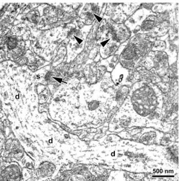
Prior to spine synapse counting, all micrographs were coded for blind analysis. An unbiased counting frame superimposed onto the electron micrographs was utilized to determine the number (RS) of asymmetric spine synapses (Fig. 1) within disectors. The estimated quantity of spine synapses in a particular sampling area was then calculated using the formula:
PSVSA
NDVD
where RS= sum of synapse counts, VSA= sampling area volume, ND= number of disectors, VD= disector volume (5.94lm3).
Long-term potentiation. With the sole purpose of verifying the effectivity of stress training in diazepam- pretreated animals, induction and stability of LTP were examined in hippocampal slice preparations as described earlier (Marosi et al., 2009; Kocsis et al., 2014). Ovariectomized females were randomly selected from the veh/ns (n= 6), veh/is (n= 5), and dz10/is (n= 6) groups to investigate functional neuroplasticity at a timepoint representing the onset of active escape testing (Table 1). Selected rats were rapidly decapitated under isoflurane anesthesia and their brains were removed and placed in an ice-cold artificial cerebrospinal
Fig. 1.Representative electron micrograph taken from the dentate gyrus stratum moleculare of a vehicle-pretreated ovariectomized female rat exposed to inescapable stress. The micrograph demon- strates a dendritic segment (d) with an emerging dendritic spine (s) that forms a spine synapse (arrow). Additional spine synapses are labeled by arrowheads. Scale bar = 500 nm.
fluid (aCSF) solution that was composed of 130 mM NaCl, 3.5 mM KCl, 1 mM NaH2PO4, 24 mM NaHCO3, 1 mM CaCl2, 3 mM MgSO4, 10 mM D-glucose, and saturated with 95% O2 and 5% CO2. Following brain removal, 350-lm coronal slices were prepared from the middle por- tion of hippocampus with a vibratome and kept in ice-cold aCSF. After vibratoming, slices were transferred to a Haas-type recording chamber and allowed to recover at room temperature for 1 h. The chamber contained a recording solution that had a flow rate of 1.5–2 ml/min and differed from the aCSF in the CaCl2 (3 mM) and MgSO4 (1.5 mM) concentrations. Following recovery, chamber temperature was set to and maintained at 34°C during the entire recording procedure.
Orthodromic stimulation of the Schaffer collateral/commissural pathway was achieved via a bipolar, concentric, stainless steel stimulating electrode (Neuronelektrod Ltd, Budapest, Hungary) inserted in the stratum radiatum at the CA1/CA2 border. Evoked field excitatory postsynaptic potentials (fEPSPs) were recorded in CA1sr using aCSF-filled glass micropipettes with a resistance of 1.5–2.5 megaX. Constant current stimuli were delivered in 0.2-ms pulses at 0.05 Hz to generate a half-maximal response that was determined from an input/output curve expressing fEPSP amplitudes against gradually increasing stimulating currents (0–100 lA). Employing the half-maximal response, amplitude stability of fEPSPs was monitored for 30 min, and then a baseline recording was acquired for 10 min.
LTP of the Schaffer collateral-CA1 synaptic response was induced by theta-burst stimulation (bursts of four impulses at 100 Hz, 100% stimulus intensity, 350 ms burst interval) (Marosi et al., 2009). After stimulation, fEPSPs were recorded for 60 min, amplified with a neu- tralized high-input-impedance preamplifier, filtered in the 1 Hz–3 kHz range, and digitized at a sampling rate of 10 kHz. Each animal provided a maximum of two record- ings that were saved to a computer and analyzed offline with the Origin 8.0 software (OriginLab Corporation, Northampton, Massachusetts).
Statistical analysis
Means were computed for each experimental outcome in each group of animals. The Shapiro–Wilks and Lilliefors tests were used to explore whether data are normally distributed and to determine a parametric or non- parametric statistical approach. The following sampling and statistical strategy was employed to evaluate the various experimental outcomes.
Behavioral and corticosterone measures. From each group, n= 10 rats were randomly selected for behavioral testing. Three measurements were obtained from every single animal: (1) number of escape failures and (2) mean of escape latencies during the active escape task, and (3) serum corticosterone concentrations at the completion of active escape testing. These three measurements were analyzed separately with two-way parametric fixed-effect ANOVA
(drug treatmentstress) followed by the Neuman–
Keuls post-hoc test.
Synapse counts. From each group, n= 4 rats were randomly selected and sacrificed at a timepoint representing the onset of active escape testing to process their brains for electron microscopic analysis.
Three measurements were obtained from every single animal: (1) number of spine synapses in CA1sr, (2) number of spine synapses in CA3sl/sr, and (3) number of spine synapses in DGsm. These three measurements were evaluated collectively with three-way parametric fixed-effect ANOVA (drug treatment, between subjects factorstress, between subjects factorsampling area, within subjects factor) followed by the Tukey–
Kramer post-hoc test.
fEPSP amplitudes. From the ovariectomized veh/ns, veh/is, and dz10/is groups of the diazepam experiment, n= 5–6 rats were randomly selected and sacrificed at a timepoint representing the onset of active escape testing to process their brains for electrophysiological analysis. Every single animal provided two independent time-series of fEPSP amplitude measurements. The amplitude data were evaluated with Scheirer-Ray-Hare two-way non-parametric fixed-effect ANOVA (grouptime) followed by the Mann–WhitneyU-test.
The significance level was conventionally set at P< 0.05.
RESULTS
Diazepam promoted stress resilience, protected hippocampal spine synapses, and muted the stress response during escape testing
Diazepam effects on helpless behaviour. At three randomly selected time-points during stress training, the experimenter briefly observed behavioral responses to inescapable stress, such as vocalizing, assuming evasive postures, and running about the cage trying to escape. Based on these behavioral responses, each animal was confirmed to be awake and perceiving footshocks.
Considering escape performance, two-way ANOVA (diazepam treatmentstress) demonstrated significant diazepam treatment effects (F2,54= 4.262, P0.02 for escape failures; F2,54= 5.005, P0.01 for escape latencies), significant stress effects (F1,54= 4.934, P0.04 for escape failures; F1,54= 11.173, P0.002 for escape latencies), and significant interaction effects (F2,54= 17.467, P0.001 for escape failures;
F2,54= 13.453, P0.001 for escape latencies) on behavioral measures in ovariectomized females; while there were significant stress effects (F1,36= 41.060, P0.001 for escape failures;F1,36= 21.687,P0.001 for escape latencies) and significant interaction effects (F1,36= 17.157, P0.001 for escape failures;
F1,36= 7.927, P0.008 for escape latencies), with the diazepam treatment effect being significant on escape failures (F1,36= 21.508, P0.001) but not on escape
latencies (F1,36= 1.906, P= 0.176) in simulated proestrus rats. Irrespective of hormonal status, diazepam treatment under non-stressed conditions did not affect escape performance (Fig. 2A, B ovariectomized females, dz5/ns vs. veh/ns,q54= 2.963, P= 0.1004 for escape failures, q54= 1.329,
P= 0.3515 for escape latencies, dz10/ns vs. veh/ns, q54= 2.586, P= 0.0731 for escape failures, q54= 1.993, P= 0.3433 for escape latencies; Fig. 2G, H simulated proestrus, dz10/ns vs. veh/ns, q36= 0.496, P= 0.7279 for escape failures, q36= 1.435, P= 0.3172 for escape latencies). On the other hand,
inescapable stress increased escape failures and escape latencies after vehicle treatment, which likewise occurred in both the absence and the presence of ovarian hormones (Fig. 2A, B ovariectomized females, veh/is vs.
veh/ns, q54= 8.566, P0.0001 for escape failures, q54= 8.502, P0.0001 for escape latencies; Fig. 2G, H simulated proestrus, veh/is vs. veh/ns,q36= 10.550, P0.0001 for escape failures,q36= 7.472, P0.0001 for escape latencies). However, these stress-induced increases were not observed in diazepam-pretreated females (Fig. 2A, B ovariectomized females, dz5/is vs.
dz5/ns, q54= 0.700, P= 0.6223 for escape failures, q54= 1.226, P= 0.3898 for escape latencies, dz10/is vs. dz10/ns, q54= 2.424, P= 0.0923 for escape failures, q54= 1.540, P= 0.2811 for escape latencies;
Fig. 2G, H simulated proestrus, dz10/is vs. dz10/ns, q36= 2.266, P= 0.1179 for escape failures, q36= 1.842, P= 0.2012 for escape latencies). As a result, diazepam treatment prior to inescapable stress caused both behavioral measures to remain significantly below the increased behavioral measures of stress- exposed controls, facilitating stress resilience in both ovariectomized and simulated proestrus animals (Fig. 2A, B ovariectomized females, dz5/is vs. veh/is, q54= 6.303, P0.0001 for escape failures, q54= 5.947, P0.0002 for escape latencies, dz10/is vs. veh/is,q54= 8.404, P0.0001 for escape failures, q54= 8.049, P0.0001 for escape latencies; Fig. 2G, H simulated proestrus, dz10/is vs. veh/is, q36= 8.780, P0.0001 for escape failures, q36= 4.196, P0.006 for escape latencies).
Diazepam effects on escape-evoked corticosterone levels. Two-way ANOVA (diazepam treatmentstress) found a significant diazepam treatment effect (ovariectomized females, F2,54= 7.799, P0.001;
simulated proestrus, F1,36= 12.831, P0.001), a significant stress effect (ovariectomized females, F1,54= 27.402, P0.001; simulated proestrus, F1,36= 31.353, P0.001), and a significant interaction effect (ovariectomized females, F2,54= 8.703, P0.001; simulated proestrus, F1,36= 22.726, P0.001) on corticosterone measurements in both ovariectomized and simulated proestrus animals.
Irrespective of hormonal status, diazepam treatment under non-stressed conditions did not affect escape-
evoked corticosterone release (Fig. 2C ovariectomized females, dz5/ns vs. veh/ns, q54= 0.783, P= 0.8451, dz10/ns vs. veh/ns, q54= 0.030, P= 0.9833; Fig. 2I simulated proestrus, dz10/ns vs. veh/ns, q36= 1.185, P= 0.4074). On the other hand, inescapable stress potentiated escape-evoked corticosterone secretion after vehicle treatment, which likewise occurred in both the absence and the presence of ovarian hormones (Fig. 2C ovariectomized females, veh/is vs. veh/ns, q54= 8.997, P0.0001; Fig. 2I simulated proestrus, veh/is vs. veh/ns, q36= 10.367, P0.0001). However, this stress-induced potentiation was not observed in diazepam-pretreated females (Fig. 2C ovariectomized females, dz5/is vs. dz5/ns, q54= 2.737, P= 0.0582, dz10/is vs. dz10/ns, q54= 1.088, P= 0.4448; Fig. 2I simulated proestrus, dz10/is vs. dz10/ns, q36= 0.832, P= 0.5598). As a result, diazepam treatment prior to inescapable stress prevented escape-evoked corticosterone release to attain the high levels of corticosterone secretion detected in stress-exposed controls, mitigating the stress response in both ovariectomized and simulated proestrus animals (Fig. 2C ovariectomized females, dz5/is vs. veh/is, q54= 5.476, P0.0004, dz10/is vs. veh/is, q54= 7.879, P0.0001; Fig. 2I simulated proestrus, dz10/is vs. veh/is.q36= 8.349,P0.0001).
Diazepam effects on hippocampal synapse counts.
Three-way ANOVA (diazepam
treatmentstresssampling area) revealed a significant diazepam treatment effect (F2,18= 34.700, P0.001), a significant stress effect (F1,18= 51.713, P0.001), a significant diazepam treatmentstress interaction effect (F2,18= 49.522, P0.001), a significant sampling area effect (F2,36= 136.063, P0.001), a significant diazepam treatmentsampling area interaction effect (F4,36= 3.107, P0.03), and a significant stresssampling area interaction effect (F2,36= 5.352, P0.009) on synapse counts in ovariectomized females, while there was a significant diazepam treatment effect (F1,12= 31.168, P0.001), a significant stress effect (F1,12= 41.435, P0.001), a significant diazepam treatmentstress interaction effect (F1,12= 15.299, P0.002), a significant sampling area effect (F2,24= 96.285, P0.001), a significant diazepam treatmentsampling area interaction effect
Fig. 2.Protective actions of diazepam treatment 30 min prior to inescapable stress in ovariectomized (Ovx, panels(A–F, M)) and simulated proestrus (ProE, panels(G–L)) animals: Effects on escape performance (panels(A, B, G, H)), escape-evoked corticosterone levels (panels(C, I)), synapse counts (panels(D–F, J–L)), and long-term potentiation of the Schaffer collateral-CA1 synaptic response (panel(M)). Box plots depict the 25th, 50th, and 75th percentiles, while whiskers show the 5th and 95th percentiles. Panels(A, G): Numbers of failed trials during the 30-trial active escape task that was performed 24 h after inescapable stress. Panels(B, H): Average escape latencies of all 30 escape trials. Panels(C, I): Stress responses to the active escape task, which were measured immediately after the completion of escape testing by assaying serum corticosterone (CS) levels. Panels(D–F, J–L): Numbers of hippocampal spine synapses, which were determined immediately before the onset of active escape testing. Panel(M): Changes in the amplitude of evoked field excitatory postsynaptic potentials (fEPSP), which were recorded at a time representing the onset of active escape testing. Each tracing point shows the average of measurements from a 10-min interval (mean ± SEM) normalized to the baseline recording (first tracing point). Treatment and stress codes: veh, vehicle-pretreated; dz5, low-dose diazepam-pretreated (5 mg/kg); dz10, high-dose diazepam-pretreated (10 mg/kg), ns, exposed to sham stress; is, exposed to inescapable stress. Significance markers (P< 0.05): (*) Significant stress effect between groups treated with the same drug; (+) significantly different from the veh/ns group; (#) Significant diazepam treatment effect among stress-exposed groups (vs. veh/is). Area abbreviations: CA1sr, CA1 stratum radiatum; CA3sl/sr, CA3 stratum lucidum/
radiatum; DGsm, dentate gyrus stratum moleculare.
3
(F2,24= 4.872,P0.02), but no stresssampling area interaction effect (F2,24= 1.031, P= 0.372) in simulated proestrus rats. Irrespective of hormonal status, diazepam treatment under non-stressed conditions did not change synapse numbers (Fig. 2D–F ovariectomized females, dz5/ns vs. veh/ns,q18= 1.928, P= 0.3804 for CA1sr, q18= 0.285, P= 0.9779 for CA3sl/sr, q18= 1.704, P= 0.4655 for DGsm, dz10/ns vs. veh/ns, q18= 0.670, P= 0.8843 for CA1sr, q18= 3.021, P= 0.1105 for CA3sl/sr, q18= 0.924, P= 0.7928 for DGsm; Fig. 2J–L simulated proestrus, dz10/ns vs. veh/ns,q12= 1.098, P= 0.4525 for CA1sr, q12= 0.805, P= 0.5796 for CA3sl/sr, q12= 2.233, P= 0.1404 for DGsm). On the other hand, inescapable stress caused a decline in the number of spine synapses in all three hippocampal areas after vehicle treatment, which likewise occurred in both the absence and the presence of ovarian hormones (Fig. 2D–F ovariectomized females, veh/is vs. veh/ns, q18= 6.132, P0.0005 for CA1sr, q18= 14.226, P0.0001 for CA3sl/sr, q18= 5.221, P0.002 for DGsm; Fig. 2J–L simulated proestrus, veh/is vs. veh/ns, q12= 7.254, P0.0004 for CA1sr, q12= 8.614, P0.0002 for CA3sl/sr, q12= 9.197, P0.0001 for DGsm). More importantly, these stress-induced declines were not observed in diazepam-pretreated rats (Fig. 2D–F ovariectomized females, dz5/is vs. dz5/ns, q18= 2.025, P= 0.1693 for CA1sr, q18= 2.177, P= 0.1413 for CA3sl/sr, q18= 2.580, P= 0.0848 for DGsm, dz10/is vs. dz10/ns, q18= 0.531, P= 0.7117 for CA1sr, q18= 2.010, P= 0.1724 for CA3sl/sr, q18= 0.108, P= 0.9400 for DGsm; Fig. 2J–L simulated proestrus, dz10/is vs. dz10/ns,q12= 3.047,P= 0.0522 for CA1sr, q12= 1.415, P= 0.3369 for CA3sl/sr, q12= 1.945, P= 0.1942 for DGsm). As a result, diazepam treatment prior to inescapable stress forced synapse counts in all areas to remain significantly above the reduced synapse levels of stress-exposed controls, protecting synapses in both ovariectomized and simulated proestrus animals (Fig. 2D–F ovariectomized females, dz5/is vs. veh/is, q18= 6.230, P0.001 for CA1sr, q18= 11.764, P0.0001 for CA3sl/sr, q18= 4.345, P0.02 for DGsm, dz10/is vs. veh/is, q18= 7.333, P0.0002 for CA1sr, q18= 13.215, P0.0001 for CA3sl/sr, q18= 4.189, P0.03 for DGsm; Fig. 2J–L simulated proestrus, dz10/is vs. veh/is,q12= 5.304, P0.003 for CA1sr, q12= 8.004, P0.0002 for CA3sl/sr, q12= 9.485,P0.0001 for DGsm).
Diazepam effects on LTP. The Scheirer-Ray-Hare two-way non-parametric ANOVA (grouptime) showed a significant group effect (H2,180= 86.224, P0.001), but no time effect (H5,180= 3.666, P= 0.598), and no interaction effect (H10,180= 3.164, P= 0.977) on the Schaffer collateral-CA1 synaptic response in ovariectomized females. Inescapable stress in vehicle- pretreated ovariectomized animals decreased theta- burst-stimulated fEPSP amplitudes during the entire 60- min recording period (Fig. 2M, veh/is vs. veh/ns, U11,10= 7, P0.0004 for 11–20 min, U11,10= 8 P0.0005 for 21–30 min, U11,10= 6 P0.0003 for
31–40 min, U11,10= 10, P0.0008 for 41–50 min, U11,10= 6 P0.0003 for 51–60 min, U11,10= 4 P0.0002 for 61–70 min). Inescapable stress similarly reduced stimulated fEPSP amplitudes in ovariectomized rats pretreated with high-dose diazepam (Fig. 2M, dz10/
is vs. veh/ns, U11,12= 109, P0.005 for 11–20 min, U11,12= 113, P0.002 for 21–30 min, U11,12= 117, P0.0008 for 31–40 min, U11,12= 116, P0.001 for 41–50 min, U11,12= 114, P0.002 for 51–60 min, U11,12= 120, P0.0004 for 61–70 min). As a result, diazepam pretreatment did not modify the effects of inescapable stress on fEPSP measurements (Fig. 2M, dz10/is vs. veh/is, U10,12= 55, P= 0.3708 for 11–
20 min, U10,12= 62, P= 0.4475 for 21–30 min, U10,12= 56, P= 0.3960 for 31–40 min, U10,12= 48, P= 0.2144 for 41–50 min, U10,12= 44, P= 0.1457 for 51–60 min, U10,12= 43,P= 0.1312 for 61–70 min).
Raw datasets are available in Mendeley Data (Huzian et al., 2021).
The protective actions of fluoxetine are limited
Fluoxetine effects on helpless behavior. Two-way ANOVA (fluoxetine treatmentstress) demonstrated significant fluoxetine treatment effects (F2,54= 15.611, P0.001 for escape failures;F2,54= 19.697,P0.001 for escape latencies), significant stress effects (F1,54= 100.551, P0.001 for escape failures;
F1,54= 121.662, P0.001 for escape latencies), and significant interaction effects (F2,54= 9.040, P0.001 for escape failures; F2,54= 4.618, P0.02 for escape latencies) on behavioral measures in ovariectomized females, while there were significant fluoxetine treatment effects (F1,36= 15.884, P0.001 for escape failures; F1,36= 11.309, P0.002 for escape latencies), significant stress effects (F1,36= 38.219, P0.001 for escape failures;F1,36= 12.433,P0.001 for escape latencies), but no interaction effects (F1,36= 3.428, P= 0.072 for escape failures;
F1,36= 1.051, P= 0.312 for escape latencies) in intact rats. Fluoxetine treatment under non-stressed conditions improved escape latencies in ovariectomized females (Fig. 3B, fx5/ns vs. veh/ns, q54= 4.553, P0.003, fx20/ns vs. veh/ns, q54= 5.580, P0.001) but not in intact rats (Fig. 3H, fx20/ns vs. veh/ns, q36= 2.337, P= 0.1071), whereas the same treatment did not affect escape failures irrespective of ovarian status (Fig. 3A ovariectomized females, fx5/ns vs. veh/ns,q54= 3.249, P= 0.0647, fx20/ns vs. veh/ns, q54= 2.830, P= 0.0504; Fig. 3G intact rats, fx20/ns vs. veh/ns, q36= 2.134, P= 0.1401). On the other hand, inescapable stress increased escape failures and escape latencies after vehicle treatment, which occurred in both the absence and the presence of ovaries (Fig. 3A, B ovariectomized females, veh/is vs. veh/ns, q54= 8.875, P0.0001 for escape failures, q54= 7.915, P0.0001 for escape latencies; Fig. 3G, H intact rats, veh/is vs. veh/ns,q36= 8.034, P0.0001 for escape failures, q36= 4.551, P0.003 for escape latencies). These stress-induced increases persisted in ovariectomized females pretreated with low-dose
fluoxetine (Fig. 3A, B, fx5/is vs. fx5/ns, q54= 12.054, P0.0001 for escape failures, q54= 12.440, P0.0001 for escape latencies, fx5/is vs. veh/is, q54= 0.070, P= 0.9608 for escape failures, q54= 0.029, P= 0.9840 for escape latencies).
Although inescapable stress increased both escape failures and escape latencies in high-dose fluoxetine- pretreated ovariectomized females as well (Fig. 3A, B, fx20/is vs. fx20/ns, q54= 3.634, P0.02 for escape
failures, q54= 6.663, P0.0001 for escape latencies), these increases were observed only in escape failures (Fig. 3G, fx20/is vs. fx20/ns, q36= 4.331, P0.005) but not in escape latencies of similarly-treated intact rats (Fig. 3H, fx20/is vs. fx20/ns, q36= 2.501, P= 0.0856).
Importantly, treatment with high-dose fluoxetine prior to inescapable stress caused both behavioral measures to remain significantly below the increased behavioral measures of stress-exposed controls, facilitating stress Fig. 3.Protective actions of a two-week fluoxetine treatment prior to inescapable stress in ovariectomized (Ovx, panels(A–F)) and intact (panels (G–L)) animals: Effects on escape performance (panels(A, B, G, H)), escape-evoked corticosterone levels (panels(C, I)), and synapse counts (panels(D–F, J–L)). Box plots depict the 25th, 50th, and 75th percentiles, while whiskers show the 5th and 95th percentiles. Panels(A, G):
Numbers of failed trials during the 30-trial active escape task that was performed 24 h after inescapable stress. Panels(B, H): Average escape latencies of all 30 escape trials. Panels(C, I): Stress responses to the active escape task, which were measured immediately after the completion of escape testing by assaying serum corticosterone (CS) levels. Panels(D–F, J–L): Numbers of hippocampal spine synapses, which were determined immediately before the onset of active escape testing. Treatment and stress codes: veh, vehicle-pretreated; fx5, low-dose fluoxetine-pretreated (5 mg/kg); fx20, high-dose fluoxetine-pretreated (20 mg/kg); ns, exposed to sham stress; is, exposed to inescapable stress. Significance markers (P< 0.05): (*) Significant stress effect between groups treated with the same drug; (+) Significant fluoxetine treatment effect among sham- stressed groups (vs. veh/ns); (#) Significant fluoxetine treatment effect among stress-exposed groups (vs. veh/is). Area abbreviations: CA1sr, CA1 stratum radiatum; CA3sl/sr, CA3 stratum lucidum/radiatum; DGsm, dentate gyrus stratum moleculare.
resilience in both ovariectomized and intact animals (Fig. 3A, B ovariectomized females, fx20/is vs. veh/is, q54= 8.071, P0.0001 for escape failures, q54= 6.832, P0.0001 for escape latencies; Fig. 3G, H intact rats, fx20/is vs. veh/is,q36= 5.837, P0.0003 for escape failures, q36= 4.388, P0.004 for escape latencies).
Fluoxetine effects on escape-evoked corticosterone release. Two-way ANOVA (fluoxetine treatmentstress) found a significant fluoxetine treatment effect (F2,54= 3.448, P0.04), a significant stress effect (F1,54= 110.346, P0.001), and a significant interaction effect (F2,54= 4.111, P0.03) on corticosterone measurements in ovariectomized females, while there was a significant fluoxetine treatment effect (F1,36= 6.030, P0.02), a significant stress effect (F1,36= 18.255, P0.001), but no interaction effect (F1,36= 3.587, P= 0.066) in intact rats. Irrespective of ovarian status, fluoxetine treatment under non-stressed conditions did not affect escape- evoked corticosterone release (Fig. 3C ovariectomized females, fx5/ns vs. veh/ns, q54= 0.666, P= 0.6395, fx20/ns vs. veh/ns, q54= 0.012, P= 0.9933; Fig. 3I intact rats, fx20/ns vs. veh/ns, q36= 0.562, P= 0.6935). On the other hand, inescapable stress potentiated escape-evoked corticosterone secretion after vehicle treatment, which likewise occurred in both the absence and the presence of ovaries (Fig. 3C ovariectomized females, veh/is vs. veh/ns, q54= 10.486, P0.0001; Fig. 3I intact rats, veh/is vs.
veh/ns, q36= 6.167, P0.0002). This stress-induced potentiation persisted in ovariectomized females pretreated with low-dose fluoxetine (Fig. 3C, fx5/is vs.
fx5/ns, q54= 9.966, P0.0001, fx5/is vs. veh/is, q54= 1.186, P= 0.4053). Although inescapable stress potentiated escape-evoked corticosterone release in high-dose fluoxetine-pretreated ovariectomized females as well (Fig. 3C, fx20/is vs. fx20/ns, q54= 5.280, P0.0005), this potentiation was not observed in similarly-treated intact rats (Fig. 3I, fx20/is vs. fx20/ns, q36= 2.379, P= 0.1013). Importantly, treatment with high-dose fluoxetine prior to inescapable stress prevented escape-evoked corticosterone secretion to reach the extents of corticosterone release encountered in stress-exposed controls, mitigating the stress response in both ovariectomized and intact animals (Fig. 3C ovariectomized females, fx20/is vs. veh/is, q54= 5.194, P0.002; Fig. 3I intact rats, fx20/is vs.
veh/is,q36= 4.349,P0.004).
Fluoxetine effects on hippocampal synapse counts.
Three-way ANOVA (fluoxetine
treatmentstresssampling area) revealed a significant fluoxetine treatment effect (F2,18= 106.089, P0.001), a significant stress effect (F1,18= 394.252, P0.001), a significant fluoxetine treatmentstress interaction effect (F2,18= 33.850, P0.001), a significant sampling area effect (F2,36= 129.389, P0.001), a significant fluoxetine treatmentsampling area interaction effect (F4,36= 6.959, P0.001), and a
significant stresssampling area interaction effect (F2,36= 32.444, P0.001) on synapse counts in ovariectomized females; while there was a significant fluoxetine treatment effect (F1,12= 34.075, P0.001), a significant stress effect (F1,12= 33.306, P0.001), a significant sampling area effect (F2,24= 16.962, P0.001), a significant fluoxetine treatmentsampling area interaction effect (F2,24= 5.201, P0.02), a significant stresssampling area interaction effect (F2,24= 4.109, P0.03), but no fluoxetine treatmentstress interaction effect (F1,12= 2.262, P= 0.158) in intact rats. Fluoxetine treatment under non-stressed conditions elicited synaptogenesis all along the trisynaptic loop in ovariectomized females (Fig. 3D–F, fx5/ns vs. veh/ns,q18= 10.053,P0.0001 for CA1sr, q18= 10.252, P0.0001 for CA3sl/sr, q18= 17.356,P0.0001 for DGsm, fx20/ns vs. veh/ns, q18= 10.867, P0.0001 for CA1sr, q18= 11.232, P0.0001 for CA3sl/sr, q18= 16.371, P0.0001 for DGsm), whereas the same treatment increased synapse numbers only in CA3sl/sr (Fig. 3K, fx20/ns vs. veh/ns, q12= 3.953, P0.02) but not in CA1sr and DGsm of intact rats (Fig. 3J, L, fx20/ns vs. veh/ns, q12= 2.156, P= 0.1533 for CA1sr, q12= 2.710, P= 0.0795 for DGsm). On the other hand, inescapable stress caused a decline in the number of spine synapses in all three hippocampal areas after vehicle treatment, which occurred in both the absence and the presence of ovaries (Fig. 3D–F ovariectomized females, veh/is vs.
veh/ns, q18= 5.696, P0.0009 for CA1sr, q18= 8.144, P0.0001 for CA3sl/sr, q18= 5.931, P0.0006 for DGsm; Fig. 3J–L intact rats, veh/is vs.
veh/ns, q12= 4.109, P0.02 for CA1sr, q12= 6.509, P0.0007 for CA3sl/sr, q12= 4.287, P0.02 for DGsm). These stress-induced declines were not affected by pretreatment of ovariectomized females with low-dose fluoxetine (Fig. 3D–F, fx5/is vs. fx5/ns, q18= 15.408, P0.0001 for CA1sr, q18= 18.396, P0.0001 for CA3sl/sr, q18= 22.662, P0.0001 for DGsm, fx5/is vs. veh/is, q18= 0.341, P= 0.9686 for CA1sr, q18= 0.000, P= 1.0000 for CA3sl/sr, q18= 0.625, P= 0.8985 for DGsm). Although inescapable stress induced synapse losses all along the trisynaptic loop in high-dose fluoxetine-pretreated ovariectomized females as well (Fig. 3D–F, fx20/is vs.
fx20/ns, q18= 10.132, P0.0001 for CA1sr, q18= 10.631, P0.0001 for CA3sl/sr, q18= 10.467, P0.0001 for DGsm), these losses were observed only in CA3sl/sr (Fig. 3K, fx20/is vs. fx20/ns, q12= 3.743, P0.03) but not in CA1sr and DGsm of similarly- treated intact rats (Fig. 3J, L, fx20/is vs. fx20/ns, q12= 2.527, P= 0.0993 for CA1sr, q12= 2.506, P= 0.1018 for DGsm). Most importantly, treatment with high-dose fluoxetine prior to inescapable stress forced synapse counts in all areas to remain significantly above the depressed synapse levels of stress-exposed controls, protecting synapses in both ovariectomized and intact animals (Fig. 3D–F ovariectomized females, fx20/is vs. veh/is, q18= 6.431, P0.0007 for CA1sr, q18= 8.745, P0.0001 for CA3sl/sr, q18= 11.836, P0.0001 for DGsm; Fig. 3J–L intact rats, fx20/is vs.
veh/is, q12= 3.738, P0.03 for CA1sr, q12= 6.719, P0.0006 for CA3sl/sr, q12= 4.491, P0.008 for DGsm).
Raw datasets are available in Mendeley Data (Huzian et al., 2021).
DISCUSSION
This study demonstrates in ovariectomized female rats that acute treatment with diazepam prior to inescapable stress promotes stress resilience in the learned helplessness paradigm, prevents the loss of hippocampal spine synapses, and reduces corticosterone secretion during escape testing. By contrast, chronic treatment with fluoxetine prior to inescapable stress, at a dose that is effective in a chronic unpredictable stress paradigm (Valentine et al., 2008), does not show any preventive effects, requiring a much higher dose of fluoxetine to partly reproduce the above-mentioned responses to diazepam. Importantly, the protective actions of both diazepam and high-dose flu- oxetine are retained in the presence of ovarian hormones.
Under non-stressed conditions, on the other hand, diaze- pam does not affect any of the analyzed endpoints irre- spective of hormonal status. Fluoxetine, by contrast, improves escape performance and exerts a powerful hip- pocampal synaptogenic response in non-stressed ovariectomized females, but these effects become masked and almost completely disappear in the presence of ovarian steroids. Moreover, in line with its limited pre- ventive capabilities, exposure to stress mostly destroys the hippocampal synaptogenic power of fluoxetine, espe- cially when lower doses are applied.
Our results concerning the remodeling of spine synapses need to be highlighted as the main novel findings of this study. There is a remarkable contrast between diazepam and fluoxetine in their hippocampal synaptic actions. Diazepam elicits robust synaptoprotective and absolutely no synaptogenic effects, while fluoxetine shows strong synaptogenic and only limited synaptoprotective actions. These behavioral and synaptic observations are in alignment with the tenets of the synaptogenic hypothesis of major depression.
The protective actions of diazepam
Our present observations with diazepam confirm and extend earlier findings most notably by providing conclusive ultrastructural evidence. This study demonstrates that diazepam, when given prior to inescapable stress, promotes stress resilience in learned helplessness, which is in agreement with a line of earlier studies (Drugan et al., 1984; De Pablo et al., 1991; Petty et al., 1992). We extend this finding by unveil- ing a synaptoprotective action of diazepam that preserves connectivity along the hippocampal neural circuitry during stress exposure. Other laboratories similarly report that benzodiazepines counter stress-induced alterations in other modalities of neuroplasticity in the hippocampus (Magarinos et al., 1999; Leussis et al., 2008; Giachero et al., 2015). By contrast, we also show that diazepam, when given without stress exposure, does not influence
any experimental outcomes, which includes the lack of effect on hippocampal synaptogenesis. Other laboratories demonstrate that benzodiazepines reduce or even com- pletely halt various modalities of synaptic plasticity as well (Evans and Viola-McCabe, 1996; Seabrook et al., 1997;
Tampellini et al., 2010; Curto et al., 2016). Considering the fact that an antidepressant response requires genera- tion of new synapses (Li et al., 2010), the lack of synapto- genic power contributes to the inability of improving escape performance, i.e., benzodiazepines possess no antidepressant efficacy (Sherman et al., 1982; Drugan et al., 1984; Maier, 1990; Naruo et al., 1993; Martin and Puech, 1996).
It has been argued that the protective actions of benzodiazepines may be attributed to their anxiolytic and hypnotic effects, causing animals not to perceive and/or not to remember stress exposure, thereby mitigating stress and negating the effectivity of stress training. Although it is not possible to discount this argument, each diazepam-pretreated rat in this study provided behavioral responses to inescapable stress, indicating that animals were awake and perceiving footshocks. The above argument is also based on the assumption that the development of helpless behavior requires stress experience and learning. Interestingly, several studies show that helpless behavior is readily induced by simple procedures, such as ovariectomy in females, and treatment with high-dose corticosterone or benzodiazepine receptor antagonists in males (Drugan et al., 1985; Hajszan et al., 2009, 2010). These simple procedures are completely devoid of stress experience and of learning the context of stress training, yet they are comparable to inescapable stress in evoking severe escape deficits. We also performed a limited LTP experi- ment in this study, which demonstrates a stress-induced LTP damage in agreement with previous reports (Kim et al., 2006). This LTP damage is not rescued by diaze- pam, indicating that a certain level of stress effectively reaches diazepam-pretreated animals (see Fig. 2M).
Our LTP experiment additionally suggests that successful coping in the active escape task does not require impec- cable functional plasticity.
Nevertheless, benzodiazepines are among the most potent suppressors of the stress response irrespective of what mechanisms are involved (File, 1982;
Pohorecky et al., 1988; de Boer et al., 1990; Matar et al., 2009). Further studies are needed, however, to reveal whether the protective actions of diazepam are mediated exclusively by mitigating stress, or additional mechanisms are also involved. Our present observation of hippocampal synaptoprotection may be especially use- ful by providing guidance for these future studies. Loss of spine synapses appears to be a neuronal defense mech- anism against glutamatergic insults, because eliminating these excitatory synapses reduces the excitatory load of neurons (Sapolsky, 2000b; McCall et al., 2013; McEwen et al., 2016; Hajszan, 2020). In light of this theory, it is noteworthy that benzodiazepines also block evoked gluta- mate release in the hippocampus (Bagley and Moghaddam, 1997; Khan et al., 1999) and decrease the excitatory load of neurons by potent hyperpolarization