Contents lists available atScienceDirect
Brain, Behavior, and Immunity
journal homepage:www.elsevier.com/locate/ybrbi
Gastrointestinal (non-systemic) antibiotic rifaximin di ff erentially a ff ects chronic stress-induced changes in colon microbiome and gut permeability without e ff ect on behavior
Dániel Kuti
a,b, Zsuzsanna Winkler
a, Krisztina Horváth
a,b, Balázs Juhász
a,b, Melinda Paholcsek
d, Anikó Stágel
d, Gabriella Gulyás
c, Levente Czeglédi
c, Szilamér Ferenczi
a, Krisztina J. Kovács
a,⁎aLaboratory of Molecular Neuroendocrinology, Institute of Experimental Medicine, Budapest, Hungary
bJános Szentágothai Doctoral School of Neurosciences, Semmelweis University, Budapest, Hungary
cDepartment of Animal Science, Faculty of Agricultural and Food Sciences and Environmental Management, University of Debrecen, Debrecen, Hungary
dDepartment of Human Genetics, Faculty of Medicine, University of Debrecen, Debrecen, Hungary
A R T I C L E I N F O
Keywords:
Maternal separation Chronic variable stress Antibiotic
LPS Clostridium
Mesenteric lymph node Tight junction Reg3b F4/80 Neophobia Ethogram
A B S T R A C T
Chronic stress is often accompanied by gastrointestinal symptoms, which might be due to stress-induced shift of gut microbiome to pathogenic bacteria. It has been hypothesized that stress alters gut permeability and results in mild endotoxemia which exaggerates HPA activity and contributes to anxiety and depression. To reveal the relationship between microbiome composition, stress-induced gastrointestinal functions and behavior, we treated chronically stressed mice with non-absorbable antibiotic, rifaximin. The“two hits”stress paradigm was used, where newborn mice were separated from their mothers for 3 h daily as early life adversity (maternal separation, MS) and exposed to 4 weeks chronic variable stress (CVS) as adults. 16S rRNA based analysis of gut microbiome revealed increases ofBacteroidetesandProteobacteriaand more specifically, Clostridium species in chronically stressed animals. In mice exposed to MS + CVS, we found extenuation of colonic mucosa, increased bacterial translocation to mesenteric lymph node, elevation of plasma LPS levels and infiltration of F4/80 po- sitive macrophages into the colon lamina propria. Chronically stressed mice displayed behavioral signs of an- xiety-like behavior and neophobia. Rifaximin treatment decreased Clostridium concentration, gut permeability and LPS plasma concentration and increased colonic expression of tight junction proteins (TJP1, TJP2) and occludin. However, these beneficial effects of rifaximin in chronically stressed mice was not accompanied by positive changes in behavior. Our results suggest that non-absorbable antibiotic treatment alleviates stress-in- duced local pathologies, however, does not affect stress-induced behavior.
1. Introduction
There is a functionally relevant communication between gastro- intestinal tract (GIT), gut microbiome and central nervous system, re- ferred to as gut-brain axis (Bravo et al., 2012; Cryan and O'Mahony, 2011). This communication occurs via enteric-, autonomic-, neu- roendocrine- and immune system pathways (Rieder et al., 2017). An increasing number of studies indicate that chronic stress affects com- position and microbial balance of gut microbiome and may lead to
serious dysbiosis (Bharwani et al., 2016; Golubeva et al., 2015; Mayer et al., 2014). In addition, chronic psychological stress and early life adversity result in specific behavioral alterations, by increasing anxiety and depressive symptoms (McCormick and Green, 2013; Slattery et al., 2012; Strekalova et al., 2004), changes in coping strategy (Cerniauskas et al., 2019; Winkler et al., 2017) and cognition (Herman et al., 1995;
Sandi, 2004). Although these stress-induced psychopathologies in human are accompanied by changes in the microbiome, the cause and effect relationship still remains unknown (Rieder et al., 2017). In
https://doi.org/10.1016/j.bbi.2019.12.004
Received 27 August 2019; Received in revised form 29 November 2019; Accepted 4 December 2019
Abbreviations:AMP, Antimicrobial peptide; BAT, Brown adipose tissue; CVS, Chronic variable stress; EDTA, Ethylenediaminetetraacetic acid; eWAT, Epididymal white adipose tissue; GAPDH, Glyceraldehyde 3-phosphate dehydrogenase; GIT, Gastrointestinal tract; H&E, Hematoxylin & eosin; IBS, Irritable bowel syndrome;
LAL, Limulus amebocyte lysate (assay); LPS, Lipopolysaccharide; MD, Minimal disease; MLN, Mesenteric lymph node; MS, Maternal separation; NCBI, National Center for Biotechnology Information; OF, Openfield test; PND, Post natal day; RIA, Radioimmunoassay; RPLP1, Ribosomal protein, large, P1; sWAT, Subcutaneous white adipose tissue; TG, Triglyceride; TJP1-3, Tight junction protein 1-3; TLR4, Toll-like receptor 4
⁎Corresponding author.
E-mail address:kovacs@koki.hu(K.J. Kovács).
Available online 07 December 2019
0889-1591/ © 2019 The Authors. Published by Elsevier Inc. This is an open access article under the CC BY-NC-ND license (http://creativecommons.org/licenses/BY-NC-ND/4.0/).
T
laboratory rodents, some of stress-induced behavioral alterations can be
“transmitted”to normal animals by fecal transplants, indicating a role of the microbiome in shaping behavioral performance (Langgartner et al., 2018).
Along with changes in the microbiome, chronic stress influence host barrier functions and result in“leaky gut”which underpins the chro- nic low-grade inflammation, observed in stress-related anxiety and de- pression (Kelly et al., 2015; Leclercq et al., 2012). Proinflammatory mediators, released in response to endotoxemia, increase hypothalamo- pituitary-adrenocortical axis activity and may exaggerate hormonal and behavioral stress responses. Though thefield advanced in the past ten years, the specific, complex relationship between stress, gut micro- biome, inflammation and brain function is still not fully revealed (Moloney et al., 2016; Scott et al., 2017; Slyepchenko et al., 2016). For instance, it is not known if overgrowth of pathogenic bacteria at the expense of commensals interferes with neuroendocrine, immune and behavioral changes during chronic stress.
Rifaximin is a non-absorbable, non-systemic, GIT-specific antibiotic, which selectively eliminates pathogenic bacteria (Kane and Ford, 2016) and relieves diarrhea associated with Clostridium difficile infections (Rubin et al., 2011). More recently, microbiome-independent anti-in- flammatory effects of rifaximin have also been reported in cases of post- infection diarrhea (Jin et al., 2018).
Emerging studies indicate that psychobiological functions can be manipulated through the microbiome (Dinan et al., 2013; Warda et al., 2019), therefore, the aim of this study was to test the hypothesis if rifaximin treatment restores chronic stress-induced gastrointestinal and inflammatory symptoms and changes in microbiome along with stress- induced changes in anxiety-like behavior.
2. Materials and methods
2.1. Experimental animals
Experiments were performed on male C57BL/6J mice. Animals were born and housed at the minimal disease (MD) level of Medical Gene Technology Unit at the Institute of Experimental Medicine. All experi- mental animals were from second litter of the breeding pairs. On PND0 the litter size was equalized to 5 by removing female offspring. After weaning at 21 days postnatal, male mice were housed 2–3/cage in 12 h light/dark cycle (lights on from 6 a.m. to 6p.m.) at 21–22 °C with hu- midity. Animals received standard pelleted rodent chow (VRF1, Special Diets Services (SDS), Witham, Essex, UK) containing 19,1 g% protein, 55,3 g% carbohydrate and 4,8 g% fat. Chow and water were provided ad libitum. Experiments were complied with the ARRIVE guidelines and performed in accordance with the guidelines of European Communities Council Directive (86/609 EEC), EU Directive (2010/63/EU) and the Hungarian Act of Animal Care and Experimentation (1998; XXVIII, Sect. 243/1998). All procedures and experiments were approved by the Animal Care and Use Committee of the Institute of Experimental Medicine, Hungarian Academy of Sciences (permit number: PEI/001/
29-4/2013).
2.2. Experimental design
In this experiment“two hits chronic stress paradigm” was used.
Starting at postnatal day 1, pups were separated from their mother (maternal separation MS) for 3 h daily for 12 days (early life stress -first hit). Pups were weaned at 21 days and kept 2–3/cage. At the age of 50 days, mice were exposed to chronic variable stress paradigm (CVS– second hit). In CVS, mice were stressed for 4 weeks, two times daily by different psychogenic stressors (social defeat, water avoidance, light/
dark changes, forced swimming, soaked bedding, slanted cages, isola- tion, crowding, shaking, restraint, foot shock, rat feces odor). Daily schedule of the stressors is found in theSupplementary material. During chronic variable stress, half of the animals received 300 mg/kg bw/day rifaximin, a non-absorbable antibiotic (Sigma). Animals from different litters were randomly assigned to rifaximine/vehicle groups. Rifaximin was dissolved in 5% hypromellose solution in drinking water. The other half of mice (controls) received 5% hypromellose to drink. Thefluid intake and body weight of the animals was monitored and rifaximin concentration in the drinking water was adjusted.
Control animals were left undisturbed except once a week change of bedding. Control mice were also randomly divided into rifaximin- treated and vehicle-treated subgroups. With this grouping, four ex- perimental groups were formed: 1: MS + CVS, rifaximin-treated; 2:
MS + CVS, vehicle treated; 3: No stress, rifaximin treated; 4: No stress, vehicle treated.
Experimental design and timeline diagram are shown onFig. 1.
2.3. Behavior tests
Sucrose consumption test was performed before CVS and one day after chronic stress. Mice had free choice for 24 h between two bottles:
one with 1% sucrose solution and the otherfilled with tap water. The position of the two bottles was switched after 12 h. Sucrose and tap water intakes were assessed by weighing bottles. The sucrose con- sumption was expressed as a percentage of total liquid intake. As we did not habituate the animals to sucrose, this test measures neophobia ra- ther than sucrose preference.
Anxiety-like behavior was studied in openfield and elevated plus maze tests. In open field test, mice were placed in the center of a 40x40x30cm, white, non-transparent plastic box and their exploration was recorded from above for 10 min and then analyzed by the Noldus EthoVision XT 10 program. The openfield was divided into 16 squares by a 4x4 grid in the software. The four inner squares of the grid were considered as central area. The apparatus was cleaned with tap water and paper towel between subjects.
Thefirstfive minutes of the mouse behavior in the openfield was analyzed by Solomon Coder software. Four different behavior elements were differentiated in this analysis: walk, survey, rearing and grooming.
The analysis was carried out by two individuals blinded to subject Fig. 1.Overview of the experimental design. MS: Maternal separation (3 h daily); CVS: Chronic variable stress (2x daily); OF: Openfield test; EPM: Elevated plus maze test.
treatment group. Walking was noted when the mouse changed its lo- cation or turned as long as the front paws moved. Surveying was noted when all paws were on thefloor and head directed upwards. Rearing was noted when two hind legs were on the floor and head directed upwards.
The elevated plus maze (arm length-30 cm, arm width-7 cm, wall height-30 cm platform height-80 cm) apparatus was made of dark-grey painted plexiglass. Open arms were surrounded by 0,3 mm high ledges.
Mice were placed into the central area of the platform facing to one of the open arms and were allowed to explore the apparatus for 5 min.
Mice were considered to enter a compartment when all four legs crossed the lines separating the compartments. Videos were analyzed by Noldus Observer software. Percentage of time spent in closed arms was used to measure anxiety-like behavior. The platform was cleaned with tap water and paper towel between subjects.
2.4. General procedure
At the end of the experiment, after decapitation (30 min after EPM) trunk blood was collected on EDTA and plasma stored at−20 °C. This blood sample was regarded as a measure of stress-induced CORT, as it was collected at the time of maximal adrenocortical CORT release provoked by EPM exposure as stressor. Adrenal glands, thymus, inter- scapular brown adipose tissue (BAT), epididymal and subcutaneous white adipose tissues (eWAT and sWAT) were collected, cleaned and weighed for each mouse. Organ weights were normalized tofinal body weight and expressed as mg/g bw. Colon, mesenteric lymph nodes, liver and colon content were harvested and stored at −70 °C until assay.
2.5. Gut permeability test in vivo
To assess gut permeability, a new set of chronically stressed and control animals (n = 22) were used. Half of these animals received vehicle or rifaxinine (300 mg/kg) as above. After overnight of fasting, all animals received FITC-labeled 4 kDa Dextran (Sigma-Aldrich) via oral gavage (dose: 44 mg/kg; 100 mg/ml). 2 h later, 250–300 µl blood was collected from the heart, centrifuged and serum was collected.
Serum samples were diluted with an equal volume of PBS and FITC concentration was measured from 100 µl of diluted serum at excitation 485 nm and emission 535 nm wavelength using Cytation 5 Cell Imaging Multimode reader (Biotek Instruments). Standard curve was obtained by serial dilution of FITC-dextran solution in PBS (range: 0–16000 ng/
ml). Equal volume of non-hemolytic serum from non- gavaged mice was added to the serial dilution before measurement. The intra- and inter- assay coefficient of variation was 5.1% and 6.7%, respectively.
2.6. Hormone, endotoxin, glucose and triglyceride measurement from plasma
Plasma corticosterone was measured from 10 µl plasma by direct RIA as described Zelena et al. (Zelena et al., 2003). The intra- and in- terassay coefficient of variation was 12.3% and 15.3%, respectively.
To determine plasma endotoxin levels, commercially available li- mulus amebocyte lysate (LAL) assay was used in accordance of the manufacturer’s instructions (Pierce LAL Chromogenic Endotoxin Quantitation Kit, Thermo Scientific). The intra- and interassay var- iances were: 5.3 and 5.6 CV% respectively.
Plasma glucose level was determined by Glucose Colorimetric Detection Kit from plasma according to the manufacturer’s protocol (Glucose Colorimetric Detection Kit, Invitrogen). For this assay, the intra- and interassay variances were: 6.1 and 9.2 CV% respectively.
Plasma triglyceride (TG) level was measured by multiparameter diagnostic device for triglycerides (MultiCare-in; Biochemical Systems International Srl).
2.7. Gene expression analysis
Frozen colon tissue samples were homogenized by Bertin Technology Minilys homogenizer in 300 µl TRI reagent. Then, total mRNA was isolated from the homogenate using a Total mRNA Mini Kit (Geneaid) according the manufacturer’s instruction. To eliminate genomic DNA contamination, DNase I (Fermentas) treatment was used.
Sample quality control and the quantitative analysis were carried out by NanoDrop (Thermo Scientific). cDNA synthesis was performed with High Capacity cDNA Reverse Transcription Kit (Applied Biosystems).
Real-Time PCR was carried out in ABI StepOnePlus instrument (Applied Biosystems) with Fast EvaGreen quantitative PCR master mix (Biotium) and gene-specific primers. Primers (Microsynth) were designed in our laboratory using Primer-BLAST software of the National Center for Biotechnology Information (NCBI). Forward and reverse primers used to quantify mRNA are listed inSupplementary table 2. Gene expression was analyzed by the 2-ΔΔCTmethod using the ABI StepOne Software v2.3 (Applied Biosystems). The amplicons were tested by melt curve analysis on ABI StepOnePlus instrument (Applied Biosystems). Relative changes in gene expression were normalized against GAPDH mRNA expression. Reference gene was selected based on the NormFinder software (Andersen et al., 2004).
2.8. Microbiome analysis
Total DNA was extracted from 200 mg colon content using QIAamp DNA Stool Mini Kit (Qiagen) according to the manufacturer’s protocol.
Total genomic DNA concentration and quality control were checked by using NanoDrop (Thermo Fisher). Targeting the bacterial 16S ribosomal RNA gene, dominant taxons of the gut microbiome were analyzed by real time quantitative PCR with Fast EvaGreen quantitative PCR master mix (Biotium) and taxon specific primers (Supplementary table 3) on ABI StepOnePlus instrument (Applied Biosystems). The position of the specific primers in 16S rRNA gene and primer references are found in Supplementary table 3. The primer specificity was tested by Melt Curve Analysis. DNA samples were diluted to the same concentration 5 ng/µl.
Quantification was done by using standard curves made from known concentrations of the respective amplicon for each set of primers. Gene expression was analyzed using ABI StepOne v2.3 program (Applied Biosystems). The results are expressed in copy number (CN) and it was calculated with the following formula: CN = A*6 × 10²³/ (L*660)*1 × 109ng/g; where A is the amount of the amplicon in ng, L is the length of the amplicon.
2.9. Bacterial load in mesenteric lymph node
Mesenteric lymph nodes (MLN) were aseptically collected and stored at−70 °C. Total DNA was isolated from MLN by Tissue Genomic DNA Mini Kit (Geneaid) according to the manufacturer. DNA con- centration and quality control were checked by using NanoDrop (Thermo Scientific). Samples were diluted to the same DNA con- centration. Then total DNA was amplified targeting bacterial 16S rRNA gene by using an universal bacterial primer (Supplementary table 2).
Amplification was processed by RT-PCR as described in 2.6.
2.10. Histology
After decapitation, a colon sample was removed, cleaned and placed immediately into 10% buffered paraformaldehyde (pH = 7.4) for 24 h.
Fixed tissue was embedded in paraffin and sectioned 5 µm thick sec- tions in two parallel series. One series was deparaffinized and stained with haematoxylin & eosin (H&E). Images of stained colon sections were captured under 20x magnification with Spot RT color digital camera on Nikon Eclipse 6000 microscope. Mucosa thickness was measured by using ImageJ software, in a blinded manner. From each mouse, five sections were randomly selected, from each section, 10
measurements were done and averaged.
F4/80 (murine macrophage marker) immunostaining was per- formed on the second series of colon sections by standard im- munohistochemical protocol. Slides were deparaffinized and rehy- drated. Antigen retrieval pretreatment was performed with proteinase K (Sigma; 10 mg/ml; diluted 1:25 in digestion puffer: 1 M Tris and 0,5M EDTA). Then, endogenous peroxidase was blocked by 0,3% H2O2.Next, slides were washed in KPBS (0,01 M potassium phosphate buffer, 0,154 M NaCl) and incubated in 2,5% normal rabbit serum for 1 h to avoid nonspecific binding. Sections were incubated in anti-mouse F4/
80 antibody made in rat (BMA Biomedicals, T-2008; 1:50) overnight at 4 °C. After washing, sections were incubated in biotinylated secondary antibody (Vector Laboratories, 1:250) for 1 h. Then, immunoreactivity was visualized with Alexa FluorTM 488 Tyramide SuperBoostTM Kit, according to the manufacturer’s instructions (Invitrogen by Thermo Fisher Scientific). Slides were coverslipped with DAPI Fluoromount-G® (SouthernBiotech) and scanned with Pannoramic® MIDI II Slide Scanner. Images were analyzed with Caseviewer 2.3. software by two different investigators, who were blinded to treatment. For quantitative analysis of the area% occupied by F4/80 immunoreactivity, ten images from each mouse were randomly selected and re-opened in Image J software. All images were set at a common threshold level and F4/80 positive areas were selected. Background subtraction procedure was performed equally in each image. The entire immunoreactive area fraction was then automatically measured by the program, with the same threshold. Area % was measured separately for submucosa and lamina propria.
2.11. Statistical analysis
Data are shown as means ± SEM. Statistical analysis was per- formed by two-way ANOVA (GraphPad Prism 7) followed by Sidak’s multiple comparison test. Sugar preference data was analyzed by re- peated-measures ANOVA followed by Sidak’s post hoc test, time being the repeated measure. In case of preCVS body weight data, unpairedt- test was used. In all cases, differences were considered statistically significant at p < 0.05.
3. Results
3.1. Behavior tests 3.1.1. Ethogram
To examine the effects of chronic stress and rifaximin on mouse behavior, we observed and quantified four distinct elements: walking, surveying, rearing and grooming during 5 min in novel environment (Fig. 2A). Compared to no-stress controls, all mice which have been exposed previously to MS + CVS displayed exaggerated locomotor- related behavior. The frequencies of all four selected behavioral ele- ments were increased significantly in stressed mice [survey: F(1, 41) = 66.76; p < 0.0001; walk: F(1, 41) = 58.67; p < 0.0001; rear:
F(1, 41) = 49.28; p < 0.0001; groom: F(1, 41) = 33.35; p < 0.0001]
(Fig. 2B). Similarly, total duration of walking, rearing and grooming were increased significantly in stressed mice, however surveying de- creased [surveying: F(1, 41) = 104.1; p < 0.0001; walking: F(1, 41) = 39.18; p < 0.0001; rearing: F(1, 41) = 37.69; p < 0.0001;
grooming: F(1, 41) = 17.38; p < 0.0002] (Fig. 2C). Rifaximin treat- ment had no effect on the behavioral pattern of control and MS + CVS mice.
3.1.2. Openfield, EPM, sucrose consumption
Increased locomotion was detected during the openfield test in MS + CVS mice. Velocity [F(1, 41) = 24.24; p < 0.0001] and dis- tance moved [F(1, 41) = 24.29; p < 0.0001] was higher in chronically stressed mice (Fig. 3A–B). In addition, stressed mice spent less time in the center [F(1, 41) = 10.38; p = 0.0025] and their first latency to
border was shorter than those of controls [F(1, 41) = 12.65;
p < 0.0010] (Fig. 3C–D).
To reveal anxiety-like behavior after MS + CVS, elevated plus maze test was performed. As shown inFig. 3.F preference for the open arms of stressed mice was significantly lower compared to control animals [F (1, 28) = 13.95; p = 0.0009]. Average entries into the open arms,first latency to open and closed arm were also analyzed, however, we did not detect significant differences in these parameters (Supplementary Fig. 1).
Neophobia was assessed by sucrose consumption tests before and after the chronic stress procedure (Fig. 3G). MS + CVS resulted in a significant decrease of sucrose consumption (novelty) in stressed mice [t = 4.148; DF = 19; p = 0.0022] and the same reduced sucrose consumption was seen in CVS + antibiotic treated mice [t = 3.251;
DF = 19; p = 0.0167], however, there was no difference in control groups. In each behavior tests (novel environment, openfield, elevated plus maze and sucrose consumption based neophobia) rifaximin treat- ment had no effect.
3.2. Changes in body and organ weights
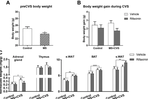
T-test revealed significant pre-CVS body weight difference between mice with postnatal maternal separation and controls [t = 2.69, df = 20, p = 0.014] (Fig. 4A). During chronic variable stress, weight gain between experimental groups was not significantly different [F (1,18) = 0.2027; p = 0.8884)] (Fig. 4B).
Normalized adrenal weight was higher in all MS + CVS exposed mice than in controls [F(1, 19) = 4.512; p = 0.047] (Fig. 4C). Two way ANOVA did not reveal drug (rifaximin) effect. Thymus weights were not different among the groups [F(1, 19) = 1.759; p = 0.2005].
Stress exposure resulted in significant increases of adipose tissue depots: normalized weights of subcutaneous- sWAT [F(1, 19) = 12.2;
p = 0.0024], epididymal- eWAT, white adipose tissues [F(1, 19) = 9.08; p = 0.0071] and interscapular brown adipose tissue, BAT [F(1, 19) = 37.71; p < 0.0001] were higher in MS + CVS mice than in controls.
3.3. Hormones and metabolic markers
During CVS procedure, basal (morning), pre-stress corticosterone levels were significantly higher in CVS exposed mice than in controls [F (1, 18) = 7.181; p = 0.0153]. Two way ANOVA indicated no drug effect [F(1, 18) = 0.1965; p = 0.6629] (Fig. 5A).
Analysis of plasma corticosterone concentrations after acute EPM exposure revealed no significant differences among the treatment groups [F(1, 19) = 0.226; p = 0.6399] (Fig. 5B).
Plasma glucose levels following acute stress were elevated in chronically stressed mice compared to controls [F(1, 13) = 22.44;
p = 0.0004] (Fig. 5C).
In addition, post hoc test revealed elevated plasma TG level in stressed mice [t = 2.627; DF = 18; p = 0.0339 compared to vehicle control], while there was no difference between the two, antibiotic treated groups (Fig. 5D).
3.4. Colon microbiome analysis
Antibiotic treatment reduced total DNA concentration in the colon content (Fig. 6A). Interestingly, colonic DNA concentration in MS + CVS + vehicle treatment group was more reduced than that of the antibiotic treated mice and it was significantly decreased compared to the control group [t = 2.558; DF = 17; p = 0.0403], suggesting that the abundance of microbiome in the colon is substantially reduced by stress treatment.
Colon microbiome diversity was analyzed at the phylum level and the results showed an increased abundance in the phylum of Bacteriodetes [F(1,17) = 5.844; p = 0.0272] and Proteobacteria in
response to stress, nevertheless rifaximin treatment significantly atte- nuated stress- inducedProteobacteriaabundance [t = 2.665; DF = 17;
p = 0.0324 compared to control, t = 2.581; DF = 17; p = 0.0385 compared to antibiotic treatment] (Fig. 6B). Changes in Firmicutes, Actinobacteria, VerrucomicrobiaandCyanobacteriawere not significant (Fig. 6B-C). Amount ofClostridiumsp. was determined at genus level.
Chronic stress resulted in increased abundance, however antibiotic treatment normalized this stress-induced elevation [t = 2.707;
DF = 17; p = 0.0297 compared to control, t = 3.233; DF = 17;
p = 0.0098 compared to vehicle] (Fig. 6D).
3.5. Colon mucosa, tight junction protein expression and gut permeability MS + CVS significantly reduced the thickness of mucosa compared to control [t = 3,082; DF = 19; p = 0.0122]. Mucosa thickness was not different in rifaximin-treated groups (Fig. 7A, B). Colonic mRNA ex- pression (Fig. 7C) of tight junction proteins, occludin [F(1, 19) = 11.27; p = 0.0033] and tight junction proteins TJP1 [F(1, 19) = 6.26; p = 0.0216] and TJP2 [F(1, 19) = 6.613; p = 0.0187] but not TJP3 was increased in both rifaximin treated groups, but remained unchanged in response to stress. In case of MUC2 mRNA there was no significant difference between the groups [F(1, 19) = 4.012;
p = 0.597]. Expression of Reg3b, a C-type lectin with antimicrobial activity, was significantly elevated in chronically stressed mice.
Rifaximin treatment restored Reg3b mRNA level to that of the controls [t = 3.811; DF = 19; p = 0.0024 compared to control, t = 4.398;
DF = 19; p = 0.0006 compared to vehicle, interaction: F(1, 19) = 10.21; p = 0.0048].
Next, we used FITC-labeled 4 kDa dextran assay to test gut perme- ability in vivo and found increased gut permeability in chronically stressed mice, while rifaximin treatment of these animals restored the normal, non-stressed values [t = 2.72; DF = 18; p = 0.0301 compared to control, t = 3.449; DF = 18; p = 0.0066 compared to vehicle, in- teraction: F(1, 18) = 6.109; p = 0.0251] (Fig. 7D).
3.6. Gut permeability, macrophage infiltration, local- and systemic bacterial load
Next, we checked whether increased gut permeability results in local piling of macrophages. In control, vehicle-treated mice im- munoreactivity corresponding to macrophage marker F4/80 was pri- marily confined to the submucosa. Essentially the same distribution was revealed in non-stressed, rifaximin-treated mice (Fig. 8A). However, in MS-CVS mice, an increase of F4/80 positive profiles and macrophage infiltration to the lamina propria was detected. Rifaximin treatment of MS-CVS animals restored the distribution of F4/80 positive macro- phages to that seen in control animals (Fig. 8A). Quantitative histolo- gical analysis of F4/80 + areas revealed significant rifaximin effect in Fig. 2.Behavioral activity of control and chronically stressed mice with or without rifaximin treatment in a novel environment (openfield arena). Each row represents one mouse (A). Frequency (B) and duration (C) of selected behavioral elements. Data were analyzed by two-way ANOVA (n = 9–14 per group).
Mean ± SEM values, **p < 0.01, ***p < 0.001, ****p < 0.0001 vs. control group.
the submucosa [F(1, 19) = 6.316; p = 0.0211]. In the lamina propria of MS-CVS animals, the area covered by F4/80 + profiles was sig- nificantly increased in stressed mice, which was reduced to that of the controls in response to rifaximin treatment [t = 14.69; DF = 19;
p < 0.0001 compared to control, t = 14.78; DF = 19; p < 0.0001 compared to vehicle, interaction: F(1, 19) = 105.4; p < 0.0001], (Fig. 8B).
Gut lymphatics are drained in mesenteric lymph nodes, which gate intestinal bacteria and pathogen-associated molecular patterns, PAMPs.
To assess bacterial translocation in chronically stressed mice, we PCR- amplified bacterial DNA from mesenteric lymph nodes using common 16S ribosomal primers. Compared to non-stressed controls, chronic stress resulted in 50% elevation of bacterial load in the MLN [t = 2.781; DF = 18; p = 0.0245]. Rifaximin administration interfered with stress-induced increase of bacterial load (Fig. 8C).
To reveal if chronic stress induces systemic endotoxemia, plasma LPS levels were measured. Higher LPS level was detected in plasma of stressed mice than in the controls [t = 2.61; DF = 18; p = 0.0351] but the increase of plasma LPS was not detected in antibiotic treated mice (Fig. 8D).
4. Discussion
In the present study we demonstrate that“two hits”chronic stress
affects colon microbiome, impairs gut barrier functions, resulting in leaky gut as demonstrated by macrophage invasion to colon mucosa and increased local- and systemic bacterial load. These changes are accompanied by signs of increased anxiety-like behavior and neophobia in stressed mice. Rifaximin, a non-absorbable, gut specific antibiotic, given to chronically stressed mice, alleviated stress-induced increase of Clostridia, improved gut permeability, mucosal erosion and inflamma- tion, however had no impact on the stress-induced behavior.
Somatic (adrenal enlargement)-, hormonal (elevated plasma CORT)- and metabolic (increased WAT depots, hyperglycemia and hyperlipi- demia) parameters validated the present MS-CVS model as chronic stress in mice (Zelena et al., 2005). Interestingly, stress-induced thymus involution was not detected, in spite of elevated basal CORT levels.
There is a well-documented mutual interaction between the gut microbiome and host stress response (Cryan and Dinan, 2012; Foster et al., 2017). Chronic stress is an important environmental factor, which triggers gut dysbiosis and in turn, altered microbiome has an impact on neuroendocrine- autonomic and behavioral aspects of the stress response. Growing evidence indicates a link between stress and microbial dysbiosis (Cryan and Dinan, 2012; De Palma et al., 2014;
Watanabe et al., 2016). Previous studies from different laboratories revealed distinct stress-induced changes of the gut microbiome com- position depending on the stressor (water avoidance (Xu et al., 2014;
Yoshikawa et al., 2017), repeated restraint (Maltz et al., 2018), social Fig. 3.Anxiety-like behavior and neophobia after MS + CVS. Openfield results: Velocity during openfield test (A), distance moved (B) center preference (C)first latency in border (D) and exploration maps of the experimental mice (E). Open arm preference in elevated plus maze test (F). Data were analyzed by two-way ANOVA (n = 9–14 per group). Mean ± SEM values, **p < 0.01, ****p < 0.0001 vs. control group. Sugar consumption of experimental mice before and after CVS procedure (G). Data were analyzed by repeated measures two-way ANOVA followed by Sidak’s multiple comparison test (n = 5–7 per group). Mean ± SEM values,
*p < 0.05, **p < 0.01 vs. control group.
defeat/disruption (Bailey et al., 2011; Langgartner et al., 2018;
Werbner et al., 2019) and origin of bacterial sampling (fecal (Gautam et al., 2018), luminal (Szyszkowicz et al., 2017) mucosal (Galley et al., 2014). General conclusion of these studies is that chronic psychological stress results in outgrowth ofBacteroidetesand decreases the abundance ofFirmicutes. Here we confirm and extend these observations to show that MS-CVS“two hits” paradigm is also capable to increaseBacter- oidetes and Proteobacteria in chronically stressed mice, which is ac- companied here with an overall decrease of total bacterial DNA con- centration in the colon content.
A number of studies have identified early life adversity as an im- portant factor affecting gut microbiome (Rincel et al., 2019). Studies in rats, mice and Rhesus monkeys revealed decrease of microbial diversity following maternal separation (Bailey and Coe, 1999) or limited nesting (Moussaoui et al., 2016). Here, we have used the“two hits”model of chronic adversity by combination of early life stress, maternal separa- tion and chronic variable stress paradigm, both of which might have an impact on gut microbiome. Future work should reveal lasting changes and sensitization of postnatally stressed microbiome to challenges in
adulthood.
Among the bacterial species studied here, the copy number of Clostridiaincreased significantly as a result of MS-CVS stress paradigm.
Thisfinding is in agreement with those of (Bailey et al., 2011; Pearson- Leary et al., 2019; Watanabe et al., 2016). Although someClostridiaare pathogenic and are responsible for diarrhea in the elderly, we did not observe serious diarrhea or watery feces in our stressed mice.
Rifaximin, which is a non-absorbable antibiotic, specifically targets Clostridiaand other Gram negative and positive bacteria. Therefore, we hypothesized that rifaximin treatment of mice exposed to MS-CVS will restore microbiome related gut-brain axis and behavioral changes to normal. Indeed, stress-inducedClostridiawere significantly attenuated by antibiotic treatment. In addition, rifaximin administration prevented stress-induced increases ofProteobacteria, but notBacteroidetes. These data confirm previousfindings that rifaximin do not significantly affect the overall composition of the human fecal microbiome (Soldi et al., 2015) and only mild changes are observed in mice (Jin et al., 2018).
More recently, rifaximin was recommended for treatment of post-in- fectious irritable bowel disease (IBS) and related abdominal discomfort Fig. 4.Effect of chronic stress and rifaximin treatment on body weight gain and organ weights. Body weight difference before CVS (at PND50) (A) and body weight gain during CVS (B). Normalized organ weights (C). Mean ± SEM. Data were analyzed by two-way ANOVA (n = 5–7 per group). In case of preCVS body weight unpairedt-test was used. *p < 0.05, **p < 0.01, ****p < 0.0001 vs. control groups.
Fig. 5.Effect of chronic stress on different plasma markers. Basal corticosterone level during CVS (A). Effect of single acute stress (EPM exposure) on corticosterone level after chronic stress (B), plasma glucose level (C), plasma triglyceride level (D). Data were analyzed by two-way ANOVA, followed by Sidak’s multiple com- parison test (n = 5–7 per group). Mean ± SEM values, *p < 0.05 vs. control group.
(Acosta et al., 2016) (Ponziani et al., 2016). In search for the me- chanism of rifaximin action, increased expression of gut tight junction proteins emerged (Jin et al., 2018). We have confirmed increased mRNA levels of tight junction proteins occludin, tjp1 and tjp2, but not tjp3, in colon samples of stressed and non-stressed, rifaximin treated mice. Increased expression of tight junction proteins along with slightly increased muc2 indicates improved gut barrier function after rifaximin administration.
“Leaky gut”is generally held as a hallmark of chronic stress-related pathologies (Camilleri, 2019; Maes et al., 2007). In the present MS-CVS model, we did not detect stress-induced changes in colonic expression of tight junction proteins (Occludin, TJP1-3) and muc2. However, we
didfind significant decrease of mucosa thickness and signs of epithelial damage. Furthermore, increased transcellular transport was detected in the gut of chronically stressed mice by FITC-dextran method, indicating impaired barrier function (Fukui, 2016). The leaky barrier should allow various feed/microbiome related antigens to initiate local immune re- sponses. Such an innate immune activation has been revealed by in- filtration of F4/80 positive macrophages into the lamina propria of MS- CVS exposed mice, which was prevented by rifaximin treatment during chronic variable stress. Gut lymphatics are drained in mesenteric lymph nodes (MLN), where a significant bacterial load has been revealed in our chronically stressed mice. Thisfinding is compatible with the report by (Velin et al., 2004) where 30 times increasedE colipassage was Fig. 6.Differences in the microbiome composition after chronic stress and rifaximin treatment. Total DNA concentration of colon content (A), copy numbers of bacterial phyla in the colon content (B,C). Copy number ofClostridiumsp. in different experimental groups (D). Data were analyzed by two-way ANOVA, followed by Sidak’s multiple comparison test (n = 5–7 per group). Mean ± SEM values, *p < 0.05 vs. control group, #p < 0.05, ##p < 0.01, vs. MS + CVS-vehicle.
Fig. 7.Effects of stress and rifaximin on colon mucosa. Representative images of hematoxylin-eosin stained colon sections (A) (scale bar: 20 µm) and bar graphs showing mean values ± SEM of mucosa thickness (B), mRNA levels of different permeability markers and antimicrobial defense-related genes from colon tissue (C), changes in gut permeability as measured by serum concentration of orally administered FITC-dextran (mean values ± SEM) (D). Data were analyzed by two-way ANOVA, followed by Sidak’s multiple comparison test (n = 5–7 per group). Mean ± SEM values, *p < 0.05, **p < 0.01, vs. non-stressed control group,
#p < 0.05, ##p < 0.01, ###p < 0.001, vs. corresponding vehicle-treated group.
detected in chronically stressed rats,ex vivo. In addition to this local bacterial translocation to MLN, significant elevation of LPS was de- tected in the plasma of MS-CVS mice, indicating systemic endotoxemia.
Rifaximin administration during CVS prevented the increase of LPS plasma levels, suggesting that rifaximin effects go beyond anti-patho- genic activity. The endotoxemia reducing effects of rifaximin has al- ready been shown in chronic liver disease (Bajaj et al., 2013), however the mechanisms of action and molecular targets remain unknown.
Another noteworthyfinding of this study is the stress-induced co- lonic upregulation of Reg3b. Reg3b is a C-type lectin, which belongs to antimicrobial peptide (AMP) family and involved in gut barrier func- tions (Shin and Seeley, 2019). Further studies are required to clarify if increased expression of Reg3b mRNA may represent an adaptive com- pensatory host mechanism to impaired barrier and increased bacterial burden.
Chronic stress exposure is a risk factor for psychiatric disorders, such as anxiety and depression, subsets of which are accompanied or driven by activated immune system (Slattery et al., 2012; Renault and Aubert, 2006). Parallel to stress-induced microbiome changes, chronic stress increases gut permeability and results in a leaky gut, through which bacterial cell wall components might reach the systemic circu- lation, trigger toll like receptors and result in low grade systemic in- flammation. LPS and related proinflammatory cytokines activate the HPA axis (Turnbull et al., 1998) and promote deterioration of stress- induced anxiety (Anisman et al., 2002).
In search for the causal role of microbiome in the development of stress-related psychopathologies, Langgartner et al. showed that stress- induced psychobiological changes can be transmitted by fecal trans- plantation from stressed to non-stressed mice (Langgartner et al., 2018).
In contrary, our present results suggest that settlement of stress-induced changes in the gut does not alleviate MS-CVS-induced locomotor hy- peractivity, anxiety-like symptoms in openfield and elevated plus maze tests and anhedonia. One explanation for the resistance of stress-in- duced behavior to improvement of gut and immune functions would be the timing of rifaximin treatment. It is likely that behavior needs more time than gut to recover from CVS. Another possibility would be that early life adversity i.e. maternal separation results in permanent
changes in the gutflora (Rincel et al., 2019) and behavior, which does not change after antibiotic treatment in adulthood. Indeed, it has been shown that early life events result in epigenetic modulation of stress/
anxiety-related genes such as hypomethylation of theCrhpromoter in the PVN of maternally deprived adult rats (Chen et al., 2012) and re- duction in histone H3 acetylation at human/rat glucocorticoid receptor (GR, NR3C1) promoter in the hippocampus (Suderman et al., 2012).
Finally, rifaximin reduced pathogenic bacteria and improved gut barrier, however, did not enforce the abundance of beneficial“psy- chobiotic”bacteria. It has been shown that administration of certain Lactobacilli and Bifidobacteria have anxiolytic/antidepressive effects (Borre et al., 2014; Bravo et al., 2011; Dinan et al., 2013).
Recent studies implicate that antibiotic usage in human and farm animals results in dysbiosis, provoke systemic inflammation, which might be responsible for long-term metabolic- (obesity), behavioral- and mental changes. However, we did not detect any significant be- havioral changes in unstressed mice treated with antibiotic for 3 weeks.
Future work should focus on the interaction between systemic anti- biotics and stress in regulation of microbiota-gut-brain axis.
In conclusion, combination of early life adversity with adult chronic variable stress (CVS) paradigm in mice results in gut dysbiosis and impaired gut barrier function along with increased locomotor activity, anxiety-like behavior and neophobia. Rifaximin treatment during CVS decreases stress-induced pathogenic bacteria, restores gut barrier functions, reduces local and systemic bacterial load, however, does not improve stress-induced behavioral changes.
Acknowledgements
The study was supported by grants from the Hungarian National Research, Development and Innovation Office 124424, and National Brain Research Program 2017-1.2.1-NKP-2017-00002 for KJK.
Publication was supported by EFOP-3.6.3-VEKOP-16-2017-00009 pro- ject. KD was supported by young investigator award from G.Richter Plc.
Fig.8.Macrophage infiltration to lamina propria, local- and systemic bacterial load. Representative images of F4/80 immunostained colon sections showing distinct macrophage distribution (green-F4/80) in stressed and rifaximin-treated animals (on blue-DAPI background). (Scale bar: 100 µm) (A). Quantitative analysis of F4/80 immunostaining in the submucosa and lamina propria (B). Bacterial load in the mesenteric lymph node expressed as % of control-vehicle group (C). Plasma LPS concentration in control, MS + CVS groups, with or without rifaximin treatment (D). Data were analyzed by two-way ANOVA, followed by Sidak’s multiple comparison test (n = 5–7 per group). Mean ± SEM values, *p < 0.05, **p < 0.01, vs. control group, #p < 0.05, ##p < 0.01, ###p < 0.001, vs.
corresponding vehicle group. (For interpretation of the references to color in thisfigure legend, the reader is referred to the web version of this article.)
Appendix A. Supplementary data
Supplementary data to this article can be found online athttps://
doi.org/10.1016/j.bbi.2019.12.004.
References
Acosta, A., Camilleri, M., Shin, A., Linker Nord, S., O'Neill, J., Gray, A.V., Lueke, A.J., Donato, L.J., Burton, D.D., Szarka, L.A., et al., 2016. Effects of rifaximin on transit, permeability, fecal microbiome, and organic acid excretion in irritable bowel syn- drome. Clin. Transl. Gastroenterol. 7, e173.
Andersen, C.L., Jensen, J.L., Orntoft, T.F., 2004. Normalization of real-time quantitative reverse transcription-PCR data: a model-based variance estimation approach to identify genes suited for normalization, applied to bladder and colon cancer data sets.
Cancer Res. 64, 5245–5250.
Anisman, H., Kokkinidis, L., Merali, Z., 2002. Further evidence for the depressive effects of cytokines: anhedonia and neurochemical changes. Brain Behav. Immun. 16, 544–556.
Bailey, M.T., Coe, C.L., 1999. Maternal separation disrupts the integrity of the intestinal microflora in infant rhesus monkeys. Dev. Psychobiol. 35, 146–155.
Bailey, M.T., Dowd, S.E., Galley, J.D., Hufnagle, A.R., Allen, R.G., Lyte, M., 2011.
Exposure to a social stressor alters the structure of the intestinal microbiota: im- plications for stressor-induced immunomodulation. Brain Behav. Immun. 25, 397–407.
Bajaj, J.S., Heuman, D.M., Sanyal, A.J., Hylemon, P.B., Sterling, R.K., Stravitz, R.T., Fuchs, M., Ridlon, J.M., Daita, K., Monteith, P., et al., 2013. Modulation of the me- tabiome by rifaximin in patients with cirrhosis and minimal hepatic encephalopathy.
PLoS One 8, e60042.
Bharwani, A., Mian, M.F., Foster, J.A., Surette, M.G., Bienenstock, J., Forsythe, P., 2016.
Structural & functional consequences of chronic psychosocial stress on the micro- biome & host. Psychoneuroendocrinology 63, 217–227.
Borre, Y.E., Moloney, R.D., Clarke, G., Dinan, T.G., Cryan, J.F., 2014. The impact of microbiota on brain and behavior: mechanisms & therapeutic potential. Adv. Exp.
Med. Biol. 817, 373–403.
Bravo, J.A., Forsythe, P., Chew, M.V., Escaravage, E., Savignac, H.M., Dinan, T.G., Bienenstock, J., Cryan, J.F., 2011. Ingestion of Lactobacillus strain regulates emo- tional behavior and central GABA receptor expression in a mouse via the vagus nerve.
Proc. Natl. Acad. Sci. U.S.A. 108, 16050–16055.
Bravo, J.A., Julio-Pieper, M., Forsythe, P., Kunze, W., Dinan, T.G., Bienenstock, J., Cryan, J.F., 2012. Communication between gastrointestinal bacteria and the nervous system.
Curr. Opin. Pharmacol. 12, 667–672.
Camilleri, M., 2019. Leaky gut: mechanisms, measurement and clinical implications in humans. Gut.
Cerniauskas, I., Winterer, J., de Jong, J.W., Lukacsovich, D., Yang, H., Khan, F., Peck, J.R., Obayashi, S.K., Lilascharoen, V., Lim, B.K., et al., 2019. Chronic stress induces activity, synaptic, and transcriptional remodeling of the lateral habenula associated with deficits in motivated behaviors. Neuron.
Chen, J., Evans, A.N., Liu, Y., Honda, M., Saavedra, J.M., Aguilera, G., 2012. Maternal deprivation in rats is associated with corticotrophin-releasing hormone (CRH) pro- moter hypomethylation and enhances CRH transcriptional responses to stress in adulthood. J. Neuroendocrinol. 24, 1055–1064.
Cryan, J.F., Dinan, T.G., 2012. Mind-altering microorganisms: the impact of the gut mi- crobiota on brain and behaviour. Nat. Rev. Neurosci. 13, 701–712.
Cryan, J.F., O'Mahony, S.M., 2011. The microbiome-gut-brain axis: from bowel to be- havior. Neurogastroenterol. Motil. 23, 187–192.
De Palma, G., Collins, S.M., Bercik, P., Verdu, E.F., 2014. The microbiota-gut-brain axis in gastrointestinal disorders: stressed bugs, stressed brain or both? J. Physiol. 592, 2989–2997.
Dinan, T.G., Stanton, C., Cryan, J.F., 2013. Psychobiotics: a novel class of psychotropic.
Biol. Psychiatry 74, 720–726.
Foster, J.A., Rinaman, L., Cryan, J.F., 2017. Stress & the gut-brain axis: Regulation by the microbiome. Neurobio.l Stress 7, 124–136.
Fukui, H., 2016. Endotoxin and other microbial translocation markers in the blood: a clue to understand leaky gut syndrome. Cell. Mol. Med. 2 (3:14), 1–14.
Galley, J.D., Yu, Z., Kumar, P., Dowd, S.E., Lyte, M., Bailey, M.T., 2014. The structures of the colonic mucosa-associated and luminal microbial communities are distinct and differentially affected by a prolonged murine stressor. Gut Microbes 5, 748–760.
Gautam, A., Kumar, R., Chakraborty, N., Muhie, S., Hoke, A., Hammamieh, R., Jett, M., 2018. Altered fecal microbiota composition in all male aggressor-exposed rodent model simulating features of post-traumatic stress disorder. J. Neurosci. Res. 96, 1311–1323.
Golubeva, A.V., Crampton, S., Desbonnet, L., Edge, D., O'Sullivan, O., Lomasney, K.W., Zhdanov, A.V., Crispie, F., Moloney, R.D., Borre, Y.E., et al., 2015. Prenatal stress- induced alterations in major physiological systems correlate with gut microbiota composition in adulthood. Psychoneuroendocrinology 60, 58–74.
Herman, J.P., Adams, D., Prewitt, C., 1995. Regulatory changes in neuroendocrine stress- integrative circuitry produced by a variable stress paradigm. Neuroendocrinology 61, 180–190.
Jin, Y., Ren, X., Li, G., Li, Y., Zhang, L., Wang, H., Qian, W., Hou, X., 2018. Beneficial effects of Rifaximin in post-infectious irritable bowel syndrome mouse model beyond gut microbiota. J. Gastroenterol. Hepatol. 33, 443–452.
Kane, J.S., Ford, A.C., 2016. Rifaximin for the treatment of diarrhea-predominant irritable bowel syndrome. Expert. Rev. Gastroenterol. Hepatol. 10, 431–442.
Kelly, J.R., Kennedy, P.J., Cryan, J.F., Dinan, T.G., Clarke, G., Hyland, N.P., 2015.
Breaking down the barriers: the gut microbiome, intestinal permeability and stress- related psychiatric disorders. Front. Cell. Neurosci. 9, 392.
Langgartner, D., Vaihinger, C.A., Haffner-Luntzer, M., Kunze, J.F., Weiss, A.J., Foertsch, S., Bergdolt, S., Ignatius, A., Reber, S.O., 2018. The role of the intestinal microbiome in chronic psychosocial stress-induced pathologies in male mice. Front. Behav.
Neurosci. 12, 252.
Leclercq, S., Cani, P.D., Neyrinck, A.M., Starkel, P., Jamar, F., Mikolajczak, M., Delzenne, N.M., de Timary, P., 2012. Role of intestinal permeability and inflammation in the biological and behavioral control of alcohol-dependent subjects. Brain Behav.
Immun. 26, 911–918.
Maes, M., Coucke, F., Leunis, J.C., 2007. Normalization of the increased translocation of endotoxin from gram negative enterobacteria (leaky gut) is accompanied by a re- mission of chronic fatigue syndrome. Neuro. Endocrinol. Lett. 28, 739–744.
Maltz, R.M., Keirsey, J., Kim, S.C., Mackos, A.R., Gharaibeh, R.Z., Moore, C.C., Xu, J., Bakthavatchalu, V., Somogyi, A., Bailey, M.T., 2018. Prolonged restraint stressor exposure in outbred CD-1 mice impacts microbiota, colonic inflammation, and short chain fatty acids. PLoS One 13, e0196961.
Mayer, E.A., Knight, R., Mazmanian, S.K., Cryan, J.F., Tillisch, K., 2014. Gut microbes and the brain: paradigm shift in neuroscience. J. Neurosci. 34, 15490–15496.
McCormick, C.M., Green, M.R., 2013. From the stressed adolescent to the anxious and depressed adult: investigations in rodent models. Neuroscience 249, 242–257.
Moloney, R.D., Johnson, A.C., O'Mahony, S.M., Dinan, T.G., Greenwood-Van Meerveld, B., Cryan, J.F., 2016. Stress and the microbiota-gut-brain axis in visceral pain: re- levance to irritable bowel syndrome. CNS Neurosci. Ther. 22, 102–117.
Moussaoui, N., Larauche, M., Biraud, M., Molet, J., Million, M., Mayer, E., Tache, Y., 2016. Limited nesting stress alters maternal behavior and in vivo intestinal perme- ability in male wistar pup rats. PLoS One 11, e0155037.
Pearson-Leary, J., Zhao, C., Bittinger, K., Eacret, D., Luz, S., Vigderman, A.S., Dayanim, G., Bhatnagar, S., 2019. The gut microbiome regulates the increases in depressive- type behaviors and in inflammatory processes in the ventral hippocampus of stress vulnerable rats. Mol. Psychiatry.
Ponziani, F.R., Scaldaferri, F., Petito, V., Paroni Sterbini, F., Pecere, S., Lopetuso, L.R., Palladini, A., Gerardi, V., Masucci, L., Pompili, M., et al., 2016. The role of antibiotics in gut microbiota modulation: the eubiotic effects of rifaximin. Dig Dis. 34, 269–278.
Renault, J., Aubert, A., 2006. Immunity and emotions: lipopolysaccharide increases de- fensive behaviours and potentiates despair in mice. Brain Behav. Immun. 20, 517–526.
Rieder, R., Wisniewski, P.J., Alderman, B.L., Campbell, S.C., 2017. Microbes and mental health: a review. Brain Behav. Immun.
Rincel, M., Aubert, P., Chevalier, J., Grohard, P.A., Basso, L., Monchaux de Oliveira, C., Helbling, J.C., Levy, E., Chevalier, G., Leboyer, M., et al., 2019. Multi-hit early life adversity affects gut microbiota, brain and behavior in a sex-dependent manner.
Brain Behav. Immun.
Rubin, D.T., Sohi, S., Glathar, M., Thomas, T., Yadron, N., Surma, B.L., 2011. Rifaximin Is Effective for the Treatment of Clostridium difficile-Associated Diarrhea: Results of an Open-Label Pilot Study. Gastroenterol. Res. Pract. 2011, 106978.
Sandi, C., 2004. Stress, cognitive impairment and cell adhesion molecules. Nat. Rev.
Neurosci. 5, 917–930.
Scott, K.A., Ida, M., Peterson, V.L., Prenderville, J.A., Moloney, G.M., Izumo, T., Murphy, K., Murphy, A., Ross, R.P., Stanton, C., et al., 2017. Revisiting Metchnikoff: Age- related alterations in microbiota-gut-brain axis in the mouse. Brain Behav. Immun.
65, 20–32.
Shin, J.H., Seeley, R.J., 2019. Reg3 Proteins as Gut Hormones? Endocrinology 160, 1506–1514.
Slattery, D.A., Uschold, N., Magoni, M., Bar, J., Popoli, M., Neumann, I.D., Reber, S.O., 2012. Behavioural consequences of two chronic psychosocial stress paradigms: an- xiety without depression. Psychoneuroendocrinology 37, 702–714.
Slyepchenko, A., Maes, M., Machado-Vieira, R., Anderson, G., Solmi, M., Sanz, Y., Berk, M., Kohler, C.A., Carvalho, A.F., 2016. Intestinal dysbiosis, gut hyperpermeability and bacterial translocation: missing links between depression, obesity and type 2 diabetes. Curr. Pharm. Des. 22, 6087–6106.
Soldi, S., Vasileiadis, S., Uggeri, F., Campanale, M., Morelli, L., Fogli, M.V., Calanni, F., Grimaldi, M., Gasbarrini, A., 2015. Modulation of the gut microbiota composition by rifaximin in non-constipated irritable bowel syndrome patients: a molecular ap- proach. Clin. Exp. Gastroenterol. 8, 309–325.
Strekalova, T., Spanagel, R., Bartsch, D., Henn, F.A., Gass, P., 2004. Stress-induced an- hedonia in mice is associated with deficits in forced swimming and exploration.
Neuropsychopharmacology 29, 2007–2017.
Suderman, M., McGowan, P.O., Sasaki, A., Huang, T.C., Hallett, M.T., Meaney, M.J., Turecki, G., Szyf, M., 2012. Conserved epigenetic sensitivity to early life experience in the rat and human hippocampus. Proc. Natl. Acad. Sci. U.S.A. 109 (Suppl 2), 17266–17272.
Szyszkowicz, J.K., Wong, A., Anisman, H., Merali, Z., Audet, M.C., 2017. Implications of the gut microbiota in vulnerability to the social avoidance effects of chronic social defeat in male mice. Brain Behav. Immun. 66, 45–55.
Turnbull, A.V., Lee, S., Rivier, C., 1998. Mechanisms of hypothalamic-pituitary-adrenal axis stimulation by immune signals in the adult rat. Ann. N.Y. Acad. Sci. 840, 434–443.
Velin, A.K., Ericson, A.C., Braaf, Y., Wallon, C., Soderholm, J.D., 2004. Increased antigen and bacterial uptake in follicle associated epithelium induced by chronic psycholo- gical stress in rats. Gut 53, 494–500.
Warda, A.K., Rea, K., Fitzgerald, P., Hueston, C., Gonzalez-Tortuero, E., Dinan, T.G., Hill, C., 2019. Heat-killed lactobacilli alter both microbiota composition and behaviour.
Behav. Brain Res. 362, 213–223.
Watanabe, Y., Arase, S., Nagaoka, N., Kawai, M., Matsumoto, S., 2016. Chronic Psychological Stress Disrupted the Composition of the Murine Colonic Microbiota and
Accelerated a Murine Model of Inflammatory Bowel Disease. PLoS One 11, e0150559.
Werbner, M., Barsheshet, Y., Werbner, N., Zigdon, M., Averbuch, I., Ziv, O., Brant, B., Elliott, E., Gelberg, S., Titelbaum, M., et al. (2019). Social-Stress-Responsive Microbiota Induces Stimulation of Self-Reactive Effector T Helper Cells. mSystems 4.
Winkler, Z., Kuti, D., Ferenczi, S., Gulyas, K., Polyak, A., Kovacs, K.J., 2017. Impaired microglia fractalkine signaling affects stress reaction and coping style in mice. Behav.
Brain Res. 334, 119–128.
Xu, D., Gao, J., Gillilland 3rd, M., Wu, X., Song, I., Kao, J.Y., Owyang, C., 2014. Rifaximin alters intestinal bacteria and prevents stress-induced gut inflammation and visceral hyperalgesia in rats. Gastroenterology 146 (484–496), e484.
Yoshikawa, K., Kurihara, C., Furuhashi, H., Takajo, T., Maruta, K., Yasutake, Y., Sato, H.,
Narimatsu, K., Okada, Y., Higashiyama, M., et al., 2017. Psychological stress ex- acerbates NSAID-induced small bowel injury by inducing changes in intestinal mi- crobiota and permeability via glucocorticoid receptor signaling. J. Gastroenterol. 52, 61–71.
Zelena, D., Barna, I., Mlynarik, M., Gupta, O.P., Jezova, D., Makara, G.B., 2005. Stress symptoms induced by repeated morphine withdrawal in comparison to other chronic stress models in mice. Neuroendocrinology 81, 205–215.
Zelena, D., Mergl, Z., Foldes, A., Kovacs, K.J., Toth, Z., Makara, G.B., 2003. Role of hy- pothalamic inputs in maintaining pituitary-adrenal responsiveness in repeated re- straint. American journal of physiology. Endocrinol. Metabolism 285, E1110–E1117.