490
I
nflammation and wound healing are determinants of disease progression and clinical outcome in atherosclerosis. They are emerging as interrelated processes with overlapping molec- ular mechanisms controlling monocyte infiltration and differ- entiation into macrophages, whose phenotype determines the stability of lesions, by controlling the balance between matrix degradation and inflammation versus matrix deposition and resolution of inflammation and wound healing.1Clinical Perspective on p 501
Monocyte/macrophage intravasation is an essential step for metabolic disease pathogenesis including athero- sclerosis. The ability of monocytes to roll and adhere to the endothelium in response to chemokines is crucial for macrophage accumulation.2 It relies on actin-dependent morphological polarization, formation of filopodia and Background—Leukocyte migration is critical for the infiltration of monocytes and accumulation of monocyte-derived macrophages in inflammation. Considering that Hck and Fgr are instrumental in this process, their impact on atherosclerosis and on lesion inflammation and stability was evaluated.
Methods and Results—Hematopoietic Hck/Fgr-deficient, LDLr−/− chimeras, obtained by bone marrow transplantation, had smaller but, paradoxically, less stable lesions with reduced macrophage content, overt cap thinning, and necrotic core expansion as the most prominent features. Despite a Ly6Chigh-skewed proinflammatory monocyte phenotype, Hck/Fgr deficiency led to disrupted adhesion of myeloid cells to and transmigration across endothelial monolayers in vitro and atherosclerotic plaques in vivo, as assessed by intravital microscopy, flow cytometry, and histological examination of atherosclerotic arteries. Moreover, Hck/Fgr-deficient macrophages showed blunted podosome formation and mesenchymal migration capacity. In consequence, transmigrated double-knockout macrophages were seen to accumulate in the fibrous cap, potentially promoting its focal erosion, as observed for double-knockout chimeras.
Conclusions—The hematopoietic deficiency of Hck and Fgr led to attenuated atherosclerotic plaque formation by abrogating endothelial adhesion and transmigration; paradoxically, it also promoted plaque instability by causing monocyte subset imbalance and subendothelial accumulation, raising a note of caution regarding src kinase–targeted intervention in plaque inflammation. (Circulation. 2015;132:490-501. DOI: 10.1161/CIRCULATIONAHA.114.012316.)
Key Words: immunology ◼ atherosclerosis ◼ cell migration assays ◼ leukocytes ◼ mobility
◼ phosphotransferases ◼ plaque, atherosclerotic
© 2015 American Heart Association, Inc.
Circulation is available at http://circ.ahajournals.org DOI: 10.1161/CIRCULATIONAHA.114.012316
Received October 1, 2012; accepted June 4, 2015.
From Experimental Vascular Pathology Group, Department of Pathology, CARIM, Maastricht University Medical Center, The Netherlands (I.M., J.S., I.W., V.H., M.G., J.C., E.A.L.B.); Division of Biopharmaceutics, Leiden Academic Center for Drug Research, Leiden University, The Netherlands (I.M., I.B., S.C.A.d.J., T.J.C.v.B.); CNRS, IPBS (Institut de Pharmacologie et de Biologie Structurale), Toulouse, France (C.C., I.M.-P.); Université de Toulouse, France (C.C., I.M.-P.); Institute for Prevention of Cardiovascular Prevention (IPEK), LMU Munich, Germany (M.D., R.R.K., Y.D., C.W., O.S.); Instituto de la Grasa, CSIC, Seville, Spain (B.B.); Department of Vascular Surgery, Orbis Hospital Sittard, The Netherlands (K.-J.S.); Department of Physiology;
Semmelweis University, Budapest, Hungary (A.M.); Department of Pathology, Academic Medical Center (AMC), Amsterdam, The Netherlands (O.S.);
and German Centre for Cardiovascular Research (DZHK), Munich Heart Alliance, Germany (O.S.).
*Drs Cougoule and Drechsler contributed equally.
†Drs Maridonneau-Parini and Soehnlein contributed equally.
The online-only Data Supplement is available with this article at http://circ.ahajournals.org/lookup/suppl/doi:10.1161/CIRCULATIONAHA.
114.012316/-/DC1.
Correspondence to Erik A.L. Biessen, PhD, Maastricht University Medical Center, P. Debyelaan 25, 6229 HX Maastricht, The Netherlands. E-mail Erik.Biessen@maastrichtuniversity.nl
Hck/Fgr Kinase Deficiency Reduces Plaque Growth and Stability by Blunting Monocyte Recruitment
and Intraplaque Motility
Indira Medina, PhD; Céline Cougoule, PhD*; Maik Drechsler, PhD*; Beatriz Bermudez, PhD;
Rory R. Koenen, PhD; Judith Sluimer, PhD; Ine Wolfs, PhD; Yvonne Döring, PhD;
Veronica Herias, PhD; Marjon Gijbels, PhD; Ilze Bot, PhD; Saskia C. A. de Jager, PhD;
Christian Weber, MD; Jack Cleutjens, PhD; Theo J.C. van Berkel, PhD;
Kees-Jan Sikkink, PhD, MD; Atilla Mócsai, PhD; Isabelle Maridonneau-Parini, PhD†;
Oliver Soehnlein, PhD, MD†; Erik A.L. Biessen, PhD
Downloaded from http://ahajournals.org by on February 28, 2019
lamellipodia, binding of integrins to endothelial adhe- sion molecules, cytoskeletal reorganization,3 and signal transduction pathways ultimately leading to the concerted loosening of adherent junctions on endothelial cells4 and monocyte transmigration across endothelial, basement membrane, and fibrous cap barriers, before their homing in expanding lesions and differentiation into pro- or antifi- brotic macrophages.
Hck and Fgr are 2 Src tyrosine kinases that display restricted coexpression in myeloid cells where they regulate β2-integrin binding to endothelial intercellular adhesion molecule-1 to facilitate cell adhesion and migration on P-selectin glycopro- tein ligand-1 (PSGL-1) and CD44 interaction with endothelial E-selectin and P-selectin.5,6 In addition, Hck and Fgr are a con- vergence point of signaling pathways initiated by a wide range of cell receptors implicated in the pathogenesis of atheroscle- rosis, including integrins, immune and growth factors, Fc-γ receptors (Fcγ) and chemokine receptors. These kinases exert their functions by the activation of several effectors including Rac/CDC42, spleen tyrosine kinase (Syk), and protein tyrosine kinase (PyK),7 which are implicated in the accumulation and trapping of macrophages in atherosclerosis.8–11 As expected from signaling molecules targeted by multiple receptors, Hck and Fgr mediate a broad spectrum of processes, ranging from cell proliferation, survival, and differentiation, to cytokine secretion, cytoskeleton dynamics, integrin-dependent cell adhesion, to the endothelium and migration.7,12–15
In light of these data, we hypothesized that Hck/Fgr defi- ciency would lead to reduced accumulation of macrophages in atherosclerosis onset and progression, a consequence of reduced diapedesis and migration. Our data imply that Hck and Fgr not only are progressively overexpressed in athero- sclerosis, but they also control critical molecular processes in monocyte influx, blood monocyte subset balance, macrophage accumulation, and the maintenance of atherosclerotic lesion stability.
Materials and Methods
Animal ExperimentsBone marrow transplantation, perivascular collar placement, and intravital microscopy experiments were approved by the local regula- tory authorities of Leiden and Maastricht, and performed in accor- dance with Dutch, French, and German government guidelines as described in the online-only Data Supplement Methods.
Cholesterol and Triglyceride Levels
Blood samples were taken by tail bleeding 1 day before and 5 weeks after the introduction of Western type diet (WTD) and at euthani- zation. Total plasma cholesterol, triglyceride, and phospholipid con- tents were measured by an enzymatic-colorimetric assay (Roche Diagnostics, Almere, The Netherlands).
Plasma Cytokine Levels
The Luminex 100 Bio-Plex cytokine assay (Bio-Rad Laboratories, Inc; Hercules, CA) was used to determine plasma levels of: inter- leukin (IL)-1α, IL-1β, IL-2, IL-4, IL-5, IL-6, IL-10, IL12(P40), IL-12(P70), IL-17, eotaxin, keratinocyte chemoattractant, monocyte chemoattractant protein-1, monocyte inflammatory protein-1α, and tumor necrosis factor-α (TNF-α). Statistical analysis was performed for the cytokines that reached the limit of detection (IL-1β, IL-12, eotaxin, monocyte chemoattractant protein-1, and IL-1α).
Blood Cell Analysis and Flow Cytometry
Blood, bone marrow, and peritoneal cells were harvested at euthaniza- tion, and single-cell suspensions were prepared. Lysis of erythrocytes was performed in ice-cold NH4Cl (8.4 g/L), NaHCO3 (1 g/L), and ethylenediaminetetraacetic acid (37 mg/L) during 3 minutes. Single- cell suspensions were stained with fluorescent label–conjugated anti- bodies for different markers and analyzed by fluorescence-activated cell sorting as detailed in the online-only Data Supplement Methods.
Whole-blood samples were analyzed on a Sysmex blood cell analyzer (XT-2000i, Sysmex Europe GmbH, Norderstedt, Germany).
Cell Culture
Bone marrow–derived macrophages (BMDM) and peritoneal mac- rophages, vascular smooth muscle cells (SMC), human aortic endo- thelial cells, and Jurkat lymphocytes were cultured as detailed in the online-only Data Supplement Methods.
Thioglycolate-Induced Peritonitis
Cells were collected for analysis by fluorescence-activated cell sort- ing and microscopic quantification by Giemsa staining 1, 3, or 5 days (as indicated) after the induction of peritonitis with a sterile solution of dehydrated Brewer complete thioglycolate broth (1 mL, 2%–3%
wt/vol, Difco Laboratories, West Molesy, UK).
Phagocytosis, Apoptosis, and Proliferation Assays
Proliferation and apoptosis assays, phagocytosis of apoptotic cells and zymosan particles, and cholesterol uptake experiments are detailed in the online-only Data Supplement Methods.
Macrophage Adhesion and Transmigration Across the Endothelium
Human aortic endothelial cells (PromoCell) were grown and prein- cubated with TNF-α (10 ng/mL) for at least 4 hours. Hck and Fgr mutant BMDM or wild-type (WT) controls were suspended at 5×10⁵ cells/mL in 1× Hanks buffer, 20 mmol/L 4-(2-hydroxyethyl)-1-piper- azineethanesulfonic acid, 0.5% human serum albumin (Baxter), and 1 mmol/L calcium and magnesium after stimulation with interferon-γ (IFN-γ; 100 U/mL Peprotech) during 16 hours. For the assessment of cell adhesion, macrophages were perfused over inflamed human aortic endothelial cell monolayers during 2 minutes at 0.1 mL/min and cells counted in 6 high-power field (100× magnification) pic- tures. Transmigration was recorded at a flow rate of 0.05 mL/min for 30 minutes in 15-second intervals by using a differential interference contrast microscope.
Macrophage Morphology, Migration, Podosome Rosette Formation, and Matrix Degradation
Assessment of macrophage morphology and 2- and 3-dimensional migration are detailed in the Methods in the online-only Data Supplement.
Gelatin Zymography and β-Hexosaminidase Release
Zymography experiments were performed as previously described.13 In brief, BMDM (1×10⁶ cells/well) were seeded overnight into 6-well fibronectin-coated plates. Conditioned cell culture medium and cell lysate were subjected to 10% (wt/vol) sodium dodecyl sulfate, 0.1 mg/mL gelatin gel electrophoresis. For β-hexosaminidase release, BMDM were seeded overnight into 6-well plates, the assay was per- formed on cell extracts obtained in 1% Triton X-100 and supernatants as previously described.15
Classical and Alternative Macrophage Polarization
BMDM (5×10⁵ cells/well) were seeded in 24-well plates and allowed to adhere overnight before immune polarization was induced by
Downloaded from http://ahajournals.org by on February 28, 2019
24-hour incubation with 100 U/mL IFN-γ (Peprotech) or 20 ng/mL IL-4 (Peprotech). RNA isolation, cDNA synthesis, and real-time polymerase chain reaction were performed as detailed in the online- only Data Supplement Methods.
SMC Collagen Synthesis and Proliferation
Cell proliferation, collagen, and noncollagenous protein extracellular deposition were assessed in vascular SMC layers by enzyme-linked immunosorbent assay (Roche, BrdU colorimetric kit) and a quantita- tive collagen and protein microassay kit (Chondrex, Inc, Redmond, WA), respectively, according to the manufacturers’ instructions.
Tissue Harvesting, Immunohistochemistry, and Plaque Morphometry
Mice were anesthetized, euthanized, and perfused before the collec- tion of hearts, aortas, common carotid arteries, peritoneal ascites, and other organs as described in the online-only Data Supplement Methods, which also contains a detailed description of cell and tissue staining and visualization procedures.
Analysis of Microarray Data
For microarray analysis, total RNA was extracted by using the guani- dine thiocyanate/CsCl gradient method17 and a NucleoSpin RNA II kit (Macherey Nagel, Duren, Germany), from early (n=13) and advanced stable (n=16) lesions obtained after autopsy (Department of Pathology, University Hospital Maastricht, Maastricht, the Netherlands) or advanced stable (n=21) and advanced unstable (n=23) lesions obtained on surgery (Department of Surgery, Maasland Hospital Sittard, Sittard, the Netherlands). RNA concentration and quality and lesion phenotype were determined as detailed in the online-only Data Supplement Methods. All human work was approved by the Ethics Committee of the University Hospital Maastricht. Written informed consent for participation in the study was obtained from all individu- als. Samples from autopsy were individually hybridized to HGU133 2.0 Plus arrays (Affymetrix, Santa Clara, CA), and samples from sur- gery were individually hybridized to Illumina Human Sentrix-8 V2.0 BeadChip (Illumina Inc, San Diego, CA).
Microarray expression data of macrophage immune polarization were obtained at the Gene Expression Omnibus Web site (www.ncbi.
nlm.nih.gov/geo) under accession number GSE18686.18 Data normaliza- tion and summarization along with statistical, cluster, and Gene Ontology analysis are described in the online-only Data Supplement Methods.
Statistical Analysis of Experimental Data
Analyses were done using MatLab’s Statistics ToolBox (Ver7.9) or INSTAT (Graphpad Software, Inc). Two-group comparisons were analyzed by the Welch Student t test to account for unequal variances (except for higher-powered data sets [n>8] with equivalent variance, where we opted for an unpaired t test). Two-sided P values of <0.05 were considered significant and denoted with 1, 2, or 3 asterisks when lower than 0.05, 0.01, or 0.001, respectively. Comparisons that did not reach significance were not highlighted by an asterisk.
Figure data are presented as mean±standard error of the mean (unless otherwise stated), whereas data in Results are given as relative change in comparison with the WT control. Regression lines were compared by analysis of covariance, using the independent variable plaque area as covariate and macrophage content or necrotic core size as outcome in a 2-group analysis of covariance. Linear regression slopes were plotted with 95% confidence intervals. Multiple com- parison analyses were analyzed by 1-way analysis of variance with Bonferroni correction at a significance threshold of 0.05.
Results
Hematopoietic Deficiency in Hck and Fgr Reduces Atherogenesis
A first indication of the participation of the src kinases Hck and Fgr in atherosclerotic lesion progression was provided by
their upregulation in advanced human atherosclerotic lesions in comparison with early ones (GSE28829; Figure IA in the online-only Data Supplement), whereas the expression of both kinases was also significantly increased in human ath- erosclerotic vulnerable lesions in comparison with stable ones (Figure IB in the online-only Data Supplement), linking them to lesion progression.
To establish active involvement of Hck and Fgr in athero- sclerosis, we generated atherosclerosis-prone chimeric mice by reconstitution of lethally irradiated LDLr−/− recipient ani- mals with Hck−/−Fgr−/− double-knockout (dKO) or WT bone marrow cells. Hck/Fgr deletion did not lead to any alteration in total body weight along the experiment nor did it affect plasma total cholesterol levels before (199.4 mg/dL versus 150.8 mg/dL for WT controls) and after (1616.1 mg/dL versus 1488.9 mg/dL, for WT controls) WTD introduction. Plasma levels of proinflammatory cytokines such as IL-1β, IL-12, eotaxin, monocyte chemoattractant protein-1, and IL−1α, as measured at euthanization, were not influenced by Hck/Fgr deficiency (Figure IC in the online-only Data Supplement).
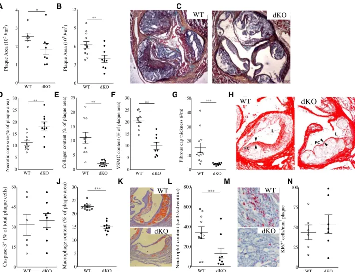
WTD-fed chimeric mice transplanted with Hck/Fgr dKO bone marrow exhibited 30% (P≤0.05) reduction in intermedi- ate atherosclerotic lesion size (Figure 1A), whereas, at later stages of plaque progression, it led to 40% smaller plaques (P≤0.01; Figure 1B and 1C). Unexpectedly, despite their reduced size, plaques from Hck/Fgr dKO chimeras exhibited a more vulnerable plaque phenotype, characterized by necrotic core expansion (+68%; P≤0.01; Figure 1D) and significant reductions in collagen and SMC (–75% and –82%, respec- tively, both P≤0.001; Figure 1E and 1F ) and fibrous cap thickness (–53%, P≤0.001; Figure 1G and 1H). The apoptotic rate in early lesions, as measured by caspase-3 staining, did not differ between dKO versus WT chimeras (Figure 1I). The diminished plaque fibrosis was coupled with 34% (P≤0.001;
Figure 1J and 1K) and 61% (P≤0.05; Figure 1L and 1M) reductions in intimal macrophage and adventitial neutrophil contents, respectively. Because plaque cell proliferation, assessed by Ki67 staining, was unchanged (Figure 1N), it is unlikely that the reduced plaque macrophage content results from Hck/Fgr deficiency–associated effects on plaque macro- phage expansion. Statistical regression analysis revealed that, although necrotic core size and plaque macrophage content were both associated with plaque size (P≤0.001 for both), nei- ther of the 2 did so in a genotype-dependent manner, indicating that the changes in plaque macrophage content and necrotic core size reflected a delayed plaque progression (Figure ID and IE in the online-only Data Supplement). The reduced presence of (F4/80+) plaque macrophages in dKO versus WT chimeras was confirmed by flow cytometry analysis of aorta (–42%, P≤0.001), whereas vascular CD3+-T lymphocyte con- tent was unchanged (Figure 2A and 2B). This aligns well with the observation that Hck and Fgr showed highest expression in myeloid cells at mRNA (Figure 2C) and protein level, as well (Figure 2D).
To address whether the diminution of plaque macrophage and neutrophil numbers was caused by a reduced availability of myeloid subsets in dKO chimeras, we studied the impact of Hck/Fgr deficiency on myeloid versus nonmyeloid subset patterns in blood, spleen, and bone marrow. The absolute and
Downloaded from http://ahajournals.org by on February 28, 2019
relative levels of circulatory, bone marrow, and spleen white blood cells, T (cytotoxic, helper, and Tregs) and B lympho- cytes, and spleen dendritic cells (resident and plasmacytoid), as well, were not disturbed in WTD-fed dKO chimeric mice (data not shown) in comparison with WT controls. Similarly, equivalent myeloid cell composition (Figure IIA in the online- only Data Supplement) and monocyte subset levels (Figure IIB in the online-only Data Supplement) were observed in bone marrow. Expression of Ly6C was not influenced by lack of Hck/Fgr in Ly6Clow and Ly6Chigh blood monocyte sub- sets. This suggests that the increased Ly6C is not attributable to the deficiency of Fgr, which was seen to bind Ly6C and activate LFA-119 (Figure IIC and IIE in the online-only Data Supplement). Similarly, absolute or relative levels of circu- latory granulocytes and monocytes were unchanged (Figure IIF through III in the online-only Data Supplement). Relative Ly6Chigh monocyte abundance (+47%, P≤0.01), however,
was significantly increased (Figure IIJ in the online-only Data Supplement), which generally is thought to be associ- ated with increased invasion into atherosclerotic lesions.
Nevertheless, significantly fewer leukocytes (Figure 2E) and, in particular, fewer monocytes/macrophages (Figure 2F and 2G) were recruited to the peritoneal cavity of dKO chimeras in a model of thioglycolate-induced peritonitis. The expres- sion of monocyte chemotaxis mediating chemokine receptors (CCR2, CCR5, CXCR1-3, and CX3CR1) by sorted Ly6Chigh bone marrow monocytes was unchanged (Figure IF in the online-only Data Supplement); likewise, dKO macrophages did not display altered expression of chemokines CCL2 and CCL5 (Figure IG in the online-only Data Supplement). This suggests that the reduced macrophage invasion into inflamed peritoneum or plaque is not attributable to aberrant chemo- taxis. No differences in the activation of macrophages were observed, as assessed by the expression of CD86, CD40, and
A B C
D E F G H
I J K L M N
WT dKO
0 10 20 30 40
50 ***
Fibrouscapthickness(m)
WT dKO
0 200 400 600
800 ***
Neutrophilcontent(cells/adventitia)
WT dKO
0 25 50 75 100
KI67+cells/mm2plaque WT dKO
0 3 6 9 12
**
PlaqueArea(105m2) WT dKO 0
1 2 3
4 *
Plaque Area (105m2)
WT dKO
0 5 10 15 20 25
30 **
Necroticcoresize(%ofplaquearea)
WT dKO 0
5 10 15 20 25
**
Collagencontent(%ofplaquearea)
WT dKO
0 15 30 45 60
Caspase-3+(% oftotalplaquecells)
WT dKO
0 5 10 15 20 25
30 ***
Macrophagecontent(%ofplaquearea)
WT dKO
0 5 10 15 20 25 30
**
VSMCcontent(% ofplaquearea)
WT dKO
WT dKO
WT
dKO
WT
dKO
µ µ µ
Figure 1. Reduced lesion size and altered lesion composition in Hck/Fgr dKO chimeras. Hck/Fgr deficiency led to reduced formation of intermediate (–29%, n=8, 10 sections analyzed per unit; A) and advanced lesions (–37%, n=13, 10 sections per unit; B) in aortic roots of Western type diet–fed LDLr–/– mice. C, Representative Movat-stained advanced plaque sections. Advanced lesions from Hck/Fgr dKO chimeras displayed features of plaque vulnerability characterized by bigger necrotic cores (+68%; D), reduced collagen (–82%;
E), reduced SMC (–75%) contents (F), and thinner fibrous caps (–53%; G; n=13, 6 sections per experimental unit). H, Representative pictures corresponding to D through G denoting lumen size (L), necrotic core (NC) expansion, fibrous cap (FC) thinning, and diminished collagen area (Picrosirius Red staining in dKO chimeras). I, Lesion caspase 3+ cell content was unchanged in Hck/Fgr deficiency. Intimal macrophages (J and K) and adventitial neutrophils (L and M), were reduced by 34% and 61%, respectively, in advanced lesions. K and M display representative slides of macrophage and neutrophil stainings, respectively. Lesions of dKO mice showed similar Ki67+ cell content, reflecting unchanged proliferation (N). WT, open circles; dKO, filled circles; *P≤0.05, **P≤0.01, ***P≤0.001. dKO indicates double- knockout; SMC, smooth muscle cells; and WT, wild type.
Downloaded from http://ahajournals.org by on February 28, 2019
major histocompatibility complex II (Figure 2H), indicating that Hck/Fgr deficiency did not perturb the activation potential of macrophages, a result that could be relevant for atheroscle- rotic lesion macrophages.
Lack of Hck/Fgr Leads to Reduced Leukocyte Adhesion to the Endothelium
Echoing the reduced accumulation of dKO macrophages in peritonitis and atherosclerotic lesions, we found that adop- tively transferred fluorescently labeled dKO BMDM dis- played profoundly reduced adhesion (–67%, P≤0.001) and almost ablated transmigration (–88%; P≤0.001) to preexist- ing collar-induced carotid artery lesions induced in WTD-fed LDLr–/– mice (Figure 3A through 3C). Next, we performed intravital microscopy analysis at the carotid artery bifur- cation of WTD-fed WT versus dKO chimeras after in situ labeling of circulating leukocytes (Rhodamine G or Rho), CD11b+ monocytes, CD11b+ Ly6Chigh monocytes, and Ly6G+ neutrophils. Concordant with the aforementioned adoptive
transfer studies, plaque neutrophil and monocyte adhesion were sharply reduced (P≤0.001 for all; Figure 3D and 3E), at which the effects on CD11b+ Ly6Chigh monocytes seemed to be most pronounced. Extending this finding, we sought to track the dynamics and more, in particular, the plaque-homing capacity of proinflammatory LyC6hi monocytes in WT versus Hck/Fgr-deficient mice. Hereto, we used the Ly6Chigh mono- cyte-specific latex labeling procedure described by Tacke et al20 and observed reduced amounts of latex bead–laden Ly6Chigh cell–derived macrophages in plaque 24 hours after bead labeling as witnessed in flow cytometry and fluorescent microscopy analysis (Figure 3F through 3H).
To be able to dissect the individual steps in monocyte recruitment to the plaque, we performed flow experiments.
dKO BMDM perfused through a monolayer of inflamed endo- thelium in vitro displayed reduced adhesion (–52%; P<0.05;
Figure 4A and 4B). However, the percentage of adherent cells able to transmigrate across the endothelium in vitro was not influenced by Hck/Fgr deficiency (Figure 4C), implying that
A B C D
E F G H
ϕ ϕ ϕ ϕϕ
Figure 2. A and B, Flow cytometry analysis of aorta-associated leukocytes in Hck/Fgr dKO vs WT bone marrow–transplanted LDLr−/−
mice. Hck/Fgr deficiency was associated with a reduced accumulation of F4/80+ macrophages (A) but had no effect on CD3+ lymphocyte contents (B) of aortas of WTD-fed LDLr−/− mice. WT, open circles; dKO, filled circles; ***P≤0.001 (n=9). C and D, Expression analysis established myeloid cell–specific expression of Hck and Fgr. C, Hck (top) and fgr (bottom) mRNA expression by monocytes (total, Ly6Chigh, and Ly6Clow), cDC, neutrophils B cells and T cells isolated by FACS from total spleen of WT and dKO chimeras; expression values are expressed relative to that of 18S (mean±SD; n=4). D, Specific expression of Fgr (bottom) and Hck (top) by monocytes and macrophages, but not B cells and dendritic cells (Fgr), was confirmed at protein level by Western blot analysis for Hck (59/61 kDa) and Fgr (55 kDa). Arrows indicate the position of 70- and 55-kDa calibration markers. β-Actin served as loading control. E through H, Reduced thioglycolate induced peritonitis in Hck/Fgr-deficient mice. E, The absolute levels of inflammatory cells recruited to the peritoneal cavity was reduced in dKO chimeras 24, 72, and 120 hours after intraperitoneal injection of thioglycolate. F, Absolute monocyte/macrophage counts in peritoneal ascites were reduced by 48.5% 120 hours after the induction of peritonitis in dKO chimeras. G, Relative monocyte/
macrophage numbers in the peritoneal cavity were unchanged. H, The expression of activation markers was not perturbed by the lack of Hck/Fgr (n=6, duplicated samples per experimental unit). WT, open circles; dKO, filled circles; *P≤0.05, ***P≤0.001. cDC indicates conventional dendritic cell; dKO, double-knockout; FACS, fluorescence-activated cell sorting; SD, standard deviation; WT, wild type; and WTD, Western type diet.
Downloaded from http://ahajournals.org by on February 28, 2019
the inhibited transendothelial macrophage migration mainly reflects the previous impairment of the adhesion mechanism.
This intriguing observation led us to investigate the (trans) migration process in closer detail. In vitro, dKO BMDM dis- played almost ablated wound invasion in a wound healing assay (–94%, P≤0.001; Figure 4D), despite that proliferation rates under baseline and lipopolysaccharide (LPS)-stimulated conditions (Figure IIIA in the online-only Data Supplement) and seeded cell densities were similar in both genotypes, indicating impaired 2-dimensional migration. In addition, dKO peritoneal macrophages presented altered morphology in vitro characterized by lack of elongation (–56%, P≤0.001) and morphological polarization (P≤0.001), whereas the cells
were also featuring sharply reduced filopodium and lamellipo- dium formation (Figure 4E through 4I), which is suggestive of dysfunctional actin network polymerization.
Taken together, these results indicate impaired adhesion and 2-dimensional crawling on the endothelium previous to diapedesis, as contributing factors to the reduced macrophage accumulation observed in atherosclerotic dKO chimeras.
Hck/Fgr-Deficient Macrophages Display Reduced 3-Dimensional Migration
We next assessed the 3-dimensional migration capacity of dKO macrophages, taking into account that, in particular, at later stages of lesion progression, extravasated cells must pass
A B C
E
D
F G H
WT dKO
0 2 4 6 8
***
Adherentcells(#cells/field
WT dKO
0 20 40 60 80
100 ***
lxbead+cells/aorticrootsection
WT dKO
0 5 10 15
20 ***
lx bead+ cells/aortic root section (% of Dapi+ cells)
WT dKO
0 5 10 15 20 25 30
***
Rho+
Cells/Field
WT dKO
0 5 10 15 20 25 30
***
CD11b+
Cells/Field
WT dKO
0 5 10 15 20 25 30
***
Ly6G+
Cells/Field
WT dKO
0 5 10 15 20 25 30
***
Ly6C+
Cells/Field
WT dKO
0 2 4 6 8
10 ***
Transmigrated cells (cells/section)
WT
dKO
CD11b
Ly6C
CD11b
WT dKO
WT Ly6C dKO
Figure 3. Impaired adhesion to the endothelium and transmigration of Hck/Fgr-deficient macrophages in vivo. A through C, DAPI- labeled Hck/Fgr dKO and DiI-labeled WT BMDM adoptively transferred to atherosclerotic LDLr–/– mice (106 BMDM/genotype/mouse) displayed reduced adhesion to (A and B) and transmigration (A and C) into preexisting atherosclerotic lesions induced by perivascular collar placement. Hck/Fgr dKO (arrowheads, blue) and DiI-labeled WT (arrows, red) BMDM are shown to adhere and home to the central atheroma 15 minutes (A, left, intravital microscopy) and 1 day (A, right, postmortem section) after cell cotransfer (scale bar, 100 μm).
D, Circulating leukocytes in atherosclerotic WT vs dKO bone marrow–transplanted LDLr–/– with were labeled in situ with Rhodamine 6G (Rho), while, in a parallel experiment, monocyte subsets and neutrophils were labeled with fluorescently tagged CD11b, Ly6C, and Ly6G antibodies immediately before intravital microscopy analysis at the carotid artery bifurcation (D; n=8). dKO chimeras clearly show reduced adhesion of Rho+ myeloid cells, CD11b+ (all) monocytes, CD11b+ Ly6Chigh monocytes, and Ly6G+ neutrophils (D). E, Representative intravital microscopy images of monocyte adhesion in WT vs dKO chimeras (red: CD11b, green Ly6C). Circulating Ly6Chigh monocytes were selectively labeled with fluorescent latex beads 72 hours after clodronate liposome–induced depletion of circulating monocytes in dKO and WT bone marrow–transplanted LDLR–/– mice. Fluorescent microscopy analysis of aortic arch plaques 24 hours after bead injection showed a reduced presence of bead-laden macrophages in plaques of dKO chimeras (F, Central panels represent overview, I and II are high-power views detailing for WT mice, whereas III and IV are high-power views of dKO mice; n=9). Quantitative analysis of plaques confirmed significant reductions in Ly6Chigh monocyte influx, both at relative (G) and absolute (H) level. WT, open circles; dKO, filled circles; *P≤0.05, **P≤0.01, ***P≤0.001. BMDM indicates bone marrow–derived macrophages; DAPI, 4′,6-diamidino-2-phenylindole;
DiI, 1,1’-dioctadecyl-3,3,3’,3’-tetramethyl-indocarbocyanine perchlorate; dKO, double-knockout; and WT, wild type.
Downloaded from http://ahajournals.org by on February 28, 2019
through collagen and SMC-rich fibrous caps. Macrophages use mesenchymal and amoeboid migration mechanisms to perform 3-dimensional infiltration21 either by protease- dependent degradation of dense extracellular matrices or by squeezing and deforming their cell body into extracellular matrix pores, respectively. In vitro, dKO BMDM displayed unimpeded amoeboid migration across type I fibrillar collagen (Figure 5A), which contrasted with their markedly inhibited mesenchymal migration through dense Matrigel (Figure 5B).
Furthermore, the addition of a cocktail of protease inhibitors inhibited the mesenchymal migration through Matrigel in WT BMDM to levels observed in untreated mutant cells. However, it failed to impact the migration capacity of mutant BMDM (Figure 5B). This suggests that Hck/Fgr deficiency–associ- ated ablation of mesenchymal migration implicates protease activity. Next, we examined whether dKO BMDM display abnormal secretion of proteases. The release of the lysosomal hydrolase β-hexosaminidase (Figure 5C), and of metallo- proteinases 2 and 9, as well, was not affected (Figure 5D)
however, excluding vesicular secretion defects to have under- lain the observed impairment of macrophage migration.21
The mesenchymal migration of macrophages requires cell adhesion and extracellular matrix–degrading structures called podosomes.22–24 As we already have shown, macrophage podosome stability and function are regulated by Hck in mac- rophages.13,16 BMDM from WT mice formed large podosome rosettes; dKO cells, in contrast, formed fewer and smaller podosome rosettes (Figure 5E and 5F). As a consequence, focal extracellular matrix degradation capacity of dKO mac- rophages as assessed by fluorescein isothiocyanate gelatin degradation, was significantly decreased in comparison with their WT counterpart (Figure 5G and 5H).
Taken together, these results indicate that Hck/Fgr gene deletion causes reduced mesenchymal migration by impairing the formation of podosome rosettes leading to diminished peri- cellular degradation of the extracellular matrix. This potentially has major implications for the invasive capacity of extrava- sated plaque macrophages. Therefore, we have histologically
A B C
D E F G H
I
WT dKO
0 500 1000 1500 2000 2500 3000 ***
Woundhealing(cells/mm2)
WT dKO
0 2 4 6 8
10 ***
Cellelongationindex
WT dKO
0 30 60 90 120
**
M forming lamellopodia (% of total)
WT dKO
0 20 40 60 80 100
***
Mformingfilopodia(%of total)
WT dKO
0 40 80 120
160 *
AdherentCells(Cells/HPMF)
WT dKO
0 10 20 30 40
Transmigration(% adherentcells)
Round P-Elong Elong
0 20 40 60 80
100 WT
dKO
*** ***
Cellmorphology(%oftotal)
WT dKO
WT dKO
ϕ ϕ
Figure 4. Hck/Fgr dKO macrophages display impaired 2-dimensional directional migration and aberrant morphology. Adhesion (A) but not transmigration (B) across monolayers of inflamed endothelium in vitro is reduced in dKO BMDM (n=3, 6 high-power microscopic field [HPMF] quantifications per sample). C, Representative differential interference contrast microscopy HPMF pictures of BMDM adherent to inflamed endothelium in vitro. D, dKO macrophages display 94% reduced 2-dimensional migration and wound healing capacity in vitro (n=3, 5 area quantifications per replicate). E and F, Morphology of peritoneal macrophages (PEM) cultured for 36 hours (n=5, 100 cells per replicate). E, Mean elongation index (EI, defined as the ratio of cell length to cell breadth). F, Percentage of rounded (EI<1.2), partially elongated cell (1.2<EI<2) and elongated (EI>2) and as judged by the EI. G and H, Percentage of PEM cultured for 16 hours, forming filopodia (G) and lamellipodia (H; n=5, 100 cells per replicate). I, Representative pictures depicting rounded cell morphology in dKO in comparison with WT PEM (Scale bar, 10 μm). WT, open circles; dKO, filled circles; *P≤0.05, ***P≤0.001. BMDM indicates bone marrow–
derived macrophages; dKO, double-knockout; and WT, wild type.
Downloaded from http://ahajournals.org by on February 28, 2019
reinspected the plaque for the presence and location of latex bead–labeled cells in the monocytes/macrophages tracking study described in Figure 3. In keeping with the impaired mesenchymal migration capacity in vitro, we observed that significantly less latex+ macrophages had migrated beyond the basal membrane into the plaque atheroma (–58%, P<0.001;
Figure 5I). Moreover, the average invasion depth of latex+ mac- rophages that had invaded into the plaque at 24 hours after label- ing was sharply reduced as well (–77%; P≤0.01; Figure 5J).
Hck/Fgr-Deficient Macrophages Display Impaired Efferocytosis and an Antifibrotic Phenotype
The subendothelial accumulation of macrophages could have contributed to the more vulnerable phenotype of dKO
chimeras versus their WT counterparts, as hallmarked by reduced fibrosis and cap thinning. Because features of plaque vulnerability are often associated with disbalanced extracel- lular matrix homeostasis, owing to the proinflammatory, col- lagen synthesis inhibitory, and erosive milieu presented by plaque macrophages, in particular, if polarized toward a clas- sically activated phenotype.1 Moreover Ly6Chigh monocytosis, as observed in dKO chimeras (Figure IIJ in the online-only Data Supplement), by itself has already been linked to prefer- ential polarization toward classically activated macrophages.25 Therefore, we investigated whether Hck/Fgr deficiency has impacted macrophage phenotype. We first assessed whether macrophage polarization itself influences Hck/Fgr expression by transcriptome analysis of differentially expressed genes
A B C D
E F G
H I J
WT dKO
0 15 30 45
60 ***
Invaded Lx+ Macrophages (% of total)
WT dKO
0 10 20 30
***
Invasiondepth(inµm)
WT dKO
0 10 20 30 40
Amoeboidmigration(%ofcells)
WT dKO
0 2 4 6 8 10
Extracellular-hexaminidase (% ofcells) 0
15 30 45 60
- - + + PI
* * o WT
dKO
Mesenchymalmigration(% ofcells)
WT dKO
0 10 20 30 40
50 ***
FITCGelatindegradation(%area)
WT dKO 0
5 10 15 20 25 ***
Largerosetteformation(%area)
WT dKO 0
5 10 15 20 25
Smallrosetteformation(%area)
WT dKO
WT dKO
β
Figure 5. Hck/Fgr-deficient macrophages have impaired 3-dimensional migration capacity in vitro and in vivo. Hck/Fgr-deficient BMDM displayed normal amoeboid (mean±SEM of n=12; A) but reduced mesenchymal migration across Matrigel transwells where proteinase inhibitors (PI) inhibited WT but not dKO BMDM migration (mean±SEM; n=9–13; B). C, β-Hexosaminidase was released at similar levels in WT and dKO BMDM (mean±SD of n=3, in triplicate). D, Metalloproteinases MMP-2 and MMP-9 secretion, assessed by gelatin zymograph, is unaffected in dKO BMDM. E, dKO BMDM form less and smaller podosome rosettes than WT controls (n=5).
Representative graphs illustrating large podosome rosettes (left) in WT BMDM and small podosome rosettes (right) in dKO BMDM, 100× magnification. F, FITC-gelatin degradation is reduced in dKO BMDM (n=3). G and H, Representative pictures of BMDM showing fewer and smaller gelatin proteolysis areas (in dark) colocalizing with podosome rosettes in dKO BMDM in comparison with WT controls (blue for cell nuclei, red for F-actin in F and H, green for Vinculin in F, and FITC-gelatin in H). Likewise, reinspection of the in vivo latex bead–aided monocyte/macrophage tracking experiment showed that 24 hours after labeling the portion of latex bead+ plaque contained macrophages that had invaded into the atheroma (defined as located at >3 μm from the endothelium) was significantly reduced in dKO chimeras (n=8; I), whereas also the average plaque invasion depth of latex+-labeled macrophages was seen to be reduced in these mice (J). WT, open circles; dKO, filled circles; *P≤0.05, **P≤0.01, ***P≤0.001. BMDM indicates bone marrow–derived macrophages; dKO, double-knockout; FITC, fluorescein isothiocyanate; Lx+, latex+; SEM, standard error of the mean; and WT, wild type.
Downloaded from http://ahajournals.org by on February 28, 2019
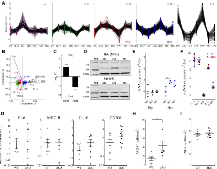
from human macrophages stimulated with TNF-α, IL-4, IL-17, LPS, IFN-γ, or LPS+IFN-γ (GSE1868618). Hck and Fgr are included in separate gene networks, as revealed by K-means cluster analysis (C-IV and C-III, respectively; Figure 6A).
Principal component analysis indicated that C-III and C-IV contained genes with upregulated expression in response to alternative (IL-4) and classic (LPS, IFN-γ, or LPS+IFN-γ) stimulants, respectively (Figure 6B). Consistently, Hck and Fgr were more than 2-fold upregulated in response to IFN-γ and IL-4, respectively (Figure 6C), suggesting their divergent participation in classic and alternatively activated macro- phage molecular networks, respectively. This was confirmed at the protein level by Western blotting on naïve and primed
BMDM from WT and dKO mice, showing a tendency toward increased Hck expression by classically activated macro- phages and increased Fgr by alternatively activated macro- phage (Figure 6D and 6E). Although conclusive evidence is lacking, classically activated macrophages are believed to represent the dominant phenotype in plaque. This therefore implies a major role for Hck in plaque macrophage function, as also suggested by the upregulation of Hck in advanced rup- ture-prone human atherosclerotic lesions (Figure IA and IB in the online-only Data Supplement)
However, the analysis of polarization marker gene expres- sion failed to demonstrate consistent Hck/Fgr deficiency–asso- ciated changes in macrophage phenotype. Baseline mRNA
G H I
WT dKO
0 20 40 60 80
iNOS+(cells/mm2)
WT dKO
0 20 40 60 80
100 *
ARG-1+(cells/mm2)
InosArg-1 MR IL12
TNF 0
4 8 12 16
20 WT
dKO
mRNAexpression(CT)
M0 M1 M2 M0 M1 M2 0
3 6 9
*
Fgr Hck
mRNAexpression(CT)
mRNA expression (in 2- Ct) IL-6 MHC-II IL-10 CD206
WT dKO
0.125 0.25 0.5 1 2 4 8 16 32
WT dKO
0.25 0.5 1 2 4 8 16
WT dKO
0.25 0.5 1 2 4 8
WT dKO
0.25 0.5 1 2 4 8
D E F
∆ ∆
∆
α
C B
A
Figure 6. Hck/Fgr are involved in separate macrophage polarization programs, but their combined deficiency does not impact
polarization marker expression. A, K-means clustering of human genes modulated on stimulation with IFN-γ, IL-17, IL-4, LPS, IFN-γ +LPS (denoted as IFN+) or TNF-α generates 5 clusters (C-I to C-V). Fgr and Hck belong to C-III and C-IV, respectively, and display opposite expression patterns. B, Principal component analysis (PCA) of modulated genes; 99.3% of the variance of the system lies within the first 3 principal components (PC1:84.8, PC2:13.2, and PC3:1.3%). C, Human Hck and Fgr expression is upregulated in response to IFN-γ and IL-4, respectively. D, This finding was confirmed by Western blotting on BMDM of WT vs dKO mice cultured in the absence (M0) or presence of IFN-γ (100 U/mL, M1) or IL-4 (20 ng/mL, M2; n=3). Quantification of Fgr (black symbols) and Hck (blue symbols) protein band intensities, corrected for the actin-loading control (mean±SD; n=3) is depicted in E. F, mRNA expression of classical (iNOS [inducible nitric oxide synthase]; IL-12; TNF-α) and alternatively activated macrophage markers (Arg-1, arginase-1; MR, mannose receptor) in nonstimulated BMDM in vitro as assessed by qPCR was not affected. Relative expression was calculated using cyclophilin as a house- keeping gene. qPCR data are presented as RE+2−(ΔΔCt−SDΔΔCt) and RE-2−(ΔΔCt+SDΔΔCt), mean±SD; n=3–4). qPCR of mRNA isolated from aorta of atherosclerotic LDLr–/– mice transplanted with WT and dKO bone marrow did not reveal any differences in expression of IL-6 (M1), MHC-II (M1) and IL-10 (M2), whereas that of CD206 (M2) was increased (G; n=6); in agreement, immunohistochemical analysis of aorta plaques showed slightly increased arg-1 (H), but unchanged iNOS staining (I; n=6–8). WT, open circles; dKO, filled circles. BMDM indicates bone marrow–derived macrophages; dKO, double-knockout; IFN-γ, interferon γ; LPS, lipopolysaccharide; MHC-II, major histocompatibility complex II; qPCR, quantitative polymerase chain reaction; TNF-α, tumor necrosis factor-α; and WT, wild type.
Downloaded from http://ahajournals.org by on February 28, 2019
expression of established classically activated macrophage markers by nonstimulated BMDM such as inducible nitric oxide synthase, IL-12, or TNF-α was essentially unchanged, as was that of the alternatively activated macrophage markers arginase-1 and the mannose receptor (Figure 6F). Likewise, polarization marker gene expression by LPS+IFN-γ or IL-4–
primed WT and dKO BMDM were largely similar (Figure IIIB and IIIC in the online-only Data Supplement, respectively).
At the protein level, dKO and WT BMDM showed equiva- lent IL-12 secretion, whereas TNF-α production was slightly increased (Figure IIID in the online-only Data Supplement) and that of nitric oxide was significantly reduced (Figure IIIE in the online-only Data Supplement). Concordant with these data, mRNA expression analysis on isolated aorta did not reveal major changes in macrophage polarization marker expression pattern, apart from a slight increase in CD206 expression (Figure 6G), whereas inducible nitric oxide syn- thase+ and arg-1+ macrophage content in intermediate lesions did not point to a shift toward classical macrophage activation in dKO versus WT chimeras either (Figure 6H and 6I).
Because the complexity of macrophage adaptive responses in vivo cannot be completely captured by the rigid dichotomy of the macrophage polarization model in vitro, we focused on Src kinase–associated differences in macrophage functions that could underlie dKO-associated plaque destabilization. First, we assessed whether Hck/Fgr deficiency impacts macrophage cell death or their ability to ingest particles, opsonized particles, and cholesterol accumulation, functions that are potentially con- trolled by src kinases and are relevant to plaque stability. Hck/
Fgr-deficient BMDM exposed normal apoptotic susceptibility (Figure IVA in the online-only Data Supplement), phagocytosis of fluorescent latex beads or opsonized particles (Figure IVB and IVC in the online-only Data Supplement), and uptake of modi- fied cholesterol, in vitro, as assessed by high-performance thin layer chromatography or fluorescent microscopy (Figure IVD and IVE in the online-only Data Supplement). Efferocytosis, defined as the macrophage capacity to process apoptotic cells, however, was considerably reduced in dKO BMDM (Figure IVF in the online-only Data Supplement), which could at least in part explain the necrotic core expansion observed in dKO chimeras.
Interestingly, incubation of vascular SMC with condi- tioned medium from nonstimulated dKO BMDM did not influence their proliferation (Figure VA in the online-only Data Supplement), but reduced their deposition of extracel- lular collagen and noncollagenous proteins akin to condi- tioned medium from LPS-primed WT macrophages and starvation medium (Figure VB and VC in the online-only Data Supplement). Apparently, Hck/Fgr deficiency favors a macrophage antifibrotic differentiation phenotype, and we propose that the impact of this effect will even be amplified by the reduced mesenchymal migration into the plaque of dKO macrophages, conducive to increased focal accumulation of antifibrotic macrophages in close proximity to the fibrous cap.
Discussion
Here, we present conclusive evidence that Hck/Fgr deficiency leads to reduced atherosclerotic lesion burden with concomi- tant reductions in macrophage accumulation and, paradoxi- cally, lesion stability. As we show, the former is attributable
to impaired adhesion of macrophages to the endothelium, whereas the latter is likely attributable to blunted mesenchy- mal migration into the plaque atheroma, resulting in the reten- tion of lytic macrophages in the plaque’s fibrous cap.
As a first hallmark of Hck/Fgr deficiency, atherosclerotic lesions displayed reduced amounts of macrophages despite the marked skewing of monocyte differentiation toward a Ly6Chigh phenotype, a subset known for its hypermigratory and proinflammatory profile and selective accumulation in atherosclerosis.20,25
With the use of intra- and extravital microscopy analy- sis of fluorescent dye, antibody and latex bead–labeled leu- kocyte subsets, we were able to firmly establish Hck/Fgr deficiency–induced impairment of monocyte and neutrophil adhesion to and diapedesis into plaque, while their chemotac- tic profile remained unaffected. This finding was confirmed by post hoc immunohistochemical analysis of plaque for the presence of latex bead–laden macrophages. The reduced presence of (Ly6Chi) monocyte–derived macrophages is espe- cially remarkable given the relative abundance of circulating Ly6Chigh monocytes in dKO chimeras. This subset is thought to be associated with higher Ly6Chigh monocyte infiltration20,25 and reduced accumulation of profibrotic and anti-inflamma- tory macrophages.26 It should be noted, however, that the Ly6Clow subset has been shown to contribute to plaque inflam- mation at later stages of disease development and has repeat- edly been linked to plaque vulnerability and fibrosis.27–29
In addition, Hck/Fgr-deficient macrophages featured an impaired morphological polarization and disrupted 2-dimen- sional migration. dKO macrophages were unable to form filopodia and lamellipodia, which is critical for those cells to adhere and establish leading and trailing poles that direct their mobilization toward higher concentrations of chemoat- tractants.30 Taken together, these results imply that Hck/Fgr deficiency results in reduced adhesion and directional crawl- ing on the endothelium and, therefore, in impaired extravasa- tion of circulating monocytes into the atherosclerotic lesion.
The 3-dimensional mesenchymal migration of macrophages depends on the formation of podosome rosettes, which release proteolytic enzymes to perform pericellular degradation of the extracellular matrix.21 In vitro, dKO macrophages exhibited an attenuated focal degradation of extracellular matrix, dis- rupted formation of podosome rosettes, and accordingly dis- rupted mesenchymal migration. Extrapolating these findings to atherosclerosis, Hck/Fgr deficiency has impacted both the adhesion of monocytes to the plaque and their mesenchymal migration across the lesional fibrous cap, as well, contributing to the striking reduction in plaque macrophage content and to subendothelial accumulation of invaded macrophages.
A second striking hallmark of Hck/Fgr deficiency in ath- erosclerosis was the paradoxical induction of necrotic core expansion and lesion vulnerability, with reduced fibrosis, SMC accumulation, and collagen deposition. This pheno- type is remarkably similar to that observed in ear excision wounds treated with Src tyrosine kinase inhibitors,31 alluding to a positive role of Hck/Fgr in macrophage profibrotic func- tions. Compatible with the latter, conditioned medium from dKO macrophages was seen to reduce collagen production by vascular SMC. The impact of this plaque-destabilizing effect
Downloaded from http://ahajournals.org by on February 28, 2019