CHAPTER X I V
Chemistry of Anterior Pituitary Hormones
B Y C H O H H A O L I A N D H E R B E R T M . E V A N S C O N T E N T S
Page
I. Gonadotrophic Hormones 633 A. Interstitial-Cell-Stimulating Hormone (ICSH, L H ) 634
1. Methods of Assay 634 a. Increase of Ovarian Weight in Normal Immature R a t s . . . . 634
b. Repair of Ovarian Interstitial Tissue in Hypophysectomized
Rats 635 c. Increase of Seminal Vesicle Weight in Normal Immature
Rats 635 d. Increase in Weight of the Ventral Lobe of the Prostate in
Hypophysectomized Male Rats 636
e. Other Methods 636 2. Methods of Isolation 636
a. Isolation of Sheep I C S H 637 b. Isolation of Swine I C S H 638 3. Comparison of Swine and Sheep I C S H 638
a. Physicochemieal Properties 638 b. Immunological Specificity 639 c. Biological Potency of Swine and Sheep I C S H 639
4. Effect of Various Agents on I C S H Activity. Chemical Differ-
ences between F S H and I C S H 640 a. Effect of Ketene 640 6. Effect of Cysteine 641 c. Effect of Protein Précipitants 641
d. Effect of Enzymes 641 B. Follicle-Stimulating Hormone (FSH) 643
1. Methods of Assay 643 a. Normal Immature Female Rats 643
b. Hypophysectomized Rats 643 2. Methods of Purification 644
a. Procedure of Fevold et al 644 b. Procedure of Fraenkel-Conrat et al 645
c. Procedure of Greep et al 645 d. Procedure of McShan and Meyer 646
3. Physicochemieal Properties 648 II. Lactogenic Hormone (Prolactin) 648
A. Methods of Assay 648 1. Crop Sac Weight Method 649
2. Minimum Stimulation Method 649 631
632 CHOH HAO LI AND HERBERT M. EVANS
3. Local Intradermal Method or Micro Method 650
B . Methods of Isolation 651 C. Physicochemical Properties 652
1. Isoelectric Point 652 2. Molecular Weight 652 3. Diffusion and Viscosity 653 4. Optical Rotation and Partial Specific Volume 653
5. Solubility 654 D . Differences in Ox and Sheep Hormones 654
1. Solubility Method 655 2. Tyrosine Method 655 E. Analytical Data 655
1. Elementary Composition 655 2. Distribution of Sulfur 656 3. Tyrosine and Tryptophan Content 656
4. Other Data 657 F. Reactions with Specific Reagents 657
1. Iodine 657 2. Reducing Agents 658
3. Ketene 658 4. Methyl Alcohol 659 ( } . Effect of Various Agents 660
1. Heat 660 2. Enzymes 660 3. Denaturing Agents 660
III. T h y r o t r o p h s Hormone 661 A. Methods of Assay 661
1. Guinea Pigs 661 2. Chicks . 662
3. Rats 662 B. Methods of Purification 662
1. Yale Procedure 663 2. California Procedure 664 C. Physicochemical Properties 664 IV. Adrenocorticotrophic Hormone ( A C T H ) 665
A. Methods of Assay 665 1. Repair Test 666 2. Maintenance Test 666 B. Methods of Isolation 667
1. Isolation of Swine A C T H 667 2. Isolation of Sheep A C T H 668 C. Physicochemical Properties 670
1. Isoelectric Point 670 2. Molecular Kinetic Data 670 3. Some Analytical Data 671
4. Solubility 671 5. Stability 671 D . Effect of Various Agents 672
1. Ketene 672 2. Nitrous Acid 672 3. Formaldehyde 673
XIV. CHEMISTRY OF ANTERIOR PITUITARY HORMONES 633
I. Gonadotropine Hormones
From the work of Smith (212), Zondek and Aschheim (236), and a number of later investigators (225), it was clear that pituitary extracts or implants produce two gonadal reactions: (a) the stimulation of follic- ular growth in the ovaries and of spermatogenic activity in the testis and (b) the final ripening of the ovarian follicles together with the exhibi- tion of estrus or heat, the rupture of the follicles and their transformation to corpora lutea, and in the testis the assumption of a functional role on the part of the Ley dig cells, which ostensibly secrete testosterone, which in turn causes the development and assumption of function on the part of the secondary sex glands. Whether or not these reactions are due to one or more hormones in the pituitary has been the subject of a number of investigations (215). Some investigators inferred from their physiological experiments that there was only one gonadotrophic hor- mone and that a difference in mode of administration or in dosage deter- mined the type of reaction observed. They would not feel that their contention is negated by the chemical fractionation of two substances in pure or almost pure form from pituitary tissue and by the experimental replacement of all gonadotrophic functions of the pituitary by the admin-
4. Iodine 673 5. Trypsin 673 E. Hydrolysis with Pepsin 673
V. Growth Hormone 674 A. Methods of Assay 674
1. B o d y Growth of Normal Female Rats 674 2. B o d y Growth of Hypophysectomized Female Rats 675
3. Tibia of Hypophysectomized Rats 675 B . M e t h o d of Isolation · 676
C. Criteria of Purity 677 1. Biological Test 677 2. Diffusion 678 3. Electrophoresis 678 4. Solubility 679 D . Biological Potency 679
E. Physicochemieal Properties 683 1. Isoelectric Point and Molecular Weight 683
2. Analytical D a t a 683 3. Diffusion and Viscosity 683 F. Effect of Various Agents 684
1. Effect of Proteolytic Enzymes 684
2. Effect of Heat 684 3. Effect of Urea 685 4. Effect of Nitrous Acid and Ketene 685
V I . Summary 686 References 688
6 3 4 CHOH HAO LI AND HERBERT M. EVANS
istration of these substances. We will not further discuss this dispute to which more extended reference is made elsewhere but will content our- selves with an account of the chemical methods which have led to the separation of two pituitary gonadotrophic substances and of the chemical characterization of those substances.
The first separation of pituitary gonadotrophic fractions into two components was obtained by Fevold, Hisaw, and Leonard ( 6 9 ) in 1931.
Subsequent work from other laboratories ( 1 1 , 1 0 3 , 2 2 7 , 2 2 8 ) confirmed the concept that two hormones are present in pituitary extracts—hormones which exhibit two kinds of gonadotrophic activity. They have been designated the follicle-stimulating hormone (FSH) or thylakentrin ( 3 7 ) and the interstitial-cell-stimulating hormone (ICSH) or metakentrin ( 1 3 7 ) . The latter was first called the luteinizing hormone or LH.
In 1940, two laboratories independently announced the isolation of ICSH in pure form from sheep ( 1 3 8 ) and swine glands ( 2 0 3 ) . The preparations appear homogeneous in electrophoretic, ultracentrifuge, and solubility studies,1 and it is particularly important that they are freed from contamination with the other gonadotrophic hormone, FSH.
On the other hand, highly purified FSH preparations have been reported ( 8 0 , 8 9 ) though this hormone has not been isolated in pure state.
Recently Evans, Simpson, Lyons, and Turpeinen ( 5 9 ) showed that lactogenic hormone can awaken or intensify corpus luteum function causing placentoma production in normal, adrenalectomized, and hypo- physectomized animals. It should therefore be considered the third member of the pituitary gonadotrophic complex. The chemistry of this hormone will be presented in another section.
A . INTERSTITIAL-CELL-STIMTJLATING HORMONE (ICSH OR LH)
1. Methods of Assay
Many methods for the determination of the potency of ICSH have been proposed from different laboratories. There is as yet no standard method to establish an international unit so that the potency of hor- monal preparations from different laboratories can be compared. In the following, the commonly accepted methods are summarized:
a. Increase of Ovarian Weight in Normal Immature Rats. This method ( 6 5 ) depends on the first-known characteristic of the hormone—
that of producing copora lutea in the ovaries of immature female rats and of increasing the weight of the ovary when injected in combination with FSH. Twenty-one-day-old female rats of 3 5 - 4 0 g. body weight are used. Before the injection of ICSH, the ovarian weight of the
1 For a discussion of criteria of purity in proteins, see (183,202).
XIV. CHEMISTRY OF ANTERIOR PITUITARY HORMONES 635
animal is caused to increase 100% over that of the control by the admin- istration of FSH for four days. Different dosages of ICSH are then injected subcutaneously twice daily over a period of five days simul- taneously with the same amount of FSH. The ovaries are weighed and examined on the sixth day. A unit of ICSH is defined as the amount of hormone which produces an additional 100% increase in the ovarian weight together with the production of corpora lutea (67). The fact that the method is based on the action of two hormones makes it difficult to determine quantitatively the content of ICSH in crude or partially purified extracts, where an unknown amount of both hormones is already present. However, the method may be useful in the standardization of the pure hormone.
b. Repair of Ovarian Interstitial Tissue in Hypophysectomized Rats.
As the name of the hormone indicated, ICSH is selectively able to repair the degenerated interstitial cells (deficiency cells) in the ovaries of hypo- physectomized animals, i.e., it causes the resumption of a normal nuclear picture, abolishing the agminated and wheel pattern of the nuclear chromatin. The method as developed by Simpson et al. (207) is based on this characteristic of the hormone. The rats are used at a standard age (26-28 days) and postoperative period (6-8 days). The routine injection procedure is once daily, intraperitoneally for three days, followed by autopsy 72 hours after the first injection. The ovaries are sectioned for histological examination. The amount of protein giving a minimal but definite effect on the "deficient" interstitial cells is called an ICSH unit. It must be mentioned that if the subcutaneous route of injection is used, the test is one fifth as sensitive.
c. Increase of Seminal Vesicle Weight in Normal Immature Rats.
The known action of ICSH in male rats is also the stimulation of the interstitial cells, which in this case produce androgen, which in turn causes a weight increase of the secondary sex organs. A test for ICSH potency in immature rats based on this principle has been proposed by Fevold (65,67). Male rats 22 days old are injected twice daily for four or five days; the animals are autopsied 24 hours after the last injection.
The seminal vesicles plus the coagulation glands are dissected and weighed. A unit is defined as the amount of hormone which produces 100% increase in these combined weights as compared with the weight of controls. Fevold found that the method could be employed to estimate the ICSH content in unfractionated extracts and believed that it gave consistent and reliable results. Although the method appears conven- ient and simple, the sensitivity of the method is unfortunately greatly influenced by the strain of animals used. In rats of the Long-Evans strain, the seminal vesicles are hardly hypertrophied after the injection
636 CHOH H A O LI A N D H E R B E R T M . E V A N S
of ICSH or unfractionated extract; the same materials, however, cause as high as 400% increase in the seminal vesicle weights in the Sprague- Dawley strain of rats (67,76).
d. Increase in Weight of the Ventral Lobe of the Prostate in Hypophy- sectomized Male Rats. Greep et al. (91) have pointed out the relatively greater reactivity of the ventral lobe of the prostate in the measurement of ICSH activity. Hypophysectomized male rats (21 days old at opera- tion, two days postoperative at the beginning of injections) are injected subcutaneously daily for four days with autopsy on the fifth day, 24 hours after the last injection. Greep et. al. propose as standard dose (unit) the amount of hormone causing a 100% increase in weight of the ventral prostate as compared with untreated controls (92). The fact that the presence of FSH does not potentiate the ICSH activity makes the method particularly valuable. We have confirmed the usefulness and reliability of the method and found further that the test becomes more sensitive when injections are made intraperitoneally instead of sub- cutaneously (207).
e. Other Methods. Witschi (234) suggests the use of the melanin reaction in the pectoral or abdominal feathers of African weaver finches as a measure of ICSH activity. The weight increase of the testes of immature pigeons or of one-day-old chicks has also been proposed for the standardization of ICSH (208).
2. Methods of Isolation
The ICSH content of pituitary tissue from different animals has been studied by a number of investigators (65,229,234). We may summarize by stating that the ICSH content of pituitaries decreases in the following order: sheep, rabbit, swine, rat, dog, horse, man, and beef. It is obvious that the pituitary glands commonly used for the isolation of ICSH are those from sheep or pigs. These glands as secured from slaughter houses may be either stored in a frozen state or desiccated with acetone.2 We have found that no loss of hormonal activity can be detected when glands are kept at — 15°C. for over a year.
The solvents generally used for the extraction of the gonadotrophic principles from the pituitary are weak aqueous alkalis or dilute alcohol—
saturated barium hydroxide (54), 0.02 Ν ammonium hydroxide (5), 2 % pyridine (65), and 40% alcohol (228). Fe void (65) has found that pyridine is not only a good extractive but serves also as a preservative.
In a later report, Fevold et al. (70) favor dilute ammonium hydroxide as the extracting agent.
2 Kupperman, Elder, and Meyer (110) have investigated various methods of reserving pituitary glands.
X I V . C H E M I S T R Y OF A N T E R I O R P I T U I T A R Y H O R M O N E S 637
a. Isolation of Sheep ICSH. The method of Li, Simpson, and Evans (138,139,140) for the isolation of ICSH from sheep glands as a pure protein is as follows:
(1) Three hundred grams acetone-desiccated whole sheep pituitary tissue is extracted with 4 L 4 0 % alcohol over a period of two days with constant stirring, and the residue is reextracted similarly using 2 1. of the alcohol. The supernatant liquids are combined and filtered through coarse folded filter paper. The clear filtrate is brought to 80 or 8 5 % alcohol b y the addition of 9 5 % alcohol and adjusted to p H 5.5 b y the addition of glacial acetic acid. The precipitate formed is removed b y centrifuging and dried with absolute ethyl alcohol and ether. The following steps are carried out at 5°C.
(2) Fifty grams of this powder are extracted with 3 1. distilled water. The super- natant is mixed with an equal volume of acetone at about p H 4.5. The precipitate is next extracted with 1 1. 1% sodium chloride solution.
(3) The 1 % saline extract is brought to 0.5 saturation with solid ammonium sul- fate. The precipitate is removed b y centrifugation and the supernatant liquid is saved for purification of the F S H .
(4) T h e 0.5 S A S3 precipitate is dissolved in distilled water and brought to 0.2 saturation with the addition of SAS. T h e supernatant liquid is next brought to 0.4 SAS.
(5) T h e 0.4 SAS precipitate is dissolved in distilled water4 and brought to 0.37 AS, and SAS is added to the supernatant to 0.4 saturation.
(6) The precipitate between 0.37 and 0.4 SAS is dissolved in distilled water and brought to 2 . 5 % trichloroacetic acid b y the addition of a 10% solution. The precipi- tate formed is dissolved in a small volume of an aqueous alkaline solution and dialyzed.
T h e I C S H is obtained in dry form b y lyophilization.
The hormone thus obtained is free of other hormonal contaminants.
When 3 mg. of the material is injected subcutaneously5 in immature hypophysectomized female rats, no histologically detectable follicular stimulation is observed. On the other hand, 0.005 to 0.01 mg. of the same substance causes repair of the ovarian interstitial tissue in such rats on intraperitoneal injection. In the same type of animal, 2 mg. of the preparation is free of thyrotrophic hormone, 10 mg. free of adreno- corticotrophic and of growth hormone. In pigeons, 10 mg. shows no crop-stimulating activity.
When the preparation is subjected to electrophoretic, ultracentrifuge, and solubility tests (140), it behaves as a homogeneous protein.
Fevold et al. (65,70) have also described a method for obtaining a highly purified sheep ICSH but their preparation contains two com- ponents in electrophoretic and ultracentrifugal analysis.
3 SAS will be used throughout this chapter as an abbreviation for saturated a m m o - nium sulfate.
4 At this step, an alternative procedure has been described for obtaining pure I C S H b y precipitation at p H 4.0 to 4.1 in 0.33 SAS solution (140).
5 See page 643 for testing F S H .
638 CHOH HAO LI AND HERBERT M. EVANS
b. Isolation of Swine ICSH. Chow, van Dyke, and co-workers (35,203) have described a method of obtaining ICSH from the glands of swine in pure form. Their procedure is outlined below.
(1) Four and one-half kg. of ground fresh swine glands are extracted with 22.5 1.
2 % sodium chloride at 4 ° C . T h e supernatant liquid is made acid with hydrochloric acid to p H 4.2 to 4.6.
(2) T o the supernatant, solid ammonium sulfate is added to saturation. The precipitate formed is dissolved in water and dialyzed. The dialyzed soluble solution is next adjusted to p H 5.1 to 5.5.
(3) T h e supernatant fluid is made to 0.33 saturation with ammonium sulfate.
T h e 0.33-SAS-soluble fraction is brought to 0.9 saturation.
(4) T h e precipitate is dissolved in water and dialyzed; ammonium sulfate is added to the dialyzed solution until 0.33 saturation. After centrifuging off the small amount of precipitate, the supernatant is adjusted to p H 7.3.
(δ) T h e precipitate formed is dissolved in water and step 4 is repeated at least seven times. T h e final pH-7.3 precipitate at 0.33 SAS is the pure hormone.
The preparation isolated shows homogeneity in electrophoretic, ultra- centrifugal, and solubility tests. The preparation was examined in immature hypophysectomized female rats for the detection of FSH, adrenocorticotrophic, and thyrotrophic contaminants and these were found to be absent in the large dose employed (0.34 mg. daily for ten days). No data however have been furnished as to the absence of the growth and lactogenic hormones.
3. Comparison of Swine and Sheep ICSH
a. Physicochemieal Properties. From osmotic pressure measure- ments, a molecular weight of 40,000 is obtained for sheep ICSH, whereas the swine hormone has a value of 100,000 as calculated from ultracentrif- ugal data. The sedimentation constants, £2o, of sheep and swine ICSH are 3.6 X 10~13 and 5.4 X 10~1 3, respectively. The latter value, if cor- rected for the viscosity and density of the solvent, becomes 6.8 X10~1 3.
Electrophoretic experiments also reveal dissimilarity in the protein hormones isolated from these two different species. At pH 7.85 and ionic strength 0.05 buffer, swine ICSH migrates with a rate of 0.52 X 10~5 cm.2/sec./v., while the sheep hormone has an electrophoretic mobility of 6.36 ΧΙΟ"5 in a buffer of pH 7.53 and ionic strength 0.05. Sheep ICSH possesses an isoelectric point at pH 4.6 and swine at pH 7.45.
This difference in the electrical properties of swine and sheep ICSH has been further verified by Chow et al. (35). They have compared the electrophoretic behavior of the swine hormone with that of a highly purified sheep ICSH prepared by Jensen. Although Jensen's prepara- tion contained two components, they were able to show that the biological activity is associated with the main component and that the isoelectric point of the main component may be estimated at about pH 4.8 to 5.0.
XIV. CHEMISTRY OF ANTERIOR PITUITARY HORMONES 639
Both sheep and swine ICSH contain carbohydrate but the content differs significantly. The sheep hormone has 4.5% mannose and 5.8%
hexosamine, while swine has 2.8% mannose and 2.2% hexosamine (93,138). The tryptophan content of the two hormones has also been found to be different. By the glyoxylic acid method, swine ICSH is shown to contain 1.0% tryptophan; the hormone isolated from sheep glands on the other hand contains 3.8% tryptophan. Table I gives a summary of the physicochemical data for ICSH as isolated from sheep and swine pituitary glands, respectively.
T A B L E I
P H Y S I C O C H E M I C A L C H A R A C T E R I S T I C S O F S H E E P AND S W I N E I C S HE
Determinations C, %
H, % N, %
Molecular weight Isoelectric point, p H
Sedimentation constant, S X 1 013
Tyrosine, % Tryptophan, % Mannose, % Hexosamine, %
° S e e (93,138,140,203).
Sheep Swine 4 9 . 3 7 6.83 14.20 14.93 40,000 100,000
4 . 6 7.45 3 . 6 5 . 4 4 . 5
1.0 3 . 8 4 . 5 2 . 8 5 . 8 2 . 2
b. Immunological Specificity. Perhaps the most sensitive test for species differences in proteins is obtained from immunological reactions.
Chou (31) has made such studies with swine and sheep ICSH. When the pure swine ICSH is injected into rabbits, the production of specific antibodies can be demonstrated both by the precipitin and complement fixation reactions. The antiserum6 thus obtained does not react with pure sheep ICSH or extracts prepared from sheep glands. It would therefore appear that the interstitial-cell-stimulating hormones isolated from swine and sheep glands are chemically different entities.
c. Biological Potency of Swine and Sheep ICSH. Since the sheep and swine hormones have been shown to be different substances it would not be unexpected that they should differ in biological potency. Some differences have been reported. Greep et al. (92) have found that the
6 Chow has further demonstrated that the swine I C S H is not only " l o b e specific"
but also ' 'hormone specific." T h e immulogical study m a y therefore serve as a highly sensitive and specific test for hormonal contaminants in any supposedly pure hormone preparation.
640 CHOH HAO LI AND HERBERT M. EVANS
sheep ICSH is far more active in the repair of the ovarian interstitial cells in hypophysectomized rats and in causing ovulation in rabbits than is the swine hormone. On the other hand, the two hormones are equally effective in stimulating the anterior prostate of hypophysectomized rats.
Table II presents a comparison of the ICSH potency of these two hor- mones as determined by different tests.
T A B L E I I
B I O L O G I C A L P O T E N C Y OF P U R E S W I N E AND S H E E P I C S HA
Test Sheep Swine
Total dose necessary to increase weight of ventral prostate from 6.39 to 9.45 mg. in hypophysec-
tomized rats 0.0134 mg.
0.005 to 0.010 0.007
0.0134 mg.
> 0 . 1 0
> 0 . 0 4 0 Total dose necessary to repair ovarian interstitial
cell in hypophysectomized rats
Total dose necessary per kg. b o d y weight to produce positive response in ovulation of all of 8 r a b b i t s . . .
0.0134 mg.
0.005 to 0.010 0.007
0.0134 mg.
> 0 . 1 0
> 0 . 0 4 0
« S e e (92).
4. Effect of Various Agents on ICSH Activity. Chemical Differences between FSH and ICSH
Since the original observation of Fe void et al. (69) that FSH is more soluble in aqueous salt solution than ICSH, a considerable number of studies has been made as to chemical differences in the two hormones on treatment with various reagents. Although almost all experiments have employed impure preparations, the results obtained are of value in our understanding of the intrinsic nature of the hormones.
a. Effect of Ketene. Ketene is considered to be a mild and specific acetylating agent for aqueous protein solutions. Three groups in pro- teins are known to react with this agent: viz. the amino, phenolic hydroxyl, and sulfhydryl groups. Both purified FSH and ICSH fractions have been subjected to ketenization at room temperature (137). After five minutes treatment, the interstitial-cell-stimulating activity is greatly reduced while the follicle-stimulating action is apparently unchanged.
Upon longer treatment (thirty minutes), both hormonal activities are almost completely destroyed. The inactivation of ICSH by ketene was later confirmed with a pure preparation (138); based on results obtained by other investigators, it is assumed that the free amino groups are essen- tial for the biological activity of ICSH. Though it is admitted that such assumptions are invalid without complete chemical analysis of the active groups in acetylated samples, nevertheless the results obtained suggest
X I V . C H E M I S T R Y OF A N T E R I O R P I T U I T A R Y H O R M O N E S 641
distinct chemical differences between ICSH and FSH in the manner of inactivation by ketene.
b. Effect of Cysteine. It is generally agreed that cysteine reduces
—S—S— cross links in proteins at an alkaline pH, i.e., thiol groups result and a corresponding amount of cysteine is oxidized to cystine. Fraenkel- Conrat et al. (79) have used this reducing agent to investigate the essen- tiality of disulfide (—S—S—) groups in gonadotrophic hormones.
When FSH and ICSH preparations are allowed to react with cysteine (forty times the amount of protein) at pH 7.7 for two days, their biological activities are greatly decreased. In a later report (81), no loss of gonado- trophic potency was noted under conditions of cysteine treatment milder than those employed for the inactivation of insulin. It appears that the disulfide groups in these gonadotrophic substances are not so readily reduced as is the case with other proteins. They conclude that the integrity of some disulfide bonds which are not easily reduced is essential for hormonal activity. The inactivation of FSH by cysteine has been confirmed by McShan and Meyer (163). The results of Bischoff's experiments (18) are also in complete agreement with those reported by Fraenkel-Conrat et al.
c. Effect of Protein Précipitants. It was noted in the course of the purification of ICSH that FSH was soluble in 2.5% trichloroacetic acid, whereas ICSH was completely precipitated in the same solution (138).
By using other protein précipitants, Fevold (64) has also observed chemi- cal differences in the two hormones; FSH was found to be inactivated by picrolonic, picric, and flavianic acids, whereas ICSH retained its activity on treatment with these reagents. While we must note that these results were not confirmed by Jensen et al. (104), it is important to remember that the disagreement could be due to differences in the assay methods employed, the strain of animals, or in the purity of the hormones. In view of our experiments with trichloroacetic acid we view it as likely that the two hormones can be observed to differ in their reactions with various reagents.
There is another protein precipitant, namely tannic acid, which has been used for some time in augmenting FSH or ICSH potency (68,76).
The augmenting effect of tannic acid and other agents on the two hor- mones is discussed elsewhere in this volume.
d. Effect of Enzymes. In 1929 Reiss and Haurowitz (189) discovered that crude trypsin destroyed gonadotrophic activity in a pituitary extract. Later, Bates, Riddle, et al. (11,194) found the destructive action of a purified trypsin preparation on FSH. However, McShan and Meyer (161) could not confirm these results; they stated that commercial trypsin destroyed almost all ICSH (88), but not FSH, activity. The
642 CHOH HAO L I AND HERBERT M. EVANS
results of Chen and van Dyke (30) and Greep (88) agreed with those of McShan and Meyer. On the other hand, Abramowitz and Hisaw (1) claimed that neither crystalline trypsin nor chymotrypsin inactivates ICSH more rapidly than FSH. It may be well to point out that all these results were obtained from assays in normal animals and further- more that the extent of enzymic digestion in no case wras determined.
T A B L E I I I
E F F E C T OF S O M E P R O T E O L Y T I C E N Z Y M E S ON G O N A D O T R O P H I C P O T E N C Y OF S W I N E P I T U I T A R Y E X T R A C T0
Enzyme Protein
digested, % F S H I C S H Crystalline carboxypepti-
dase 5 (5 hr.) Unaffected Unaffected
5-12 (30-41 hr.) Unaffected or reduced Reduced Crystalline c h y m o t r y p s i n . . . 18-35 Reduced Absent
68-80 Absent Absent
Crystalline trypsin 12-48 Reduced Absent 61-75 Usually absent Absent Merck's trypsin 10 Unaffected Unaffected
35-46 Unaffected or reduced Reduced 61-75 Unaffected or reduced Absent
Papain 6-31 Unaffected Unaffected
60-65 Reduced Reduced
Crystalline pepsin 10-34 Unaffected or reduced Reduced?
58-80 Absent Reduced?
α Taken from Chow, Greep, and van D y k e (34).
The controversies concerning the trypsin experiments finally become clear. Chow, Greep, and van Dyke (34) have reinvestigated the effect of crystalline trypsin and commercial trypsin (Merck) on the gonado- trophic activity of pituitary (swine) extract in a most careful and thor- ough manner. The rate of hydrolysis of the extract was estimated by determining the decrease of protein precipitable by trichloroacetic acid, and the destruction of gonadotrophic potency was followed by assays in immature hypophysectomized male or female rats. Using crystalline trypsin, Chow et al. found that with over 60% digestion all gonadotrophic activity is lost while when the amount of digestion is between 12-48%, FSH appears to be more resistant than ICSH. If commercial trypsin is used, the disappearance of 10% of the protein is not followed by any destruction of FSH or ICSH potency, but, when higher percentages of protein are digested, ICSH appears to be selectively destroyed. The interesting results with crude trypsin cannot be explained by the presence of crystalline trypsin. Chow et al. conclude that "the proteolytic
XIV. CHEMISTRY OF ANTERIOR PITUITARY HORMONES 643
activity of Merck's trypsin depends to a major extent on the presence of enzyme(s) other than trypsin or chymotrypsin." Chow, Greep, and van Dyke (34) have also studied the effect of crystalline pepsin on FSH and ICSH potency. Their results are summarized in Table III.
Another difference in the reactivity of the two gonadotrophic hor- mones toward enzymic digestion is shown by the experiments of McShan and Meyer (161,162), who found that ptyalin (saliva) abolished the follicle-stimulating activity, the luteinizing activity being relatively resistant.
B. FOLLICLE-STIMULATING HORMONE (FSH)
1. Methods of Assay
a. Normal Immature Female Rats. Since FSH stimulates follicular growth, it is obvious that one may use increase in ovarian weights as contrasted with those of controls to determine rat units (63,70). Fevold and co-workers have employed this method routinely for the quantitative estimation of FSH potency. It must be emphasized that FSH is poten- tiated by the presence of ICSH and that, if the animal's own pituitary is present or if FSH fractions are not free from ICSH, the assay results obtained in normal immature animals do not represent absolute units of FSH.
Another procedure, based on the fact that FSH exerts an augmenta- tion effect on the activity of the gonadotrophic principle in pregnant women's urine (CG, chorionic gonadotropin) in immature female rats, has been reported by Evans et al. (61). A unit is defined as the minimal amount of material which, given subcutaneously to 24-26-day-old female rats in combination with a standard amount of CG, augments the effect of the latter by 100%. The difficulties of this method are: the presence of ICSH in FSH preparations will render the test less sensitive and it gives no information regarding ICSH contamination. It is therefore advisable to assay FSH preparations in hypophysectomized animals for quantitative data.
b. Hypophysectomized Rats. (1) Female. In hypophysectomized animals FSH causes the enlargement of the ovarian follicles while leaving the interstitial tissue in the deficient condition if, of course, the prepara- tion is not contaminated with the luteinizing factor. In the assay of FSH in these animals, either increase of ovarian weights or histological exami- nation for beginning follicular development may be taken for measure- ment of the hormone. One rat unit represents the minimal total amount which, injected subcutaneously once daily over a period of three days into hypophysectomized rat (26-28 days old at operation, six- to eight-
644 CHOH HAO LI AND HERBERT M. EVANS
day postoperative interval before injection), causes the occurrence of healthy (nonatretic) follicles with small antra, as evidenced 72 hours after beginning the injection (61). For routine laboratory assay, three animals per group can furnish a reliable answer to hormonal potency The method is very sensitive and has the further advantage that the presence of ICSH can be observed simultaneously if there is repair of the interstitial tissue.
(2) Male. The follicle-stimulating hormone is known to stimulate the epithelium of the seminiferous tubules. As shown by Greep et al.
(90,92), FSH causes an increment in testis weights proportional to the dose injected without any stimulation of the secondary sex organs in hypophysectomized male rats. The animals employed were hypophy- sectomized at 21 days of age (35-47 g. body weight); after two days postoperative, injections began once daily for four days and the animals were autopsied on the day following the last injection. If ICSH is a contaminant in the follicle-stimulating preparation, the test becomes unreliable, for ICSH alone is able to increase the weight of the testes
(209).
Other methods have been used by investigators for the assay of FSH such as the increase of the uterine weights (115), the production of oestrous vaginal smears (234), etc.
2. Methods of Purification
Sheep and swine pituitaries are rich in follicle-stimulating substance and they are therefore commonly used to obtain a potent FSH prepara- tion. The extraction of pituitary tissue (fresh or acetone-dried material) is made either with saline or alcoholic solutions like those employed for the isolation of ICSH. Chemically the follicle-stimulating hormone is in one respect unique in that it is the only known anterior hypophyseal hormone soluble in half saturated ammonium sulfate. Highly purified FSH possesses a high carbohydrate content. In addition, as has been discussed, the hormone is resistant to tryptic digestion when a commercial enzyme preparation is employed. The follicle-stimulating hormone has not been isolated in pure form. There are methods which enable one to obtain a so-called "biologically pure" preparation, i.e., a preparation free from other active contaminants. Subjoined, we give a few methods which appear to be satisfactory in preparing potent follicle-stimulating preparations.
a. Procedure of Fevold et al. (70): One kg. of frozen sheep glands are finely ground and extracted with 2 1. of dilute aqueous ammonium hydroxide at pH 8.0. The supernatant liquids are brought to 0.25 M
X I V . C H E M I S T R Y OF A N T E R I O R P I T U I T A R Y H O R M O N E S 645
ammonium sulfate and to pH 5.4. After the removal of the precipitate formed by centrifugation, the supernatant is adjusted to pH 7.0 and fractionated with ammonium sulfate. The fraction soluble at 2.4 M but precipitated at 2.7 M ammonium sulfate is the follicle-stimulating substance. As assayed by increase in the ovarian weights of immature female rats, the product obtained contains 20 R.U./mg. No experi- mental data were given by Fevold et al. as to possible contamination with other active components.
In an earlier report (65), Fevold used 2 % pyridine as the extractant.
The gonadotrophic substances are adsorbed by benzoic acid; the ICSH fraction is next removed by precipitation at pH 4.2 in 0.2 SAS, the soluble material containing the FSH. Inert substances in the FSH preparation are further removed by basic lead acetate. FSH preparations thus obtained have a potency of 50 and 75 R.U./mg.
b. Procedure of Fraenkel-Conrat et al. (80): From the fraction soluble in 0.5 SAS as described in step 3 for the isolation of sheep ICSH (see page 637), the supernatants are brought to 0.67 saturation with ammonium sulfate. The precipitate obtained between 0.5 and 0.67 SAS contains the follicle-stimulating substance. It can be further purified in the following ways: precipitation at pH 4.8 in 35% acetone, precipita- tion in 48% alcohol in the presence of a few drops of saturated sodium chloride solution, and removal of contaminating proteins at pH 4.1 in saturated sodium chloride solution.
The product obtained causes beginning ovarian follicular development at a total dose of 0.004 mg. when injected subcutaneously in hypophy- sectomized rats. Higher doses of the preparation (0.04 to 0.06 mg.) show indications of ICSH contamination, i.e., luteinization of the follicu- lar walls and repair of the interstitial tissue. In addition, oestrous uteri are caused by 0.016 mg. of the hormone.
c. Procedure of Greep et al. (89) : Greep, van Dyke, and Chow (89) have described a method for obtaining a so-called "biologically pure"
follicle-stimulating hormone from swine pituitary glands. The method, which is based on the fact that FSH is soluble in a pH 4.4 acetate buffer containing 20.5% sodium sulfate whereas ICSH is insoluble in this solvent, is outlined as follows:
(1) Fresh hog pituitaries are extracted with 2 % sodium chloride at p H 4.2. T h e supernatant is saturated with ammonium sulfate. T h e precipitate is dialyzed and the clear dialyzed solution is adjusted to p H 5.1. (2) T h e supernatant is brought to 5 0 % saturation with ammonium sulfate at p H 4.2. T h e supernatant is further brought to 0.9 SAS. (3) The precipitate is dialyzed until saltfree. One volume of 1 M acetate buffer of p H 4.41 and 2 volumes of 4 1 % sodium sulfate are added.
T h e precipitate contains no F S H and is used for the isolation of I C S H . (4) T h e supernatant is made to 4 0 % ammonium sulfate b y adding 40 g. ammonium sulfate/
646 CHOH HAO LI AND HERBERT M. EVANS
100 ml. (5) The precipitate is dialyzed until free of salt. Steps 8 and 4 are repeated until no turbidity is observed in step 8.
The final precipitate (92) does not cause enlargement of the ventral prostate or stimulation of the ovarian interstitial cells in hypophysec- tomized rats when injected at a total dose of 2.77 mg. protein (assuming the preparation%contains 13% nitrogen), but 0.0154 mg. produces an increase of 50% in ovarian weights over those of untreated controls. The preparation did not give uterine stimulation at the highest dose tested (a total dose of 1.92 mg. in ten days). It is apparent that the FSH pre- pared by Greep et al. was free from ICSH contamination. Unfortu- nately, they did not give data to show that the preparation contained no adrenocorticotrophic, thyrotrophic, lactogenic, or growth activity so that its biological "purity" cannot be regarded as established.
In a later communication, Chow (33) reported that ultracentrifugal and electrophoretic experiments indicated definitely the heterogeneity of their FSH preparation. However, Greep et al. (89) stated earlier that solubility studies in one solvent show "no evidence of contaminating proteins if the concentration of the solid phase is five times that saturat- ing the solution, though an increase in the amount of protein Ν is observed if the solid phase is hundred times that necessary for saturation."
d. Procedure of McShan and Meyer (163): As already mentioned, commercial trypsin preparations destroy only ICSH activity in gonado- trophic extracts and FSH is apparently resistant to the enzyme digestion.
McShan and Meyer utilize this fact and develop a method to prepare a
"biologically pure" follicle-stimulating substance. Their method may be described in the following steps:
(1) Acetone-dried sheep pituitary powder is extracted with water. T h e gonado- trophic activity is precipitated from the supernatant b y the addition of four volumes of acetone. (2) T h e precipitate is further extracted with water; the supernatant is digested at 38°C. for 35 hours at p H 8 with 40 mg. trypsin (Fairchild)/g. of original pituitary powder. (3) Insoluble material formed during digestion is centrifuged off;
the supernatant is placed in a 75° water bath for twenty minutes. (4) The digest is dialyzed against 0.1 Af acetate buffer of p H 4.0. T h e precipitate formed is discarded.
(5) The supernatant is mixed with four volumes of 9 5 % alcohol; the fine white precipi- tate is dried with alcohol and acetone.
The final product is found to cause only follicular development in normal and hypophysectomized immature female rats in most cases.
The preparation is showrn to be free from lactogenic and thyrotrophic activities as assayed in pigeons and chicks. No physicochemieal purity is given. It is probable that the preparation contains a high percentage of inactive contaminants as judged by its ability to increase the ovarian wreights of normal immature rats when compared with the preparation
XIV. CHEMISTRY OF ANTERIOR PITUITARY HORMONES 647
T A B L E I V
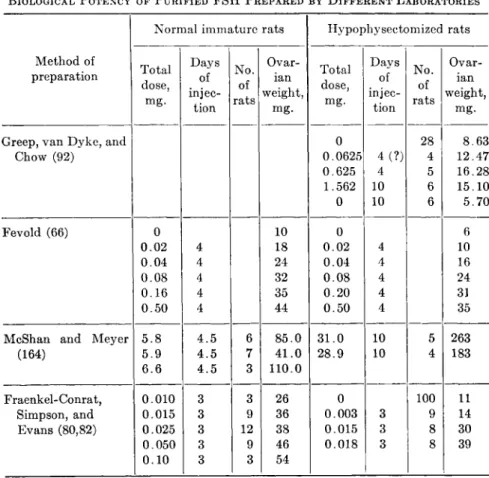
B I O L O G I C A L P O T E N C Y O F P U R I F I E D F S H P R E P A R E D B Y D I F F E R E N T L A B O R A T O R I E S
Normal immature rats Hypophysectomized rats Method of
preparation Total dose, mg.
Days of injec-
tion N o .
of rats
Ovar- ian weight,
mg.
Total dose, mg.
D a y s of injec-
tion N o .
of rats
Ovar- ian weight,
mg.
Greep, van D y k e , and 0 28 8.63
Chow (92) 0.0625
0.625 1.562 0
4 ( ? ) 4 10 10
4 5 6 6
12.47 16.28 15.10 5.70
Fevold (66) 0 10 0 6
0 . 0 2 4 18 0 . 0 2 4 10 0 . 0 4 4 24 0 . 0 4 4 16 0 . 0 8 4 32 0 . 0 8 4 24 0 . 1 6 4 35 0 . 2 0 4 31 0 . 5 0 4 44 0 . 5 0 4 35 McShan and M e y e r 5 . 8 4 . 5 6 8 5 . 0 3 1 . 0 10 5 263
(164) 5.9
6 . 6
4 . 5 4 . 5
7 3
4 1 . 0 110.0
2 8 . 9 10 4 183
Fraenkel-Conrat, 0 . 0 1 0 3 3 26 0 100 11 Simpson, and 0.015 3 9 36 0.003 3 9 14 Evans (80,82) 0.025 3 12 38 0.015 3 8 30 0 . 0 5 0 3 9 46 0.018 3 8 39 0 . 1 0 3 3 54
Table IV summarizes the biological potency of FSH fractions pre- pared by different laboratories. It is clear that the swine FSH prepared by Greep et al. has a much lower follicle-stimulating potency than sheep hormone as reported by Fevold and Fraenkel-Conrat et al., but the sheep FSH gives an ICSH reaction at very low doses while the swine FSH is apparently free from ICSH action. Some investigators attribute these differences to species characteristics (82,164) while others (33) feel that they are due to contamination of sheep FSH by ICSH. The final answer to the problem must await the isolation of FSH in pure form.
obtained by Fraenkel-Conrat et al. (80). The preparation has been shown to cause local reactions at the site of injection when used in human subjects but, in a later report (164), McShan and Meyer introduce a procedure to remove the toxic substance.
648 CHOH HAO LI AND HERBERT M. EVANS
3. Physicochemical Properties
Gurin (93) reported that swine FSH obtained by the method of Greep, van Dyke, and Chow contains 4.5% mannose and 4.4% hexose- amine. Evans et al. (53) found that the potent FSH from sheep glands is rich in carbohydrate and glucoseamine. McShan and Meyer (162,163) stated that their FSH preparations contain about 20% glucose. In ad- dition, FSH activity is destroyed by certain amylase preparations (45).
It is probable that pure FSH is a glycoprotein. It is of interest to note that all gonadotrophic hormones including those of nonpituitary origin seem to contain carbohydrate (93).
The preparation as described by Fraenkel-Conrat et al. (80) contains 1.3.1% nitrogen. It is generally agreed that FSH proteins are very soluble in water; in the absence of electrolytes, they are soluble in 50%
acetone, 70% alcohol (80), and 50% dioxane (63). The follicle-stimulat- ing potency is comparatively stable; in solutions of pH 7 to 8 the activity is retained at 75°C. for thirty minutes (163), but it is destroyed at 60°C.
for fifteen minutes in 50% alcoholic solution (63). Chow (33) has esti- mated the isoelectric point of his FSH preparation from swine glands to be about 4.8.
The effects of enzymes and of other chemical agents on the follicle- stimulating activity of pituitary extracts have already been discussed in this chapter.
II. Lactogenic Hormone (Prolactin)
The first indication of the existence of a lactogenic substance in extracts of the anterior pituitary came from the experiments of Strieker and Grueter (216), who found that extracts initiate lactation in ovariec- tomized pseudopregnant rabbits. The conclusion was later confirmed by Corner (44) and others (155,174,191). In 1932, Riddle et al. (192,193) discovered the stimulating action of the lactogenic principle on the crop sac of the pigeon and suggested the name prolactin. Two other names have been proposed: galactin (86) and mammotropin (151).
A . METHODS OF ASSAY
In estimating lactogenic potency two groups of methods have been employed; one depends on the crop sac reaction in the pigeon and the other on the response of the mammary gland of the "conditioned" rabbit or guinea pig. The latter methods (86,152,154,173), are less quantita- tive and are laborious; they will not be discussed here.
X I V . C H E M I S T R Y OF A N T E R I O R P I T U I T A R Y H O R M O N E S 649
1. Crop Sac Weight Method
This is the original method proposed by Riddle, Bates, and Dykshorn (193). They found that the combined weights of the two excised crop sacs are proportional to the amount of lactogenic principle injected.
Pigeons (six to ten weeks after hatching) are injected intramuscularly once daily for four days and autopsied about 96 hours after the first injection. Under these conditions Riddle et al. found that the crop weight is a linear function of the logarithm of the dosage. The pigeons used must have approximately the same body weight because the crop sac weight depends on the size of the bird (197). If the crop weight is calculated in terms of a unit of body weight, i.e., relative crop weight, a more consistent response-dosage relationship is obtained. According to Folley et al. (74) the accuracy of the weight method depends on three factors: " ( 1 ) the standard deviation of a single observation, (2) the number of birds used in each group and (3) the slope of the dose-response curve."
Bates and Riddle (10) have found a seasonal variation in the response of crop sacs; maximum response occurs in winter and summer. It is therefore essential to keep a standard preparation at hand and always compare the potency of an unknown with that of the standard. To obtain uniform results, the birds must be of the same STRAIN and race.
Bates et al (12) reported that the crop sac response changes with racial and strain differences. Folley, Dyer, and Coward (74) investigated the effect of light and temperature on the crop sac response of pigeons; they found that light exerts no influence on the response and that a maximum response is achieved at an equable temperature in the region of 15°C.
The effectiveness of different routes of injections has been studied by Bates and Riddle (8). Subcutaneous injections are most effective, intraperitoneal ones least effective.
2. Minimum Stimulation Method
In a footnote in their paper, Lyons and Catchpole (155) stated that
"one need not depend upon a weight increase in the crop-gland over and above the control to determine a positive reaction, since beginning growth changes may be seen in crops that weigh less than the average normal, as early as 48 hours after the injection of potent hormone." They suggest a qualitative test for lactogenic activity by merely holding the crop gland to the light to examine beginning recognizable stimulation. McShan and Turner (165) proposed a quantitative assay method based on the suggestions of Lyons and Catchpole and defined a pigeon unit as "the total amount of hormone injected during a period of 4 days which cause a
650 CHOH H A O LI A N D H E R B E R T M . E V A N S
minimal but definite proliferation of the crop glands of 50 ± 11 per cent in common pigeons weighing 300 ± 40 gm." In our laboratory, we have employed this method routinely to estimate lactogenic potency and have found that, even using only three birds per group, satisfactory results can be obtained. Silver King pigeons, four to five weeks from hatching and weighing 400 to 550 g., are injected subcutaneously once daily for four days with 0.5 ml. of the hormonal solution; 24 hours after the last injec- tion, the crop is dissected and examined against the light for a positive reaction. If two out of three birds give a positive response, the amount of hormone used is considered to be one unit.
3. Local Intradermal Method or Micro Method
For the estimation of a minute quantity of lactogenic hormone, e.g., that in urine, Lyons and Page (156) introduced a highly sensitive test by intracutaneous injection in the neck skin directly over the pigeon's crop sac. According to Lyons (152), the hormone solution (0.1 ml.) is injected intradermally within the skin covering the crop sacs, daily for four days with a 27-gage hypodermic needle. The birds are sacrificed on the fifth day; the sacs are dissected off and held slightly stretched against the light when a positive response can be seen with the naked eye. The injections are generally carried out over one crop sac and the sac on the opposite side may serve as a control or be injected with a different dose level. The volume of injection fluid has some influence on the sensi- tivity of the method (9). It is therefore essential for quantitative results to utilize a constant volume. It has been shown that the method will detect 1/10,000 of a unit obtained from the minimum stimulation test.
McQueen-Williams (159) and others (185,186) have applied this method to detect the presence of lactogenic hormone in a single rat pituitary implanted directly over the crop sac.
In 1938 arrangements were made for the establishment of an inter- national unit (I.U.) of the lactogenic hormone during the Conference on the Standardization of Hormones (25). In the following year the inter- national standard preparation of the hormone was issued; the inter- national unit is defined as: "the specific activity contained in 0.10 mg.
of the standard preparation" (26). It is therefore possible to state the potency of any lactogenic preparation in terms of the international unit.
Lyons (154) has made careful studies on the potency of the international standard material and found that it is about one half as potent as the pure lactogenic hormone in the guinea pig assay and about one third as potent by the crop weight response method.
XIV. CHEMISTRY OF ANTERIOR PITUITARY HORMONES 651
B. METHODS OF ISOLATION
The content of lactogenic hormone in pituitary glands of different species has been reported by a number of investigators. Bates and Riddle (7) have found that ox and sheep glands contain much higher concentrations of the hormone than do glands obtained from swine. A later report by Chance et al. (29) showed that the hormone content7 is progressively smaller in the following order: sheep, ox, man, swine, and horse. The content in horse glands amounts to only 4 % of the amount found in sheep or ox. Therefore the starting material for the isolation of the lactogenic hormone is usually either ox or sheep glands.
The solvents which have been used by different investigators for the extraction of the lactogenic substance from pituitary tissue, are: aqueous acid solutions of pH 2 (191), aqueous alkali solutions of pH 9 (86,192,193), 66% acetone in acid at pH 2 (155), 60-70% alcohol at pH 9-10 (7), and aqueous saline solution (22). Bergman and Turner (16) have made a comparison of these methods of extraction and conclude that the method of Bates and Riddle (7) is superior both for total yield and potency/mg.
extracted substance. But the acid-acetone extraction of Lyons (152,153) has constituted the initial step in the isolation of the hormone in pure form. A prime advantage of Lyon's extract is that it contains mainly only the lactogenic and adrenocorticotrophic hormones, the other hor- mones being either not extractable with the solvent or destroyed by the acidic acetone.
A highly purified and potent lactogenic hormone preparation was first described by Lyons (152,153) in 1937. In the same year, White et al.
(233) announced the preparation of a crystalline protein possessing crop- stimulating activity; in the preliminary note, there was, however, no data concerning the biological and chemical purity of the crystalline preparation. It was not until 1942 that White, Bonsnes, and Long (232) published a satisfactory identification of the crystalline protein with the hormone. In the meantime (1940-1941) Li, Lyons, and Evans (129,130,132,133) showed that their lactogenic hormone preparation behaved like a pure protein as judged by electrophoretic and solubility studies.
The original method of Lyons (153) for the isolation of lactogenic hormone in pure form as slightly modified by Li et al. (129,130,132,133) may be described in the following steps:
7 For the content of lactogenic hormone in pituitaries of ox, mice, rats, guinea pigs, rabbits, and cats, see Reece and Turner (187).
652 CHOH HAO LI AND HERBERT M. EVANS
(1) One kg. of ground sheep pituitaries is extracted with 4 1. acetone to which has been added 100 ml. 12 M hydrochloric acid. The extracted material is next precipi- tated out b y the addition of 5 1. acetone. (2) Dissolve the precipitate in 200 ml. 5 0 % acetone and reprecipitate b y adding 800 ml. acetone. The precipitate formed is dissolved in 300 ml. of approximately 1 0 % aqueous ammonium hydroxide. (3) The solution is allowed to stand at room temperature for three hours. T w o volumes of acetone are added and if a precipitate forms this is removed b y centrifugation. (4) The supernatant is mixed with one volume of acetone and 10 ml. 12 Ν hydrochloric acid. T h e precipitate is dissolved in 200 ml. water with the aid of 1 M sodium hydrox- ide to obtain a clear solution. ( 5 ) T h e solution is adjusted to p H 6.5 with 1 M hydro- chloric acid and the precipitate removed b y centrifugation. (6) The supernatant is brought to p H 5.5 and kept at — 15°C. for a few hours. After thawing, the solution is centrifuged. ( 7 ) The precipitate is dissolved in slightly alkaline solution and steps 5 and 6 are repeated until the final p H 5.5 precipitate behaves as a single substance in electrophoretic experiments.
The hormone thus isolated contains about 30 I.U./mg. as assayed by the minimum stimulation method in pigeons. The preparation is free from other active components. Electrophoretic studies (129,130,132) in buffers of pH from 2 to 8 show that the preparation migrates in the electrical field as a single protein. Evidence for the purity of the prepara- tion is also furnished by solubility experiments in three different solvents.
The pure hormone can also be obtained by an alternate method as described by Li, Simpson, and Evans (141) or by the method of White et al. (232).
C. PHYSICOCHEMICAL PROPERTIES
1. Isoelectric Point
From the pH at which the hormone becomes least soluble, it has been speculated that prolactin must possess an isoelectric point at approxi- mately pH 5.5. The exact isoelectric point of the hormone as estimated by the moving-boundary method in electrophoresis (129) is found to be pH 5.73 in buffer solutions of ionic strength 0.055. White et al. (230,232) reported a value of pH 5.65 using the micro electrophoresis technique of Abramson (2) in buffers of ionic strength 0.10. The small difference between these two values is most probably due to the different ionic strengths employed and they should be considered to be in satisfactory agreement.
2. Molecular Weight
The report of Lyons and Page (156) and others (49,99,113) on the detection of prolactin in urine would lead one to expect that the molec- ular weight of the hormone is comparatively low. The first molecular weight data were obtained from osmotic pressure measurements (132) and indicated that the hormone has indeed a low molecular weight, i.e., 26,500. From analytical data, the molecular weight may also be esti-
X I V . C H E M I S T R Y OF A N T E R I O R P I T U I T A R Y H O R M O N E S 653 mated to be about 25,000. Diffusion and viscosity data suggest a value of 22,000 (see below).
The sedimentation constant of prolactin as determined in the analy- tical air-driven ultracentrifuge has been reported by White, Bonsnes, and Long (232) to be 2.8 X 10~13 cm./sec./dyne. Preliminary data on the sedimentation and diffusion constants as secured in the laboratory of J. W. Williams were also reported by them to be: S20 = 2.65 X 10~1 3, D20 = 7.5 X 10~7; from these values, a molecular weight of 32,000 for prolactin was computed. Since no complete data were given, the value 32,000 can only be assumed tentatively as the molecular weight of pro- lactin when determined by ultracentrifuge. At any rate, the difference in these two values (26,500 and 32,000) is hardly surprising, for many experiments (179) have shown that molecular weight determinations as obtained by osmotic pressure or by ultracentrifugation methods need not be in close agreement. The comment made by White (230) that the value 26,500 is "considerably too l o w " would appear to be gratuitous.
3. Diffusion and Viscosity
The viscosity of prolactin solutions (117) has been determined in an Ostwald viscometer. A straight-line relationship exists between the viscosity and the protein concentration up to 1.0% solution. From the slope of such a straight line, the hormone molecule is demonstrably far from spherical. If one assumes that it is a prolate ellipsoid the ratio of the long to the short axis as computed from Simha's equation (206) is 5.7. The shape of a protein molecule may also be expressed by the frictional constant, / / /0, which can be computed by Perrin's equation (181) if one knows the ratio of the long to the short axis of a prolate ellipsoid of revolution. Thus, f/f0 for prolactin is calculated to be 1.29 (117).
The membrane diffusion method of Northrop and Anson (178) was employed to determine the diffusion coefficient of the hormone (108).
After making the correction as suggested by Mehl (170), D2o = 9.0 X 10~7 cm.2/sec. From a combination of this constant with the fric- tional ratio, the molecular weight of prolactin may be estimated to be 22,000, which is in fair agreement with that obtained by osmotic pressure measurements.
4. Optical Rotation and Partial Specific Volume
The optical rotation of prolactin solutions is found to be a linear function of the concentration (117). From the observed rotation of 1.0% solution at 25°C, the specific rotation of the lactogenic hormone is
— 40.5°. The partial specific volume of the hormone is calculated by
654 CHOH HAO LI AND HERBERT M . EVANS
determining the density of the solution containing different weight frac- tions of the protein and found to be 0.721. This value is as expected for ordinary proteins.
5. Solubility
Lactogenic hormone has some interesting solubility characteristics.
The pure hormone is soluble in absolute methyl or ethyl alcohol in the presence of a small amount of acid (71,121). It is extremely insoluble in water when no electrolytes are present. The hormone isolated from ox glands has a solubility of 0.102 g./l. at 7-8°C. (133). In aqueous acid solution, prolactin is easily salted out in a low concentration of sodium chloride.
T A B L E V
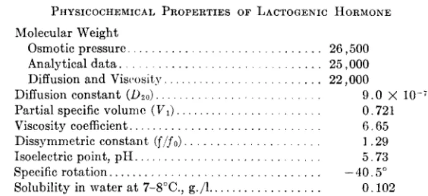
P H Y S I C O C H E M I C A L P R O P E R T I E S OF L A C T O G E N I C H O R M O N E Molecular Weight
Osmotic pressure 26,500 Analytical data 25,000 Diffusion and Viscosity 22,000 Diffusion constant (L>20) 0 X 1 0~7 9
Partial specific volume (Vi) 0· 721
Viscosity coefficient 6-65 Dissymmetric constant
(f/f
0)
1 ·29Isoelectric point, p H 5. 7 3
Specific rotation —40.5 Solubility in water at 7-8°C., g./l 0 . 1 0 2
Table V summarizes the physicochemical properties of the lactogenic hormone.
D . DIFFERENCES IN O X AND SHEEP HORMONES
The hormone isolated from either ox or sheep pituitaries shows no difference in crop-sac-stimulating potency. Bischoff and Lyons (19) were unable to differentiate the ox and sheep hormone through the use of precipitin, anaphylaxis, or the Dale and Arthus reactions. It was further found that the ox and sheep hormones can not be distinguished in elec- trophoretic experiments; they apparently migrate with the same mobility in buffers from pH 2 to 9 and have identical isoelectric points (129). The two hormones have the same stereochemical structure as judged by their optical rotation properties (117); there are also no differences in molec- ular weight and the content of tryptophan, arginine, cystine, and methionine (118,132). However, the hormone isolated from ox as contrasted with that isolated from sheep pituitary tissue can be differ- entiated by the following two methods.
XIV. CHEMISTRY OF ANTERIOR PITUITARY HORMONES 655
1. Solubility Method
The solubility method has been shown to be a sensitive test for distinguishing species specificity of proteins (112). Using this method, Li et al. (134,135) found that ox and sheep lactogenic hormone are not identical proteins. For instance, in 0.357 M sodium chloride solution at pH 2.25, the sheep hormone has a solubility of 0.506 g./l. of the solvent at 25°C, whereas the solubility of the ox preparation is only 0.316 g. In citrate buffer (1 M> pH 6.36), the ox hormone is more soluble than the sheep. It is further shown that when sheep hormone is added to a saturated solution of the ox hormone, more protein is dissolved indicating that the two substances are not the same.
The hormones isolated from these two species can also be demon- strated to be different entities by their behavior when salted out with sodium chloride. Cohn (38) has shown that the solubility of a protein is defined by an equation of the form: log S = β — Κ8μ, where μ is the ionic strength/1000 g. water, S the solubility in g./L, and Ks and β con- stants. It has been showrn (133) that the hormone from ox or sheep glands has an almost identical K8 but different values for β are obtained when the salting-out studies are made with sodium chloride in 0.01 M hydrochloric acid solution. This means that in 0.01 M hydrochloric acid sheep hormone is more soluble than is the ox protein and that they are different proteins.
In alcoholic solution, ox hormone is more soluble than the sheep (71).
2. Tyrosine Method
We have observed that the tyrosine content of ox lactogenic hormone is consistently higher than that of sheep; ox protein has 5.73% tyrosine, whereas sheep contains 4.53% (131,132). It is not likely that the differ- ence in the solubility behavior of these two proteins can be completely explained by their tyrosine content and further determinations of other amino acids will be necessary to explain this phenomenon.
E. ANALYTICAL D A T A
1. Elementary Composition
The elementary analysis (121) of a pure lactogenic hormone prepara- tion yields the following results: C, 50.72%; H, 6.63%; N, 15.86%; S, 1.79%; P, nil. Earlier investigators (51,160) had already found no phosphorus in purified lactogenic preparations. The nitrogen content of the hormone was reported by White et al. as 14.38%, 16.49%, and 16.84% (232,233).