FUMONISIN B
1EXPOSURE INCREASES Hsp70 EXPRESSION IN THE LUNG AND KIDNEY OF RATS WITHOUT INDUCING
SIGNIFICANT OXIDATIVE STRESS
Dániel J. KÓCSÓ1*, Judit SZABÓ-FODOR1, Miklós MÉZES1,2, Krisztián BALOGH2, Szilamér FERENCZI3, András SZABÓ1,2, Brigitta BÓTA1 and Melinda KOVÁCS1,4 1MTA-KE Mycotoxins in the Food Chain Research Group, Kaposvár University, Guba S. u. 40, H-7400 Kaposvár, Hungary; 2Department of Nutrition, Faculty of Agricultural and Environmental Sciences, Szent István University, Gödöllő, Hungary;
3Institute of Experimental Medicine of the Hungarian Academy of Sciences, Budapest, Hungary; 4Mycotoxins in the Food Chain Research Group, Faculty of Agricultural
and Environmental Sciences, Kaposvár University, Kaposvár, Hungary (Received 13 February 2018; accepted 25 July 2018)
The objective of this experiment was to determine whether fumonisin B1
(FB1) added to the diet of rats in a dose of 50 mg/kg changes the production of heat shock protein 70 (Hsp70) in the lungs and kidney of rats. We also studied the effect of this mycotoxin on the antioxidant system of the body. Mature (8 weeks old) male Wistar Crl:WI BR rats (n = 6/group) were fed the toxin-containing diet for 5 days. FB1 resulted in a 7% body weight reduction without significantly changing the feed intake. Western blot analysis of the lungs and kidney demon- strated a substantial (1.4-fold and 1.8-fold, respectively) increase in Hsp70 ex- pression. Alterations could not be detected in the clinical chemical parameters (to- tal protein, albumin, total cholesterol, glucose, creatinine and urea concentrations, and aspartate aminotransferase activity). There was no statistically significant change in malondialdehyde concentrations and the measured antioxidant parame- ters (the amount of reduced glutathione, GSH and glutathione peroxidase activity, GPx) in the blood plasma, lung and kidney tissue. Thus, it can be concluded that FB1 did not induce oxidative stress in the lungs and kidney, but increased Hsp70 production.
Key words: Fumonisin B1, rat, Hsp70, clinical chemical parameters, oxi- dative stress, antioxidants
*Corresponding author; E-mail: kocso.daniel@ke.hu; gfapgfap@gmail.com;
Phone: 0036 (82) 505-800
Open Access. This is an open-access article distributed under the terms of the Creative Commons Attribution- NonCommercial 4.0 International License (https://creativecommons.org/licenses/by-nc/4.0/), which permits un- restricted use, distribution, and reproduction in any medium for non-commercial purposes, provided the origi- nal author and source are credited, a link to the CC License is provided, and changes – if any – are indicated.
The mycotoxin fumonisin B1 (FB1) is primarily produced by the moulds Fusarium verticillioides and F. proliferatum, which are regarded as natural con- taminants of maize and maize-based feeds all over the world. FB1 is a polar compound, and the backbone of its structure is a carbon chain containing hy- droxyl and methyl groups. FB1 accounts for 70–80% of the fumonisin analogues (groups A, B, C and P). Due to its structure, it disturbs the biosynthesis of sphin- golipid (one of the important membrane constituents) by inhibiting the activity of the sphinganine N-acyltransferase enzyme (Wang et al., 1991; Merrill et al., 1993). As a result of this, one of the toxic effects of FB1 manifests itself in de- creasing the amounts of sphingolipids, increasing the levels of the cytotoxic sphinganine (Sa) and sphingosine (So), and inhibiting the degradation processes of sphingolipids (Wang et al., 1991; Yoo et al., 1992; Riley et al., 1994a,b). In addition, FB1 influences the metabolism of phospholipids and polyunsaturated fatty acids, and may induce apoptosis in various tissues (Voss et al., 1996;
Tolleson et al., 1996; Bucci et al., 1998). The International Agency for Research on Cancer (IARC) classifies FB1 as a Group 2B carcinogen, i.e. it is possibly carcinogenic to humans. It is the direct causative agent of porcine pulmonary oe- dema (PPE) (Kriek et al., 1981) and equine leukoencephalomalacia (ELEM) (Marasas et al., 1988). Human oesophageal cancer has also been associated with the consumption of maize containing high levels of FB1 (Thiel et al., 1992;
Marasas et al., 1993).
Numerous observations have been reported on the renotoxic effects of FB1
depending on the duration of exposure and the magnitude of the doses applied.
Such effects include e.g. apoptosis and necrosis, as well as the development of adenoma and carcinoma in which the cytotoxic action of FB1 plays a role. The early experiments conducted by Kriek et al. (1986) on rats suggested that the secondary metabolites of Fusarium moniliforme might induce pathological chang- es in the renal tissue. This was subsequently confirmed by subchronic (7–30 days) feeding experiments conducted by Voss et al. (1989, 1993) with 20–150 mg/kg FB1, in which histopathological signs indicative of varying degrees of reversible renal toxicity (such as increased tubular basophilia and cytoplasmic vacuolation, hyperplasia of the tubular epithelial cells and their infiltration into the lumen, as well as necrotic processes) were observed. In addition to tubular nephrotic chang- es, FB1 also caused morphological damage associated among others with hyper- plasia in the liver cells and the biliary tract, which was confirmed by the elevated alanine transaminase (ALT), aspartate transaminase (AST) and alkaline phospha- tase (ALKP) activities and bilirubin concentration of the blood serum.
During their 26-month-long experiments conducted on rats to reveal the pulmotoxic, hepatotoxic and renotoxic effects of FB1 fed in a concentration of 50 mg/kg, Gelderblom et al. (1991) observed macroscopically visible atrophy in the kidneys. The kidneys had lighter colour and irregular surface, and contained numerous cortical and medullary cysts. Fibrosis, interstitial lymphocytic infiltra-
tion and the appearance of retention cysts in the kidney were described, together with vacuolar and hyaline degeneration and necrosis of the proximal tubular epi- thelial cells. Numerous glomerular effects were also noted, such as atrophy of the glomerular bundle, thickening of the glomerular loops, and thickening of the wall of the Bowman’s capsule due to fibrotic changes. AST and gamma- glutamyl transferase (GGT) activities and (conjugated and unconjugated) biliru- bin concentrations were elevated, indicating the hepatotoxic effects of FB1.
After subchronic (≤ 90 days) exposure of rats to FB1-contaminated feed (0, 5, 15, 50 and 150 mg/kg), Voss et al. (1996) described mild cellular-level changes in the kidneys and increased apoptotic processes and morphological changes in the cells of proximal tubules located in the area of the so-called corti- comedullary junction, on the external side of the medulla. After a single intrave- nous administration of FB1 to rats in a dose of 1.25 mg/kg, Lim et al. (1996) re- ported apoptotic and necrotic changes together with cell proliferation processes that could be brought into association with the appearance of the so-called non- genotoxic carcinogenic effects of FB1. By histological examination of the kidneys of rats fed increasing doses (99, 163, 234 and 484 mg/kg) of FB1 for 28 days, Tolleson et al. (1996) demonstrated an increase in the apoptotic processes of tubular epithelial cells.
In a 104-week dose-response feeding study, FB1 did not prove to be car- cinogenic in female F344/N/Nctr BR rats even in a dose as high as 100 mg/kg feed, while in male rats dietary FB1 concentrations of 50 and 150 mg/kg induced adenoma and carcinoma involving the renal tubules (Howard et al., 2001).
Much less information is available on the effects of FB1 on the pulmonary tissues of rats. Gelderblom et al. (1991) fed BD IX male rats a diet containing 50 mg/kg purified FB1 for 26 months. In 66% of the rats examined between the 18th and the 26th month of the study hepatocellular carcinoma was found, which produced metastases to the lungs, heart and kidneys.
In their 5-day experiments, He et al. (2002) administered FB1 subcutaneous- ly to mice in a dose of 2.25 mg/kg body weight. They found increased tumour ne- crosis factor alpha (TNFα) mRNA expression and elevated levels of the sphingoid bases (sphingosine and sphinganine), which was different in the lungs and the heart. After the intraperitoneal injection of FB1 to mice in a dose of 10 mg/kg body weight over a period of 5 days, Kim et al. (2006) observed significant elevations in the levels of Sa and Sa-1-P in the lung, kidney, liver, heart and brain tissues, with- out any pathological changes in the tissues. The apoptotic and pathological chang- es attributable to the presence of mycotoxins trigger cytoprotective molecular pro- cesses associated with the increased production of heat shock proteins (Jolly and Morimoto, 2000; Hassen et al., 2005). Heat shock proteins constitute the most an- cient and evolutionarily extremely well conserved intracellular defence system of eukaryotic cells. They are present in the cytoplasm, nucleus and mitochondria, and one of their main tasks is to protect proteins against harmful effects (e.g. oxidative
stress). Their increased expression can be an early marker of cytotoxic effects.
Studying the relationship between oxidative stress caused by mycotoxins and Hsp70 production, El Golli-Bennour and Bacha (2011) classified fumonisins as mycotoxins having a moderate oxidative effect. However, there is a scarcity of da- ta on the effect of FB1 on Hsp production, and most of the available data were de- rived from in vitro experiments performed in cell cultures.
Therefore, the aim of this work was to study the acute effects of FB1 admin- istered in a dose of 50 mg/kg of diet for 5 days, focusing on oxidative stress and the production of heat shock protein 70 (Hsp70) in hitherto less investigated or- gans, the lungs and the kidney.
Materials and methods Accommodation and feeding of rats, sampling
Mature (8 weeks old) male Wistar Crl:WI BR rats weighing approximately 200–250 g (n = 6/group, FB1 vs. control, total n = 12) and kept in individual metabolic cages (Tecniplast, Castronno, Italy) were used in the experiments. In the experimental room, light (8:00–20:00) and dark (20:00–8:00) periods were alternated at 12-h intervals daily (at 20 °C and 50% relative humidity). The commercial diet (Ssniff, R/M-Z+H) was ground, and an FB1-containing fungal culture was added to it to provide a finished feed containing 50 mg/kg FB1 for the experimental group. After homogenisation, the ground diet was regranulated and then dried at 40 °C. The diet fed to the control group was also ground and then regranulated, but no fungal culture was added to it. During the experiment, drinking water and feed were provided ad libitum, the daily feed intake was rec- orded, and the body weights of the rats were measured at the start and at the end of the experiment. After the treatments lasting for 5 days, blood samples were taken from the retroorbital plexus of the rats in diethyl ether narcosis, then the rats were decapitated and exsanguinated after cervical dislocation for postmor- tem sample collection. To determine Hsp70 expression, approximately 0.5-g samples were taken from the lungs and the kidneys of all rats, from the same ana- tomical locations, and the samples were stored at –80 °C until analysed. Blood se- rum samples were assayed for clinical chemical parameters while lung and kid- ney samples for parameters indicative of oxidative stress.
The animal experiment was authorised by the Food Chain Safety and An- imal Health Directorate of Somogy County Government Office under permission no. SOI/31/1679-11/2014.
Production of FB1
Fumonisin B1 was produced by the application of Fusarium verticillioides strain MRC 826 according to the method of Fodor et al. (2008). The homoge-
nised fungal culture contained FB1 at a concentration of 3.44 g/kg. This fungal culture was mixed into the basal diet of the experimental animals, to provide feed contaminated with 50 mg/kg FB1 toxin. The mycotoxin concentrations of the control and the experimental diets were determined by LC-MS (Shimadzu, Kyo- to, Japan). The lower limit of detection (LOD) for FB1 was 3 μg/kg. The diet fed to the control group did not contain detectable amounts of FB1. The absence of deoxynivalenol, zearalenone and T-2 toxin was checked and confirmed.
Western blot analysis
Kidney and lung tissue samples (n = 6/group) were homogenised in 500 μl lysis buffer (1% NP40, 1% sodium deoxycholate, 0.1% SDS, 15 mM NaCl, 10 mM phosphate buffer, 2 mM EDTA, 2 mg/ml aprotinin, 0.5 mg/ml leupeptin, 2 mM sodium vanadate, 20 mM NaF, 0.5 mM DTT, 1 mM PMSF) for 3 min.
Subsequently the cell lysate was centrifuged (13,000 rpm, 30 min, 4 °C) and the pellet collected. Total protein concentration of the samples was determined with the BCA™ Protein Assay KIT (Thermo-Fisher, Budapest, Hungary). Thirty to 35 µg protein/sample quantities were applied to 10% SDS polyacrylamide gels [30% Acrylamide/Bis-acrylamide, 1.5 M Tris (pH 8.8), 1.0 M Tris (pH 6.8), 100 g/L SDS, 100 g/L APS, TEMED] and transferred to nitrocellulose (0.45 µm) membranes. The membranes were washed for 3×5 minutes with TBS-T (1× TBS pH 7.6, 0.1% Tween 20), then blocked in phosphate-buffered saline (10× PBS) containing 5% nonfat dried milk powder, 1% BSA and 0.1% Tween 20. Subsequently the membranes were incubated with primary anti-Hsp70 anti- bodies (1:1000; Sigma, Budapest) at 4 °C for 12 h. As internal control, anti-β- actin antibodies (1:10,000; Sigma, Budapest, Hungary) were used. After another 3×5-min washing with TBS-T (pH 7.5), secondary antibodies conjugated with horseradish peroxidase (HRP) were used in 1:500 dilution (Biomarker, Budapest, Hungary) to quantify the binding of the primary antibodies. After a repeated 3×5-min washing with TBS-T, the light emission of the blotted proteins was en- sured by the use of a WesternBright Enhanced Chemiluminescent HRP substrate detection system (Biomedica, Budapest, Hungary), and the proteins were detect- ed either on CL-XPosure clear-blue X-ray films or using a FluorChem Q Imag- ing system imaging program (ProteinSimple, Santa Clara, CA, USA). The densi- tometric analysis of the chemiluminescent signals thus obtained was performed with an ImageJ software. The data were presented by pixel density in arbitrary units ± SEM (P < 0.05).
Clinical chemistry and antioxidant parameters
The concentrations of plasma total protein (TP), albumin (ALB), total cho- lesterol (tCHOL), glucose (GLU), creatinine (CREA) and urea, as well as the ac- tivities of aspartate aminotransferase (AST), were determined in a veterinary la-
boratory (Vet-Med Laboratory Ltd., Budapest, Hungary), using a Roche Hitachi 912 Chemistry Analyzer (Hitachi, Tokyo, Japan) with commercial diagnostic kits (Diagnosticum Ltd., Budapest, Hungary).
For the determination of lipid peroxidation, blood plasma, kidney and lung samples were stored at –80 °C until analysed. Lipid peroxidation was determined by the quantification of malondialdehyde (MDA) levels by the 2-thiobarbituric acid method (Placer et al., 1966) in the blood plasma and in 1:9 homogenates of tissue samples in physiological saline. Among the components of the antioxidant system, the amount of reduced glutathione (GSH) measured in the blood plasma and 10,000 g supernatant fraction of tissue homogenates by the method of Sedlak and Lindsay (1968) and the activity of glutathione peroxidase (GPx) according to Lawrence and Burk (1978). GSH content and GPx activity were calculated to protein content which was determined by the biuret method in the blood plasma (Weichselbaum, 1948) and with Folin phenol reagent in tissue homogenates (Lowry et al., 1951).
Statistical analysis
For the comparison of group means (body weight, Hsp70, antioxidant pa- rameters, clinical chemical parameters), unpaired t-test was used with the GraphPad Prism 7.00 software. Normality was tested with GraphPad Prism 7.00 according to the Kolmogorov–Smirnov method.
Results Body weight
Although the difference was not significant, the average initial body weight of the toxin-treated animals was slightly higher compared to controls (Table 1). During the 5-day-long feeding the control group showed a body weight gain while the FB1 group a body weight decrease. The weekly average feed intake was not significantly different in the two groups. However, the daily feed intake data show that the toxin-fed rats consumed less feed from the second day of the experiment onwards, although the difference was significant only on day 4 (Table 2).
Western blot analysis
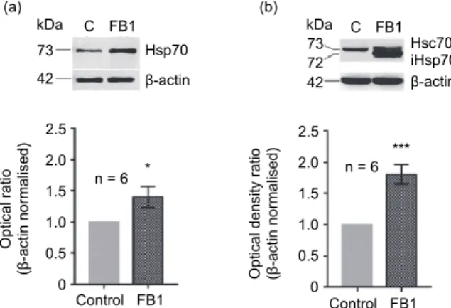
By Western blot analysis we determined the changes in the quantity of Hsp70 protein in the lung and kidney tissues of the rats treated with FB1 (Fig. 1).
During the five-day FB1 feeding trials the Hsp70 (iHsp70, Hsp72) protein expression of the rat lung significantly increased as compared to the lung tissue of the control rats (Fig. 1a). Also, there was a significant difference between the
toxin-fed and the control rats in the quantitative changes of inducible Hsp70 (iHsp70) in the kidney tissue as a result of the 5-day treatment (Fig. 1b).
Table 1
Somatic traits of the control group and the group fed fumonisin B1 (mean ± SEM, n = 6 per group) Somatic traits Control Fumonisin B1 P (t-test)
Daily feed consumption, g 14.1 ± 1.6 13.0 ± 0.5 NS
Initial body weight, g 210.4 ± 12.4 242.3 ± 10.0 NS
Final body weight, g 217.3 ± 13.1 223.7 ± 23.0 NS
Weight gain, g/5 days 6.92 ± 2.55 –18.6 ± 7.6 0.007
NS = not significant
Table 2
Daily feed intake (g/day/animal) of the control group and the group fed fumonisin B1 (mean ± SEM, n = 6 per group)
Day Control Fumonisin B1 P (t-test)
Day 1 14.6 ± 2.9 17.7 ± 4.1 NS
Day 2 14.6 ± 3.3 12.0 ± 2.4 NS
Day 3 14.5 ± 3.8 11.9 ± 1.8 NS
Day 4 14.2 ± 1.1 12.4 ± 1.5 0.04
Day 5 14.2 ± 2.3 11.7 ± 2.5 NS
NS = not significant
Peroxidation and antioxidant parameters
No statistically significant differences were found in MDA concentrations and in the measured antioxidant parameters (GSH, GPx) of the blood plasma, lung and kidney in the treated and in the control groups (Table 3).
Clinical chemical parameters
There were no statistically significant differences in the clinical chemical parameters, which were in the reference range, indicating that FB1 administered in this dose for such a short period of time did not cause serious pathological al- terations in kidney and liver function (Table 4).
Discussion
Considering the animals’ body weight and feed intake, the 50 mg/kg of feed FB1 dose was equivalent with about 3 mg/kg of body weight (bwkg) oral FB1 exposure. Taking into account the low absorption rate of FB1 (about 4%), this corresponded to about 0.12 mg/bwkg parenteral toxin intake.
Fig. 1. Changes of Hsp70 protein expression induced by fumonisin B1 treatment. The pictures above the diagrams represent the characteristic electrophoretic profiles of proteins isolated from the
lungs (a) and the kidney (b). The band of β-actin as normalisation control (42 kDa) can be seen in the lower rows, under the bands of the Hsp70 (constitutive Hsp70) and iHsp70 (inducible Hsp70) proteins. The constitutive Hsp proteins can be seen in the ~73 kDa band of the upper row, while the inducible Hsp variants in the lower, ~72 kDa row (Hsp72). As a result of FB1 treatment, the quanti-
ty of Hsp70 increased significantly in both the lungs (a) and the kidney (b). Data are presented as mean ± SEM. ***P < 0.001 and *P < 0.05
FB1 did not induce expressed feed refusal but resulted in significant body weight loss. In a study using rats, the oral intake of 88.6 mg/kg FB1 (as fungal culture) over a period of 10 days did not cause weight loss or reduced feed intake (Riley and Voss, 2006); however, the mean body weight and feed consumption was 10 and 20% lower, respectively, compared to the control group (the differ- ence was not significant). The intake of > 10 mg/kg FB1 in feed (0.82 mg/bwkg) lowered the growth performance of rats after 35 days because of impaired feed utilisation (Gbore et al., 2010). According to Gelderblom et al. (1994), FB1 re- duces the feed intake in the first 14 days of exposure but subsequently this effect disappears as a result of adaptation at high (250, 500 and 750 mg/kg diet) levels of FB1. Considering the above findings, it is a novel result that FB1 fed in a dose of 50 mg/kg of diet reduced the body weight by about 7% within 5 days. Taking the feed intake data into consideration, the body weight loss was presumably not only attributable to less feed consumption, but also to the impaired digestibility of nutrients as shown by Gbore et al. (2010). As that was the only study related to FB1 and feed utilisation, further investigations are needed to support these re- sults.
The cell-level mechanism of action of FB1 is not known in every detail even today. With respect to its effects exerted on phospholipids, the constituents of cell membranes, its structural analogy with sphingoid bases serving as the backbone of sphingolipids is of decisive importance. Phospholipids play a fun-
C FB1
Control FB1 2.5
2.0 1.5 1.0 0.5 0 Optical ratio (β-actin normalised)
n = 6 *
***
n = 6
Optical density ratio (β-actin normalised) 2.5 2.0 1.5 1.0 0.5 0
Control FB1 Hsc70 iHsp70 β-actin Hsp70
β-actin 73
42
73 72 42 C FB1
(a) (b)
kDa kDa
damental role in cellular signal transduction processes, cell proliferation and dif- ferentiation, as well as in apoptosis (Merrill et al., 1997).
Table 3
Malondialdehyde concentration and antioxidant parameters in the blood plasma, lung and kidney tissue of the control rats and the rats fed fumonisin B1 (mean ± SEM, n = 6 per group)
Parameters Control Fumonisin B1
Plasma
GSH1 2.82 ± 0.08 2.89 ± 0.09
GPx2 2.82 ± 0.35 3.30 ± 0.13
MDA3 33.79 ± 1.02 36.61 ± 3.09
Lung
GSH 2.67 ± 0.14 3.04 ± 0.34
GPx 2.58 ± 0.32 3.10 ± 0.38
MDA 92.58 ± 9.92 106.40 ± 8.14
Kidney
GSH 1.87 ± 0.11 2.09 ± 0.08
GPx 0.77 ± 0.04 0.79 ± 0.082
MDA 109.60 ± 6.16 89.30 ± 4.02
1glutathione (micromoles/g protein), 2glutathione peroxidase (IU/g protein),
3malondialdehyde (micromoles/g) Table 4
Clinical chemical parameters of the control rats and the rats fed fumonisin B1 (mean ± SEM, n = 6 per group)
Clinical chemical parameters Control Fumonisin B1 P
Albumin (g/l) 35.53 ± 0.32 34.48 ± 0.55 0.367
AST (IU/l) 123.30 ± 8.12 118.2 ± 10.94 0.818
Glucose (mmol/l) 5.26 ± 0.75 4.967 ± 0.5 0.335
Urea (mmol/l) 4.98 ± 0.51 7.6 ± 2.99 0.080
Creatinine (µmol/l) 39.00 ± 1.91 45.± 4.06 0.325
Total cholesterol (mmol/l) 1.28 ± 0.11 1.367 ± 0.14 0.489
Total protein (g/l) 64.98 ± 0.93 63.2 ± 1.46 0.788
Acting as a competitive inhibitor of N-acyltransferase (ceramide synthase), FB1 decreases the amount of complex sphingolipids (Zieglerheitbrock et al., 1992;
Wood et al., 1992), which may manifest itself in the accumulation of sphingoid bases and their metabolites, the disturbances of cell proliferation and differentia- tion, and in increased apoptosis (Harel and Futerman, 1993; Schroeder et al., 1994; Yoo et al., 1996). TNF-α signalling as one of the messenger systems trig-
gering apoptosis (Wallach, 1996) forms part of the cytotoxicity of FB1 (Yoo et al., 1996). The binding of TNFα to appropriate death receptors (DR) may stimu- late extrinsic, primarily non-oxidative signalling, as pathways leading to apopto- sis (Mahmood and Shukla, 2010). Numerous aspects of the role played by oxida- tive stress in the cytotoxicity of FB1 and in the cause and effect relationships in- volved are still unclear. As in the present experiment we did not find significant changes in the antioxidant parameters either in the kidney or in the lungs, it is likely that the stimulus triggering the increased synthesis of heat shock proteins was some factor not affecting the mitochondrial respiratory chain. All these are consistent with earlier statements according to which FB1 belongs to the moder- ately oxidative toxins (Galvano et al., 2002; Mobio et al., 2003; Domijan and Abramov, 2011).
Abel and Gelderblom (1998) as well as Lemmer et al. (1999) described li- pid peroxidation as a later step of cytotoxicity than the primary damage caused by FB1. This is also supported by the fact that Marnewick et al. (2009) found in- creased GPx activity in the kidney of rats, but only after exposure to a high FB1
dose (250 mg/kg of feed) fed for 21 days.
Despite the DNA damage caused by FB1, Galvano et al. (2002) did not ob- serve a significant increase either in reactive oxygen species (ROS) production or in cytotoxicity and extracellular lactate dehydrogenase (LDH) activity.
We consider it possible that FB1 exposure affected the mitochondria but the applied dose and exposure time did not cause sufficient damage in the mito- chondrial membranes to allow the release of cytochrome c and other apoptogenic factors into the cytoplasm.
To confirm either of these alternatives, it would be necessary to demon- strate the presence or absence of other ‘helper’ proteins or different components of the signalling pathways associated with oxidative damage. It is also possible that FB1 activated that molecular mechanism of the so-called extrinsic apoptotic signal transduction which, by avoiding the mitochondria and thus ROS release, amplified the activation of caspase-8 (from the elements of the caspase cascade), thus triggering an alternative signalling mechanism leading to apoptosis (Jones et al., 2001), and the amount of Hsp70 may have increased when all these events were blocked.
On the basis of our results we can state that dietary exposure of rats to 50 ppm FB1 for 5 days led to an increase in the quantity of Hsp70, which pre- sumably indicates alteration of the physiological signal transduction processes taking place in the kidney and lungs and augmentation of the cytoprotective mechanisms, though this assumption has to be supported by additional investiga- tions.
The molecular mechanisms utilised by different xenobiotics such as FB1, leading to the increase of Hsp70 expression, are associated with structural modi- fications of cellular proteins induced by toxin treatment (Salminen et al., 1996,
1998). The activity increase found during the relatively short, 5-day mycotoxin treatment used in this study is not surprising, as the physiological synthetic pro- cesses tend to change already within a few hours after harmful stress effects ex- erted on cells, and they may remain inhibited for several days (Rao and Engel- berg, 1965). Parallel to all these changes, the quantity of stress proteins also in- creases, and the degree and duration of the stress effect influence the intensity and duration of the stress response.
During exposure to different environmental stressors, the protein aggregates and/or denatured proteins accumulating in the cytosol activate the heat shock tran- scription factor (HSF) through heat shock factor type 1, and this may be one of the initial steps of the developing stress response. After its trimerisation, phosphory- lated HSF-1 becomes capable of migrating into the cell nucleus and, through stim- ulation of the promoter region of the heat shock components, inducing Hsp gene expression (Otaka et al., 2006). Although after the intraperitoneal treatment of rats with 0.5 mg/kg FB1 for 2–7 days, Rumora et al. (2007) measured lower-intensity Hsp70 activity in the kidneys, suggesting the inhibitory effect exerted by FB1 on HSF-1 as a possible reason. Based upon the heat shock response (HSR) given to the treatment applied in the present experiment, it can be assumed that some other molecular mechanisms may underlie this enhanced expression.
Under the present experimental conditions (50 mg/kg feed FB1 oral expo- sure for 5 days) the actual intensity of the molecular chaperon network could be affected. In order to get a more accurate picture on the underlying mechanisms responsible for the different Hsp expressions, we need further investigations to reveal other factors involved in the cellular defence system.
Acknowledgements
This research was supported by the Hungarian Academy of Sciences (within the framework of the MTA-KE ‘Mycotoxins in the Food Chain’ Research Group), the GINOP Excellence program ref. no.: GINOP-2.3.2-15-2016-00046 and the EFOP-3.6.3- VEKOP-16-2017-00008 project. The authors thank Dr. Viktória Dénes (Department of Experimental Zoology and Neurobiology, Institute of Biology, Faculty of Sciences, Pécs University) for her help in evaluating the results of the Western blot analysis.
References
Abel, S. and Gelderblom, W. C. (1998): Oxidative damage and fumonisin B1-induced toxicity in primary rat hepatocytes and rat liver in vivo. Toxicology 131, 121–131.
Bucci, T. J., Howard, P. C., Tolleson, W. H., Laborde, J. B. and Hansen, D. K. (1998): Renal ef- fects of fumonisin mycotoxins in animals. Toxicol. Pathol. 26, 160–164.
Domijan, A-M. and Abramov, A. Y. (2011): Fumonisin B1 inhibits mitochondrial respiration and deregulates calcium homeostasis – Implication to mechanism of cell toxicity. Int. J. Bio- chem. Cell Biol. 43, 897–904.
El Golli-Bennour, E. and Bacha, H. (2011): Hsp70 expression as biomarkers of oxidative stress:
Mycotoxins’ exploration. Toxicology 287, 1–7.
Fodor, J., Balogh, K., Weber, M., Mézes, M., Kametler, L., Pósa, R., Mamet, R., Bauer, J., Horn, P., Kovács, F. and Kovács, M. (2008): Absorption, distribution and elimination of fumonisin B1 metabolites in weaned piglets. Food Addit. Contam. Part A 25, 88–96.
Galvano, F., Campisi, A., Russo, A., Galvano, G., Palumbo, M., Renis, M., Barcellona, M. L., Pe- rez-Polo, J. R. and Vanella, A. (2002): DNA damage in astrocytes exposed to fumonisin B1. Neurochem. Res. 27, 345–351.
Gbore, F. A., Yinusa, R. I. and Salleh, B. (2010): Evaluation of subchronic dietary fumonisin B1 on nutrient digestibility and growth performance of rats. Afr. J. Biotechnol. 9, 6442–6447.
Gelderblom, W. C., Cawood, M. E., Snyman, S. D. and Marasas, W. F. (1994): Fumonisin B1 do- simetry in relation to cancer initiation in rat liver. Carcinogenesis 15, 209–214.
Gelderblom, W. C., Kriek, N. P. J., Marasas, W. F. O. and Thiel, P. G. (1991): Toxicity and car- cinogenicity of the Fusarium moniliforme metabolite, fumonisin B1, in rats. Carcinogenesis 12, 1247–1251.
Harel, R. and Futerman, A. H. (1993): Inhibition of sphingolipid synthesis affects axonal out- growth in cultured hippocampal neurons. J. Biol. Chem. 268, 14476–14481.
Hassen, W., El Golli, E., Baudrimont, I., Mobio, T. A., Ladjimi, M., Creppy, E. E. and Bacha, H.
(2005): Cytotoxicity and Hsp70 induction in Hep G2 cells in response to zearalenone and cytoprotection by sub-lethal heat shock. Toxicology 207, 293–301.
He, Q., Bhandari, N. and Sharma, R. P. (2002): Fumonisin B1 alters sphingolipid metabolism and tumor necrosis factor alpha expression in heart and lung of mice. Life Sci. 71, 2015–2023.
Howard, P. C., Eppley, R. M., Stack, M. A., Warbritton, A., Voss, K. A., Lorentzen, R. J., Kovach, R. M. and Bucci, T. J. (2001): Fumonisin B1 carcinogenicity in a two-year feeding study using F344 rats and B6C3F1 mice. Environ. Health Perspect. 109, 277–282.
Jolly, C. and Morimoto, R. (2000): Role of the heat shock response and molecular chaperones in oncogenesis and cell death. J. Natl Cancer Inst. 92, 1564–1572.
Jones, C., Ciacci-Zanella, J. R., Zhang, Y., Henderson, G. and Dickman, M. (2001): Analysis of fumonisin B1-induced apoptosis. Environ. Health Perspect. 109, 315–320.
Kim, D. H., Yoo, H. S., Lee, Y. M., Kie, J. H., Jang, S. and Oh, S. (2006): Elevation of sphin- ganine 1-phosphate as a predictive biomarker for fumonisin exposure and toxicity in mice.
J. Toxicol. Environ. Health 69, 2071–2082.
Kriek, N. P. J., Kellerman, T. S. and Marasas, W. F. O. (1981): A comparative study of the toxicity of Fusarium verticillioides (F. moniliforme) to horses, primates, pigs, sheep and rats.
Onderstepoort J. Vet. Res. 48, 129–131.
Kriek, N. P. J., Marasas, W. F. O., van Rensburg, S. J., Finchem, J. E., Yagen, B. and Joffe, A. Z.
(1986): Chronic pathological effects of some fusarial toxins. In: Steyn, P. S. and Vleggaar, R. (eds) Mycotoxins and Phycotoxins. Elsevier, Amsterdam. pp. 525–534.
Lawrence, R. A. and Burk, R. F. (1978): Species, tissue and subcellular distribution of non Se- dependent glutathione peroxidase activity. J. Nutr. 108, 211–215.
Lemmer, E. R., de la Motte-Hall, P., Omori, N., Omori, M., Shephard, E. G., Gelderblom, W. C.
A., Cruse, J. P., Barnard, R. A., Marasas, F. O., Kirsch, R. E. and Thorgeirsson, S. S.
(1999): Histopathology and gene expression changes in rat liver during feeding of fumonisin B1, a carcinogenic mycotoxin produced by Fusarium moniliforme. Carcinogene- sis 20, 817–824.
Lim, C. W., Parker, H. M., Vesonder, R. F. and Haschek, W. M. (1996): Intravenous fumonisin B1 induces cell proliferation and apoptosis in the rat. Natural Toxins 4, 34–41.
Lowry, O. H., Rosenbrough, N. J., Farr, A. L. and Randall, R. J. (1951): Protein measurement with the Folin phenol reagent. J. Biol. Chem. 193, 265–275.
Mahmood, Z. and Shukla, Y. (2010): Death receptors: targets for cancer therapy. Exp. Cell Res.
316, 887–899.
Marasas, W. F. O., Kellerman, T. S., Gelderblom, W. C. A., Coetzer, J. A. W., Thiel, P. G. and van der Lugt, J. J. (1988): Leukoencephalomalacia in a horse induced by fumonisin B1 isolated from Fusarium moniliforme. Onderstepoort J. Vet. Res. 55, 197–203.
Marasas, W. F. O., Thiel, P. G., Gelderblom, W. C. A., Shephard, G. S., Sydenham, G. S. and Rheeder, J. P. (1993): Fumonisins produced by Fusarium moniliforme in maize: foodborne carcinogens of Pan African importance. Afr. Newsl. Occup. Health Saf. 2, 11–18.
Marnewick, J. L., van der Westhuizen, F. H., Joubert, E., Swanevelder, S., Swart, P. and Gelder- blom, W. C. (2009): Chemoprotective properties of rooibos (Aspalathus linearis), honey- bush (Cyclopia intermedia) herbal and green and black (Camellia sinensis) teas against cancer promotion induced by fumonisin B1 in rat liver. Food Chem. Toxicol. 47, 220–229.
Merrill, A. H. Jr., Schmelz, E. M., Wang, E., Dillehay, D. L., Rice, L. G., Meredith, F. and Riley, R. T. (1997): Importance of sphingolipids and inhibitors of sphingolipid metabolism as components of animal diets. J. Nutr. 127, 830–833.
Merrill, A. H. Jr., Wang, E. and Riley, R. T. (1993): Method of altering sphingolipid metabolism and detecting fumonisin ingestion and contamination. Official Gazette of the United States Patent and Trademark Office 1153, 384.
Mobio, T. A., Tavan, E., Baudrimont, I., Anane, R., Carratu, M-R., Sanni, A., Gbeassor, M. F., Shier, T. W., Narbonne, J. F. and Creppy, E. E. (2003): Comparative study of the toxic ef- fects of fumonisin B1 in rat C6 glioma cells and p53-null mouse embryo fibroblasts. Toxi- cology 183, 65–75.
Otaka, M., Odashima, M. and Watanabe, S. (2006): Role of heat shock proteins (molecular chaper- ones) in intestinal mucosal protection. Biochem. Biophys. Res. Commun. 348, 1–5.
Placer, Z. A., Cushman, L. L. and Johnson, B. C. (1966): Estimation of product of lipid peroxida- tion (malonyl dialdehyde) in biochemical systems. Anal. Biochem. 16, 359–364.
Rao, P. N. and Engelberg, J. (1965): HeLa cells: effects of temperature on the life cycle. Science 148, 1092–1094.
Riley, R. T. and Voss, K. A. (2006): Differential sensitivity of rat kidney and liver to fumonisin toxicity: organ-specific differences in toxin accumulation and sphingoid base metabolism.
Toxicol. Sci. 92, 335–345.
Riley, R. T., Hinton, D. M., Chamberlain, W. J., Bacon, C. W., Wang, E., Merrill, A. H. and Voss, K. A. (1994a): Dietary fumonisin B1 induces disruption of sphingolipid metabolism in Sprague-Dawley rats: a new mechanism of nephrotoxicity. J. Nutr. 124, 594–603.
Riley, R. T., Voss, K. A., Yoo, H. S., Gelderblom, W. C. A. and Merrill, A. H. (1994b): Mecha- nism of fumonisin toxicity and carcinogenesis. J. Food Prot. 57, 638–645.
Rumora, L., Domijan, A. M., Grubisić, T. Z. and Peraica, M. (2007): Mycotoxin fumonisin B1 al- ters cellular redox balance and signalling pathways in rat liver and kidney. Toxicology 242, 31–38.
Salminen, W. F., Richard, V. and Roberts, S. M. (1996): Induction of Hsp70 in HepG2 cells in re- sponse to hepatotoxicants. Toxicol. Appl. Pharmacol. 141, 117–123.
Salminen, W. F., Voellmy, R. and Roberts, S. M. (1998): Effects of N-acetyl cysteine on heat shock protein induction by acetaminophen in mouse liver. J. Pharmacol. Exp. Ther. 286, 519–524.
Schroeder, J. J., Crane, H. C., Xia, J., Liotta, D. C. and Merrill, A. H. Jr. (1994): Disruption of sphingolipid metabolism and stimulation of DNA synthesis by fumonisin B1: A molecular mechanism for carcinogenesis associated with Fusarium moniliforme. J. Biol. Chem. 269, 3475–3481.
Sedlak, J. and Lindsay, R. H. (1968): Estimation of total, protein-bound, and nonprotein sulfhydryl groups in tissue with Ellman’s reagent. Anal. Biochem. 25, 192–205.
Thiel, P. G., Marasas, W. F. O., Sydenham, E. W., Shephard, G. S. and Gelderblom, W. C. A.
(1992): The implications of naturally occurring levels of fumonisins in corn for human and animal health. Mycopathology 117, 3–9.
Tolleson, W. H., Dooley, K. L., Sheldon, W. G., Thurman, J. D., Bucci, T. J. and Howard, P. C.
(1996): The mycotoxin fumonisin induces apoptosis in cultured human cells and in liver and kidneys of rats. Adv. Exp. Med. Biol. 392, 237–250.
Voss, K. A., Chamberlain, W. J., Bacon, C. W. and Norred, W. P. (1993): A preliminary investiga- tion on renal and hepatic toxicity in rats fed purified fumonisin B1. Nat. Toxins 1, 222–228.
Voss, K. A., Norred, W. P., Plattner, R. D. and Bacon, C. W. (1989): Hepatotoxicity and renal tox- icity in rats of corn samples associated with field cases of equine leukoencephalomalacia.
Food Chem. Toxicol. 27, 89–96.
Voss, K. A., Riley, R. T., Bacon, C. W., Chamberlain, W. J. and Norred, W. P. (1996): Subchronic toxic effects of Fusarium moniliforme and fumonisin B1 in rats and mice. Nat. Toxins 4, 16–93.
Wallach, D. (1996): Suicide by order: Some open questions about the cell killing activities of the TNF ligand and receptor families. Cytokine Growth Factor Rev. 7, 211–221.
Wang, E., Norred, W. P., Bacon, C. W., Riley, R. T. and Merrill, A. H. (1991): Inhibition of sphin- gosine biosynthesis by fumonisins. J. Biol. Chem. 266, 14486–14490.
Weichselbaum, T. E. (1948): An accurate and rapid method for the determination of protein in small amounts of serum and plasma. Am. J. Clin. Pathol. 16, 40–43.
Wood, L. C., Jackson, S. M., Elias, P. M., Grunfeld, C. and Feingold, K. R. (1992): Cutaneous bar- rier perturbation stimulates cytokine production in the epidermis of mice. J. Clin. Invest.
90, 482–487.
Yoo, H. S., Norred, W. P., Showker, J. and Riley, R. T. (1996): Elevated sphingoid bases and complex sphingolipid depletion as contributing factors in fumonisin-induced toxicity. Tox- icol. Appl. Pharmacol. 138, 211–218.
Yoo, H. S., Norred, W. P., Wang, E., Merrill, A. H. and Riley, R. T. (1992): Fumonisin inhibition of de novo sphingolipid biosynthesis and cytotoxicity are correlated in LLC-PK cells. Tox- icol. Appl. Pharmacol. 114, 9–15.
Zieglerheitbrock, H. W. L., Kafferlein, E., Haas, J. G., Meyer, N., Strobel, M., Weber, C. and Flieger, D. (1992): Gangliosides suppress tumor necrosis factor production in human mon- ocytes. J. Immunol. 148, 1753–1758.