The role of microRNAs
in renal ischemia reperfusion injury:
therapeutic application of RNA interference
PhD thesis
Tamás Kaucsár MD
Basic Medicine Doctoral School Semmelweis University
Supervisor: Dr. Péter Hamar MD, Ph.D, D.Sc Official reviewers: Dr. Zoltán Giricz Ph.D
Dr. Tamás Csont MD, Ph.D
Head of the Final Examination Committee:
Dr. Barna Vásárhelyi MD, Ph.D, D.Sc Members of the Final Examination Committee:
Dr. Andrea Fekete MD, Ph.D Dr. Anita Rácz MD, Ph.D
Budapest 2014
1
Introduction
Micro RNAs (miRNAs)compose a large family: about 1% of the genes encoded in the genome belong to the miRNA family. First described in 1993 miRNAs are short, being composed of only 18-25 nucleotides and play an important role in post-transcriptional regulation of gene-expression, through RNA interference (RNAi). The hypothesis, that miRNAs exert their regulatory function in networks is supported by the high number of non-coding RNAs which are functionally active. Emerging knowledge surrounding the role of miRNAs in the regulation of post-transcriptional gene expression has dramaticallyaltered the view of how target genes are regulated.
A single microRNA may alter the expression of a large number of target genes, thus influencing a specific pathology by regulating whole disease-specific pathways and signaling cascades rather than a single gene. This unique function underlines the immense importance of these small molecules. A functional investigation of selected miRNAs is ongoing and research is now trying to influence expression of miRNA in different disease states. Presently, experimental strategies aimed at interfering with miRNAome are based on transfection of small, pre-determined nucleic acid sequences into target cells.
The most effective miRNA inhibitors like the antisense oligonucleotides (ASO) act on the mature miRNA. Chemical modification (e.g. locked nucleic acids (LNA)) is usually applied to stabilize the ASOs against nuclease degradation, improve affinity for target miRNA and to promote tissue uptake for in vivo delivery.
The role of miRNAs is currently under intense investigation in many disease areas. Acute kidney injury (AKI) is a frequent complaint in clinical nephrology, thus represents a major socioeconomic health problem. Prerenal etiology (e.g. hypoperfusion in circulatory shock or during cardiac surgery) through ischemia-reperfusion (I/R) injury of the kidney is one of the primary causes of AKI. Moreover, it is commonly associated with the transplantation
2
procedure and thus an unavoidable phenomenon in transplanted kidneys. AKI is also increasingly recognized as a cause of chronic kidney disease. Currently, a targeted and specific therapy for this important clinical disorder is not available.
The role of miRNAs in the I/R-induced AKI, though under intensive research is yet unclear. Besides their involvement in the pathological processes, several miRNAs were also evaluated as biomarkers in renal I/R induced AKI. The disease-specific miRNA alterations may provide future diagnostic tools and therapeutic targets for AKI.
3
Objectives
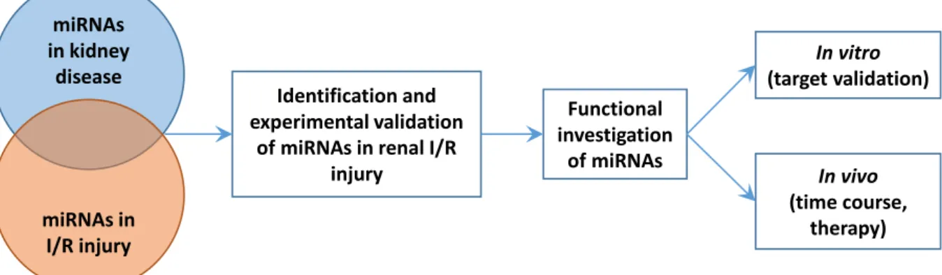
The objective of the thesis was to gain insight and understanding into the involvement of miRNAs in renal ischemia-reperfusion injury. Therefore our aims were (Figure 1):
1. to estimate which miRNA could be changed in renal ischemia- reperfusion injury through a comprehensive literature investigation;
2. to identify and validate selected miRNAs in ischemic acute kidney injury;
3. to analyze the function of the validated miRNAs, by:
a. time-course study, to investigate in which phase of AKI are the miRNAs upregulated;
b. evaluating the efficiency of a miRNA based therapy.
miRNAs in kidney
disease
miRNAs in I/R injury
Identification and experimental validation
of miRNAs in renal I/R injury
Functional investigation
of miRNAs
In vivo (time course,
therapy) In vitro (target validation)
Figure 1. Objectives and work-plan.
Identification of the miR-17 family in a murine renal I/R injury model and the time-course study of several miRNAs, carried out at Semmelweis University resulted in an original research article (further referred to as: miR-17 study). The investigation of miR-24 in ischemic AKI, containing target validation and in vivo evaluation of a miR-24 based therapeutic strategy, was carried out at Hannover Medical School and lead to another research article (further referred to as: miR-24 study).
4
Methods
Kidney ischemia-reperfusion injury
Acute kidney injury was induced by renal ischemia-reperfusion (I/R) injury in male C57BL/6 mice. After anesthesia the left renal pedicle was prepared and clamped. Sham operated control mice were also prepared in the same manner as those subjected to ischemia except that the renal pedicle was not clamped. The durations of reperfusion varied between 1 day and 7 days, in function of the experiment. For the survival analysis bilateral renal I/R injury was performed.
LNA-modified miRNA oligonucleotides therapies
Mice were dosed with intraperitoneal injections (i.p.) of a locked nucleic acid (LNA) modified oligonucleotide targeting miR-24 (LNA-24) as well as control LNA (directed against a microRNA expressed in Caenorhabditis elegans) (LNA-CTR) at a concentration of 10 mg/kg 24 hours before the operation.
Ex vivo cell purification/sorting
The cellular origin of miR-24 following induction of I/R-injury was investigated by fluorescence-activated cell sorting (FACS) analysis using specific antibodies: anti-CD31 for endothelial cells, Lotus tetragonolobus agglutinin (LTA) for proximal epithelium and anti-Tim1 for injured proximal epithelium. PDGF-Receptor beta+ pericytes were separated from kidneys by MACS magnetic bead separation.
Plasma Urea and NGAL ELISA
Renal function was evaluated by determination of blood urea nitrogen (BUN) retention. Urea values were divided by 2.14 to obtain BUN levels.
5
The levels of neutrophil gelatinase associated lipocalin (NGAL), a sensitive marker of tubular epithelial damage was determined with ELISA. The NGAL concentrations were calculated using a four parameter logistic curve-fit.
Histology and immunohistochemistry
Renal tissue samples fixed in buffered formaldehyde were dehydrated and embedded in paraffin wax (FFPE) for histology and immunohistochemistry.
Blocks of 70-sample tissue microarrays (TMA) were made from each FFPE kidney. Renal tubular necrosis and regeneration were evaluated in Periodic acid- Schiff (PAS) stained TMA sections. Renal tubular cell damage was evaluated also by NGAL immunostaining. For inflammatory cell influx the following primary antibodies were used: anti-F4/80 for macrophages, anti-CD45 for leucocytes, anti-Ly-6G/Gr-1 for neutrophils, anti-CD4 for T helper lymphocytes. Capillary rarefaction in the outer medulla was evaluated with anti- CD31 antibody. A fluorescein in situ cell death detection kit was used for Terminal deoxynucleotidyl transferase dUTP nick end labeling (TUNEL) assay.
Cell Culture experiments
For in vitro analyses immortalized human kidney proximal tubular epithelial cells (HK-2) were used. Transient liposomal transfection of miRNAs was carried out using specific and control miRNA and Lipofectamine 2000.
Scratch wound healing assay was also performed: after the scratches in the cell monolayer were generated the cells were photographed at 0, 8, and 24 hours and the cell free area was calculated.
RNA preparation
Total RNA was extracted from the upper third of the kidney with TRI- Reagent. The RNA concentration and purity was inspected by the 260 nm / 280 nm absorbance ratio. RNA samples were also electrophoresed on agarose gel and the 28S and 18S ribosomal RNA fraction integrity was examined.
6
Multiplex analysis of the microRNA profile
The expression of 46 microRNAs was determined with the Luminex® 200™ System. First, the extracted RNA was poly (A) tailed and biotinylated and then sample RNA hybridization with the bead mix and microRNA detection was performed. All samples were tested in duplicates and the background median fluorescence intensity (MFI) was subtracted before further calculations. MicroRNAs with low median fluorescence intensity were excluded from the statistical analysis.
Quantitative real-time PCR analysis of miRNAs and gene expression in renal tissue
MicroRNA expressions were evaluated with TaqMan probes and real- time quantitative PCR (qPCR). Gene mRNA levels were measured by dsDNA dye based SYBR Green qPCR. All samples were measured in duplicates and expressions were calculated using the relative quantification (ΔΔCq) method.
The qPCR reaction efficiency was also verified with standard curves. Gene array analysis was performed with Affymetrix GeneChip.
MicroRNA target prediction
MicroRNA databases and in silico target prediction tools were used to identify potential microRNA targets. Targets predicted by at least two prediction data bases and containing a miR-24-8mer seed match in the respective 3’UTR region were considered.
Statistical analysis
Continuous variables were compared using unpaired T-test or one-way analysis of variance (ANOVA), followed by the Dunnett's multiple comparison post hoc test versus the (sham) control group or Tukey's multiple comparisons test. Linear correlation was assessed with Pearson product-moment correlation coefficient. The null-hypothesis was rejected if the two-sided p-value reached statistical significance (* = p<0.05, ** = p<0.01, *** = p<0.001).
7
Results
miR-17 study
Lethal renal ischemia-reperfusion injury markers
After lethal, 30 min I/R injury the main histological lesions were found in the outer stripe of the outer medulla, and to a lesser degree in the cortical region.
Immunohistochemistry of the renal tubular damage marker NGAL demonstrated a strong NGAL specific tubular staining after I/R injury in the outer stripe and cortex. Whole kidney NGAL mRNA, plasma NGAL and BUN level increased compared to the sham operated group.
Kinetics of sublethal renal ischemia-reperfusion injury markers
Twenty-four hours after 20 min ischemia all kidney damage markers increased significantly. Thereafter, histologic signs of tubular regeneration appeared and renal damage markers started to decrease. On the fourth day of reperfusion all damage markers returned close to sham values.
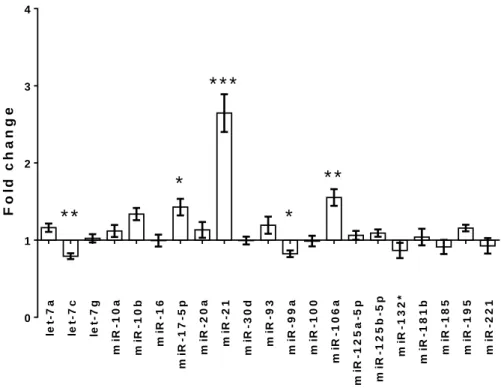
Micro RNA expression changes and time-course of renal miR-17-5p, miR- 106a and miR-21 expressions after renal ischemia-reperfusion injury
Five (miR-21, miR-17-5p, miR-106a, let-7c and miR-99a) miRNAs had a significantly different expression relative to the sham operated group (Figure 2).
However, only miR-21, miR-17-5p, and miR-106a changed more than 30% after I/R injury. The qPCR analysis confirmed the significance of the results regarding miR-17, miR-21 and miR-106a. After sublethal ischemia miR-17-5p increased significantly after one day of reperfusion, and remained significantly elevated until the third day. On the other hand, renal miR-21 expression was first elevated after three days of reperfusion, and remained upregulated on the fourth day, when miR-17-5p expression already returned to the sham-operated level.
8
let-7a let-7c let-7g miR-10a miR-10b miR-16 miR-17-5p miR-20a miR-21 miR-30d miR-93 miR-99a miR-100 miR-106a miR-125a-5p miR-125b-5p miR-132* miR-181b miR-185 miR-195 miR-221
0 1 2 3 4
* *
*
*
* *
* * *
Fold change
Figure 2. The miRNA expression profile of renal I/R injury, measured on the Luminex multiplex platform (fold changes observed after 24 hours of reperfusion following 30 min ischemia, compared to the sham-operated group).
Correlation between renal miR-17-5p and miR-21 expressions
Significant correlations were detected between renal miR-17-5p and miR- 21 expressions at every studied time point in both the ischemia-reperfusion and sham-operated groups. However, the slopes of the regression lines were significantly different between the ischemia-reperfusion and sham-operated groups on the third and fourth days of reperfusion. Moreover, the slopes became steeper as the reperfusion time increased.
9
miR-24 study
miR-24 in renal I/R-injury
Levels of miR-24 were increased in mouse kidneys at 1 and 7 days after induction of I/R injury compared to contralateral control kidneys. Cell sorting analysis after I/R-injury revealed a specific enrichment of miR-24 in tubular epithelial and endothelial cells at 1 day of reperfusion. At a reperfusion time of 7 days after I/R-injury miR-24 was up-regulated in injured tubular epithelial cells, although in healthy tubules changed to levels of controls. A slight, non- significant decrease in miR-24 expression was detected in pericytes. In kidney transplant biopsies of patients with prolonged cold ischemia time an increase in miR-24 was detected.
Functional role of miR-24 in tubular epithelial cells
Intriguingly, transfection of cells with miR-24 precursors without any additional cellular stressors culminated in an increase in apoptosis as assessed by TUNEL staining. Scratch migration analysis following miR-24 enrichment indicated a defect in tubular epithelial migratory capacity.
Targets of miR-24 in vitro
After a global messenger RNA expression analysis in proximal tubular epithelial cells following overexpression of miR-24 precursors 1822 genes were down-regulated compared to cells transfected with a pre-negative control oligonucleotide. In our subsequent analyses we focused on sphingosine-1- phosphate receptor 1 (S1PR1), H2A histone family, member X (H2A.X) and Heme Oxygenase-1 (HO-1).
Markers of kidney damage and endothelial activation in I/R-injury after miR- 24 silencing
Treatment of mice with an LNA-modified antimiR targeting miR-24 (LNA-24) before unilateral I/R injury not only resulted in a better survival, but also in a marked reduction of kidney injury marker (NGAL and KIM-1) gene
10
expression and amelioration of renal function parameters (serum-creatinine and -urea). Epithelial cell injury and capillary rarefaction on day 1 after I/R-injury was significantly improved in animals treated with an LNA-modified antimiR targeting miR-24.
Kidney morphology, infiltration of immune cells, level of apoptosis after miR- 24 silencing
LNA-24 treatment resulted in a significant improvement of kidney morphology on day 1 after I/R-injury. Infiltration of leucocytes, macrophages, T-cells, and neutrophils significantly decreased following LNA-24 treatment at all investigated time points. Tubular cell apoptosis was significantly lower in LNA-24 treated animals at 1 day of reperfusion.
Target regulation of miR-24 in I/R-injury in vivo
We found HO-1 to be significantly up-regulated in the outer medulla after anti-miR-24 LNA as compared to control LNA (LNA-CTR) treated animals.
H2A.X was also found to be up-regulated by miR-24 inhibition, though not to a statistically significant level. S1PR1 was not regulated in vivo.
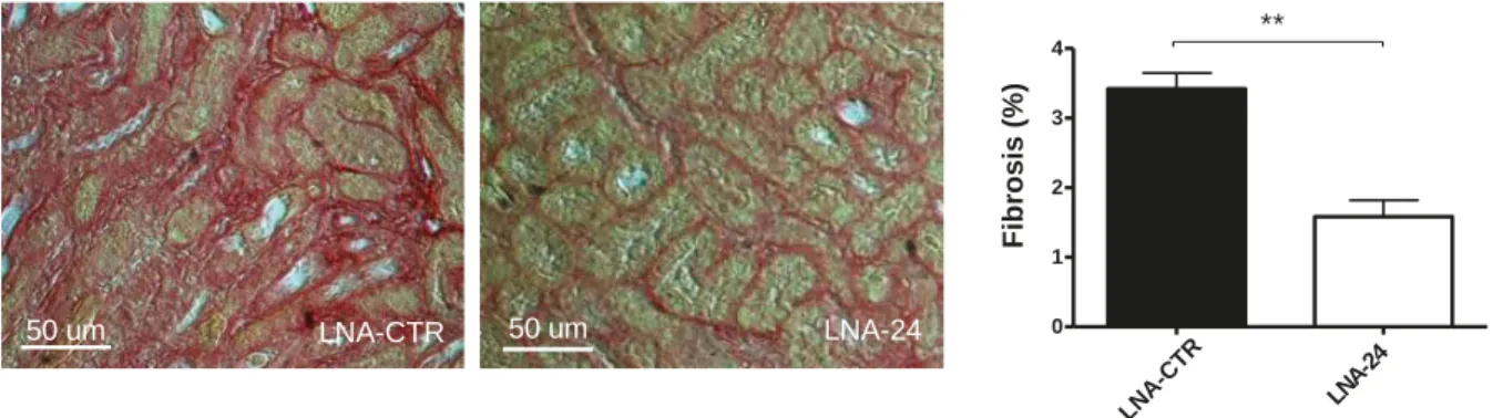
MiR-24 in the progression from acute kidney injury to chronic kidney disease At a reperfusion time of 7 days the level of developing fibrosis and expression of fibrosis-associated genes was highly reduced in LNA-24 treated mice subjected to unilateral I/R-injury (Figure 3).
50 um LNA-24
LNA-CTR 50 um
LNA-CTR LNA-24 0
1 2 3 4
Fibrosis (%)
**
Figure 3. Fibrosis development (sirius red-staining) analysis in mice with renal I/R injury treated with control LNA (LNA-CTR) and LNA-24 and quantification of results at reperfusion for 7 days.
11
Conclusions
The presented results demonstrated that miR-17-5p, miR-21, miR-106a and miR-24 are involved in the pathophysiologic processes of the ischemia- reperfusion (I/R)-induced acute kidney injury (AKI).
The miR-17 study showed that miR-17 upregulation occurred during the maintenance phase and was followed later by miR-21 upregulation, which in turn lasted until the recovery phase. The expression of these miRNAs correlated with each other, which finding should be further investigated to get a deeper understanding of a possible relationship between miR-21 and miR-17. The timing of miRNA up-regulations suggests that miR-17 and miR-21 could play a role in the recovery phase of the I/R-induced AKI. Considering the progresses made in miRNA research in the kidney and miRNA based therapeutic approaches miR-17-5p, miR-21 and miR-106a could represent novel targets in the treatment of the ischemia-induced AKI.
The miR-24 study revealed that miR-24 contributes to renal I/R-injury by influencing tubular epithelial and endothelial apoptosis through regulation of anti-apoptotic HO-1 and H2A.X. Here we provide clear evidence of the pro- apoptotic role of miR-24 in renal I/R-injury. Silencing miR-24 ameliorates the apoptotic response in vivo leading to suppressed tubular epithelial and endothelial apoptosis, which is associated in turn with enhanced capillary density and reduced tubulo-interstitial fibrosis and the potential of blunting the AKI (acute kidney injury) to CKD (chronic kidney disease) continuum. Of note, this is the first evidence demonstrating that pharmacological miRNA inhibition might be a viable therapeutic option in the treatment of patients with this life- threatening clinical disorder. Intriguingly, miR-24 is also enriched in transplant kidneys of patients with prolonged cold ischemia time, indicating its potential role in human renal I/R- injury. This study highlights that miR-24 modulation might ultimately lead to the first targeted clinically applicable therapy of patients with AKI.
12
Candidate’s publications
Related to the theme of the PhD thesis
Kaucsar T, Racz Z, Hamar P. (2010) Post-transcriptional gene-expression regulation by micro RNA (miRNA) network in renal disease. Adv Drug Deliv Rev, 62(14): 1390-401. IF: 13.577
Rácz Z, Kaucsár T, Hamar P. (2011) The huge world of small RNAs: regulating networks of microRNAs (review). Acta Physiol Hung, 98(3):243-51. IF: 0.821
Kaucsár T, Révész C, Godó M, Krenács T, Albert M, Szalay CI, Rosivall L, Benyó Z, Bátkai S, Thum T, Szénási G, Hamar P. (2013) Activation of the miR- 17 family and miR-21 during murine kidney ischemia-reperfusion injury.
Nucleic Acid Ther, 23(5):344-54. IF: 2.888
Lorenzen JM, Kaucsar T, Schauerte C, Schmitt R, Rong S, Hübner A, Scherf K, Fiedler J, Martino F, Kumarswamy R, Kölling M, Sörensen I, Hinz H, Heineke J, van Rooij E, Haller H, Thum T. (2014) MicroRNA-24 Antagonism Prevents Renal Ischemia Reperfusion Injury. J Am Soc Nephrol, 25. DOI:
10.1681/ASN.2013121329. IF(2013): 9.466
Unrelated to the theme of the PhD thesis
Kaucsár T, Bodor C, Godó M, Szalay C, Révész C, Németh Z, Mózes M, Szénási G, Rosivall L, Sőti C, Hamar P. (2014) LPS-induced delayed preconditioning is mediated by Hsp90 and involves the heat shock response in mouse kidney. PLoS One, 9(3):e92004. IF(2013): 3.534
Paşca SP, Dronca E, Nemeş B, Kaucsár T, Endreffy E, Iftene F, Benga I, Cornean R, Dronca M. (2010) Paraoxonase 1 activities and polymorphisms in autism spectrum disorders. J Cell Mol Med, 14(3):600-7. IF: 4.608
Paşca SP, Dronca E, Kaucsár T, Craciun EC, Endreffy E, Ferencz BK, Iftene F, Benga I, Cornean R, Banerjee R, Dronca M. (2009) One carbon metabolism disturbances and the C677T MTHFR gene polymorphism in children with autism spectrum disorders. J Cell Mol Med, 13(10):4229-38. IF: 5.228
13
Acknowledgements
First I would like to thank my supervisor, Dr. Péter Hamar for his efforts in teaching and supporting me during my PhD studies, pointing out the importance of the in vivo experiments.
I am also grateful to all my colleagues from the Institute of Pathophysiology:
Prof. Dr. László Rosivall, Head of the Basic Medicine Doctoral School for the opportunity to carry out my PhD studies; Dr. Gábor Szénási for his indispensable advices and for sharing me his broad research experience. Mária Godó, Csaba Révész, Ágnes Cser, Dr. Csaba Szalay, Dr. Zsuzsanna Rácz and Csaba Bodor for their kind help in the experiments and for teaching me numerous methods. Dr. Gábor Kökény for his valuable suggestions serving to the improvement of the present thesis.
Special thanks to Prof. Dr. Thomas Thum, Dr. Johan Lorenzen and Dr. Sándor Bátkai and all the members of the Institute of Molecular and Translational Therapeutic Strategies (Hannover Medical School, Germany) for their support during my KAAD Scholarship and for the possibility to be involved in and learn from their innovative research.
I would also like to thank my previous supervisors: Dr. Ioana Berindan Neagoe and Dr. Maria Dronca (Iuliu Haţieganu University of Medicine and Pharmacy, Romania) for initiating me in biomedical research and for supporting my application for PhD studies.
Many thanks to the community of the ReNaissance House (and to Ulrich Kiss SJ) for being my “second family” during my PhD studies in Budapest.
Nevertheless I am very grateful to my parents, my sister and my grandparents for trusting me and for the unconditional support and encouragement during my professional trainings.
Research in this thesis was supported by U.S. National Institutes of Health Research grant #R03 TW07069 funded by the Fogarty International Center and the National Institute of Diabetes and Digestive and Kidney Diseases. Further support was provided by the Hungarian Scientific Research Fund (OTKA: K81972, NF69278), the Health Science Council (ETT:
011-07/2009) and the Else-Kröner-Fresenius Foundation awarded to J.L. and T.T. The assistance of the Cell Sorting Core Facility of the Hannover Medical School supported in part by Braukmann-Wittenberg-Herz-Stiftung and Deutsche Forschungsgemeinschaft is also acknowledged. My research in Germany was supported by the scholarship of the Catholic Academic Exchange Service (KAAD).