The role of renal venous pressure in the development of postoperative acute kidney injury
Main findings in the thesis Csaba Kopitkó, MD.
Semmelweis University
Doctoral School of Pathological Sciences
Supervisor: Tibor Gondos, MD., CSc.
Official reviewers:
Zsolt Molnár, MD., DSc.
Zsolt Iványi, MD., PhD Final Examination Committee:
Head: Endre Zima, MD., PhD.
Members: Edit Király, MSc., PhD.
István Hornyák, MSc., PhD.
Budapest 2019
Introduction
Acute kidney injury (AKI) is still a leading cause of intensive care mortality / morbidity following major abdominal surgery, partly due to changes in sepsis, and in part to changes in pressure on glomerular perfusion.
The background of postoperative AKI is mainly due to circulatory causes; by optimizing the mean arterial pressure (MAP) and the abdominal perfusion pressure (MAP – intraabdominal pressure [MAP)], the risk of AKI can be reduced. The IAP and the driving pressure of filtration in glomeruli can differ significantly due to the tight capsule of the kidney. Studies conducted with renal perfusion calculated by MAP – central venous pressure (CVP), which better represents the intracapular pressure, have been shown in cardiology patients.
Intensive care unit (ICU) patients often require ventilatory support, which increases the risk of AKI. The increase in risk cannot be attributed to the positive end expiratory pressure (PEEP) on the basis of literature data. No published data are available for other airway pressure values.
Objectives
The incidence of AKI is on an upward trend, significantly increasing ICU mortality. Its early diagnosis is very insistent, because it is still based on the measurement of serum creatinine with slow kinetics and the rate of urine output. Biomarkers failed the expectations of them, their use did not spread.
The aim of our study was to determine:
• the abdominal perfusion pressure at which does not develop AKI in the certain hemodynamic constellation
• the effect of CVP on AKI after major abdominal surgery
• the effect of invasive ventilation on the onset of AKI, with particular reference to the mean airway pressure (Pmean)
• the optimal way to calculate the renal fusion pressure using the parameters listed above.
Materials and Methods
Between 5 January 2015 and 15 October 2015, a multicentre study was carried out with the participation of nine Hungarian ICUs (4 university, 4 regional and 1 city hospitals). Patients over 18 years of age, who have been undergone major abdominal surgery, admitted to ICU for at least 48 hours in the immediate postoperative period, were included in the study. The study was authorized by resolution No. 039320/2014 / OTIG of the Hungarian Health Registration and Training Center. Before recruiting in the trial, we have received written informed consent from the patient / their legal guardians or next-of-kin.
Patients were categorized into two large groups after the end of data collection, depending on whether they had acute renal failure after 48 hours or not.
Patients were excluded from the study
a) who have end-stage (dialysis-dependent) renal failure on admission to the ICU;
b) who required suprarenal crossclamping during surgery;
c) whose kidneys have been affected by surgery, thus reducing the number of nephron, thus preventing the diagnostic utility of serum creatinine;
d) where the surgery also affected the bladder, so intra- abdominal pressure measurement became uncertain.
The urine volume, MAP, IAP and CVP, PEEP and Pmean were recorded on ICU admission and at 6, 12, 24, and 48 hours thereafter. Blood urea and creatinine levels were measured after 24 and 48 hours.
IAP was measured in the bladder in a supine position at the end of the expiration. The zero point was the intersection of the iliac crest and the midaxillary line. Measurements were done 30-60 seconds after administration of 25 ml of saline solution, with no active abdominal activity. The result was
given in mmHg
(1 mmHg = 1.36 H2Ocm).
AKI was defined by the AKIN criteria system.
Data analysis was performed after data collection was completed. The data were presented as median and interquartile range (IQR). In order to find the optimal
method of calculating renal perfusion pressure, venous pressures reducing MAP were examined in all possible combinations. The median data of the different groups were compared with the Mann-Whitney U-test and the prevalence rates were compared with the χ2-test. Receiver Operating Curve (ROC) analysis was performed for parameters significantly discriminating AKI and no-AKI groups. The optimal threshold was determined based on both the Youden index and the root mean squared error (RMSE).
Since it cannot be ruled out on a theoretical basis that CVP, PEEP/Pmean and IAP influence each other, linear regression analysis was performed as an independence test.
We accepted the parameters as independent, and they were released for further analysis only if r <0.3.
Results
In the study, 84 patients (with normal kidney function: 45 persons - 53.6%, kidney failure: 39 people - 46.4%, of which AKI I: 21 persons - 25%, AKI II: 9 persons - 10.7%, AKI III 9 persons - 10.7%) could be evaluated. The number of patients, who were finally successfully included, only allowed detailed statistical processing of the differences between the no-AKI and AKI groups. Due to the low number of cases, statistical analysis according to the individual stages of AKI was not meaningful.
The lower limit of MAP’s interquartile range was above 65 mmHg in both groups, at each measuring point, and the median was between 70 and 80 mmHg. No significant difference was found between the no-AKI / AKI group, except for the difference observed at the time of admission (89 vs. 82 mmHg). The other pressure values (CVP, IAP, PEEP, Pmean) also showed no significant difference between the two groups, each were in physiological and tolerable ranges.
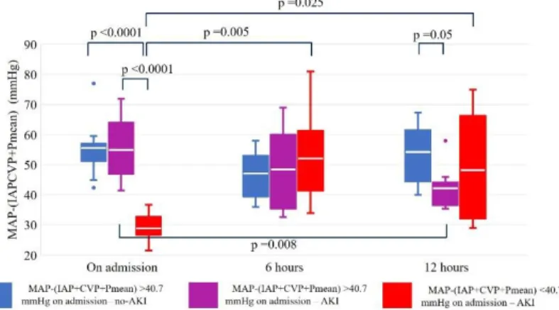
Of the formulas for the renal fusion pressure, MAP – (IAP + CVP + Pmean) proved to be the most predictive for AKI.
ROC analyses were performed:
• 1-AUC measured at the time of admission: 0.794; LR +:
2.743; LR: 0.282; sensitivity: 80%, specificity 71%; p <0.01
• 1-AUC measured after 12 hours: 0.810; LR +: 3,000; LR:
0.200; sensitivity: 86%, specificity 71%; p <0.05
• 1-AUC measured at 6-12 hours change: 0.796; LR +:
4.500; LR: 0.176; sensitivity: 86%, specificity 81%; p
<0.05.
Patient data were divided based on the median values of MAP – (IAP + CVP + Pmean) on ICU admission (40.7 mmHg). Below this level, AKI developed in all patients. In patients with renal perfusion pressure above median level, the development of AKI was associated with a tendency of renal perfusion: patients having rising perfusion pressure in the first 12 hours (especially between 6 and 12) in ICU were protected against AKI; but the AKI remained unavoidable
in almost all patients, at whom renal perfusion has worsened.
1. Figure. The course of MAP-(IAP+CVP+Pmean) in the first 12 hours in ICU after major abdominal surgery. The p values within or between groups
not shown were not statistically significant.
Discussion / Conclusions
Postoperative AKI is known to be due to circulatory causes.
Proper monitoring of kidney perfusion has many difficulties. In the absence of readily available direct methods, indirect measurements must be inferred. Among the influencing hemodynamic parameters, routine invasive monitoring of MAP, IAP and CVP is generally accepted. Of the respiratory parameters, the recording of the PEEP that may affect renal venous pressure is a common practice in ICUs, but monitoring of the Pmean is nowadays unusual.
Numerous summaries mention the role of venous dysfunction in kidney perfusion, but none of them serve as a basis for monitoring.
In our study, observations were made about the near- physiological pressure values, suggesting that the unrelated variations may result in deterioration of the glomerular perfusion. It can be assumed that our findings are valid, or that they may be subject to higher statistical power with more abnormal parameters. In our study, patients with preoperatively known end-stage renal insufficiency were excluded, but no persons with significantly elevated serum
creatinine levels were included. It may be assumed that in patients with pre-existing renal impairment, patients could benefit more from the opportunities offered by renoprotective monitoring.
Overall, our research focused on a thrilling area of intensive therapy, with many open questions that, by responding to the shift from global perfusion parameters to regionality and to the personalized care, results the patient to spend shorter time in the intensive care unit and provide better quality of life for patients afterwards.
List of own publications
LIST OF PUBLICATIONS RELATED TO THESIS
Csaba Kopitko, Laszlo Medve, Tibor Gondos. (2016) Pathophysiology of renal blood supply. New Med, 20(1):
27-29.
Csaba Kopitko, Laszlo Medve, Tibor Gondos. (2017) Renal blood supply and fluid therapy. New Med, 21(1):
21-24.
Csaba Kopitko, Laszlo Medve, Tibor Gondos. (2018) Renoprotective postoperative monitoring: what is the best method for computing renal perfusion pressure? – an observational, prospective, multicentre study. Nephron, 139: 228-236.
Kopitkó Csaba, Medve László, Gondos Tibor. (2018) A szeptikus heveny vesekárosodás kialakulása, kezelési lehetőségek. Aneszteziológia és Intenzív Terápia, 48(3): 5- 16.
Kopitkó Csaba, Medve László, Gondos Tibor. (2018) A vénás pangás rontja a veseműködést – fikció vagy valóság?
Lege Artis Medicinae, 28(10): 451-457.
Csaba Kopitkó, László Medve, Tibor Gondos (2019) The value of combined hemodynamic, respiratory and intra- abdominal pressure monitoring in predicting Acute Kidney Injury after major intraabdominal surgeries. Renal Failure, doi: 10.1080/0886022X.2019.1587467.
LISTOFPUBLICATIONSNOTRELATEDTOTHESIS
Molnár Miklós, Kopitkó Csaba, Szőcs Katalin, Hertelendy Ferenc. (1997) A tamoxifen fokozza az angiotenzin II vérnyomásemelő hatását. In: Magyar Élettani Társaság 62.
Vándorgyűlése. Konferencia helye, ideje: Pécs, Magyarország, Paper 40.
Katalin Szöcs, Csaba Kopitkó, Miklos Molnár, Frank Hertelendy. (1997) Estrogen and nitric oxide are critical in maintaining refractoriness to the pressor action of
angiotensin II in pregnancy. J Soc Gynecol Investig, 4:(1 S) Paper 679.
Kopitkó Csaba, Szőcs Katalin, Molnár Miklós. (1996) A tamoxifen fokozza az angiotenzin II vérnyomásemelő hatását. In: XXVI. Membrán-Transzport Konferencia Sümeg. Konferencia helye, ideje: Sümeg, Magyarország, Paper 60.
Kopitkó Csaba, Szőcs Katalin, Molnár Miklós. (1996) Tamoxifen, Increases Angiotensin II Pressor Responsiveness and Decreases Nitrate in Pregnant Rats. In:
Magyar Élettani Társaság LXI. vándorgyűlése. Konferencia helye, ideje: Szeged, Magyarország, Paper 98.
Cecconi M, Hofer C, Teboul JL, Pettila V, Wilkman E, Molnar Zs, Rocca GD, Aldecoa C, Artigas A, Jog S, Sander M, Spies C, Lefrant JY, De Backer, D. and on behalf of the FENICE Investigators and the ESICM Trial Group. (2015) Fluid challenges in intensive care: the FENICE study: A global inception cohort study. Intensive Care Med, 41(9), 1529-1537. – mint vizsgáló.