Slit Ventricle as a Neurosurgical Emergency: Case Report and Review of Literature
Zoltan Mencser1, Zsolt Kopniczky1,2, David Kis1, Pal Barzo1
INTRODUCTION
Symptomatic slit ventricle is one of the most challenging complications of shunt surgery in children.1-4 The clinical triad features headache, slow refilling of the valve, and narrow ventricles on imaging.
Most authors agree that the most likely underlying mechanism is chronic cere- brospinal fluid (CSF) overdrainage. How- ever, clinical signs and symptoms may appear along with a wide range of intra- cranial pressure (ICP) values. Probably, as the Rekate classification suggests,4 multiple pathophysiologic mechanisms can be involved to produce these symptoms. The common features are previous hydrocephalus treated with shunt insertion and headache.4,5
The following 5 distinct syndromes were identified by Rekate:1) intermittent, extremely low-pressure headaches that are analogous to spinal headaches; 2) inter- mittent proximal obstruction; 3) shunt failure with small ventricles (normal vol- ume hydrocephalus); 4) intracranial
hypertension with working shunts (hy- drocephalic pseudotumor); and 5) head- aches unrelated to shunt function.
The clinical appearance is marked with chronic or intermittent headache, associ- ated nausea, vomiting, lethargy, and different degrees of consciousness or irritability.6
Patients in the Rekate groups 2, 3, and 4 are at major risk when elevated ICP war- rants urgent actions.7 Early recognition and adequate therapy are especially important in cases of groups 3 and 4, which are characterized by intermittent shunt failure, causing increased ICP either in a form of spike waves or as a progressive elevation of baseline pressure. Urgent intervention is needed
in a classic clinical form of raised ICP, such as confusion, drowsiness, slow psychomotor reactions, and vegetative reflexes (bradycardia and hypertension).4,8
CASE PRESENTATION
At 6 months of age, the patient presented squeal for several hours accompanied with sunset eyes sign and bulging anterior fontanel. The cranial ultrasound and head computed tomography scan showed dilated ventricles under tension of all 4 ventricles with left-sided porencephaly.
She was diagnosed with hydrocephalus, and prompt insertion of a ventriculoper- itoneal shunt system was performed with a programmable valve (Sophysa SM8
-BACKGROUND:Symptomatic slit ventricle is one of the most challenging complications of shunt surgery in children. Clinical signs and symptoms may appear with a wide range of intracranial pressure (ICP) values. We report the case of a 10-year-old girl, who did not present the classic clinical features of extremely elevated ICP, which was proven by multiple invasive ICP recordings, performed during shunt revisions.
-CASE DESCRIPTION:At the age of 6 months, the patient presented squeal for many hours, accompanied with sunset eyes, bulging anterior fontanel, and dilated ventricles of all 4 ventricles on computed tomography scan. Acute ventriculoperitoneal shunt insertion was performed with adjustable valve.
During the following 9 years, she was regularly seen and medically treated for intermittent headache, with nausea and vomiting. From 9 years of age, she was hospitalized for severe (10/10 on the visual analog scale), unbearable headache, agitation, and screaming on multiple occasions. Altogether, we had to revise the shunt system 5 times throughout 1 year. Radiologic imaging always showed narrow ventricles. Ophthalmologic examination of the fundus never revealed signs of raised ICP. Perioperative monitoring of the ICP with intraparenchymal sensor showed unexpected high values of 40e45 mm Hg. However, repetitive shunt revisions were successful only temporarily because the symptoms always returned. Only bilateral shunting of the ventricular system was able to eliminate the symptoms permanently.
-CONCLUSIONS:Images of slit ventricle can be associated either with low or extremely high ICP needing urgent surgical consideration, including ICP moni- toring. Bilateral shunt insertion can be effective treatment for slit ventricle syndrome.
Key words
-Hydrocephalus
-Raised ICP
-Shunt failure
-Slit ventricle
Abbreviations and Acronyms CSF: Cerebrospinal fluid ICP: Intracranial pressure
From the1Department of Neurosurgery, Faculty of Medicine, Albert Szent-Györgyi Clinical Center, University of Szeged, Szeged; and2Department of Neurotraumatology, Péterfy HospitaleNational Institute of Traumatology, Budapest, Hungary
To whom correspondence should be addressed:
Zoltan Mencser, M.D.
[E-mail:mencser67@gmail.com]
Citation: World Neurosurg. (2019) 130:493-498.
https://doi.org/10.1016/j.wneu.2019.07.006
Journal homepage:www.journals.elsevier.com/world- neurosurgery
Available online:www.sciencedirect.com 1878-8750/$ - see front matterª2019 Elsevier Inc. All rights reserved.
[Sophysa, Orsay, France]). Acute clinical signs and symptoms improved as raised ICP returned to a normal level.
During the following 9 years, she was occasionally seen at our department for intermittent headache, which sometimes was associated with an upper respiratory tract infection and could be managed medically. She was admitted at the age of 4 years, when headache set on suddenly, and she was screaming because of intol- erable pain. The pain disappeared after a deep sleep, but later reappeared again.
However, resetting the valve opening pressure at a higher level and enhancing fluid intake relieved her symptoms without need for surgery.
During the following years, occasional episodes of headaches, a short period of drowsiness, and difficulty coping with school were reported, but follow-up im- aging always showed a normal to narrow ventricle system, and the patient’s com- plaints always ceased a few days later, spontaneously. Multiple ophthalmologic examinations could not identify any ab- normalities regarding the fundi, and papillary edema was never reported.
From the age of 6 years, episodes of sudden, severe headaches along with screaming became more and more frequent; however, they resolved just as suddenly, in a few minutes or after some sleep. The patient was hospitalized 3 consecutive times because of the clinical scenarios previously described, but sur- gery was not indicated, and the symptoms resolved in 24e48 hours. Retrospectively, from the clinical reports, we know that the opening valve pressure needed multiple readjustments from a low (30 mm H2O) to a higher level.
At 9 years of age and onward, the pa- tient was hospitalized on multiple occa- sions for strong (visual analog scale score 10 of 10), unbearable headaches accom- panied with agitation and screaming. She never presented a progressively reduced level of consciousness or confusion, only moderate, temporary drowsiness. Alto- gether, we had to revise the shunt system 5 times throughout a year.
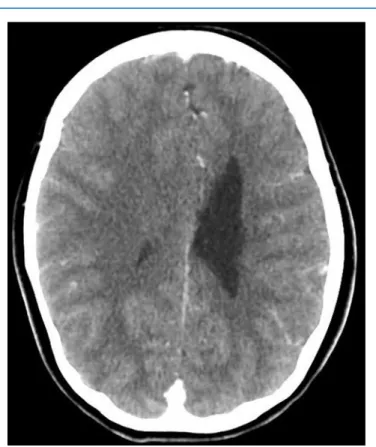
Radiologic imaging always showed narrow ventricles. (Figure 1).
Ophthalmologic examinations did not reveal signs of raised ICP.
In 3 of the 5 surgeries, we measured
and monitored ICP with an
intraparenchymal sensor (Pressio [Sophysa, Orsay, France]), which was inserted at the beginning of the opera- tions. Each case gave unexpectedly high values (40e50 mm Hg).
During the surgeries, we never found obstruction of any part of the right ven- triculoperitoneal shunt system. Cautious movement of the proximal drain resulted in promptflow of CSF, or a new ventric- ular drain always restored a satisfactory shunt function for a while. In 2 revisions out of the 5, the reposition of the ven- tricular catheter was enough to restore the function of the catheter. In the other 3 cases, once a valve replacement was per- formed, and twice we changed the ven- tricular catheters.
A long-term solution could only be achieved by adding another shunt system on the opposite (left) side, with a medium-pressurefixed valve.
In the past 5 years, the patient has experienced no similar symptoms, and follow-up magnetic resonance imaging scans show bilateral narrow ventricles.
DISCUSSION
In the presented case, the clinical signs and symptoms of raised ICP could not be matched with the radiologic images of narrow/slit ventricles and normal ophthalmologic findings. However, the patient did not show the classical symp- toms of raised ICP, whereas the objective values measured with an intraparenchymal sensor were extremely high, and imaging remained insignificant. Thesefindings are contradictory and raise the question of what the rational description of the un- derlying physiologic events could be.
Regarding the lack of ventricular dila- tion in case of high-pressure shunt failure,
Figure 1. (AeC) Cranial computed tomography (CT) scans. Three axial CT scans taken in the periods of acute strong headaches show no signs of dilated ventricles. The intracranial pressure measured during surgery each time raised above 40 mm Hg.
the literature puts forward several theories.
Venous Outflow Disturbances
The brain matter can only change its vol- ume by mobilizing itsfluid content. This depends on the venous outflow capacity.
Because the venous outflow is limited or blocked, the brain becomes rigid and incompressible; therefore, the ventricular system cannot enlarge. In adults, pseu- dotumor cerebri is often associated with morphologically proven venous outflow disturbances,2,9-14 raised ICP without enlarged ventricles. In the presented case, we could not identify any abnormalities in the venous outflow system: the venous sinuses were intact and had no stenosis or obstruction (Figure 2), but the venous hypertension is the most likely part of the pathomechanism.
Arterial Pulsatility Vector Changes The drainage of CSF by a shunt system substantially alters the spread of arterial pulsatility waves onto the CSF space. The arterial pulsatility vector may be abolished with the subsequent loss of the diastolic recurrent wave of the subarachnoid space and its effect at the cortical surface.
Capillary venous outflow of the cortex will be changed because of a lack of diastolic waves. Once the level of ICP reaches the venous pressure of the bridging vessels, they will collapse, and the venous outflow resistance will rise.15 The simultaneous increase of venous pressure and ICP in children with hydrocephalus has been reported for many years,1,16-18 especially in those who, as a response to shunt malfunction, present with slit ventricle syndrome.19 These findings were interpreted as an early collapse of bridging veins or obstruction of the sinuses.12,13
Isolated Ventricle
Chronic shunting may lead to ventricular asymmetry, and even to the collapse of the ipsilateral ventricle and a functional blockage of the proximal drain. This consequently may cause contralateral ventricular dilation (Figure 3), and if the foramen of Monro becomes closed because of displacement of the septum pellucidum, it may become isolated.3,20,21 Typically, this mechanism would cause intermittent, fluctuating clinical symptoms, like in the presented case.
The right-side ventricles were always
collapsed, whereas the left-side ventricles were moderately dilated. Retrospectively, this mechanism is proven by the long- term benefit of shunt insertion into the left ventricle (where porencephaly was also present).
Theory of Capillary Absorption Laziness This concept argues that chronically altered CSF absorption pathways at the capillary level will ultimately damage normal CSF outflow dynamics, and will lead to high-pressure slit ventricle syn- drome.22 The normal transcapillary absorption is practically abolished because of an easier outflow offered by shunt insertion. A large part of CSF is drained by the functioning shunt, and the transmural CSF pressure is minimized, which progressively results in the blockage of transcapillary absorption, causing rigidity and incompressibility of brain parenchyma. Our patient’s long- lasting abnormal physiologic conditions might have contributed to these mecha- nisms as well.
Our examinations proved that there was no morphologic venous outflow obstruc- tion, but the venous hypertension could persist without abnormal dural venous anatomy,14 and all other mechanisms could have played a role in the clinical evolution of this case. However, most probably, the low opening pressure of the valve (30 mm H2O) must have caused chronic CSF overdrainage with all its harmful consequences.3,18 We cannot fully understand why the valve setting was changed to repeatedly low values.
Furthermore, isolated ventricle syn- drome also developed during critical acute periods. The temporary blockage of the foramen of Monro because of the collapse of midline structures made the left-sided ventricle close, the CSF become trapped, and the ventricle become isolated. From this moment, the retrogradeflow will be impaired from the subarachnoid spaces to the ventricle and from the left side to the right. This is what we could identify on the follow-up CT scans on occasions of com- plaints: the left-side ventricle was moder- ately dilated, whereas the structures on the right side collapsed.
The pressure-volume dynamic features are rather similar to those of patients with severe traumatic brain injuries: a small
Figure 2. Venous phase magnetic resonance angiography showing patent venous structures of the brain.
add up (of CSF) to intracranial volume increases the ICP disproportionately.23-25
Presumably, children with a shunt sys- tem probably have a different capacity of autoregulation, which may ensure appro- priate cerebral perfusion even with elevated ICP.17,18,24 However, in cases of slit ventricle syndrome, the autoregulation capacity might be exhausted, and minimal increase in CSF volume may provoke a sharp increase in ICP. These pressure- volume reactions are very similar to those experienced in severe traumatic brain injury, depending on the segment of the pressure-volume curve of the patient’s vascular compliance.8
We think in the presented case that a significant, long-term drainage of CSF (overdrainage) stopped the CSF absorp- tion into the dural venous sinuses by keeping the ICP low and prompted the consequent disturbance in venous circu- lation. The complex process and the effect of long-term shunt treatment 1) caused ventricular asymmetry with recurrent
obstructions of the foramen of Monro leading to pressure differences and blocking of the CSF flow, 2) caused insufficient capillary absorption, or 3) induced a collapse of bridging veins with facilitation of an increase in venous hy- pertension, and eventually the slit ventricle syndrome.3,17,18,22
The management of symptomatic slit ventricle widely varies from pain manage- ment26 and repeated shunt revisions27 to different extracerebral decompressive surgeries (subtemporal craniectomy and cranial expansion).28,29 In the literature, shunt revision generally stands for the change of the ventricle catheter, an insertion of a new valve of higher- pressure threshold or antisiphon de- vice.27 The literature reports successful decompressive procedures, aiming to reduce the pressure exercised to the brain and the extra-intracerebral CSF pathways. The underlying idea is to keep the ventricles open around the catheter and prevent its isolation. By normalizing
the pressure of extracerebral CSF spaces, the collapse of the bridging veins and secondary venous stagnation can also be prevented.
Albright and Tyler-Kabara30 performed craniotomy and craniectomy for early ossification of the skull because of shunt overdrainage, which successfully treated the symptoms of slit ventricle. Others found this type of management to be effective only for a limited period of time, which warranted another decompressing intervention, either a combined, ventriculo- and lumboperitoneal shunt31 or cisterna magna-peritoneal shunt inser- tion. Finally, application of a combined cisterna-magna and ventriculoperitoneal shunt could ensure permanent recovery of the patient.32
Retrospectively, we think that left-side porencephaly and brain atrophy pre- vented the lateral ventricle from collapse, which could have an effect similar to extracerebral decompression (lumboper- itoneal shunt or craniectomy). Normal CSF outflow could ensure equal pressure conditions between the 2 lateral ventricle systems. The elimination of the difference in pressure (with both sides of shunt placement) could reopen the foramen of Monro and prevent recurrent blockage.
The benefit from the insertion of another shunt system into the left ventricular sys- tem justifies this idea because the patient has presented no symptoms since then, she only has occasional headaches without neurologic signs. This theory has been confirmed by a dog model: when CSF was withdrawn from one lateral ventricle, dogs showed intraventricular pressure differen- tials. The foramen of Monro acts as a valve mechanism that usually closes in response to CSF withdrawal.
Since the contralateral ventricles were shunted 5 years ago, the presented patient has been doing well. The beneficial effect of the contralateral shunting may be attributed to the fact that avoidance of ventricular isolation keeps the way to shunts that provide CSF drainage. When a surgeon is developing surgical strategies to treat patients with slit ventricle syn- drome, all of these mechanisms should be taken into consideration.
We have to acknowledge that the nar- row ventricles, the lack of a significant fall in the level of consciousness, and the lack
Figure 3. Cranial computed tomography scan. During headache, the left lateral ventricle shows only moderate dilatation.
of papillary edema delayed us in recog- nizing the extremely high ICP.
In fact, in slit ventricle syndrome cases, the real diagnostic challenge is to define high (critical) ICP without ventricular dilation.
As previously discussed, raised ICP can evolve without progressive deterioration in consciousness.
CONCLUSIONS
We aimed to highlight the importance of ICP measurement33,34 to define the path- ophysiologic background of shunt mal- function and to give an overview of the literature about possible mechanisms of slit ventricle syndrome, which is a special case of shunt failure.
The presented case, when mild-to- moderate clinical signs and irrelevant radiologic findings did not appropriately reflect the highly abnormal pressure condi- tions, was rather challenging to manage.
Because radiologic images of the slit ventricle can equally be associated with either low or extremely high ICP, the clinical observation and ophthalmologic findings must be completed with ICP. Only its values can indicate adequate surgical management.
Children having had previous shunt insertion and showing a sudden onset of irritability or a decreased level of con- sciousness might have threateningly high ICP, even without any evidence of ven- tricular dilation, and might need urgent surgical consideration. Although many medical and surgical treatment modalities are recommended in the literature, the slit ventricle syndrome can effectively be managed also by bilateral shunting of the ventricular system.
REFERENCES
1. Engel M, Carmel PW, Chutorian AM. Increased intraventricular pressure without ventriculomegaly in children with shunts:“normal volume”hydro- cephalus.Neurosurgery. 1979;5:549-552.
2. Bateman GA. Hypertensive slit ventricle syn- drome: pseudotumor cerebri with a malfunction- ing shunt?J Neurosurg. 2013;119:1503-1510.
3. Ros B, Iglesias S, Martín Á, Carrasco A, Ibáñez G, Arráez MA. Shunt overdrainage syndrome: review of the literature.Neurosurg Rev. 2018;41:969-981.
4. Rekate HL. Shunt-related headaches: the slit ventricle syndromes. Childs Nerv Syst. 2008;24:
423-430.
5. Rekate HL. Classification of slit-ventricle syn- dromes using intracranial pressure monitoring.
Pediatric Neurosurg. 1993;19:15-20.
6. Chernov MF, Kamikawa S, Yamane F, Ishihara S, Hori T. Neurofiberscope-guided management of slit-ventricle syndrome due to shunt placement.
J Neurosurg. 2005;102(3 suppl):260-267.
7. Eide PK. Quantitative analysis of continuous intracranial pressure recordings in symptomatic patients with extracranial shunts.J Neurol Neurosurg Psychiatry. 2003;74:231-237.
8. Marmarou A, Shulman K, Lamorgese J.
Compartmental analysis of compliance and outflow resistance of the cerebrospinalfluid sys- tem.J Neurosurg. 1975;43:523-534.
9. Pickard JD, Czosnyka Z, Czosnyka M, Owler B, Higgins JN. Coupling of sagittal sinus pressure and cerebrospinal fluid pressure in idiopathic intracranial hypertension - a preliminary report.
Acta Neurochir Suppl. 2008;102:283-285.
10. Dwyer CM, Prelog K, Owler BK. The role of venous sinus outflow obstruction in pediatric idiopathic intracranial hypertension. J Neurosurg Pediatr. 2013;11:144-149.
11. Sainte-Rose CS, Lacombe J, Pierre-Kahn A, Renies D, Hirsch J-F. Intracranial venous sinus hypertension: cause or consequence of hydro- cephalus in infants?J Neurosurg. 1984;60:727-736.
12. Bateman GA, Lechner-Scott J, Copping R, Moeskops C, Yap SL. Comparison of the sagittal sinus cross-sectional area between patients with multiple sclerosis, hydrocephalus, intracranial hypertension and spontaneous intracranial hypo- tension: a surrogate marker of venous transmural pressure?Fluids Barriers CNS. 2017;14:18.
13. Bateman GA, Smith RL, Siddique SH. Idiopathic hydrocephalus in children and idiopathic intra- cranial hypertension in adults: two manifestations of the same pathophysiological process?
J Neurosurg. 2007;107(6 suppl):439-444.
14. Karahalios DG, Rekate HL, Khayata MH, Apostolides PJ. Elevated intracranial venous pres- sure as a universal mechanism in pseudotumor cerebri of varying etiologies. Neurology. 1996;46:
198-202.
15. Preuss M, Hoffmann KT, Reiss-Zimmermann M, et al. Updated physiology and pathophysiology of CSF circulation—the pulsatile vector theory.Childs Nerv Syst. 2013;29:1811-1825.
16. Fried A, Shapiro K. Subtle deterioration in shun- ted childhood hydrocephalus. A biomechanical and clinical profile.J Neurosurg. 1986;65:211-216.
17. Shapiro K, Fried A. Pressure-volume relationships in shunt-dependent childhood hydrocephalus.
The zone of pressure instability in children with acute deterioration.J Neurosurg. 1986;64:390-396.
18. Di Rocco C, Massimi L, Tamburrini G. Shunts vs endoscopic third ventriculostomy in infants: are there different types and/or rates of complica- tions? A review.Childs Nerv Syst. 2006;22:1573-1589.
19. Shapiro K, Fried A, Marmarou A. Biomechanical and hydrodynamic characterization of the hydro- cephalic infant.J Neurosurg. 1985;63:69-75.
20. Linder M, Diehl JT, Sklar FH. Significance of postshunt ventricular asymmetries. J Neurosurg.
1981;55:183-186.
21. Rekate HL, Williams FC Jr, Brodkey JA, McCormick JM, Chizeck HJ, Ko W. Resistance of the foramen of Monro.Pediatr Neurosci. 1988;14:
85-89.
22. Jang M, Yoon SH. Hypothesis for intracranial hypertension in slit ventricle syndrome: new concept of capillary absorption laziness in the hydrocephalic patients with long-term shunts.
Med Hypotheses. 2013;81:199-201.
23. Haubrich C, Czosnyka M, Diehl R, Smielewski P, Czosnyka Z. Ventricular volume load reveals the mechanoelastic impact of communicating hydro- cephalus on dynamic cerebral autoregulation.
PLoS One. 2016;11:e0158506.
24. Adams H, Donnelly J, Czosnyka M, et al. Tem- poral profile of intracranial pressure and cere- brovascular reactivity in severe traumatic brain injury and association with fatal outcome: an observational study.PLoS Med. 2017;14:e1002353.
25. Szczepanski TA, Weiser A, Zub WL, Jarmundowicz W, Kozba-Gosztyła M, Czapiga B.
Assessment of cerebral bloodflow during infusion test in the diagnosis of normal pressure hydro- cephalus.Adv Clin Exp Med. 2012;21:55-61.
26. Obana WG, Raskin NH, Cogen PH, Szymanski JA, Edwards MS. Antimigraine treatment for slit ventricle syndrome.Neurosurgery. 1990;27:760-763 [discussion: 763].
27. Czosnyka M, Czosnyka Z. Overdrainage of cere- brospinal fluid and hydrocephalus shunts. Acta Neurochir (Wien). 2017;159:1387-1388.
28. Epstein FJ, Fleischer AN, Hochwald GM, Ransohoff J. Subtemporal craniectomy for recur- rent shunt obstruction secondary to small ventri- cles.J Neurosurg. 1974;41:29-31.
29. Buxton N, Punt J. Subtemporal decompression:
the treatment of noncompliant ventricle syn- drome.Neurosurgery. 1999;44:513-518 [discussion:
518-519].
30. Albright AL, Tyler-Kabara E. Slit-ventricle syn- drome secondary to shunt-induced suture ossifi- cation.Neurosurgery. 2001;48:764-770.
31.Le H, Yamini B, Frim DM. Lumboperitoneal shunting as a treatment for slit ventricle syn- drome.Pediatr Neurosurg. 2002;36:178-182.
32. Rekate HL, Nadkari T, Wallace D. Severe intra- cranial hypertension in slit ventricle syndrome managed using a cisterna magna-ventricle-peri- toneum shunt. J Neurosurg. 2006;104(4 suppl):
240-244.
33. Sæhle T, Eide PK. Intracranial pressure moni- toring in pediatric and adult patients with hydro- cephalus and tentative shunt failure: a single- center experience over 10 years in 146 patients.
J Neurosurg. 2015;122:1076-1086.
34. Eide PK, Sorteberg W. Diagnostic intracranial pressure monitoring and surgical management in idiopathic normal pressure hydrocephalus: a 6-
year review of 214 patients.Neurosurgery. 2010;66:
80-91.
Conflict of interest statement: The authors declare that the article content was composed in the absence of any commercial or financial relationships that could be construed as a potential conflict of interest.
Received 14 April 2019; accepted 1 July 2019
Citation: World Neurosurg. (2019) 130:493-498.
https://doi.org/10.1016/j.wneu.2019.07.006
Journal homepage:www.journals.elsevier.com/world- neurosurgery
Available online:www.sciencedirect.com 1878-8750/$ - see front matterª2019 Elsevier Inc. All rights reserved.