Pharmacological Activation of Soluble Guanylate Cyclase Protects the Heart Against Ischemic Injury
Sevil Korkmaz, PhD*; Tamás Radovits, MD, PhD*; Eniko˝ Barnucz, MD; Kristóf Hirschberg, MD;
Philipp Neugebauer; Sivakkanan Loganathan; Gábor Veres, MD;
Szabolcs Páli; Beatrice Seidel; Stefan Zöllner; Matthias Karck, MD; Gábor Szabó, MD, PhD
Background—The role of the nitric oxide/cGMP/cGMP– dependent protein kinase G pathway in myocardial protection and preconditioning has been the object of intensive investigations. The novel soluble guanylate cyclase activator cinaciguat has been reported to elevate intracellular [cGMP] and activate the nitric oxide/cGMP/cGMP– dependent protein kinase G pathway in vivo. We investigated the effects of cinaciguat on myocardial infarction induced by isoproterenol in rats.
Methods and Results—Rats were treated orally twice a day for 4 days with vehicle or cinaciguat (10 mg/kg). Isoproterenol (85 mg/kg) was injected subcutaneously 2 days after the first treatment at an interval of 24 hours for 2 days to produce myocardial infarction. After 17 hours, histopathological observations and left ventricular pressure-volume analysis to assess cardiac function with a Millar microtip pressure-volume conductance catheter were performed, and levels of biochemicals of the heart tissues were measured. Gene expression analysis was performed by quantitative real-time polymerase chain reaction. Isolated canine coronary arterial rings exposed to peroxynitrite were investigated for vasomotor function, and immunohistochemistry was performed for cGMP and nitrotyrosine. The present results show that cinaciguat treatment improves histopathological lesions, improves cardiac performance, improves impaired cardiac relaxation, reduces oxidative stress, ameliorates intracellular enzyme release, and decreases cyclooxygenase 2, transforming growth factor-, and-actin mRNA expression in experimentally induced myocardial infarction in rats.
In vitro exposure of coronary arteries to peroxynitrite resulted in an impairment of endothelium-dependent vasorelax- ation, increased nitro-oxidative stress, and reduced intracellular cGMP levels, which were all improved by cinaciguat.
A cardioprotective effect of postischemic cinaciguat treatment was shown in a canine model of global ischemia/reperfusion.
Conclusion—Pharmacological soluble guanylate cyclase activation could be a novel approach for the prevention and treatment of ischemic heart disease.(Circulation. 2009;120:677-686.)
Key Words: contractility
䡲
genes䡲
myocardial infarction䡲
nitric oxideM
yocardial infarction (MI) is the rapid development of myocardial necrosis that occurs when a coronary artery is severely blocked so that there is a significant imbalance between the oxygen supply and the demand of the myocar- dium, causing damage or death of a portion of the myocar- dium. A better understanding of the processes involved in myocardial injury has stimulated the search for new drugs that could limit the myocardial damage. It has been proposed that the nitric oxide (NO)/soluble guanylate cyclase (sGC)/cGMP/cGMP– dependent protein kinase G pathway may play a pivotal role in myocardial protection and preconditioning.
In the healthy endothelium, vascular NO binds to the ferrous
heme iron (Fe2⫹) and activates a key signal transduction enzyme, sGC, resulting in cGMP generation. This activation promotes various actions such as vasodilation, inhibition of platelet aggregation, and growth inhibition.1 Cardiovascular disease is frequently associated with impaired NO/sGC/
cGMP signaling. The concept has been advanced that under physiological conditions sGC exists as a pool of oxidized (or heme-free) NO-insensitive sGC and reduced NO-sensitive sGC and that this NO-insensitive sGC pool is increased under pathophysiological conditions.2 Organic nitrates act as a source of NO, but their efficacy is limited by the development of tolerance after sustained administration,3 the absence of
Received February 23, 2009; accepted June 19, 2009.
From the Department of Cardiac Surgery, University of Heidelberg, Heidelberg, Germany (S.K., T.R., E.B., K.H., P.N., S.L., S.P., B.S., S.Z., M.K., G.S.), and Heart Center (T.R., K.H., S.P.) and Department of Cardiovascular Surgery (G.V.), Semmelweis University, Budapest, Hungary.
*Drs Korkmaz and Radovits contributed equally to this work.
The online-only Data Supplement is available with this article at http://circ.ahajournals.org/cgi/content/full/CIRCULATIONAHA.
109.870774/DC1.
Correspondence to Dr Sevil Korkmaz, Department of Cardiac Surgery, University of Heidelberg, INF326 OG2, 69120 Heidelberg, Germany. E-mail korkmaz_sevil@hotmail.com
© 2009 American Heart Association, Inc.
Circulationis available at http://circ.ahajournals.org DOI: 10.1161/CIRCULATIONAHA.109.870774
677
clinically relevant antiplatelet activity,4 and the inability to activate NO-insensitive sGC. Therefore, compounds that activate NO-insensitive sGC with a profile of activity similar to that of organic nitrates but devoid of the problem of tolerance and the potential cytotoxic actions of NO may offer a new approach for treating cardiovascular disease. A novel sGC activator, cinaciguat (BAY 58 –2667), which activates sGC in its NO-insensitive, oxidized (or heme-deficient) state, induces cGMP generation and vasodilation preferentially in diseased vessels.5 Cinaciguat exhibits a potent vasorelaxant effect on isolated vessels, in vitro and in vivo antiplatelet activity, a strong blood pressure–lowering effect, and a hemodynamic profile comparable to that of organic nitrates.6 Its pharmacokinetic and pharmacodynamic properties, toler- ability, and safety have recently been assessed by phase I clinical trials.7A phase IIb clinical study for the indication of acute decompensated heart failure is currently underway.8
Clinical Perspective on p 686
Isoproterenol, a synthetic catecholamine and-adrenergic agonist, has been found to induce MI in rats,9and the acute phases of myocardial necrosis and repair mimic those that occur in patients: changes in serum enzymes,10ECG alter- ations,11 and histological changes.12 The rat model of isoproterenol-induced MI offers a standardized noninvasive technique for studying the effects of various cardioprotective therapeutic attempts.
In the present study, the effects of cinaciguat in isoproterenol-induced MI in rats were evaluated. We focus on the correlated histopathological, biochemical, and functional changes and on cardiac performance-related gene expression.
In the pathogenesis of isoproterenol-induced MI, it has been proposed that reactive oxygen-derived free radicals play a crucial role.13The secondary aim of our experiment was to examine the effects of cinaciguat on vascular dysfunction induced by nitro-oxidative stress on isolated canine coronary arterial rings. Additionally, a clinically relevant canine model of global reversible cardiac ischemia/reperfusion injury was used to evaluate the effects of a postischemic treatment protocol with cinaciguat.
Methods
AnimalsMale Sprague-Dawley rats (250 to 350 g; Charles River, Sulzfeld, Germany) and foxhound dogs of both sexes were used in these experiments. Rats were housed in a room at a constant temperature of 22⫾2°C with 12-hour light/dark cycles and were fed a standard laboratory rat diet and water ad libitum. The rats were acclimatized for at least 1 week before experiments and were randomly assigned to different groups. All animals received humane care in compliance with thePrinciples of Laboratory Animal Careformulated by the National Society for Medical Research and theGuide for the Care and Use of Laboratory Animalsprepared by the Institute of Labo- ratory Animal Resources and published by the National Institutes of Health (NIH Publication No. 86 –23, revised 1996). The experiments were approved by the ethics committee of the Land Baden- Württemberg for Animal Experimentation.
Rat Model of Isoproterenol-Induced MI Induction of Myocardial Injury
Isoproterenol was injected subcutaneously into rats (85 mg/kg) daily for 2 consecutive days to induce MI,14and the mortality rate of rats
(within 30 minutes and between 30 minutes and 24 hours) in different dosage groups was assessed.
Experimental Groups
Rats were randomly divided into 5 groups. The control group received methylcellulose for 4 days and sterile distilled water on days 3 and 4. The isoproterenol-group received methylcellulose for 4 days and isoproterenol on days 3 and 4. The cinaciguat plus isoproterenol group received cinaciguat for 4 days and isoproterenol on days 3 and 4. The cinaciguat group rats received cinaciguat for 4 days and sterile distilled water on days 3 and 4. The isoproterenol plus cinaciguat group received cinaciguat 12 hours after the first isoproterenol injection and at the same time as the second injection of isoproterenol. The experiment was stopped 17 to 22 hours after the last administration of drugs.
Hemodynamic Measurements
Left ventricular (LV) pressure-volume (PV) analysis to assess cardiac function with a Millar (Houston, Tex) microtip PV- conductance catheter was performed as described previously15(see the online-only Data Supplement).
Canine Model of Global Reversible Myocardial Ischemia/Reperfusion
General Management and Surgical Procedure
Anesthetized dogs underwent global cardiac ischemia/reperfusion in the setting of cardiopulmonary bypass with hypothermic cardiac arrest. After initiation of cardiopulmonary bypass, the ascending aorta was cross-clamped, and the hearts were arrested with crystal- loid cardioplegia (Custodiol). After 60 minutes, the aortic cross- clamp was released, and the reperfusion phase was initiated. Each animal underwent 60 minutes of cardiac ischemia and 60 minutes of reperfusion16(see the online-only Data Supplement).
Experimental Groups
Two groups of dogs were studied. The control group (n⫽6) received placebo; the cinaciguat group (n⫽6) received cinaciguat in a dose of 12.5g in a short infusion for 10 minutes starting 5 minutes before the initiation of reperfusion.
Hemodynamic Measurements
Heart rate (HR), mean arterial pressure (MAP), cardiac output, coronary blood flow, and PV loop– derived (conductance catheter) contractility indexes were assessed at baseline and at 60 minutes of reperfusion16(see the online-only Data Supplement).
In Vitro Model of Vascular Nitro-Oxidative Stress Preparation of Isolated Canine Coronary Arterial Rings Hearts were excised from anesthetized healthy control dogs, and the coronary arteries were isolated and placed in cold (4°C) Krebs- Henseleit solution (118 mmol/L NaCl, 4.7 mmol/L KCl, 1.2 mmol/L KH2PO4, 1.2 mmol/L MgSO4, 1.77 mmol/L CaCl2, 25 mmol/L NaHCO3, and 11.4 mmol/L glucose; pH⫽7.4). After dissection of surrounding tissue, segments (length, 4 mm) of coronary arteries were placed in 30 mL Krebs-Henseleit solution supplemented with 10 mmol/L HEPES buffer, incubated for 20 minutes with vehicle or cinaciguat (10⫺9mol/L), and then exposed to vehicle or peroxynitrite (150mol/L) to induce endothelial injury
Experimental Groups, Measurement Protocol
The experimental groups were as follows: control group (vehicle treatment, exposure to vehicle), peroxynitrite group (vehicle treat- ment, exposure to peroxynitrite), cinaciguat plus peroxynitrite group (cinaciguat treatment, exposure to peroxynitrite), and cinaciguat group (cinaciguat treatment, exposure to vehicle). After the incuba- tion time, a vessel ring from each group was used for functional isometric tension measurements as described previously.17Coronary preparations were preconstricted with U46619 (5⫻10⫺7mol/L), and relaxation responses were examined by the addition of cumulative concentrations of acetylcholine (10⫺9to 5⫻10⫺5mol/L). For testing
relaxing responses of smooth muscle cells, sodium nitroprusside (10⫺10to 10⫺4mol/L) was used. Relaxation is expressed as percent of contraction induced by U46619.
Biochemical Estimations
Blood collected from the rats in EDTA tubes was immediately centrifuged, and plasma was separated. Plasma cGMP and cAMP concentrations were determined by competitive enzyme immunoas- say with commercial kits (GE Healthcare, Buckinghamshire, UK).
Lactate dehydrogenase activity was estimated with a commercial kit (Biotrend Chemikalien GmbH, Cologne, Germany), and the thiobar- bituric acid reactive substances (TBARS) were measured by a commercial kit (ZeptoMetrix Corp, Buffalo, NY).
Quantitative Real-Time Polymerase Chain Reaction
Total RNA was isolated from apex of the hearts (including both infarct and healthy areas) with the RNeasy Fibrous Tissue Mini Kit (Qiagen, Hilden, Germany). RNA concentration and purity were determined photometrically (260, 280, and 230 nm). Reverse tran- scription was performed with the QuantiTect Reverse Transcription Kit (Qiagen) using 500g RNA in a volume of 20L. Quantitative real-time polymerase chain reactions were performed on the Light- Cycler480 system with the LightCycler480 Probes Master and Universal ProbeLibrary probes (Roche, Mannheim, Germany) (see the online-only Data Supplement).
Histopathological Process
Hearts from rats and canine coronary arterial segments from each experimental group were fixed in buffered paraformaldehyde solu- tion (4%) and embedded in paraffin. Then, 5-m-thick sections were placed on adhesive slides. Rat heart samples were stained with hematoxylin and eosin; for identification of intracellular cGMP,18 cGMP immunohistochemistry was performed. According to previ- ously described methods,19 we performed immunohistochemical staining on coronary arterial rings for nitrotyrosine, and for cGMP.
Terminal deoxynucleotidyl transferase-mediated dUTP nick-end la- beling (TUNEL) assay was performed to detect DNA strand breaks (see the online-only Data Supplement).
Chemical Reagents
Cinaciguat (BAY 58 –2667; (4-[((4-carboxybutyl){2-[(4- phenethylbenzyl)oxy]phenethyl}amino methyl[benzoic]acid) was provided by Bayer HealthCare (Wuppertal, Germany). For a detailed description of preparation and application, see the online-only Data Supplement. Peroxynitrite (Calbiochem, San Diego, Calif) was diluted with 4.7% NaOH. Custodial was purchased from Dr Franz
Köhler Chemie GmbH (Alsbach-Hähnlein, Germany). Isoproterenol hydrochloride was dissolved in sterile distilled water; acetylcholine and sodium nitroprusside, in normal saline; and thromboxane A2- receptor agonist U46619 (9,11-dideoxy-11␣,9␣-epoxy- methanoprostaglandin F2␣), in ethanol. These reagents were bought from Sigma-Aldrich (Steinheim, Germany).
Statistical Analysis
All values were expressed as mean⫾SEM. In the case of canine hemodynamic parameters, a paired ttest was used to compare 2 means within a group (comparison of “before” and “after” values).
Means between the groups were compared by an unpaired 2-sided Studentttest (comparison of the control and cinaciguat groups). In all other cases, means between groups were compared by 1-way ANOVA followed by an unpairedttest with Bonferroni correction for multiple comparisons. Values of P⬍0.05 were considered significant.
Results
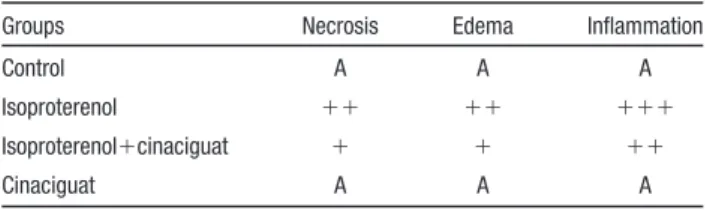
Histopathological Examination of Cardiac Tissues Compared with the control group (Figure 1A), myocardial tissues from isoproterenol-treated rats revealed myofibrillar degeneration, which is related to a dense inflammatory infiltrate and interstitial edema (Figure 1B). Cinaciguat treat- ment exhibited a decreased degree of necrosis with less fragmentation of fibers and inflammation after isoproterenol administration (Figure 1C). Drug-treated groups showed the normal appearance of cardiac muscle fibers (Figure 1D).
Table 1 shows the effect of cinaciguat on the degree of histological changes in myocardial tissues in control and isoproterenol-induced rats.
Effect of Cinaciguat on Plasma Lactate Dehydrogenase Activity and TBARS Concentration
Increased plasma lactate dehydrogenase activity after isopro- terenol treatment in rats was significantly reduced by ci- naciguat administration, indicating amelioration of intracel- lular enzyme release (Figure 2A). Increased plasma TBARS concentration after isoproterenol treatment was significantly prevented by cinaciguat treatment, indicating reduced oxida- tive stress (Figure 2B).
Table 1. Effect of Cinaciguat on the Degree of
Histopathological Changes in Myocardium in the Rat Model of Isoproterenol-Induced MI
Groups Necrosis Edema Inflammation
Control A A A
Isoproterenol ⫹⫹ ⫹⫹ ⫹⫹⫹
Isoproterenol⫹cinaciguat ⫹ ⫹ ⫹⫹
Cinaciguat A A A
A indicates no changes;⫹, mild changes;⫹⫹, moderate changes; and
⫹⫹⫹, marked changes.
Figure 1.Histological examinations of myocar- dium (hematoxylin and eosin staining; magnifi- cation⫻100) in each group. Iso indicates isoproterenol.
Figure 2.Effect of cinaciguat on plasma lactate dehydrogenase activity (LDH; A) and TBARS (B) in isoproterenol (Iso) -induced MI in rats. All values are expressed as mean⫾SEM. *P⬍0.05 vs control; #P⬍0.05 vs isoproterenol.
Effect of Cinaciguat on Cardiac Function in MI Compared with the control group, isoproterenol treatment in rats was associated with significantly decreased systolic performance. The slope Emaxof the end-systolic PV relation- ship and preload recruitable stroke work showed a marked reduction in isoproterenol-treated rats. Cinaciguat treatment resulted in a significant increase in these parameters, indicat- ing improved LV contractility (Figure 3A and 3B). In contrast, the end-diastolic PV relationship was increased in isoproterenol-treated rats, indicating increased end-diastolic stiffness. Values of the end-diastolic PV relationship of cinaciguat plus isoproterenol–treated rats did not differ com- pared with the control group (Figure 3C). Isoproterenol treatment was associated with impaired cardiac relaxation, as
reflected by prolonged, which was significantly improved by cinaciguat (Figure 3D). Isoproterenol treatment was asso- ciated with significantly increased HR and decreased ejection fraction and stroke volume, which were reversed to the level of controls after cinaciguat treatment. Isoproterenol treatment resulted in increased LV end-diastolic pressure, decreased stroke work and LV systolic pressure, and minimum pressure development (⫺dP/dt). Cinaciguat had no effect on these parameters. Rats treated with cinaciguat plus isoproterenol had lower MAP and LV systolic pressure. Cinaciguat treat- ment alone had no effect on any of the hemodynamic parameters studied (Table 2). In the postinfarction cinaciguat treatment group, we detected significantly improved cardiac relaxation and a tendency toward improved systolic function (without reaching the level of statistical significance) com- pared with the isoproterenol group (Figure 3E through 3H;
see Table I of the online-only Data Supplement).
Effect of Cinaciguat on Mortality in MI
There were no deaths in the control and cinaciguat-treated groups. The rate of mortality (within 30 minutes) in the isoproterenol- and cinaciguat plus isoproterenol–treated rats was 6% and 0%, respectively. During the experimental period, the mortality rate of rats was 56% for the isoproter- enol group and 33% for the cinaciguat plus isoproterenol group (see Table II of the online-only Data Supplement).
Similarly, the mortality rate was markedly reduced after postinfarction cinaciguat treatment (56% versus 25%) (see Table III of the online-only Data Supplement).
Effect of Cinaciguat on Plasma cGMP and cAMP Concentrations and on Myocardial cGMP Level Significantly increased levels of plasma cGMP were ob- served in isoproterenol- and cinaciguat plus isoproterenol–
treated rats compared with control and cinaciguat-treated rats (Figure 4A), whereas plasma cAMP level remained un- changed (Figure 4B). In the cinaciguat-treated group, the myocardial cGMP score was significantly higher compared with the control group (Figure 4C).
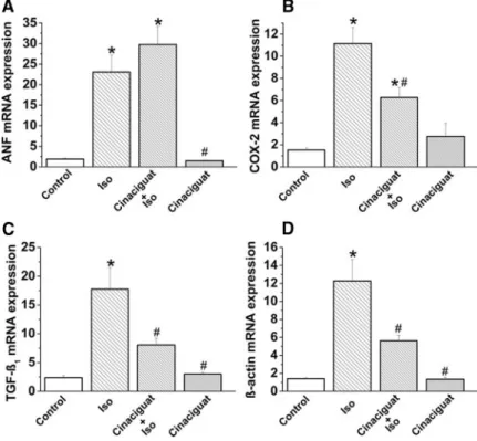
Effect of Cinaciguat on Gene Expression
Quantitative real-time polymerase chain reaction from apical myocardium RNA extracts revealed that mRNA expression for atrial natriuretic factor (ANF), cyclooxygenase 2 (COX- 2), transforming growth factor-1(TGF-1), and-actin was significantly increased in the isoproterenol-treated group (Figure 5A through 5D), whereas-myosin heavy chain and endothelial NO synthase mRNA expression remained un- changed (data not shown). Cinaciguat treatment was shown to prevent the increase in COX-2, TGF-1, and-actin mRNA expression in isoproterenol-treated rats, whereas the ANF mRNA level was not affected.
Effect of Cinaciguat on Cardiac Function After Global Ischemia/Reperfusion
Table 3 shows the in vivo effects of postischemic cinaciguat treatment on global cardiac ischemia/reperfusion injury in our canine model. Baseline values did not differ between groups.
MAP showed a decrease in both groups after ischemia/
Figure 3.Effect of cinaciguat on LV contractility, end-diastolic stiffness, and cardiac relaxation in isoproterenol (Iso) -induced MI in rats. A and E, Slope (Emax) of the LV end-systolic PV rela- tionships; B and F, preload recruitable stroke work (PRSW); C and G, end-diastolic PV relationship (EDPVR); D and H, time constant of LV pressure decay (). A through D, Preinfarction cinaciguat treatment group; E through H, postinfarction cinaciguat treatment group. All values are expressed as mean⫾SEM. *P⬍0.05 vs control; #P⬍0.05 vs isoproterenol.
reperfusion. HR and cardiac output showed no differences between groups and over time. Coronary blood flow de- creased significantly in the control group after ischemia/
reperfusion, whereas it remained unchanged in the cinaciguat group. After ischemia/reperfusion injury, significantly de- creased contractility (shown by decreased end-systolic PV relationship and preload recruitable stroke work) was ob- served in the control group, which was reversed by postis- chemic cinaciguat treatment.
In Vitro Effects of Cinaciguat on Vascular Function of Coronary Arterial Rings Exposed to Peroxynitrite
A marked impairment of endothelial function after exposure of isolated rings to the reactive oxidant peroxynitrite was demonstrated in our in vitro organ bath experiments.
Acetylcholine-induced maximal relaxation was significantly attenuated after exposure of isolated segments to peroxynitrite, which indicates endothelial dysfunction. Cinaciguat significantly improved the acetylcholine-induced, endothelium-dependent, NO-mediated vasorelaxation after exposure of coronary rings to peroxynitrite (Figure 6A). Endothelium-independent vas- cular smooth muscle function, indicated by the vasorelaxation of coronary arteries to sodium nitroprusside, a direct NO donor, was not impaired by peroxynitrite. Incubation of rings with peroxynitrite only significantly enhanced the relaxation to 10⫺8 mol/L sodium nitroprusside (Figure 6B). Contractile responses to U46619 did not show any significant differences in the groups studied (data not shown).
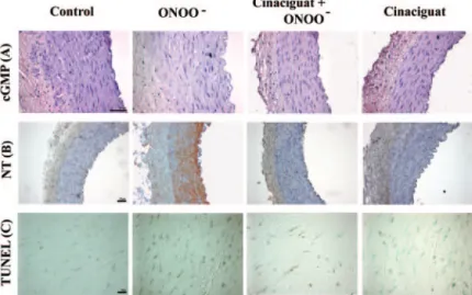
In Vitro Effects of Cinaciguat on cGMP Levels, Nitro-Oxidative Stress, and DNA Strand Breaks in Coronary Arterial Rings Exposed to Peroxynitrite A significantly lower score of cGMP staining (decreased red staining) was observed in the media of peroxynitrite- exposed rings compared with control. Cinaciguat treatment resulted in significantly higher immunoreactivity for cGMP but did not reach the level of statistical significance (Figures 7A and 8A).
Increased nitrotyrosine formation observed after exposure of coronary arteries to peroxynitrite was reduced after ci- naciguat treatment, as evidenced by decreased brown staining Table 2. Effect of Cinaciguat on HR, MAP, Maximal LV Pressure, LV End-Diastolic Pressure, Ejection Fraction, Maximum Pressure Development, Minimum Pressure Development, End-Diastolic Volume, End-Systolic Volume, Stroke Volume, Efficiency, Systemic Vascular Resistance, Arterial Elastance, Stroke Work, and End-Diastolic PV Relationship in Isoproterenol-Induced MI in Rats
Control Isoproterenol Cinaciguat⫹Isoproterenol Cinaciguat
HR, bpm 427⫾9 485⫾12* 448⫾12 413⫾10†
MAP, mm Hg 102⫾3 89⫾3 64⫾3*† 115⫾7†
LVSP, mm Hg 133⫾4 102⫾3* 88⫾4*† 138⫾4†
LVEDP, mm Hg 5.9⫾0.3 10.5⫾1.0* 9.9⫾0.8* 6.1⫾0.3†
EF, % 56⫾6 41⫾4* 46⫾4 54⫾6
LV dP/dt, mm Hg/s 8372⫾389 7607⫾370 8660⫾548 8426⫾520
LV⫺dP/dt, mm Hg/s 12 063⫾770 6474⫾457* 6608⫾430* 12 087⫾545†
EDV,L 280⫾42 329⫾39 348⫾29 246⫾16
ESV,L 213⫾26 270⫾33 230⫾21 171⫾10†
SV,L 192⫾12 137⫾16* 165⫾15 155⫾12
Efficiency, % 61.8⫾4.0 63.0⫾4.2 61.3⫾3.2 54.0⫾4.6
SVR, mm Hg䡠mL⫺1䡠min 1.75⫾0.22 1.85⫾0.17 1.17⫾0.22† 1.79⫾0.13
Ea, mm Hg/L 0.92⫾0.12 1.02⫾0.10 0.69⫾0.09† 1.07⫾0.09
SW, mm Hg䡠L 16 057⫾1172 9711⫾1211* 10 338⫾1141* 13 005⫾1345
⫹dP/dt–EDV, mm Hg䡠s⫺1䡠L⫺1 46.6⫾6.4 35.7⫾2.9 42.0⫾9.9 43.7⫾3.4
LVSP indicates maximal LV pressure; LVEDP, LV end-diastolic pressure; EF, ejection fraction; dP/dt, maximum pressure development;⫺dP/dt, minimum pressure development; EDV, end-diastolic volume; ESV, end-systolic volume; SV, stroke volume; SVR, systemic vascular resistance; SW, stroke work; and EDPVR, end-diastolic PV relationship. All values are expressed as mean⫾SEM.
*P⬍0.05 vs control; †P⬍0.05 vs isoproterenol.
Figure 4.Effect of cinaciguat on plasma cGMP (A), plasma cAMP (B), and myocardial cGMP (C) levels in isoproterenol (Iso) -induced MI in rats. All values are expressed as mean⫾SEM.
*P⬍0.05 vs control; #P⬍0.05 vs isoproterenol.
(Figures 7B and 8B). We found pronounced DNA damage in the wall of peroxynitrite-exposed rings, as reflected by the quantitative assessment of TUNEL-positive cells. Cinaciguat treatment tended to decrease peroxynitrite-induced DNA strand breaks (Figures 7C and 8C).
Discussion
The present study evaluated the effects of cinaciguat, an NO- and heme-independent sGC activator, on experimentally induced MI in rats. Our study showed that cinaciguat im- proves cardiac performance, prevents an increase in oxidative stress, and ameliorates histopathological changes and intra- cellular enzyme release in isoproterenol-induced MI. We also showed that cinaciguat has protective effects on coronary arteries and enhances endothelium-dependent vasorelaxation in an in vitro model of vascular nitro-oxidative stress.
Effects of Cinaciguat Pretreatment on Cardiac Dysfunction in MI
We show that isoproterenol treatment is characterized by decreased systolic performance accompanied by delayed relaxation and increased diastolic stiffness of the heart.
Although we found that isoproterenol treatment was associ- ated with decreased ejection fraction (known to be influenced by both preload and afterload), it cannot be used reliably to assess the contractile function in the models in which both preload and afterload are altered. End-systolic PV relation- ship (Emax) and preload recruitable stroke work (2 well- established load-independent contractility indexes)20 were decreased in isoproterenol-treated rats, suggesting decreased systolic performance. Another PV-derived index, dP/dt– end- diastolic volume, which is known to be sensitive to contrac- tile changes and insensitive to preload, also has a tendency to be depressed in isoproterenol-treated rats. Impaired ventric- Figure 5.Effect of cinaciguat on gene expression in isoproterenol (Iso) -induced MI in rats. ANF (A), COX-2 (B), TGF-1(C), and-actin (D) mRNA expression. All values are expressed as mean⫾SEM. *P⬍0.05 vs control; #P⬍0.05 vs isoproterenol.
Table 3. Effect of Postischemic Cinaciguat Treatment on HR, MAP, Cardiac Output, Coronary Blood Flow, End-Systolic PV Relationship, and Preload Recruitable Stroke Work in the Canine Model of Global Cardiac Ischemia/Reperfusion
Before Ischemia/Reperfusion After Ischemia/Reperfusion
Control Cinaciguat Control Cinaciguat
HR, bpm 111⫾6 131⫾9 120⫾12 139⫾3
MAP, mm Hg 89.2⫾3.5 90.1⫾2.3 63.0⫾4.9† 59.2⫾2.8†
CO, L/min 2.99⫾0.34 2.65⫾0.30 2.39⫾0.32 2.75⫾0.58
CBF, mL/min 39.63⫾5.35 33.83⫾3.24 24.00⫾2.59† 40.00⫾4.13*
ESPVR, mm Hg/mL 5.04⫾0.55 6.13⫾0.78 2.62⫾0.18† 4.27⫾0.70*
PRSW, mm Hg 57.69⫾3.28 75.11⫾10.39 31.48⫾1.88† 59.98⫾12.01*
CO indicates cardiac output; CBF, coronary blood flow; ESPVR, end-systolic PV relation; and PRSW, preload recruitable stroke work. All values are expressed as mean⫾SEM.
*P⬍0.05 vs control; †P⬍0.05 vs before ischemia/reperfusion.
ular relaxation (as reflected by the decreased⫺dP/dt, pro- longed , and increased LV end-diastolic pressure) and increased end-diastolic stiffness (as reflected by the increased end-diastolic PV relationship) were also detected in isoproterenol-treated groups. Relaxation, as an active process, depends mostly on Ca2⫹uptake by the sarcoplasmic reticulum during diastole, and end-diastolic stiffness is affected pre- dominantly by alterations in myocardial structural compo- nents. Consistent with diastolic dysfunction, LV end-diastolic pressure was also increased in this group of rats, which may be influenced by changes in preload, and its increase may have resulted from increased venous return. Most of these parameters return to near-control levels after cinaciguat treatment, showing improvement in contractile pump func- tion, LV relaxation, and diastolic stiffness.
Arterial elastance, Ea, directly affects systolic function and is considered the most reliable index of LV afterload. In rats with MI, values of Ea were not different from those in the control group, whereas cinaciguat significantly decreased afterload. When rats with MI are treated with cinaciguat, MAP is reduced (and presumably the LV afterload), and the ventricle can eject blood more rapidly, which increases stroke volume. After cinaciguat treatment, the ventricle will not fill to the same end-diastolic volume (preload) found before the afterload reduction because less blood remains in the ventri- cle after systole. During MI, stroke volume often falls because of a depression of LV contractility, resulting in a compensatory increase in afterload to maintain blood pres- sure within a physiological range. It has been shown that in experimental heart failure in dogs, intravenous administration of cinaciguat resulted in a reduction in cardiac preload and
afterload.21Deteriorating myocardial status after isoprotere- nol administration is expected to affect HR. Increased HR observed in isoproterenol-treated rats, which reduces the time for diastole filling, can reduce the myocardial blood supply and cause ischemia. By decreasing afterload through its vasodilatory effect (shown by decreased MAP in cinaciguat plus isoproterenol–treated rats), cinaciguat may improve myocardial oxygen supply and decrease oxygen demand.
Effect of Postischemic Cinaciguat Treatment on Cardiac Dysfunction in MI
When applying a postinfarction treatment with cinaciguat, we observed an improvement only in diastolic function; LV contractility was not significantly ameliorated. In contrast, in the canine model of reversible global ischemia/reperfusion, systolic performance after postischemic cinaciguat treatment was significantly improved. The different results in these models might be explained by the type of ischemia (irrevers- ible or reversible) and the timing of the treatment (because of the repetitive isoproterenol administration, the development of myocardial necrosis lingers in the rat MI model, so an exact timing was possible only in the canine model).
Effect of Cinaciguat Pretreatment on Myocardial Ischemic Tissue Damage
Myocardial tissue damage induced by isoproterenol was reduced by cinaciguat, as shown by decreased inflammatory infiltrate, degeneration, and interstitial edema. These data showed the cardioprotective action of cinaciguat in rats with MI. The increased activity of the cardiac enzyme lactate dehydrogenase in the plasma of isoproterenol-treated rats was
Figure 6.Effect of cinaciguat on peroxynitrite (ONOO⫺) -induced vascular dysfunction in canine coronary arterial rings. A, Acetylcholine (ACh) -induced endothelium-dependent vasorelaxation. B, Sodium nitroprusside (SNP) –induced endothelium- independent vasorelaxation in each group. Values represent mean⫾SEM. *P⬍0.05 vs control; #P⬍0.05 vs ONOO⫺.
Figure 7.Representative photomicrographs of cGMP and nitrotyrosine (NT) immunohistochemis- try staining and TUNEL assay in the vessel wall of coronary arterial rings in each group. A, cGMP (red staining; magnification⫻400); B, nitrotyrosine (brown staining; magnification⫻200); C, TUNEL (brown staining; magnification⫻1000). Note the decreased red and increased brown staining, indi- cating decreased cGMP immunoreactivity, nitroty- rosine formation, and DNA damage in the peroxynitrite-exposed rings.
decreased by cinaciguat. When myocardial cells are damaged or destroyed as a result of deficient oxygen supply or glucose, the cell membrane becomes permeable or may rupture, which results in the leakage of this enzyme from damaged tissues into the bloodstream.22 In this study, mRNA expression of
-actin (a cytoskeletal protein important in the maintenance of cytoarchitecture) was markedly increased by isoproterenol and significantly downregulated by cinaciguat. These obser- vations suggest that -actin mRNA expression could be important in mediating the structural alterations that occur in the damaged myocardium.
Mechanisms of Cardioprotective Effects of Cinaciguat Against Ischemic Injury
In animal models of MI, gene expression of ANF is report- edly upregulated23and correlates strongly with diastolic wall stress and stretch.24,25Our present results on myocardial ANF mRNA content showed a marked increase in this factor in isoproterenol- and cinaciguat plus isoproterenol–treated rats, suggesting end-diastolic wall stress. Recent studies also showed that production of cGMP by activation of natriuretic peptides receptor just before therapeutic reperfusion has a significant anti-infarct effect in both animals26and humans.27 Our results also showed a correlation between increased myocardial ANF mRNA expression and plasma cGMP levels in these groups of rats, which could be a phenomenon that opposes MI. In heart failure, increased cGMP concentrations in extracellular fluids, including plasma28 and urine,29 are believed to result from its passage through the cellular membrane30 as plasma cGMP is seen as an overspill of intracellular cGMP. However, plasma cGMP levels do not necessarily reflect intracellular cGMP levels or the activation of sGC by intracellular cinaciguat because the overspill from the intracellular space to the plasma might also be interpreted
as an intracellular overproduction. Any relevant biological activity of cGMP in plasma remains to be elucidated.
In this study, we observed increased myocardial cGMP levels in cinaciguat-treated rats. The absence of increased myocardial cGMP content in cinaciguat plus isoproterenol–
treated rats could be explained by the short half-life of cinaciguat5 and the relative long interval (17 to 22 hours) between the last treatment and the experiment. According to the unchanged cAMP levels, a role for cyclic nucleotide cross-regulation in the observed cardioprotective effects of cinaciguat seems rather unlikely.
The cardioprotective effect of the sGC activator could be mediated by several other mechanisms. Cinaciguat induces cGMP generation and evokes vasodilation preferentially in diseased vessels.5 The markedly increased COX-2 mRNA content in isoproterenol-treated rats was significantly down- regulated after cinaciguat, suggesting the decrease of proin- flammatory prostanoid production and ischemic inflamma- tion. COX-2 is induced only in cardiomyocytes in response to stress such as ischemia31 and therefore is not expressed in healthy myocardium. It has been shown that TGF-, a locally generated cytokine, is upregulated after acute MI in rats and has a deleterious role in the response of the heart to injury.32 In the present study, increased TGF-1mRNA content (which could be due to inflammatory changes) in isoproterenol- treated rats was significantly downregulated by cinaciguat.
These findings support the concept that cGMP-activated pathways interfere with TGF- signaling, presumably by cGMP-mediated inhibition of TGF- expression, as previ- ously proposed.33
Enormous amounts of reactive oxygen species are pro- duced during MI and are involved in membrane damage, leading to increased TBARS levels. We also showed a significant elevation of plasma TBARS concentration after isoproterenol that was prevented by cinaciguat, indicating reduced oxidative stress. It is also known that COX-2 may generate reactive oxygen species.34 In the present study, increased COX-2 mRNA content after isoproterenol was significantly downregulated after cinaciguat, suggesting de- creased reactive oxygen species generation. In conditions associated with increased free radical release and oxidative stress, intracellular cGMP accumulation has been shown to reduce tissue injury.35
Protective Effect of Cinaciguat on Endothelial Dysfunction Induced by Peroxynitrite
In coronary arteries exposed to peroxynitrite, we observed that relaxation to acetylcholine was diminished; in contrast, sodium nitroprusside–induced relaxation remained un- changed. Thus, responsiveness of the vascular smooth muscle cell to NO does not appear to be impaired. Our findings suggest that the release of NO after activation of muscarinic receptors in endothelial cells is blunted, whereas the cGMP- mediated signal transduction system and contractile apparatus in vascular smooth muscle cells are functionally preserved in arteries exposed to peroxynitrite. Szabo et al36also showed that exposure of isolated vessel rings to high concentrations of peroxynitrite caused a marked impairment of the endothe- lium-dependent relaxations. In accordance, our immunohis- Figure 8.Scoring of cGMP and nitrotyrosine immunohistochem-
istry and TUNEL-assay. Immunohistochemical scores for cGMP (A) and nitrotyrosine (B) of the coronary artery wall of control, peroxynitrite-exposed (ONOO⫺; 150mol/L), and cinaciguat- treated ONOO⫺-exposed coronary arterial rings. C, Average number of TUNEL-positive cell nuclei in a microscopic field. Values represent mean⫾SEM. *P⬍0.05 vs control; #P⬍0.05 vs ONOO⫺.
tochemical staining for cGMP supports reduced NO bioavail- ability in coronary arteries exposed to peroxynitrite.
Improvement in endothelial function could be due mainly to the cytoprotection of endothelium by cinaciguat.
Study Limitations
Supramaximal doses of isoproterenol, a-adrenergic agonist and well-known inducer of MI,14are widely used as a model of evaluating cardioprotective drugs.37 Although this rat model does not exactly reflect the clinical situation in terms of coronary occlusion and regional MI, animals develop
“infarct-like” lesions when injected with isoproterenol. In a rabbit model of coronary artery ligation, Krieg et al38recently showed that cinaciguat elicited an infarct-reducing effect. Our canine model does not reflect acute MI but routine cardiac surgery with extracorporal circulation and cold cardioplegic arrest leading to a myocardial and endothelial dysfunction.
Conclusions
The present results indicate that the sGC activator cinaciguat has a significant effect in the protection of the heart against MI in rats. Our results show that cinaciguat improves cardiac function, improves histopathological lesions, reduces oxida- tive stress, ameliorates intracellular enzyme release, and decreases COX-2, TGF-1, and-actin mRNA expression in experimentally induced MI. We also show that cinaciguat enhances the endothelium-dependent vasorelaxation in ca- nine coronary arterial rings exposed to peroxynitrite, de- creases nitro-oxidative stress, and increases intracellular cGMP levels. Although results of animal experimental stud- ies should not be directly extrapolated to human biology, pharmacological sGC activation could be a novel approach for the prevention and treatment of ischemic heart disease.
Acknowledgments
The expert technical assistance of Heike Ziebart is gratefully acknowledged. The authors acknowledge Bayer HealthCare AG for the donation of cinaciguat.
Source of Funding
This work was supported by the Land Baden-Württemberg.
Disclosures
None.
References
1. Moro MA, Russel RJ, Cellek S, Lizasoain I, Su Y, Darley-Usmar VM, Radomski MW, Moncada S. cGMP mediates the vascular and platelet actions of nitric oxide: confirmation using an inhibitor of the soluble guanylyl cyclase.Proc Natl Acad Sci U S A. 1996;93:1480 –1485.
2. Stasch JP, Schmidt PM, Nedvetsky PI, Nedvetskaya TY, H S AK, Meurer S, Deile M, Taye A, Knorr A, Lapp H, Muller H, Turgay Y, Rothkegel C, Tersteegen A, Kemp-Harper B, Muller-Esterl W, Schmidt HH. Tar- geting the heme-oxidized nitric oxide receptor for selective vasodilatation of diseased blood vessels.J Clin Invest. 2006;116:2552–2561.
3. Munzel T, Genth-Zotz S, Hink U. Targeting heme-oxidized soluble gua- nylate cyclase: solution for all cardiorenal problems in heart failure?
Hypertension. 2007;49:974 –976.
4. Feelisch M. The use of nitric oxide donors in pharmacological studies.
Naunyn Schmiedebergs Arch Pharmacol. 1998;358:113–122.
5. Evgenov OV, Pacher P, Schmidt PM, Hasko G, Schmidt HH, Stasch JP.
NO-independent stimulators and activators of soluble guanylate cyclase:
discovery and therapeutic potential. Nat Rev Drug Discov. 2006;5:
755–768.
6. Stasch JP, Schmidt P, Alonso-Alija C, Apeler H, Dembowsky K, Haerter M, Heil M, Minuth T, Perzborn E, Pleiss U, Schramm M, Schroeder W, Schroder H, Stahl E, Steinke W, Wunder F. NO- and haem-independent activation of soluble guanylyl cyclase: molecular basis and cardiovascular implications of a new pharmacological principle.Br J Pharmacol. 2002;
136:773–783.
7. Frey R, Muck W, Unger S, Artmeier-Brandt U, Weimann G, Wensing G.
Pharmacokinetics, pharmacodynamics, tolerability, and safety of the soluble guanylate cyclase activator cinaciguat (BAY 58 –2667) in healthy male volunteers.J Clin Pharmacol. 2008;48:1400 –1410.
8. Schmidt HH, Schmidt PM, Stasch JP. NO- and haem-independent soluble guanylate cyclase activators.Handb Exp Pharmacol. 2009:309 –339.
9. Rona G. Catecholamine cardiotoxicity. J Mol Cell Cardiol. 1985;17:
291–306.
10. Wexler BC. Myocardial infarction in young vs old male rats: pathophys- iologic changes.Am Heart J. 1978;96:70 – 80.
11. Wexler BC, Greenberg BP. Effect of exercise on myocardial infarction in young vs. old male rats: electrocardiograph changes.Am Heart J. 1974;
88:343–350.
12. Chagoya de Sanchez V, Hernandez-Munoz R, Lopez-Barrera F, Yanez L, Vidrio S, Suarez J, Cota-Garza MD, Aranda-Fraustro A, Cruz D.
Sequential changes of energy metabolism and mitochondrial function in myocardial infarction induced by isoproterenol in rats: a long-term and integrative study.Can J Physiol Pharmacol. 1997;75:1300 –1311.
13. Singal PK, Dhalla AK, Hill M, Thomas TP. Endogenous antioxidant changes in the myocardium in response to acute and chronic stress conditions.Mol Cell Biochem. 1993;129:179 –186.
14. Rajadurai M, Stanely Mainzen Prince P. Preventive effect of naringin on cardiac markers, electrocardiographic patterns and lysosomal hydrolases in normal and isoproterenol-induced myocardial infarction in Wistar rats.
Toxicology. 2007;230:178 –188.
15. Radovits T, Bomicke T, Kokeny G, Arif R, Loganathan S, Kecsan K, Korkmaz S, Barnucz E, Sandner P, Karck M, Szabo G. The phosphodiesterase-5 inhibitor vardenafil improves cardiovascular dys- function in experimental diabetes mellitus.Br J Pharmacol. 2009;156:
909 –919.
16. Szabo G, Soos P, Mandera S, Heger U, Flechtenmacher C, Bahrle S, Seres L, Cziraki A, Gries A, Zsengeller Z, Vahl CF, Hagl S, Szabo C.
INO-1001 a novel poly(ADP-ribose) polymerase (PARP) inhibitor improves cardiac and pulmonary function after crystalloid cardioplegia and extracorporal circulation.Shock. 2004;21:426 – 432.
17. Veres G, Radovits T, Schultz H, Lin LN, Hutter J, Weigang E, Szabolcs Z, Szabo G. Effect of recombinant aprotinin on postoperative blood loss and coronary vascular function in a canine model of cardiopulmonary bypass.Eur J Cardiothorac Surg. 2007;32:340 –345.
18. Hirschberg K, Radovits T, Loganathan S, Entz L, Beller CJ, Gross ML, Sandner P, Karck M, Szabó G. Selective phosphodiesterase-5 inhibition reduces neointimal hyperplasia in rat carotid arteries after surgical end- arterectomy.J Thorac Cardiovasc Surg. 2009;137:1508 –1514.
19. Liaudet L, Soriano FG, Szabo E, Virag L, Mabley JG, Salzman AL, Szabo C. Protection against hemorrhagic shock in mice genetically deficient in poly(ADP-ribose)polymerase. Proc Natl Acad Sci U S A.
2000;97:10203–10208.
20. Kass DA, Maughan WL, Guo ZM, Kono A, Sunagawa K, Sagawa K.
Comparative influence of load versus inotropic states on indexes of ventricular contractility: experimental and theoretical analysis based on pressure-volume relationships.Circulation. 1987;76:1422–1436.
21. Boerrigter G, Costello-Boerrigter LC, Cataliotti A, Lapp H, Stasch JP, Burnett JC Jr. Targeting heme-oxidized soluble guanylate cyclase in experimental heart failure.Hypertension. 2007;49:1128 –1133.
22. Sabeena Farvin KH, Anandan R, Kumar SH, Shiny KS, Sankar TV, Thankappan TK. Effect of squalene on tissue defense system in isoproterenol-induced myocardial infarction in rats. Pharmacol Res.
2004;50:231–236.
23. Shimoike H, Iwai N, Kinoshita M. Differential regulation of natriuretic peptide genes in infarcted rat hearts.Clin Exp Pharmacol Physiol. 1997;
24:23–30.
24. Buttrick PM, Kaplan M, Leinwand LA, Scheuer J. Alterations in gene expression in the rat heart after chronic pathological and physiological loads.J Mol Cell Cardiol. 1994;26:61– 67.
25. Schultz JE, Witt SA, Nieman ML, Reiser PJ, Engle SJ, Zhou M, Pawlowski SA, Lorenz JN, Kimball TR, Doetschman T. Fibroblast growth factor-2 mediates pressure-induced hypertrophic response.J Clin Invest. 1999;104:709 –719.
26. Yang XM, Philipp S, Downey JM, Cohen MV. Atrial natriuretic peptide administered just prior to reperfusion limits infarction in rabbit hearts.
Basic Res Cardiol. 2006;101:311–318.
27. Kitakaze M, Asakura M, Kim J, Shintani Y, Asanuma H, Hamasaki T, Seguchi O, Myoishi M, Minamino T, Ohara T, Nagai Y, Nanto S, Watanabe K, Fukuzawa S, Hirayama A, Nakamura N, Kimura K, Fujii K, Ishihara M, Saito Y, Tomoike H, Kitamura S. Human atrial natriuretic peptide and nicorandil as adjuncts to reperfusion treatment for acute myocardial infarction (J-WIND): two randomised trials.Lancet. 2007;
370:1483–1493.
28. Fujio N, Ohashi M, Nawata H, Kato K, Ibayashi H, Matsuo H. Rela- tionship between atrial natriuretic polypeptide and cyclic 3⬘5⬘-guanosine monophosphate in human plasma.J Lab Clin Med. 1987;109:706 –710.
29. Michel JB, Mercadier JJ, Galen FX, Urbain R, Dussaule JC, Philippe M, Corvol P. Urinary cyclic guanosine monophosphate as an indicator of experimental congestive heart failure in rats.Cardiovasc Res. 1990;24:
946 –952.
30. Hamet P, Pang SC, Tremblay J. Atrial natriuretic factor-induced egression of cyclic guanosine 3⬘:5⬘-monophosphate in cultured vascular smooth muscle and endothelial cells. J Biol Chem. 1989;264:
12364 –12369.
31. Shinmura K, Xuan YT, Tang XL, Kodani E, Han H, Zhu Y, Bolli R.
Inducible nitric oxide synthase modulates cyclooxygenase-2 activity in the heart of conscious rabbits during the late phase of ischemic precon- ditioning.Circ Res. 2002;90:602– 608.
32. Thompson NL, Bazoberry F, Speir EH, Casscells W, Ferrans VJ, Flanders KC, Kondaiah P, Geiser AG, Sporn MB. Transforming growth factor beta-1 in acute myocardial infarction in rats.Growth Factors. 1988;1:
91–99.
33. Saura M, Zaragoza C, Herranz B, Griera M, Diez-Marques L, Rodriguez-Puyol D, Rodriguez-Puyol M. Nitric oxide regulates trans- forming growth factor-beta signaling in endothelial cells.Circ Res. 2005;
97:1115–1123.
34. O’Banion MK. Cyclooxygenase-2: molecular biology, pharmacology, and neurobiology.Crit Rev Neurobiol. 1999;13:45– 82.
35. Dias-Junior CA, Souza-Costa DC, Zerbini T, da Rocha JB, Gerlach RF, Tanus-Santos JE. The effect of sildenafil on pulmonary embolism- induced oxidative stress and pulmonary hypertension. Anesth Analg.
2005;101:115–120.
36. Szabo C, Cuzzocrea S, Zingarelli B, O’Connor M, Salzman AL. Endo- thelial dysfunction in a rat model of endotoxic shock: importance of the activation of poly (ADP-ribose) synthetase by peroxynitrite.J Clin Invest.
1997;100:723–735.
37. Karthick M, Stanely Mainzen Prince P. Preventive effect of rutin, a bioflavonoid, on lipid peroxides and antioxidants in isoproterenol- induced myocardial infarction in rats. J Pharm Pharmacol. 2006;58:
701–707.
38. Krieg T, Liu Y, Rutz T, Methner C, Yang XM, Dost T, Felix SB, Stasch JP, Cohen MV, Downey JM. BAY 58 –2667, a nitric oxide-independent guanylyl cyclase activator, pharmacologically post-conditions rabbit and rat hearts.Eur Heart J. 2009;30:1607–1613.
CLINICAL PERSPECTIVE
Although clinical care is improved, public awareness is raised, and health innovations are widely used, myocardial infarction remains the leading cause of death worldwide. The nitric oxide/soluble guanylate cyclase/cGMP/cGMP–
dependent protein kinase G signaling cascade has been shown to be an important sequence in both preconditioning and postconditioning. Alteration of this pathway has been thought to be involved in the pathogenesis of various cardiovascular diseases and is subject to the development of new therapeutic agents. Cinaciguat, the novel nitric oxide– and heme-independent activator of soluble guanylate cyclase, is currently being evaluated for the treatment of chronic heart failure in early clinical trials and appears to be well tolerated. Its nitric oxide independence may make it an ideal candidate for a pharmacological approach in acute myocardial infarction. In this study, we demonstrate that in a rat model of myocardial infarction induced by isoproterenol, cinaciguat treatment improves histopathological lesions, improves cardiac performance, improves impaired cardiac relaxation, reduces oxidative stress, and ameliorates intracellular enzyme release.
Furthermore, we demonstrate cardioprotective effect of postischemic cinaciguat treatment in a clinically relevant canine model of global cardiac ischemia/reperfusion injury. Collectively, these findings make cinaciguat a candidate for the prevention and treatment of acute myocardial ischemia in humans.