O R I G I N A L A R T I C L E
Pharmacokinetics, pharmacodynamics, and safety of moss-aGalactosidase A in patients with Fabry disease
Julia B. Hennermann1 | Laila Arash-Kaps1 | György Fekete2 | Andreas Schaaf3 | Andreas Busch3 | Thomas Frischmuth3
1Villa Metabolica, Department of Pediatric and Adolescent Medicine, University Medical Center Mainz, Mainz, Germany
2II. Department of Pediatrics, Semmelweis University, Budapest, Hungary
3Greenovation Biotech GmbH, Freiburg, Germany
Correspondence
Julia B. Hennermann, University Medical Center Mainz, Villa Metabolica, Department of Pediatric and Adolescent Medicine, Langenbeckstr. 1, 55131 Mainz, Germany.
Email: julia.hennermann@unimedizin- mainz.de
Communicating Editor:Robin Lachmann
Funding information Greenovation Biotech GmbH
Abstract
Moss-aGalactosidase A (moss-aGal) is a moss-derived version of human α-galactosidase developed for enzyme replacement therapy in patients with Fabry disease. It exhibits a homogenous N-glycosylation profile with >90% mannose- terminated glycans. In contrast to mammalian cell produced α-galactosidase, moss-aGal does not rely on mannose-6-phosphate receptor mediated endocytosis but targets the mannose receptor for tissue uptake. We conducted a phase 1 clinical trial with moss-aGal in six patients with confirmed diagnosis of Fabry disease dur- ing a 28-day schedule. All patients received a single dose of 0.2 mg/kg moss-aGal by i.v.-infusion. Primary endpoints of the trial were safety and pharmacokinetics;
secondary endpoints were pharmacodynamics by analyzing urine and plasma Gb3 and lyso-Gb3 concentrations. In all patients, the administered single dose was well tolerated. No safety issues were observed. Pharmacokinetic data revealed a stable nonlinear profile with a short plasma half-life of moss-aGal of 14 minutes. After one single dose of moss-aGal, urinary Gb3 concentrations decreased up to 23%
7 days and up to 60% 28 days post-dose. Plasma concentrations of lyso-Gb3 decreased by 3.8% and of Gb3 by 11% 28 days post-dose. These data reveal that a single dose of moss-aGal was safe, well tolerated, and led to a prolonged reduction of Gb3 excretion. As previously shown, moss-aGal is taken up via the mannose receptor, which is expressed on macrophages but also on endothelial and kidney cells. Thus, these data indicate that moss-aGal may target kidney cells. After these promising results, phase 2/3 clinical trials are in preparation.
K E Y W O R D S
Agalsidase, alpha-galactosidase, Fabry disease, mannose receptor, moss,Physcomitrella
1
|I N T R O D U C T I O N
Fabry disease (FD) (Online Mendelian Inheritance in Man 301500) is an X-linked progressive multi-organ disease caused by mutations in the GLA gene. These mutations
result in decreased or deficient levels of the lysosomal enzyme α-galactosidase A (EC 3.2.1.22), thus leading to accumulation of globotriaosylceramide (Gb3) and related glycosphingolipids in many cell types, for example, vascular endothelial cells, podocytes, and cardiomyocytes. The
DOI: 10.1002/jimd.12052
This is an open access article under the terms of the Creative Commons Attribution License, which permits use, distribution and reproduction in any medium, provided the original work is properly cited.
© 2019 The Authors.Journal of Inherited Metabolic Diseasepublished by John Wiley & Sons Ltd on behalf of SSIEM
J Inherit Metab Dis.2019;42:527–533. wileyonlinelibrary.com/journal/jimd 527
affection of vascular endothelial cells results in a vasculopa- thy of small vessels, mainly in kidney, heart, and central ner- vous system. The prevalence in hemizygous male FD- patients is estimated at 1 in 40 000. Heterozygous female carriers are also affected by the disease. The clinical picture is broad and symptoms progress with increasing age. Typi- cal clinical symptoms include neuropathic pain, hypohidro- sis, angiokeratoma, cornea verticillata, diarrhea, chronic kidney disease, cardiomyopathy, arrhythmia, and stroke.1
Specific treatment for FD consists either of enzyme replacement therapy (ERT) or chaperone therapy, mainly in reducing storage material in the endothelial cells of different organs. Early start of treatment results in a reduction of life- threatening events. In Europe, two different ERTs for biweekly i.v.-application are approved: Agalsidase alfa and Agalsidase beta.2 Since 2016, a chaperone therapy with Migalastat is approved in patients with FD aged 16 years and older who are carrying certain amenable mutations.3
Cellular uptake of mammalian cell produced α-galactosidase occurs mainly via mannose-6-phosphate (M6P) receptor mediated endocytosis.4 However, there is increasing evidence for additional alternative uptake routes of which the mannose receptor (MR) is a promising example.5,6 MR recognizes terminal mannose, fucose, and N- acetylglucosamine residues of glycoproteins and is expressed in dendritic, endothelial, smooth muscle, and renal mesangial cells.7 Due to this expression pattern, MR is an interesting new target for a potentially improved FD therapy. Moss-aGal is produced in the mossPhyscomitrella patensand exhibits a very homogenous glycosylation profile with exclusively man- nose or N-acetylglucosamine residues targeting MR.6Poten- tially immunogenic plant specific xylose and fucose residues have been eliminated through genetic engineering.8
Previous in vitro and in vivo testing of moss-aGal con- firmed its efficient uptake and its ability to degrade accumu- lated Gb3 in the target organs kidney and heart.6 Results from the murine FD model suggested that the MR pathway might play an important role targeting Agalsidase into the kidney.6
The here presented data of the first human clinical trial demonstrates the safety of a single dose of moss-aGal and represents the first step for a full clinical development of moss-aGal as a new therapeutic option for FD.
2
|M A T E R I A L S A N D M E T H O D S 2.1
|Moss-aGal
Human alpha-galactosidase was produced in the mossPhys- comitrella patensas described previously.6 The study drug was manufactured, tested, and released by Glycotope Bio- technology GmbH (Heidelberg, Germany), according to
Good Manufacturing Practice and local regulations. Moss- aGal preparations were provided in 10 mL glass vials with 5 mL aqueous solution for infusion at a concentration 0.35 mg/mL.
2.2
|Study design
The trial was designed as an open-label, single-arm, phase 1 study in patients with Fabry disease with sequential adminis- tration of one dose of moss-aGal. Eligible patients received a single dose of 0.2 mg/kg body weight moss-aGal over an infusion period of 60 ± 5 minutes on day 1. Total infusion volume was 250 mL. For safety reasons, patients were hos- pitalized during the infusion and at least until 24 hours after end of the infusion. Follow-up visits were scheduled on days 2, 7, 14, and 28. A total of six patients were planned to be enrolled. Treatment occurred sequentially.
Primary endpoints of the trial were safety and evaluation of pharmacokinetics. Secondary endpoint was the evaluation of pharmacodynamics by analyses of Gb3 and globotriaosyl- sphingosine (lyso-Gb3) concentrations in plasma and urine.
Inclusion criteria were female or male patients aged 18-65 years with confirmed diagnosis of FD, either by con- firmation of deficientα-Gal A activity or by confirmation of a disease-causing mutation in the GLA gene, which was mandatory for women. Patients had to be treatment naïve or paused ERT for at least 3 months. Only patients with ele- vated plasma lyso-Gb3 concentrations were enrolled.
Patients had to present at least one clinical manifestation of FD, for example, neuropathic pain, angiokeratoma, cornea verticillata, cardiomyopathy, hypo- or anhydrosis, abdomi- nal pain, diarrhea, serum creatinine >1.0 mg/dL, or protein- uria >300 mg/24 hours. Patients of childbearing age or with partners of childbearing age had to use a medically accept- able method of contraception. All patients gave written informed consent.
Patients with increased anti-α-Gal A immunoglobulin G (IgG) concentrations at screening, known allergy, or intoler- abilities to ERT, kidney disease requiring dialysis or a kid- ney transplant, malignancies, or other severe diseases were excluded.
This study was conducted in compliance with the ethical principles originating in or derived from the Declaration of Helsinki and in compliance with the institutional review board, informed consent regulations, and International Con- ference on Harmonization Good Clinical Practices Guide- lines. All local regulatory requirements were followed. The study was approved by the Ethics Committee of the State Chamber of Medicine in Rhineland-Palatinate and the Ethics Committee for Clinical Pharmacology KFEB, Budapest.
This clinical study has been registered in clinicaltrials.gov (NCT02995993).
2.3
|Statistical methods
Due to the small number of patients, only descriptive statisti- cal analysis was performed. Analyses included all patients that received an infusion of moss-aGal. Pharmacokinetic variables were evaluated using standard noncompartmental analyses and were descriptively summarized using spaghetti plots. Gb3 and lyso-Gb3 concentrations were described as change from baseline using box-plots for visualization (plot- ting minimum, maximum, 1st and 3rd quartile, median).
2.4
|Safety assessment
Safety was assessed by evaluation of adverse events, physi- cal examination, vital signs, 12-lead electrocardiography, routine laboratory tests (hematology, coagulation tests, clini- cal chemistry, and urine analysis), urine pregnancy test for female patients of childbearing potential, and anti-aGal IgG antibodies.
2.5
|Measurement of anti-alpha-galactosidase antibodies
Measurement of anti-aGalIgG antibodies was performed by enzyme-linked immunosorbent assay as previously described.9 Limit of detection was 6.25 antigen units (AU)/mL, but cross- reaction with other antibodies were only excluded at titers
>20 AU/mL. Antibodies were determined at screening and 28 days. Analyses were performed by Prof. Dr. K. J. Lackner and Dr. K. Bruns, Central Lab of the University Medical Center Mainz.
2.6
|Quality of life and brief pain inventory
Quality of life was assessed by the 36-item Short Form Health Survey questionnaire (SF36; Quality metrics). Levels of pain over the past 24 hours were assessed by using the validated Brief Pain Inventory (BPI).10For method details and results see Supplementary Tables S1-S2, Supporting Information.2.7
|Measurement of Gb3- and lyso- Gb3-concentrations
Gb3 was measured by liquid chromatography-mass spec- trometry and tandem mass spectrometry (LC-MS/MS) in plasma and morning urine at screening, baseline, day 7, 14, and 28; lyso-Gb3 was measured by LC-MS/MS in plasma at screening, baseline, day 7, 14, and 28. Plasma and urinary concentrations of Gb3 isoforms Gb3-C24-0, Gb3-C16-0, Gb3-C18-0, Gb3-C24-1 as well as lyso-Gb3 concentrations in plasma were measured as described before.11,12
2.8
|Pharmakokinetics
Moss-aGal serum concentrations were determined by vali- dated enzyme activity assay using 4-methylumbelliferyl alpha-D-galactopyranoside as artificial substrate. The assay was validated for moss-aGal serum concentrations in the range 50-50 000 ng/mL. Serum for pharmacokinetic ana- lyses was taken before start of the infusion, 55 minutes after start of infusion, and 15 minutes, 30 minutes, 1, 2, 4, 8, and 24 hours post infusion.
3
|R E S U L T S
3.1
|Patient population
Nine patients (8 females, 1 male) were screened of whom two did not meet the inclusion/exclusion criteria; one patient withdrew its consent. The remaining six patients were eligi- ble for study participation and were included in the study.
Patients included in the study were all females of Caucasian origin with a median age of 50.5 years (range 38-59). Geno- type of these patients is included in Supplementary Table S3. Four patients were recruited from the study center in Mainz, two from the center in Budapest.
3.2
|Dosing and adverse events
During the trial, no serious adverse events were observed. In total, five adverse events (AEs) in four patients were reported (Table 1). Only one AE, dysgeusia, was considered being possibly related to the study medication. Headache was the only AE considered to be an infusion-associated reaction. Both events resolved within 24 hours. No patient early terminated the trial.
3.3
|Clinical examination and laboratory evaluation
There were no clinically relevant abnormal physical findings and no clinically relevant changes of vital parameters. No relevant changes in laboratory parameters were observed T A B L E 1 Overview of all adverse events (AEs)
Adverse event # of AEs (N)
# Of patients with AEs (N)
Vulvitis 1 1
Limb discomfort 1 1
Dysgeusia 1 1
Headache 1 1
Oropharyngeal pain 1 1
Total 5 4
after administration of the study drug. Urine pregnancy tests were negative in all female patients before administration of moss-aGal. No relevant changes were seen in electrocardio- gram analyses.
3.4
|Formation of anti-aGal antibodies
All patients were negative for anti-aGal IgG antibodies at screening and at close out visit on day 28. In one patient anti-aGal antibody concentration was slightly elevated up to 7.4 AU/mL and did not change 28 days after a single dose of moss-aGal. However, these concentrations remained below the defined threshold of 20 AU/mL, at which unspe- cific cross reactions of other antibodies can be excluded.
4
|P H A R M A C O K I N E T I C S A N D P H A R M A C O D Y N A M I C S
4.1
|Pharmacokinetic
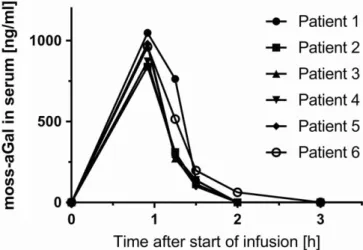
Measurement of pharmacokinetic revealed a nonlinear stable profile in all six patients (Figure 1). The maximum observed
serum concentration (C max) of moss-aGal was reached 55 minutes after start of the infusion, with a mean maximum concentration of 946 ng/mL. Maximum concentrations dropped to half the levels (t½) within 14 minutes (range 11-15 minutes). The mean steady state volume of distribu- tion (Vss) was 0.13 L/kg and the apparent terminal volume of distribution (Vz) was at 0.08 L/kg slightly smaller.
Table 2 summarizes the main pharmacokinetic parameters.
4.2
|Plasma Gb3 and lyso-Gb3 concentrations
Before treatment, all patients had elevated plasma concentra- tions of lyso-Gb3 with a median of 5.25 ng/mL (range 1.43-12.00). After one single dose of moss-aGal, median lyso-Gb3 plasma concentrations decreased by 3.77% from baseline to day 28 (Supplementary Figure S1). Plasma Gb3 (Gb3-C24-0) concentrations decreased from baseline to day 28, with a median decrease of 11% (Figure 2A). Other Gb3 isoforms also showed a slight decrease in plasma with a median decrease of 7.2% for Gb3-C16-0, 4.9% for Gb3-C18-0, and 0.8% for Gb3-C24-1 (Supplementary Figure S2).4.3
|Urine Gb3 concentrations
After one single dose of moss-aGal, Gb3 in morning urine decreased continuously in all patients from baseline to day 28. The mean decrease from baseline of urinary Gb3 (Gb3-C24-0) was 23% at day 7, 54% at day 14, and 60% at day 28 post-dose (Figure 2B). Urinary Gb3-C24-0 concen- trations showed the largest decrease but urinary Gb3-C16-0, Gb3-C18-0, and Gb3-C24-1 concentrations also decreased from baseline to day 28 (Supplementary Figure S3).
5
|D I S C U S S I O N
This is the first clinical trial with moss derived human alfa galactosidase in humans with FD, and the first trial ever with F I G U R E 1 Serum concentration of moss-aGal after i.v.-infusion.
Serum concentrations of moss-aGal were measured at indicated time points using a validated enzyme activity assay
T A B L E 2 Overview of pharmacokinetic parameters
Cmax
(ng/mL)
tmax
(h)
AUClast
(h ng/mL)
AUC0-inf
(h ng/mL)
t1/2
(h)
MRT (h)
CL (L/h/kg)
VZ
(L/kg)
VSS
(L/kg)
N 6 6 6 5 5 5 5 5 5
Mean 946 0.92 789 789 0.23 0.51 0.26 0.08 0.13
Minimum 840 0.92 677 720 0.18 0.49 0.21 0.07 0.12
Median 965 0.92 738 747 0.23 0.51 0.27 0.08 0.13
Maximum 1049 0.92 970 937 0.25 0.56 0.28 0.10 0.14
Abbreviations: AUC0−inf, area under the serum concentration curve (AUC) extrapolated to infinity; AUClast, AUC between the time of dosing and the last measurable (positive) concentration; Cmax, maximum observed serum concentration; CL, total serum clearance; MRT, main residence time extrapolated to infinity; t1/2, terminal elimination half-life; tmax, time to Cmax; Vz, apparent terminal volume of distribution; VSS, steady state volume of distribution.
a biological drug manufactured in moss. It demonstrates the tolerability of a single administration of moss-aGal.
The moss system is used for the production of a variety of different recombinant proteins, as different growth factors and complement factor H.13The application of a moss-derived protein on humans is possible as the basic structures of N- glycosylation are conserved between humans and plants.
However, in contrast to mammalian cell produced α-galactosidase charges, which show a heterogeneous glyco- sylation profile with 25-40% of phosphorylated carbohydrate residues, moss-aGal has a more homogenous glycosylation profile with exclusively mannose and N-acetylglucosamine terminated glycans.6,14Thus, moss-aGal is not endocytosed by M6P receptors but targets the MR for tissue uptake. It
has been shown that the expression of MR is not only restricted to macrophages, but that MR are also expressed on a variety of different cell types, such as endothelial, kidney mesangial, dendritic, tracheal smooth muscle, and retinal pigment epithelium cells.7
5.1
|Safety
A single administration of moss-aGal revealed a good safety profile as it was tolerated without any severe adverse events.
Headache as an adverse event was evaluated as potentially infusion related and has been reported as ERT-related before.15 The adverse event of dysgeusia was evaluated as possibly drug-related and has never been reported before on ERT in FD. However, further clinical studies are required to evaluate the safety profile of moss-aGal upon repeated administration.
5.2
|Antibodies
None of the patients developed anti-aGal IgG antibodies after one single dose of moss-aGal. This result was expected since formation of IgG antibodies against Agalsidase alfa and beta mainly affects male patients and requires repeated exposure to Agalsidase. In the majority of patients, anti- bodies develop during the first 3 months of ERT.16 The slightly elevated anti-aGal antibody concentration in one patient did not alter after application of moss-aGal, and, thus, was interpreted as an unspecific cross-reaction. The immunogenicity profile of moss-aGal will be evaluated in an upcoming phases 2-3 clinical trial which will involve male patients and a repeated dosing over at least 6 months.
5.3
|Pharmacokinetics
Data on pharmacokinetics demonstrated a stable profile in all patients with a short plasma half-life of the infused enzyme of only 14 minutes. In contrast, mammalian cell produced α-galactosidase charges have a much longer plasma half-life time: plasma half-life of Agalsidase alfa is 108 ± 17 minutes in males and 89 ± 28 minutes in females,17of Agalsidase beta 80-120 minutes.18
A potential sink for moss-aGal is the uptake of the enzyme by macrophages which express mannose receptors, as it has been demonstrated for mannose terminating gluco- cerebrosidase.19However, the prolonged decrease in urinary Gb3 concentrations even 28 days after a single dose of moss-aGal is an indication for targeting of the kidney, espe- cially as the reduction of Gb3 concentrations was much higher in urine as in blood. The assumption that macro- phages are not the only sink for moss-aGal is supported by previous in vitro and in vivo studies. In the mouse model, moss-aGal and Agalsidase alfa had comparable efficacy in F I G U R E 2 Relative change to baseline of Gb3 concentrations in
plasma (A) and urine (B) after moss-aGal dosing. Shown are Gb3 (Gb3-C24-0 isoform) concentrations. For urinary values, Gb3 levels were measured at indicated time points and related to urinary creatinine level. Length of the box represents the interquartile range (25th and 75th percentile). The line represents the median and the cross the mean.
Whiskers show minimum to maximum values
reducing Gb3 concentrations in the kidney, reflecting a simi- lar effectiveness of cellular uptake in this organ.6As previ- ously shown by in vitro and in vivo experiments, moss-aGal has a higher uptake in cultured endothelial cells than Agalsi- dase alfa.6 Thus, the short plasma half-life of moss-aGal may be a combination of uptake by macrophages and endo- thelial cells via the MR.
5.4
|Pharmacodynamics
Regarding plasma substrate degradation, after one single dose of moss-aGal no relevant changes in plasma Gb3 and lyso-Gb3 concentrations were found. Plasma Gb3 and lyso- Gb3 concentrations were measured at baseline and only 7 days after the application of moss-aGal. Thus, a decrease of these metabolites in plasma might have occurred shortly after the application of moss-aGal and might have been missed due to the long time-period of 7 days between appli- cation and determination. However, in consecutive trials, plasma Gb3 and lyso-Gb3 concentrations should be deter- mined already within 24 hours application of moss-aGal.
The decrease of urinary Gb3 excretion after one single dose of moss-aGal demonstrates that moss-aGal targets the kidney and is enzymatically active. The prolonged decrease of Gb3 excretion seems to reflect a long half-life of the enzyme in the renal cells and a slow turnover of tubular cells.20 These may lead to a continuous substrate degrada- tion resulting in a prolonged reduction of urinary Gb3 excre- tion. The effect of moss-aGal on urinary Gb3 excretion was much higher than previously reported for mammalian cell producedα-galactosidase.20
After these promising results, phase 2/3 clinical trials are in preparation.
6
|C O N C L U S I O N
In conclusion, these data sustain the hypothesis that mannose-terminated enzymes may be effective also in lyso- somal storage disorders in which nonmacrophage cells are mainly affected. This study demonstrated that a single infusion of moss-aGal was safe, well tolerated and led to a prolonged reduction of urinary Gb3 excretion.
After these promising results, phase 2/3 clinical trials for moss-aGal are in preparation.
A C K N O W L E D G M E N T S
The study was supported by Greenovation, Biotech GmbH.
We thank Prof. Dr. K. J. Lackner and Dr. K. Bruns, Central Laboratory of the University Medical Center Mainz, for ana- lyses of anti-alpha-galactosidase IgG antibodies. Further- more, we thank all the patients for participating in the trial.
C O N F L I C T S O F I N T E R E S T
The study was supported by Greenovation, Biotech GmbH.
J.B.H. received educational grants from Shire Global, as well as travel support and/or honoraria from Shire Deutsch- land GmbH, Amicus Therapeutics GmbH, and Greenovation Biotech GmbH. L.A.-K. received travel support and/or hon- oraria from Shire Deutschland GmbH, Amicus Therapeutics GmbH. G.F. has no conflicts of interest. A.S., A.B., and T.F. are staff members and T.F. is CEO of Greenovation Biotech GmbH. Greenovation Biotech develops and markets moss-based biopharmaceuticals.
A U T H O R C O N T R I B U T I O N S
J.B.H. was involved in conception and design of the study, in analysis and interpretation of the data, and in drafting the article. L.A.-K. and G.F. were involved in performing the trial, in analysis of the data, and in revising the article criti- cally for important intellectual content. A.S. and A.B. were involved in conception and design of the study, in analysis and interpretation of the data, and contributed to the writing of the manuscript. T.F. was involved in conception and design of the study, and in revising the manuscript critically for important intellectual content. All authors read and approved the final version of the manuscript.
R E F E R E N C E S
1. Mehta A, Hughes DA. In: Adam MP, Ardinger HH, Pagon RA, et al., eds.GeneReviews®. Seattle, WA: University of Washington, Seattle; 1993–2018 2002 Aug 5 [updated 2017 Jan 5].
2. El Dib R, Gomaa H, Carvalho RP, et al. Enzyme replacement ther- apy for Anderson-Fabry disease. Cochrane Database Syst Rev.
2016;7:CD006663.
3. Markham A. Migalastat: first global approval. Drugs. 2016;76:
1147-1152.
4. Ghosh P, Dahms NM, Kornfeld S. Mannose 6-phosphate recep- tors: new twists in the tale. Nat Rev Mol Cell Biol. 2003;4:
202-212.
5. Prabakaran T, Nielsen R, Larsen JV, et al. Receptor-mediated endocytosis of α-galactosidase A in human podocytes in Fabry disease.PLoS One. 2011;6:e25065.
6. Shen J-S, Busch A, Day TS, et al. Mannose receptor-mediated delivery of moss-made α-galactosidase A efficiently corrects enzyme deficiency in Fabry mice.J Inherit Metab Dis. 2015;39:
293-303.
7. Stahl PD, Ezekowitz RAB. The mannose receptor is a pattern rec- ognition receptor involved in host defense.Curr Opin Immunol.
1998;10:50-55.
8. Koprivova A, Stemmer C, Altmann F, et al. Targeted knockouts of Physcomitrella lacking plant-specific immunogenic N-glycans.
Plant Biotechnol J. 2004;2:517-523.
9. Ries M, Clarke JTR, Whybra C, et al. Enzyme-replacement ther- apy with agalsidase alfa in children with Fabry disease.Pediatrics.
2006;118:924-932.
10. Radbruch L, Loick G, Kiencke P, et al. Validation of the German version of the Brief Pain Inventory. J Pain Symptom Manage.
1999;18:180-187.
11. Johnson B, Mascher H, Mascher D, et al. Analysis of lyso- globotriaosylsphingosine in dried blood spots. Ann Lab Med.
2013;33:274-278.
12. Lukas J, Giese A-K, Markoff A, et al. Functional characterisation of alpha-galactosidase a mutations as a basis for a new classifica- tion system in Fabry disease.PLoS Genet. 2013;9:e1003632.
13. Reski R, Parsons J, Decker EL. Moss-made pharmaceuticals: from bench to bedside.Plant Biotechnol J. 2015;13:1191-1198.
14. Lee K, Jin X, Zhang K, et al. A biochemical and pharmacological comparison of enzyme replacement therapies for the glycolipid storage disorder Fabry disease.Glycobiology. 2003;13:305-313.
15. Goker-Alpan O, Longo N, McDonald M, et al. An open-label clin- ical trial of agalsidase alfa enzyme replacement therapy in children with Fabry disease who are naïve to enzyme replacement therapy.
Drug Des Devel Ther. 2016;10:1771-1781.
16. Deegan PB. Fabry disease, enzyme replacement therapy and the significance of antibody responses.J Inherit Metab Dis. 2012;35:
227-243.
17. European Medicine Agency (2018a) Replagal EPAR—ANNEX I SUMMARY OF PRODUCT CHARACTERISTICS. EMEA/H/C/
000369-IAIN/0096
18. European Medicine Agency (2018b) Fabrazyme EPAR—ANNEX I SUMMARY OF PRODUCT CHARACTERISTICS. Fabrazyme- EMEA/H/C/000370-N/0104
19. Tekoah Y, Tzaban S, Kizhner T, et al. Glycosylation and function- ality of recombinantβ-glucocerebrosidase from various production systems.Biosci Rep. 2013;33:771-781.
20. Schiffmann R, Murray GJ, Treco D, et al. Infusion of α-galactosidase A reduces tissue globotriaosylceramide storage in patients with Fabry disease.Proc Natl Acad Sci. 2000;97:365-370.
S U P P O R T I N G I N F O R M A T I O N
Additional supporting information may be found online in the Supporting Information section at the end of the article.
How to cite this article: Hennermann JB, Arash- Kaps L, Fekete G, Schaaf A, Busch A, Frischmuth T.
Pharmacokinetics, pharmacodynamics, and safety of moss-aGalactosidase A in patients with Fabry disease.
J Inherit Metab Dis. 2019;42:527–533.https://doi.
org/10.1002/jimd.12052