ESTABLISHMENT OF METHODOLOGY FOR NON-INVASIVE ELECTROCARDIOGRAPHIC MEASUREMENTS IN TURTLES
AND TORTOISES
Anna Linda NÓGRÁDI1* and Márton BALOGH2
1Department of Exotic Animal and Wildlife Medicine and 2Department of Internal Medicine, University of Veterinary Medicine, István u. 2, H-1078 Budapest, Hungary
(Received 2 March 2018; accepted 25 July 2018)
The lack of knowledge on how to perform species-specific electrocardio- graphic (ECG) measurements in chelonians makes ECG evaluations difficult. The aim of this study was to establish non-invasive methods for ECG sample collec- tion in different species of non-anaesthetised chelonians, focusing on adhesive and clamp electrodes. A total of 72 turtles and tortoises from 20 species and vari- ous sizes were used for the study. Body weight ranged from 32 g to 65 kg. From the aspect of specimen fixation, dorsal recumbency proved to be the most useful.
Both adhesive and clamp electrodes yielded results when applied to the plastron and skin folds. Pre-emptive results suggest an indirect correlation with plastron thickness, the presence of a hinge, habitat and measurable ECG wave amplitude.
ECG wave recordings are more likely in aquatic chelonians and species with a hinge. With size the plastron also thickens, making wave detection impossible.
ECG waves were detected in 41 of the 72 specimens, complete PQRST complex- es in 19 animals, with the rest showing absent P waves in all leads. ECG ampli- tudes were below 1 mV, with an average of 0.15 mV R wave amplitude.
Key words: Electrocardiography, heart, cardiac evaluation, chelonian, rep- tile
The reptilian heart can be classified as crocodilian and non-crocodilian. In the non-crocodilian heart, the ventricles are not physically divided and the pul- monary and systemic blood flow are separated and regulated by functional plas- ticity, divided by incomplete muscular ridges (White, 1976; Farrell et al., 1998;
Schilliger, 2012).
The non-crocodilian heart has formerly been thought of as a three- chambered organ, but is now considered an atypical four-chambered heart, since the sinus venosus is considered an additional chamber. It is composed of four main cavities (a single ventricle, two atrial chambers and the sinus venosus), three atrial trunks (the aortic arch, the right aortic arch and the pulmonary trunk), four
*Corresponding author; E-mail: anna.nogradi@gmail.com; Phone: 0036 (30) 856-8256
afferent veins and a well-developed coronary vascularisation (White, 1976; Far- rell et al., 1998; Murray, 2006a,b; Wyneken, 2009).
With more than 9000 species of reptiles little is known about the cardiac anatomy and physiology of most species (Farrell et al., 1998).
In chelonians the width of the heart is almost the same as its length (Far- rell et al., 1998). In most chelonians it is located in the ventral midline, where the humeral, pectoral and abdominal scutes of the plastron intersect, but the position of scutes varies between species (Kik and Mitchell, 2005).
The cavum venosum contains both oxygenated and deoxygenated blood.
The proportion of mixed blood depends on how well the interventricular septum and the muscular ridge are developed (Hicks, 1998).
In non-crocodilians, when anoxia occurs, like in cases of apnoea, or the pressure of the pulmonary artery increases, the perfusion of vital organs is en- sured by a right to left intracardiac shunt, which may occur while the blood pres- sure in the pulmonary veins decreases and the pulmonary outflow resistance grows (Hicks, 1998; Wyneken, 2009). A left to right shunt occurs when systemic blood oxygen levels drop, in case of pulmonary resistance or when the metabo- lism rate increases (Hicks, 1998; Wyneken, 2009).
The function and physiological role of cardiac shunting are not yet totally understood and agreed upon. The degree of cardiac shunting differs with species.
Freshwater turtles with poorly subdivided ventricles have large cardiac shunts (Schilliger, 2012).
Contractions of the reptile heart, since there is no ‘pacemaker’ system or specialised Purkinje fibre-based cardiac conduction system like in mammals, are initiated by cardiac muscle fibres in the sinus venosus of the right atrium and myofibre arrangements in the surrounding and openings of the chambers. These spread to the left and then caudally (White, 1976; Farrell et al., 1998; Wyneken, 2009). Depolarisation of the ventricle starts at its base and then spreads left. Re- polarisation spreads from the base to the apex to the right and left (White, 1976;
Murray, 2006b). Ventricular systole tends to be long and diastole is short (Wyneken, 2009).
Reptiles are poikilothermic animals, so body temperature must always be considered as an important factor when examining these animals (Wyneken, 2009). During basking, the heart rate also increases (Kik and Mitchell, 2005).
Apart from body temperature, the heart rate is also influenced by other noncardi- ac factors like body size, age, activity, respiratory rate, hypovolaemia, digestion, gravidity, and sensory stimulations (Farrell et al., 1998; Murray, 2006a,b).
A complete cardiovascular examination should be performed on all reptile patients, regardless of the limitations (Kik and Mitchell, 2005; Murray, 2006a,b).
The diagnostic tools most used for examining the reptilian heart are auscultation, Doppler probe, electrocardiography, radiography, ultrasound, and CT (Murray, 2006b). Cardiac diseases are difficult to diagnose in turtles and tortoises, and
most are not discovered until necropsy (Hnizdo and Pantchev, 2011). Direct aus- cultation is not possible in chelonians, but Doppler probes help determine the pulse frequency (Murray, 2006b; Schilliger, 2012).
Electrocardiography (ECG) could be helpful in diagnosing cardiac disease in the reptile patient (Murray, 2006b). Standardised ECG techniques according to Einthoven have been used in the past (Murray, 2006b; Schilliger, 2012). Unfor- tunately, standard ECG parameters are not established for many species (Schil- liger, 2012). Interpretation is difficult due to the limited amount of reference ma- terial and the limited degree of sensitivity of the equipment available (Kik and Mitchell, 2005).
Correct placement of the probe is important, and bad positioning of the electrodes can influence ECG readings. The electrodes in reptiles can be placed as self-adhering skin electrodes, stainless steel hypodermic needles, stainless steel suture material through the skin or alligator clips. Placement usually pur- sues the traditional four limb lead placement, but can vary according to species (Murray, 2006b; Schilliger, 2012).
Placement of the electrodes on chelonians is often problematic (Murray, 2006b). According to Murray (2006 b), four limb placement is not appropriate in chelonians due to the low surface/signal voltage generated, and interpreting ECG results is not possible (Murray, 2006b). ECG electrodes have yielded results when attached to the axillary and femoral areas with alligator clips in the past (Murray, 2006b; Schilliger, 2012). Drilling holes in the carapace and attaching the leads to the dermal bone may give better results as better electrical contact is achieved in chelonians, but it is extremely invasive (Murray, 2006b).
The low electric amplitudes (usually < 0.1mV) often seen while perform- ing ECG in reptiles make interpretation and readings of diagnostic quality diffi- cult (Kik and Mitchell, 2005; Murray, 2006b). Equipment with good sensitivity or pre-amplification is required for ECG measurements. Cardiac waveforms may also be unclear due to skeletal muscle activity (Murray, 2006b).
Like in mammals, ECGs in reptiles have P, QRS and T complexes. An SV wave may precede the P wave, and represents the depolarisation of the sinus venosus and the terminal posterior vena cava at its termination at the sinus veno- sus, which take part in the systole of the atria. The impulse begins in the centre of the sinus venosus. The SV wave indicates sinal contraction, the P wave indi- cates atrial depolarisation and contraction, the QRS complex indicates ventricu- lar depolarisation and contraction and the T wave indicates ventricular repolari- sation (Mullen, 1967; McDonald, 1976; White, 1976; Cook and Westrom, 1979;
Murray, 2006b).
Ventricular systole is usually long, and diastole is short (Wyneken, 2009).
In the preferred optimum temperature zone (POTZ), the QT interval can be elon- gated and the TP interval shortened (Wyneken, 2009).
The reptilian ECG has many limitations, but its use in reptile cardiology may hoist other diagnostic methods and could help in the diagnosis of cardiac disease (Murray, 2006b). It can also help monitor the reptile patient during an- aesthesia, even though a reptile heart may continue to beat after death, so just a sinus rhythm is not reliable enough (Murray, 2006b).
While numerous cardiac diseases can be diagnosed, and anaesthesia can be monitored with the help of ECG in mammals, the lack of normal reference values in reptiles makes evaluation of the various components of the ECG not possible at the moment. In chelonians the problem is not only the lack of refer- ence values, but the lack of established methodology of adequate sample collec- tion that can be evaluated, due to the shell and the diverse anatomy of turtles and tortoises.
With the increasing abundance of compact, digital, long-term ECG moni- toring equipment, their applicability in turtles and tortoises is yet to be deter- mined. The aim of this study was to establish non-invasive methods for ECG sample collection in different species of non-anaesthetised chelonians, focusing on adhesive and clamp electrodes, so normal electrocardiographic reference val- ues can be determined in the future.
To the authors’ knowledge no study has been published on placing adhe- sive electrodes on the plastron of chelonians and on whether or not it is a useful way of electrocardiography in turtles and tortoises.
Materials and methods
A total of 72 turtles and tortoises of various sizes, belonging to 20 species, were used for the study. Body weight ranged from 32 g to 65 kg. The animals were kept in enclosures with appropriate species-specific husbandry and diets, and were only removed out of their preferred optimal temperature zone (POTZ) when the examinations could not be performed inside the enclosure. The animals underwent a complete physical examination and only the apparently healthy ones were included in the study. Each animal was weighed and sexed, whenever this was possible.
Different, non-invasive methods of fixation were investigated for exami- nation. These methods included lifting the animal parallel to the ground, holding them head up in a 90 degree angle, and dorsal recumbency (Fig. 1).
Fixation of the electrodes was achieved by the use of adhesive and clamp electrodes. Adhesive electrodes were applied to the plastron (Fig. 2), the cara- pace and the skin folds (Fig. 3), where possible. Clamp electrodes were applied to the extremities, the skin folds, the plastron (Fig. 4) and the carapace, when possible. Both methods of electrode fixation were applied to all the sites men- tioned above, whenever this was possible.
Fig. 1. Placing chelonians in dorsal recumbency for measurements yields results
Fig. 2. Adhesive electrodes applied on the plastron of a big-headed turtle (Platysternon megaceph- alum)
Fig. 3. Adhesive electrodes applied on the skin folds of a common snapping turtle (Chelydra ser- pentina)
Mobile, Bluetooth ECG monitoring equipment (InnoBase pico, Innomed Ltd., Hungary) was used to record ECG samples in seven leads (I, III, III, V, AVR, AVL, AVF).
2 3
Fig. 4. Clamp electrodes applied to the plastron of an Asian leaf turtle (Cyclemys dentata)
The collected data were analysed using the software provided, to measure pulse, P wave length and amplitude, QRS complex length, R wave amplitude and PR intervals in all standard seven lead ECGs (I, III, III, V, AVR, AVL, AVF).
Results
Out of the different, non-invasive methods of fixation, positioning the an- imals in dorsal recumbency proved to be the most useful. Hand motions of the holding person could be excluded and the animals stayed still for a while when lying on their backs. In other positions the signal was not identifiable due to the constant movement of the extremities. The examined 6 Aldabra giant tortoises (Aldabrachelys gigantea) were exceptions due to their size and anatomy, which allowed a different electrode placement site.
The 66, non-Aldabra specimens ranged from 32 grams to 16 kilograms (average: 1756.7 ± 3455.4 grams). Those with complete detectable complexes ranged from 513 to 9500 grams (average: 1871.7 ± 2922.1 grams).
The preliminary results suggest an indirect correlation with plastron thick- ness, the presence of a hinge, aquatic and terrestrial habitat and measurable ECG wave amplitude. The authors managed to detect ECG waves in 41 of the 72 spec-
imens and complete PQRST complexes in 19 animals with the rest showing ab- sent P waves in all leads. Excluding the examined 6 Aldabra giant tortoises the 35 patients from which ECG waves could also be detected ranged from 252 grams to 11 kilograms in weight, with an average of 2097.3 ± 2992.3 grams.
Those with complete detectable complexes ranged from 513 to 9500 grams (av- erage: 1871.7 ± 2922.1 grams). Both adhesive and clamp electrodes produced re- sults depending on the species and placement, respectively.
Full complexes were achieved with both adhesive and clamp electrodes when applied to the plastron and the skin folds. Partial results were obtained when adhesive electrodes were applied to the plastron, the carapace and the skin folds and clamp electrodes were applied to the extremities, the skin folds and the plastron. The 6 examined Aldabra giant tortoises were exceptions, since they all yielded detectable ECG waves with clamps applied to the skin folds. Species dis- tribution showed 12 aquatic and 8 terrestrial animals. In agreement with the liter- ature, the authors found ECG amplitudes below 1 mV, with an average R wave amplitude of 0.15 mV. Table 1 summarises the results of the study.
Discussion
Electrocardiography in reptiles has great potential but many limitations as well. It could help diagnose cardiac disease and monitor the reptile patient during anaesthesia (Murray, 2006b). Non-invasive ECG methods need to be determined and normal reference values established. With the special anatomy and diversity of chelonians, a single type of method is unlikely to yield results in all species.
Since chelonians are poikilothermic animals, true physiological measurements can only be made in animals housed at optimal environmental temperature, since POTZ influences the body temperature and the chelonian heart (Holz and Holz, 1995; Kik and Mitchell, 2005). Heart rate hysteresis (change with temperature) is normal in reptiles. Heart rate increases during basking and lowers during cooling (Seebacher, 2000; Franklin and Seebacher, 2003). The readings will truly reflect the values that can be evaluated with the reference values of the species only when species-specific normal values have been established. Normal ECG values exist only for a few chelonian species (Holz and Holz, 1995). This study made it possible to examine animals that were in the POTZ, but often by the time reptiles arrive at a veterinary clinic their body temperature has dropped due to inadequate transportation. This does not only give false results when examining the heart, but increases the risk of anaesthesia. It is vital for the owner and the veterinarian to keep the chelonian as close to the POTZ as possible.
Body size influences the heart rate, since it is slower in smaller reptiles (Farrell et al., 1998). Body size also plays a crucial role in ECG measurements because in very small specimens reading is not possible due to the very low elec-
tric amplitude, while in very big chelonians the plastron thickness does not allow signals to be interpreted by the ECG equipment.
Digestion and pregnancy also influence the heart rate (Seebacher, 2000;
Franklin and Seebacher, 2003). In the present study, the authors did not perform ECG measurements on gravid animals.
One study showed that handling caused tachycardia for several minutes in examined turtles (Cabanac and Bernieri, 2000).The authors had every step of the examination prepared to reduce the influence of handling on the results. Activity and ventilation also increases the heart rate, while apnoea slows it (Munns et al., 2004, 2005). In some freshwater turtles the heart rate can double during ventila- tion compared to the resting rate in apnoea (Galli et al., 2004). Fast and precise ECG measurements are important to get accurate results.
Low-voltage waves, arrhythmias in unfed animals, uncommon Q and S waves, and sex differences with the P–R interval being shorter in females have also been determined (Kaplan and Schwartz, 1963). Due to the different species and sizes and the relatively low number of individuals in one species the authors could not draw conclusions regarding the correlation between sex and ECG values.
Significant differences were also described in the surgical stage of anaes- thesia (Kaplan and Schwartz, 1963). A study on nine anaesthetised red-eared sliders (Trachemys scripta elegans) was performed with three cutaneous elec- trodes to record lead II traces. The study found lower ECG amplitude waveforms than in mammals and a longer repolarisation phase with longer ST and QT inter- vals and shorter TP intervals, with a correlation between a lower heart rate need- ing a longer period of repolarisation without prolonged depolarisation (Holz and Holz, 1995). The aim of the authors was to perform measurements on conscious animals. Unfortunately studies that have been conducted and published were mostly done on anaesthetised animals. In the future, reference values should also be obtained on non-anaesthetised animals when possible.
Both adhesive and clamp electrodes produced results depending on the species and placement, respectively. Because of the large variety in chelonians, the authors were not expecting to find one electrode placement type that would work on all turtles and tortoises. Applying electrodes to the skin folds yields re- sults in Aldabra giant tortoises but not in African spurred tortoises (Centrochelys sulcata). This makes sense if one compares the habitat of the two species. Aldab- ra giant tortoises live on islands and have basically no predators, so they have relatively thin and soft skin compared to their size and do not pull themselves back in their shell. African spurred tortoises have to face predators on mainland Africa, have a thick skin and can basically close themselves off as a defence mechanism. Because of this, performing ECG on the latter did not yield results while the authors could easily apply clamp electrodes on Aldabra giant tortoises with good results.
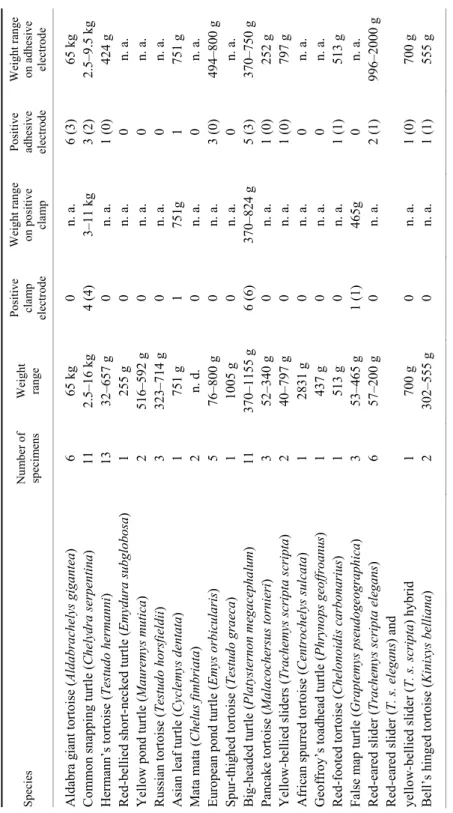
Table 1 Characteristics of turtles and tortoises included in the study SpeciesNumber of specimensWeight range Positive clamp electrode Weight range on positive clamp Positive adhesive electrode
Weight range on adhesive electrode Aldabra giant tortoise (Aldabrachelys gigantea)6 65 kg0 n. a. 6 (3)65 kg Common snapping turtle (Chelydra serpentina)11 2.5–16 kg4 (4)3–11 kg3 (2)2.5–9.5 kg Hermann’s tortoise (Testudo hermanni)13 32–657 g0 n. a. 1 (0)424 g Red-bellied short-necked turtle (Emydura subglobosa)1 255 g0 n. a. 0 n. a. Yellow pond turtle(Mauremys mutica)2 516–592 g0 n. a. 0 n. a. Russian tortoise(Testudo horsfieldii))3 323–714 g0 n. a. 0 n. a. Asian leaf turtle (Cyclemys dentata)1 751 g1 751g 1 751 g Mata mata(Chelus fimbriata)2 n. d. 0 n. a. 0 n. a. European pond turtle (Emys orbicularis)5 76–800 g0 n. a. 3 (0)494–800 g Spur-thighed tortoise (Testudo graeca)1 1005 g0 n. a. 0 n. a. Big-headed turtle (Platysternon megacephalum))11 370–1155 g6 (6)370–824 g5 (3)370–750 g Pancake tortoise (Malacochersus tornieri)3 52–340 g0 n. a. 1 (0)252 g Yellow-bellied sliders (Trachemys scripta scripta)2 40–797 g0 n. a. 1 (0)797 g African spurred tortoise (Centrochelys sulcata)1 2831 g0 n. a. 0 n. a. Geoffroy’s toadhead turtle (Phrynops geoffroanus)1 437 g0 n. a. 0 n. a. Red-footed tortoise (Chelonoidis carbonarius)1 513 g0 n. a. 1 (1)513 g False map turtle (Graptemys pseudogeographica)3 53–465 g1 (1)465g 0 n. a. Red-eared slider (Trachemys scripta elegans)6 57–200 g0 n. a. 2 (1)996–2000 g Red-eared slider (T. s. elegans) and yellow-bellied slider (T. s. scripta) hybrid1 700 g0 n. a. 1 (0)700 g Bell’s hinged tortoise (Kinixys belliana)2 302–555 g0 n. a. 1 (1)555 g Weights are presented in grams unless otherwise indicated. In positive cases fully positives are presented in parentheses. n. a.: not applicable, n. d.: no data recorded
It was important for the authors to place electrodes in a non-invasive man- ner. Drilling holes in the carapace to achieve better electrical contact has been done in the past (Murray, 2006b), but it is invasive, painful and, according to the authors, not suitable for the everyday veterinary practice.
Due to the lack of relevant literature the authors wanted to know if adhe- sive electrodes work on the plastron or carapace of chelonians. As mentioned in the results, they do yield results when used on the plastron in some species and sizes. This is a new and easily accessible way to perform electrocardiography in chelonians in everyday veterinary practice, although not on all species and sizes.
This study suggests that ECG wave recordings are more likely in aquatic chelonians and species with a hinge. Weight also plays a role, since animals un- der 252 grams were too small to produce measurable results, while turtles and tortoises above 11 kg in weight had a growth in plastron thickness that did not make wave detection possible.
An increasing availability of compact, digital long-term ECG monitoring equipment in veterinary practices can be seen. Veterinarians are held back from using it in chelonians because ECG probe placement sites are not determined and reference values are not available. This study showed some sites that yield results with adhesive and clamp electrodes. Pre-emptive results suggest that smaller (approx. 500–1000 g) specimens yield results with adhesive electrodes applied to the plastron (although there is a high variance between the species, suggesting that the aquatic ones are more likely to provide detectable ECG waves with this method), whereas in the case of larger animals, clamps applied to the skin folds show detectable waves. Chelonian ECG would have clinical applications for the practitioner. Just like small mammals, chelonians also have cardiac diseases, which could be determined by the use of ECG, and patient monitoring during an- aesthetic episodes, which is not an easy task at the moment, would also be easier with the help of ECG.
References
Cabanac, C. and Bernieri, C. (2000): Behavioural rise in body temperature and tachycardia in han- dling of a turtle (Clemmys insculpta). Behav. Proc. 49, 61–68.
Cook, R. A. and Westrom, W. (1979): Cardiac anatomy, cardiac physiology and electrocardiology of reptiles. Am. Assoc. Zoo Vet. An. Proc., pp. 16–22.
Farrell, A. P., Gamperl, A. K. and Francis, E. T. B. (1998): Comparative aspects of heart morphology.
In: Gans, C. and Gaunt, A. S. (eds) Biology of the Reptilia. Volume 19, Morphology G.
Visceral Organs. Society for the Study of Amphibians and Reptiles, Ithaca. pp. 375–424.
Franklin, C. E. and Seebacher, F. (2003): The effect of heat transfer mode on heart rate responses and hysteresis during heating and cooling in the estuarine crocodile Crocodylus porosus. J.
Exp. Biol. 206, 1143–1151.
Galli, G., Taylor, E. W. and Wang, T. (2004): The cardiovascular responses of the freshwater turtle Trachemys scripta to warming and cooling. J. Exp. Biol. 207, 1471–1478.
Hicks, J. W. (1998): Cardiac shunting in reptiles: mechanisms, regulation and physiological func- tions. In: Gans, C. and Gaunt, A. S. (eds) Biology of the Reptilia. Volume 19, Morphology G. Visceral Organs. Society for the Study of Amphibians and Reptiles, Ithaca. pp. 425–483.
Hnizdo, J. and Pantchev, N. (2011): Medical Care of Turtles & Tortoises. Edition Chimaira, Frank- furt. pp. 329–336.
Holz, R. M. and Holz, P. (1995): Electrocardiography in anaesthetised red-eared sliders (Trache- mys scripta elegans). Res. Vet. Sci. 58, 67–69.
Kaplan, H. M. and Schwartz, C. (1963): Electrocardiography in turtles. Life Sci. 2, 637–645.
Kik, M. J. L. and Mitchell, M. A. (2005): Reptile cardiology: A review of anatomy and physiology, diagnostic approaches, and clinical disease. Sem. Av. Exo. Pet Med. 14, 52–60.
McDonald, H. S. (1976): Methods for the physiological study of reptiles. In: Gans, C. and Dawson, W. R. (eds) Biology of the Reptilia. Volume 5, Physiology A. Academic Press, New York.
pp. 19–126.
Mullen, R. K. (1967): Comparative electrocardiography of the squamata. Physiol. Zool. 40, 114–126.
Munns, S. L., Hartzler, L. K., Bennett, A. F. and Hicks, J. W. (2004): Elevated intra-abdominal pressure limits venous return during exercise in Varanus exanthematicus. J. Exp. Biol. 207, 4111–4120.
Munns, S. L., Hartzler, L. K., Bennett, A. F. and Hicks, J. W. (2005): Terrestrial locomotion does not constrain venous return in the American alligator, Alligator mississippiensis. J. Exp.
Biol. 208, 3331–3339.
Murray, M. J. (2006a): Cardiopulmonary anatomy and physiology. In: Mader, D. R. (ed.) Reptile Medicine and Surgery, 2nd edition. Saunders Elsevier, St. Louis. pp. 124–134.
Murray, M. J. (2006b): Cardiology. In: Mader, D. R. (ed.) Reptile Medicine and Surgery, 2nd edi- tion. Saunders Elsevier, St. Louis. pp. 181–195.
Schilliger, L. (2012): Reptile Cardiology. In: Proceedings of the International Conference on Rep- tile and Amphibian Medicine. Cremona, May 13–15, 2012. pp. 62–71.
Seebacher, F. (2000): Heat transfer in a microvascular network: the effect of heart rate on heating and cooling in reptiles (Pogona barbata and Varanus varius). J. Theor. Biol. 3, 97–109.
White, F. N. (1976): Circulation. In: Gans, C. (ed.) Biology of the Reptilia. Volume 5, Physiology A. Academic Press, New York. pp. 275–334.
Wyneken, J. (2009): Normal reptile heart morphology and function. Vet. Clin. North Am. Exot.
Anim. Pract. 12, 51–63.