R E S E A R C H A R T I C L E
Effects of substrate, ceramic thickness, translucency, and cement shade on the color of CAD/CAM
lithium-disilicate crowns
Alexandra Czigola DMD
1| Emese Abram DMD
1| Zoltan I. Kovacs DMD
2|
Krisztina Marton DMD, PhD
2| Peter Hermann DMD, PhD
1| Judit Borbely DMD, PhD
11Department of Prosthodontics, Faculty of Dentistry, Semmelweis University, Budapest, Hungary
2Department of General Dental Preclinical Practice, Faculty of Dentistry, Semmelweis University, Budapest, Hungary
Correspondence
Alexandra Czigola, DMD, Department of Prosthodontics, Faculty of Dentistry, Semmelweis University, Szentkirályi Street 47, 1088 Budapest, Hungary.
Email: czigola.alexandra@dent.semmelweis- univ.hu
Abstract
Objective:The aim of this in vitro study was to evaluate the effects of substrate colors, different levels of ceramic thickness and translucency, and cement shades on the color difference from a reference color of lithium-disilicate crowns.
Materials and Methods:A premolar tooth preparation was made on a study model for 1.0 and 1.5 mm thick full-ceramic crowns. Digital impressions were taken (3Shape TRIOS) and crowns designed in a CAD program (DentalDesigner). Shade A1 crowns were milled (Everest, Kavo) from high- translucency (HT) and low-translucency IPS e.max (Ivoclar Vivadent) blocks. Twelve substrates were made of different colors and materials (Natural Die Material, Co-Cr, zirconia, and gold-colored alloy).
Three different shades of try-in pastes were used to simulate the effect of cements (Variolink Esthetic try-in paste; Ivoclar). Shade measurement was done three times for each crown by a spectrophotome- ter (VITA Easyshade Advance); averages were compared to a reference crown (A1, HT, 1.5 mm, ND2 abutment, neutral try-in paste) withΔE00(CIEDE2000, according to the CIE latest standard) calculated.
Results:All the examined parameters influenced theΔE00of the crowns. The weakest effect was exerted by the try-in paste.
Conclusions:All examined parameters influenced the final color of e.max CAD lithium-disilicate ceramic crowns.
Clinical Significance
Matching the shade of ceramic crowns to the natural tooth color is a great challenge in dentistry.
To meet patients' increasing esthetical expectations, CAD/CAM methods are very popular for full- ceramic crowns. However, several factors such as the shade of the abutment, luting cement color, ceramic thickness, and translucency may influence the final color. Our objective was to measure the optical effect of these factors on the final shade of CAD/CAM lithium-disilicate ceramic crowns.
K E Y W O R D S
CAD/CAM, ceramic, colorimetry, crown, optical phenomena
1 | I N T R O D U C T I O N
New CAD/CAM (Computer Aided Design/Computer Aided Manufactur- ing) technologies are being introduced in the dental marketplace, and most companies investing into restorative dentistry are also on the mar- ket to develop materials for digital systems. CAD/CAM systems are
based on three factors: data collection, data processing, and manufactur- ing. Nowadays, open systems make it possible to use the constituent parts separately.1
Based on a 2015 AACD (American Academy of Cosmetic Dentistry) survey, 1/3 of dentists currently use a CAD/CAM system in their prac- tice, while another 1/3 are considering to invest into such technology.2 DOI: 10.1111/jerd.12470
This is an open access article under the terms of the Creative Commons Attribution License, which permits use, distribution and reproduction in any medium, provided the original work is properly cited.
© 2019 The Authors.Journal of Esthetic and Restorative Dentistrypublished by Wiley Periodicals, Inc.
J Esthet Restor Dent.2019;31:457–464. wileyonlinelibrary.com/journal/jerd 457
The abundance of new systems facilitates the use of superior dental ceramics besides conventional techniques. This strictly con- trolled industrial ceramic processing means increased micro- structural uniformity, higher density, lower porosity, and decreased residual stress. CAD/CAM systems have the potential to improve clinical predictability. These ceramic materials are perfect for manufacturing all types of all-ceramic restorations, such as inlays, onlays, crowns, and brid- ges.3The final goal is a quick, reliable, and predictable esthetic result.
Together with the development of CAD/CAM systems, intraoral scanners are also available on the dental market. With laboratory scanners, it is possible to create a 3D model of the oral cavity without taking an impression and digitizing a model. Technicians can use these intraoral scan based digital models to design the restorations.4Digital impressions offer speed, efficiency, storability of captured informa- tion, and an easier way of communication between the dental office and the laboratory through digital images.5
Newer generations of all-ceramic systems and adhesive cements allow dentists to use a minimally invasive approach and make thinner restorations (1.0-1.5 mm). It is a great task to preserve as much tooth structure as we can and obtain a superior esthetic result. The goal is to achieve the desired color, especially the VITA Classical A1 shade, which is the most commonly selected shade tab for ceramic restorations.6
By allowing greater light transmission, all-ceramic materials improve the translucency of the restoration; however, a perfect natural-like color cannot be ensured.7Lithium disilicate-reinforced glass-ceramic restora- tions are in the focus of interest due to their low refractive index, which makes the material very translucent despite its high crystalline content.
This characteristic makes them suitable for full contour restorations and the highest of esthetic demands.8,9In the 2015 AACD survey, when the question“which restoration material would you use in your own mouth?” was asked, the majority (84%) of responding dentists chose lithium-disilicate (eg, IPS e.max) with zirconia trailing.2
Natural tooth color is defined by the optical properties of enamel and dentin. It is a great challenge in dentistry to match the color of natural teeth with ceramic restorations.
It has been described that the shade of the restorations is highly influenced by the prepared die. If a ceramic restoration is placed on a dark underlying tooth structure, for example, an endodontically treated tooth, the color beneath the crown might result in discoloration and shadowing of the restoration, particularly in the cervical areas.9
In addition to the prepared die, luting cements can also be a modi- fying factor. Composite resin cements are produced in different shades by manufacturers to influence the final appearance of full-ceramic res- torations. Their aim is to enhance the final appearance of the crowns or veneers. It has been demonstrated that controlling the thickness of the ceramic might allow clinicians to manage the overall translucency of the restoration, while the choice of cement color has less of an effect.9In another study, using different shaded try-in pastes did not bring the crowns a perceptible color change from their original crown color.10
The purpose of this study therefore was to evaluate the color difference of different substrate colors, different levels of ceramic thickness and translucency, and different cement shades of lithium- disilicate crowns (IPS e.max CAD, Ivoclar Vivadent, Schaan, Liechten- stein) produced by CAD/CAM technology based on intraoral scans (3shape TRIOS, Copenhagen, Denmark) from a reference color.
2 | M A T E R I A L S A N D M E T H O D S
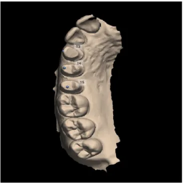
An upper right first premolar tooth #14 on a study model was prepared with a chamfer finishing line for 1.0 and 1.5 mm thick full-ceramic crowns. A silicone index was made of the upper right quadrant to control the depth of the preparation.
Digital impression was taken using a 3Shape TRIOS intraoral scanner (Figure 1). Pre-preparation scan11 was made in intercuspal position of the jaws for occlusion. The digital impression of the origi- nal tooth shape was used to design the ceramic crowns.
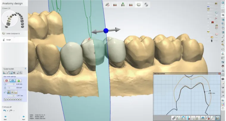
Crowns were designed for the prepared tooth #14 and for the adjacent teeth #13 and 15 with the DentalDesigner (3Shape, Copenha- gen, Denmark) CAD program in the Dental Laboratory of the Univer- sity. The CAD/CAM method ensured that all crowns were identical in shape and size.
Even thicknesses of 1.0 and 1.5 mm were secured on the buccal sides of the ceramic crowns (Figure 2), taking into consideration a 5-mm diameter of the Easyshade probe (VITA Easyshade Advance 4.0).
The influence of different cement shades on the final color was also investigated. The marginal gap for the cement was set to 0.01 mm; a uniform layer of 0.04 mm gap (3Shape DentalDesigner program original set up) was used on the inner surface (Figure 3).
Test crowns were milled from polymethyl-methacrylate material to check the design.
When satisfied with all settings and parameters, the CAD files were sent to an Everest KaVo CAM unit (KaVo, Bieberach, Germany).
The crowns were milled from IPS e.max CAD (Ivoclar Vivadent) lithium-disilicate ceramic blocks.
Maxillary right first premolar (14): 10 copies of 1.0 mm and 10 copies of 1.5 mm thick A1 crowns were milled from low-translucency (LT) and high-translucency (HT) blocks.
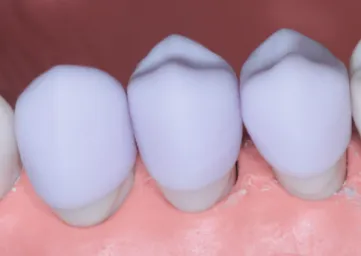
Adjacent teeth #13 and 15 crowns were also milled to secure the approximal contact points. Figure 4 shows the crowns before crystallization.
FIGURE 1 Scan of prepared dies in the DentalDesigner CAD program
Crystallization firing and glazing of the crowns was done according to the manufacturers' instructions.
IPS Natural Die (Ivoclar Vivadent) composite material of nine dif- ferent shades was used to create the core part of the substrates fused together with replaceable plastic posts of the study model. CAD/CAM method was used to mill Co-Cr alloy, gold painted alloy, and sup- erwhite zirconia substrate cores. Substrates were made in 12 different colors (Figure 5).
Variolink Esthetic (Ivoclar Vivadent) try-in cement was used for cementation. The advantage of the try-in paste is that it can be easily wiped off crowns. Opaque light plus, yellowish warm, and translucent neutral shades were used.
VITA Easyshade Advance 4.0 device was used for shade measure- ment. To measure the color, spectrophotometers can be used. The VITA company's first spectrophotometer was the Easyshade in 2004.
Dozic et al found that Easyshade was the most reliable instrument of FIGURE 2 The buccal surface designed and controlled for equal thicknesses (1.0 mm and 1.5 mm)
FIGURE 3 The cementation gap (40μm)
shade matching in both in vitro and in vivo circumstances.12The posi- tion of the Easyshade probe was standardized on the buccal surface of the crowns. Each crown-shade was measured three times. CIELAB values were recorded. Easyshade measurements were performed by two trained dental students. The spectrophotometer was calibrated before each measurement. External light sources such as daylight were not excluded during all measurements and they were taken in a room with artificial lightening conditions. According to the Commission internationale de l'éclairage (CIE) standard Easyshade uses the D65 (6500 K, daylight) illuminant for shade matching.12As it is described by CIE in 1931 for determining the color of an object the size of tooth the 2 Observer is needed. The 2Observer is used in the Easyshade for CIELAB color space.13
Easyshade advance 4.0 screen can display theL a bcoordinates for the measured shade. We can calculate a modified chromaC0 and hue angleh0fromL0,a0, andb0coordinates to define the numerical color dif- ference (ΔE00) between two pairs of samples by using CIEDE2000 equitation.14
ΔE00 (color difference in the coordinate system according to CIEDE2000) was calculated by comparison to the reference crown:
high translucent, A1, 1.5 mm thick crown placed on the ND2 substrate with neutral try-in paste. Many studies have selected shade A1 for their research, as this tooth shade is the most commonly selected for
ceramic restorations.6,19,22–25,28,30For reference abutment we have chosen ND2 shade acting as a non-discolored prepared healthy dentin and neutral try-in paste, that we would not want to affect the final color of the crown by a colored luting cement.
CIEDE 2000 (ΔE00) equation15 was used to calculate color difference:
ΔE00=
ffiffiffiffiffiffiffiffiffiffiffiffiffiffiffiffiffiffiffiffiffiffiffiffiffiffiffiffiffiffiffiffiffiffiffiffiffiffiffiffiffiffiffiffiffiffiffiffiffiffiffiffiffiffiffiffiffiffiffiffiffiffiffiffiffiffiffiffiffiffiffiffiffiffiffiffiffiffiffiffiffiffiffiffiffiffiffiffiffiffiffiffiffiffiffiffiffiffi ΔL0
kLSL
2
+ ΔC0 kCSC
2
+ ΔH0 kHSH
2
+RTΔC0 kCSC
ΔH0 kHSH: s
CIELAB is classically the standard parameter for total color differ- ence between two objects, but to improve the correction between computed and perceived color differences, it is recommended to use the CIEDE2000 color-difference formula (ΔE00). CIEDE2000 (ΔE00) includes specific corrections for nonuniformity of CIELAB space (so- called weighting functionsSL,SC, andSH) and the parametric factors for the influence of illuminating and viewing conditions in color-difference evaluation (KL, KC, and KH). The values of KL, KC, and KH in the CIEDE2000 formula were all set to 1.15RTmeans rotation function, the interaction between chroma and hue differences in the blue region.16
The CIEDE2000 formula is the latest international standard according to CIE.15It provides better adjustments in color-difference evaluation.17,18
For CIEDE 2000 formula, the perceptibility threshold (PT) was set at 0.8 and an acceptability threshold (AT) ofΔE00= 1.8 was set.19
Measurement procedure steps taken included cement application, crown positioning on the substrate, cement excess removal, spectro- photometer calibration and measurement.
3 | S T A T I S T I C A L A N A L Y S I S
We used the four-way interaction model for statistical analysis. We estimated the goodness of the model's fit by subtracting the ratio of the residual sum of squares and the total sum of squares from one (ie, calculating the coefficient of determination,R2), and also looked at the proportion of residuals under the perceptibility and ATs. The model's fit was remarkably good atR2= 0.9798 forΔE00(withR2= 0.9860 achiev- able in a complete four-way interaction structure). Residuals were under the PT/ATs in 91.5%/99.7% of observations. The parameter FIGURE 4 IPS e.max CAD crowns before crystallization
FIGURE 5 The 12 different substrates
values ofCandhfor each observation were calculated fromL,a, and b following published formulae.L,a,b,C, and hwere then averaged across each measurement triplet. This averaged dataset was then used to calculateΔE00. A crown identifier was generated to group observa- tions of identical crown thickness and translucency (but varying sub- strate and cement material). Multilevel mixed-effects linear regression was used to evaluate the effects of technical parameters on color match. The outcome variable wasΔE00based on the CIEDE2000 for- mula. Fixed-effect explanatory variables included crown translucency, crown thickness, substrate type, and cement type (all categorical), plus interaction terms between: thickness and translucency; thickness and substrate; translucency and substrate; cement and substrate; translu- cency and thickness and substrate (three-way). Interactions between cement and thickness, and between cement and translucency, were not used because they were observed to be negligible size compared to other interactions. The model included the random effect of crown identifier and allowed heteroskedastic variability across different sub- strates. Modeling results were expressed as adjusted predictions with 95% confidence intervals (CI) or adjusted effects with 95% CI. The sig- nificance criterion was set atα= 0.05. The statistical package Stata20 was used for data handling and analysis.
4 | R E S U L T S
Working with the recent CIEDE 2000 equation (Figure 6) 41 of the 144 measured combinations were within the acceptable range (under AT); however, only 13 of these were below the PT.
The smallestΔE00values were measured on 1.5 mm thick LT crowns. With Co-Cr and gold alloy substrates, there was no combi- nation under the PT. The greatest color difference compared to the
reference crown could be detected on 1 mm HT crowns on Co-Cr substrates.
Negative range data presented in Figure 7 indicate that the respectiveΔE00values (ie, shade discrepancy) were lower for 1.5 mm than for 1.0 mm crowns. It can be concluded that thicker crowns pro- vide better coverage, as expected, unless the material is highly translu- cent, in which case thickness seems to play a less accentuated role.
It is interesting to note that thickness has no effect when applying HT crowns with zirconium dioxide substrates. However, in case of LT crowns, inferior results were obtained with the thicker formulation.
As it is represented in Figure 8, crowns with greater translucency have higher ΔE00 values resulting in a lower quality shade match.
Nonetheless, the results of HT crowns on yellowish substrates show decreasing ΔE—despite the decreasing lightness of the shade (ND1-ND6)—thoughΔE00increase was expected since HT crowns are more translucent. This phenomenon demonstrates the complexity of the optical properties of these materials.
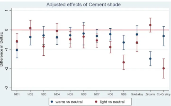
In Figure 9, the Values in the negative range indicate better results having been achieved with a type of cement different from the neutral.ΔE00differences greater in absolute value than 1 can be con- sidered clinically significant; such effects are exclusive to the ND9 and Co-Cr substrates when applying light plus cement, and to the zirconia substrates when applying warm cement.
5 | D I S C U S S I O N
Results of the present study showed that the color difference (ΔE00) of a CAD/CAM glass-ceramic lithium-disilicate full-ceramic crown is influenced by ceramic thickness and translucency, substrate color, and cement color. Data of this investigation are in agreement with previ- ous studies in the literature.9,10,21–31
FIGURE 6 Model predictions as a function of restoration technical parameters (ΔE00 based on CIEDE2000 formula). They-axis representsΔE shade of substrate. Markers indicate point estimate and 95% confidence interval. The green and black lines are the acceptability threshold and the perceptibility threshold, respectively. The reference crown (A1 crown/low translucency/1.5 mm thick/neutral cement/ND2 substrate) is also represented here
If a ΔE00value less than 0.8 (with the recent CIEDE2000 for- mula) is regarded a clinically imperceptible color change, only 13 of the measured combinations of LT crowns were below this visibility threshold. None of the HT crowns was in this range. The reason might be the material's optical properties: LT blocks have more lithium-disilicate crystals than HT blocks. Crystals reduce the internal scattering of light as it passes through the material. That means when the substrate has a dark color or the underlying tooth is highly dis- colored, the application of a CAD/CAM glass-ceramic lithium dis- ilicate with a HT ceramic block may result in limited success.9,24,28,29
However, translucent ceramics have been more frequently used to fabricate anterior restorations than opaque ones, for example, medium-opacity and low-opacity (LO) blocks. Pires et al compared HO (high-opacity) and LO ceramics and foundΔE00values of ceramic HO to be lower than those of ceramic LT. Clinicians should consider increasing the thickness and opacity of the ceramic to mask the underlying color.30In many studies, increasing ceramic thickness is accompanied by better color results.9,21,22,28We found that ceramic thickness has less of an effect if the material is highly translucent (HT crowns, Figure 7).
FIGURE 7 Comparison of 1.0 and 1.5 mm ceramic thickness. Thex-axis represents substrate shades whileyrepresentsΔE00differences of 1.5 mm crowns in relation to their 1.0 mm counterparts. The red reference line represents mean values for 1.0 mm crowns
FIGURE 8 Low-translucency (LT) crowns' ceramic translucency compared to high-translucency (HT) crowns. The red line demonstrates the values of the LT crowns
Besides translucency, crown chromacity also plays an important role in final color. A limitation of our study therefore is that we mea- sured A1 shaded crowns only. Al Ben Ali et al confirmed that increasing chromacity (high-opacity ceramics) can reduce the color effect of the underlying tooth structure.22
Previous studies agreed that the underlying substructure affects the final color of the restoration.9,10,21–23,28Chaiyabutr et al found that dark-colored abutment teeth had the greatestΔE values com- pared to other configurations.9Changing the underlying color from a lighter to a darker background resulted in increased color differ- ences.9,22Ge et al found that using gold shaded posts and cores did not influence the color of 1.5 mm thick full-ceramic crowns (Empress 2), with all measurements being under the patients' average percep- tion level (ΔE= 1.8). The greatest color change was caused by the Ni- Cr post core.10The present study found that there was no combina- tion under the AT (ΔE00= 1.8) with gold alloy substrates, and only one measured combination was below the AT (ΔE00= 1.8) with Co-Cr sub- strates (1.5 mm LT crown, light plus try-in paste). As we expected, the lowestΔEvalues were found with LT 1.5 mm thick crowns (Figure 6).
Only a few studies can be found about the color modifying effect of luting cements, but they agree that the choice of cement color has less of an effect on the final color of restorations.9,23,28
According to Niu, the final shade of a 1.5-mm thick lithium- disilicate crown is affected not only by the cement's shade but also by its layer thickness (300, 100, 50μm). In this study, white opaque cements (Multilink white opaque; Nexus3 white opaque) demon- strated better masking ability than cements of other colors.31When we used opaque cement (Variolink Esthetic light plus), it significantly influenced the crown-shade on ND9 and Co-Cr substrates (Figure 9).
Another parameter of interest is luting cement layer thickness.
Increasing white opaque cement layer thickness from 100 to 300μm did not affect the shade of lithium-disilicate restorations.31Neither any measured ceramic layer thickness (1.0, 1.5, 2.0, 2.5 mm), nor increased cement thickness (300 vs 100μm) had significant modifying effects.9
It should be plausible that the layer thickness we used (40μm) results in no more than a minuscule color change. However,
increasing the amount of luting cement causes a loss of bond strength between the crown and the die, and increases the possibil- ity of ceramic fractures.
6 | C O N C L U S I O N
Within the limitations of this study, all examined parameters play an important role in changing the color of a monolithic CAD/CAM full- ceramic crown compared to the reference crown. In case of HT crowns, the crown wall thickness has limited influence. Data show that the opti- cal properties of these ceramic materials are highly complex, but of the two examined e.max CAD block translucency levels (LT and HT), LT crowns produced better color outcomes. Cement color has less of an effect, but in some cases (ND9, Co-Cr substrates) opaque cement shades can help mask the underlying darker substrate color.
O R C I D
Alexandra Czigola https://orcid.org/0000-0002-3543-5237
R E F E R E N C E S
1.van Noort R. The future of dental devices is digital.Dent Mater. 2012;
28:3-12.
2.American Academy of Cosmetic Dentistry. Survey 2015. http://www.
aacd.com/proxy/files/Publications%20and%20Resources/AACD%20 State%20of%20the%20Cosmetic%20Dentistry%20Industry%202015.
pdf. Accessed April 10, 2018.
3.McLaren EA, Hyo L. CAD/CAM update: technologies and materials and clinical perspectives. Inside Dentistry. 2006:98-103.
4.Ender A, Mehl A. Accuracy of complete-arch dental impressions: a new method of measuring trueness and precision. J Prosthet Dent.
2013;109:121-128.
5.Kim SY, Kim MJ, Han JS, Yeo IS, Lim YJ, Kwon HB. Accuracy of die- scaptured by an intraoral digital impression system using para- llelconfocal imaging.Int J Prosthodont. 2013;26:161-163.
6.Bagis B, Turgut S. Optical properties of current ceramics systems for laminate veneers.J Dent. 2013;41:24-30.
7.Wee AG, Monaghan P, Johnston WM. Variation in color between intended matched shade and fabricated shade of dental porcelain.
J Prosthet Dent. 2002;87:657-666.
8.Giordano R, McLaren EA. Ceramics overview: classification by micro- structure and processing methods.Compend Contin Educ Dent. 2010;
31:682-684. 686, 688.
9.Chaiyabutr Y, Kois JC, Lebeau D, Nunokawa G. Effect of abutment tooth color, cement color, and ceramic thickness onthe resulting opti- cal color of a CAD/CAM glass-ceramic lithium disilicate reinforced crown.J Prosthet Dent. 2011;105:83-90.
10. Ge J, Wang XZ, Feng HL. Influence of different post core materials on the color of Empress 2 full ceramic crowns.Chin Med J (Engl). 2006;
119:1715-1720.
11. 3Shape TRIOS. TRIOS-2014-1-1.3.4.1-A-EN. http://www.danedentallab.
com/uploads/7/5/9/2/7592248/_trios_3_shape_manual.pdf. Accessed April 10, 2018.
12. Dozic AKC, El-Zohairy A, Feilzer AJ, Khashayar G. Performance of five commercially available tooth color-measuring devices.J Prosthodont.
2007;16:93-100.
13. VITA Easyshade. Operating Manual. https://www.dt-shop.com/fileadmin/
media/ga/21886_ga_enu.pdf. Accessed December 11, 2018.
14. Luo MR, Cui G, Rigg B. The development of the CIE2000 color differ- ence formula.Color Res Appl. 2001;26:340-350.
15. CIE. Colorimetry. Report No.: CIE Pub. No. 15. Vienna: CIE Central Bureau; 2004.
FIGURE 9 Cement shade effect compared to the reference neutral shade. Thex-axis represents substrate shades while they-axis represents the estimatedΔE00of test cements in comparison to the neutral cement, with the red line (zero) representing meanΔE00
values obtained with neutral cements
16. Pecho OE, Ghinea R, Alessandretti R, Pérez MM, Della Bona A. Visual and instrumental shade matching using CIELAB and CIEDE2000 color difference formulas.Dent Mater. 2016;32:82-92.
17. Gómez-Polo C, Portillo MM, Lorenzo Luengo MC, et al. Comparison of the CIELab and CIEDE2000 color difference formulas.J Prosthet Dent. 2016;115:65-70.
18. Ghinea R, Pérez MM, Herrera LJ, et al. Color difference thresholds in dental ceramics.J Dent. 2010;38:57-64.
19. Paravina RD, Ghinea R, Herrera LJ, et al. Color difference thresholds in dentistry.J Esthet Restor Dent. 2015;27:1-9.
20. StataCorp. Stata Statistical Software: Release 11. College Station, TX:
StataCorp LP; 2009.
21. Niu E, Agustin M, Douglas RD. Color match of machinable lithium dis- ilicate ceramics: effects of foundation restoration.J Prosthet Dent.
2013;110:501-509.
22. Al Ben Ali A, Kang K, Finkelman MD, et al. The effect of variations in translucency and background on color differences in CAD/CAM lith- ium disilicate glass ceramics.J Prosthodont. 2014;23:213-220.
23. Dozic A, Tsagkari M, Khashayar G, Aboushelib M. Color management of porcelainveneers: influence of dentin and resin cement colors.
Quintessence Int. 2010;41:567-573.
24. Kürklü D, Azer SS, Yilmaz B, Johnston WM. Porcelain thickness and cement shade effects on the colour and translucency of porcelain veneering materials.J Dent. 2013;41:1043-1050.
25. Ozturk O, Uludag B, Usumez A, et al. The effect of ceramic thickness and number of firings on the color of two ceramic systems.J Prosthet Dent. 2008;100:99-106.
26. Turgut S, Bagis B, Ayaz EA. Achieving the desired colour in dis- coloured teeth, using leucite-based CAD-CAM laminate systems.
J Prosthet Dent. 2014;42:68-74.
27. Wang F, Takahashi H, Iwasaki N. Translucency of dental ceramics with different thicknesses.J Prosthet Dent. 2013;110:14-20.
28. Vichi A, Ferrari M, Davidson CL. Influence of ceramic and cement thickness on the masking of various types of opaque posts.J Prosthet Dent. 2000;83:412-417.
29. Chang J, Da Saliva JD, Sakai M, et al. The optical effect of composite luting cement on all ceramic crowns.J Dent. 2009;37:937-943.
30. Pires LA, Novais PM, Araújo VD, Pegoraro LF. Effects of the type and thickness of ceramic, substrate, and cement on the optical color of a lithium disilicate ceramic.J Prosthet Dent. 2017;117:144-149.
31. Niu E, Agustin M, Douglas RD. Color match of machinable lithium dis- ilicate ceramics: effects of cement color and thickness.J Prosthet Dent.
2014;11:42-50.
How to cite this article: Czigola A, Abram E, Kovacs ZI, Marton K, Hermann P, Borbely J. Effects of substrate, ceramic thickness, translucency, and cement shade on the color of CAD/CAM lithium-disilicate crowns. J Esthet Restor Dent.
2019;31:457–464.https://doi.org/10.1111/jerd.12470