UN CORRECTED PR
OOF
https://doi.org/10.1007/s10989-018-9748-z
Effect of PACAP on Bacterial Adherence and Cytokine Expression in Intestinal Cell Cultures
A. Illes1,2 · G. Horvath1 · E. Schafer3 · M. Kerenyi4 · O. Karadi5 · B. Opper1 · G. Toth6 · D. Reglodi1
Accepted: 16 August 2018
© Springer Nature B.V. 2018
Abstract
Bacterial adhesion is a crucial event of intestinal pathological conditions evoked by bacterial infections. Pituitary adenylate cyclase activating polypeptide (PACAP) is an endogenous neuropeptide having a widespread distribution throughout the entire body including the digestive tract. It has diverse physiological functions in the gastrointestinal system, including pro- tective effects in several models of intestinal inflammatory conditions. However, its effects on bacterial adherence and the inflammatory reactions as a result of that have not been elucidated yet. The aim of our study was therefore to investigate the effect of PACAP on bacterial adherence and cytokine expression upon lipopolysaccharide (LPS) exposure. Small intestinal INT407 and colonic Caco-2 cells were treated with PACAP prior to exposure to bacteria (Escherichia coli, Salmonella Typhimurium, Klebsiella pneumoniae, Enterococcus faecalis) and colonies were counted. PACAP had no significant influ- ence on bacterial adhesion, as it did not change the number of colonies of investigated bacteria. However, PACAP was able to counteract the LPS-induced increases in the expression of the cytokines IL-8 and CXCL-1 in INT407 cells, as assessed by cytokine array. These results indicate that while PACAP has no direct effect on bacterial adherence, it can influence the cytokine expression of intestinal cells upon endotoxin-induced exposure, possibly contributing to the known anti-inflam- matory actions of PACAP in the intestinal system.
Keywords Bacterial adherence · Pituitary adenylate cyclase activating polypeptide · Intestinal cell cultures · Lipopolysaccharide · Cytokine expression
Introduction
Pituitary adenylate cyclase-activating polypeptide (PACAP) is a pleiotropic neuropeptide belonging to the secretin/
glucagon/vasoactive intestinal peptide family. It was first
identified in ovine hypothalamus based on the efficacy on influencing adenylate cyclase activity (Miyata et al. 1989).
PACAP has a widespread distribution in the body and exerts a wide range of physiological effects also in the gastrointes- tinal system (Somogyvari-Vigh and Reglodi 2004; Horvath et al. 2016; Reglodi et al. 2018). The occurrence and func- tions of PACAP and its receptors can be well demonstrated in neuroendocrine and interstitial cells, in the myenteric and submucosal plexus in the entire length of the gastrointes- tinal tract and also in the pancreas, gall bladder and liver (Arciszewski et al. 2015; Ji et al. 2013; Koves et al. 1993;
Oh et al. 2005; Vu et al. 2016; Zhang et al. 2006; Reglodi et al. 2018). Among others, PACAP influences motility of the intestinal wall (Fujimiya and Inui 2000), inhibits pace- maker activity of interstitial cells of Cajal (Wu et al. 2015) and regulates sphincter function (Farre et al. 2006). PACAP affects release of brain-derived neurotrophic factor in intes- tinal smooth muscle cells and influences gastric juice secre- tion (Al-Qudah et al. 2015; Reglodi et al. 2018).
* G. Horvath
gabriella.horvath@aok.pte.hu
1 Department of Anatomy, MTA-PTE PACAP Research Team, University of Pecs, Pécs, Hungary
2 1st Department of Internal Medicine, University of Pecs, Pécs, Hungary
3 Department of Gastroenterology, Medical Centre, Hungarian Defence Forces, Budapest, Hungary
4 Department of Medical Microbiology and Immunology, University of Pecs, Pécs, Hungary
5 Department of Oncotherapy, University of Pecs Medical School, Pécs, Hungary
6 Department of Medical Chemistry, University of Szeged, Szeged, Hungary
AQ1
AQ2 1 2
3
4 5
6 7 8 9 10 11 12 13 14 15 16 17 18 19
20 21
22
23 24 25
26 27 28 29 30 31 32 33 34 35 36 37 38 39 40 41 42 43 44 A1
A2 A3 A4 A5 A6 A7 A8 A9 A10 A11 A12 A13 A14
UN CORRECTED PR
OOF
Several in vivo and in vitro studies confirmed the general cytoprotective, antiapoptotic, antioxidant, and anti-inflam- matory effects of PACAP (Reglodi et al. 2012; Ferencz et al.
2009; Horvath et al. 2010; Vaudry et al. 2009). Our previous studies investigating cytoprotective effects have revealed that PACAP is able to exert ambivalent effects on cell viability in the small intestinal INT407 cells depending on the applied stressor and the timing of application (Illes et al. 2017). The anti-inflammatory activity of PACAP can be ascribed to inhi- bition of immune and inflammatory cells. PACAP decreases the release of inflammatory chemokines and cytokines such as TNF-α and IL-6, inhibits chemotaxis and phagocytosis.
Hence, it is an important endogenous immunomodulatory peptide in many different models of inflammatory diseases (Gomariz et al. 2006). In humans, several studies have pre- viously shown changes in PACAP level in colon diseases.
An earlier study found significantly lower levels of PACAP in sigmoid colon and rectum tumors compared to normal healthy tissue (Szanto et al. 2012). Another study described significantly higher PACAP levels in patients with symp- tomatic diverticular disease (Simpson et al. 2009). On the contrary, investigations of colon mucosa of children with ulcerative colitis found decreases in nerve fibers contain- ing PACAP (Kaminska et al. 2006, 2007). Furthermore, our previous investigations have found marked increase in levels of both PACAP isoforms in patients suffering from ulcera- tive colitis (Horvath et al. 2016). The protective effects of endogenous PACAP can also be detected in the colon: in dextran sulfate sodium-induced colitis the symptoms of the disease in PACAP-deficient mice, such as weight loss, bleed- ing, diarrhea were markedly more severe than in wild type animals (Azuma et al. 2008). Higher morbidity of colorec- tal tumors in PACAP-deficient mice indicate the possible regulatory role of endogenously present PACAP (Nemetz et al. 2008). In case of acute ileitis caused by experimental Toxoplasma gondii infection in mice, PACAP prophylaxis improved survival and anti-inflammatory cytokine response (Heimesaat et al. 2014). Besides the beforementioned data, our previous studies have shown differences in microbiota composition in PACAP deficient mice compared to wildtype mice: Bifidobacteria are virtually absent in the knockouts (Heimesaat et al. 2017). A recent study has revealed direct antimicrobial effect of PACAP38 against Gram-positive and Gram-negative bacteria (Starr et al. 2018). All these results indicate that PACAP influences directly and indirectly the intestinal flora and bacterial colonization, which might be a link with increased susceptibility of PACAP deficient mice to intestinal inflammatory diseases and tumors.
Adherence of bacteria to the surface of intestinal epi- thelial cells is a crucial step in intestinal bacterial infec- tions. Despite the available data on the antimicrobial and intestinal effects of PACAP, its role in bacterial adherence has not been elucidated yet. Therefore, the aim of our study
was to explore the role of PACAP in bacterial adhesion in small intestinal INT407 and colon adenocarcinoma Caco-2 cell lines. For this purpose, four different bacteria (Escheri- chia coli, Enterococcus faecalis, Klebsiella pneumoniae, Salmonella Typhimurium) were used. We chose these bac- teria because Escherichia coli, Enterococcus faecalis and Klebsiella pneumonia are commensal bacteria in the gut.
Although Klebsiella pneumonia is member of the gut flora, it can lead to progression of gastrointestinal diseases such as Crohn’s disease and ulcerative colitis (Kaur et al. 2018).
Furthermore, Salmonella Typhimurium was chosen based on its pathogenic effect in the small intestine.
In addition, our studies were expanded to obtain further information on PACAP’s effects on inflammatory processes in the intestinal system using INT 407 cell culture.
Materials and Methods
INT 407 Cell CultureThe INT 407 cell line isolated originally from human embry- onic intestinal tissue was purchased from ATCC. INT 407 cells were cultured in Roswell Park Memorial Institute (RPMI 1640) medium (Lonza, Switzerland) supplemented with 10% fetal bovine serum (Biosera, USA) and 1% peni- cillin–streptomycin (Biosera, USA). Cells were passaged by trypsinization (Trypsin/EDTA; Biosera, USA), followed by dilution in RPMI medium containing 10% fetal bovine serum. Experiments started 24 h after incubation in humified 95% air and 5% CO2 mixture at 37 °C in the medium.
Caco‑2 Cell Culture
The Caco-2 cell line derived from human colon adenocar- cinoma cell line was from ATCC. Caco-2 cells were cul- tured in DMEM high glucose/F-12 supplemented with 10%
fetal bovine serum and 1% penicillin–streptomycin (Bios- era, USA). Cells were passaged by trypsinization (Trypsin/
EDTA; Biosera, USA), followed by dilution in DMEM medium containing 10% fetal bovine serum. Experiments started 24 h after incubation in humified 95% air and 5%
CO2 mixture at 37 °C in the medium.
Determination of Bacterial Adherence to INT 407 and CaCo‑2 With and Without PACAP
Before determination of bacterial adhesion, the living INT 407 and Caco-2 cells were counted by trypan blue and 3 × 10⁵ cells/
well were plated into 24-well tissue culture plates. Both the small intestinal INT407 cells and the large intestinal Caco-2 cells were grown in their media supplemented with 10% fetal bovine serum without antibiotics in 5% CO2 at 37 °C. Half of
45 46 47 48 49 50 51 52 53 54 55 56 57 58 59 60 61 62 63 64 65 66 67 68 69 70 71 72 73 74 75 76 77 78 79 80 81 82 83 84 85 86 87 88 89 90 91 92 93 94 95 96 97
98 99 100 101 102 103 104 105 106 107 108 109 110 111 112
113
114
115 116 117 118 119 120 121 122 123 124
125
126 127 128 129 130 131 132 133 134
135 136
137 138 139 140 141 142
UN CORRECTED PR
OOF
the 24-well tissue culture plates contained cells, which were cultured in the above described medium supplemented with 400 ng/ml PACAP. Next day the confluent monolayers were washed three times with 1 ml Dulbecco’s Phosphate-Buffered Saline (DPBS) before infection with 3 × 10⁸ bacterial cells in DPBS. After incubation for 3 h at 37 °C in 5% CO2 each well was washed three times with 1 ml of DPBS. To recover adher- ent bacterial cells, washed INT 407 and Caco-2 cells in each well were treated with 1 ml of 0.1% Triton-X100 and 0.25%
trypsine in PBS for 10 min at room temperature. Each lysate was homogenized by repeated pipetting and 10 µl of their ten- fold serial dilutions were plated on Mueller–Hinton agar plates and incubated at 37 °C for 24 h. The following day, the colo- nies were counted to determine the number of bacteria that had adhered to the small and large intestinal cells. Adherent bacterial counts were obtained from three independent assays with each assay performed in triplicate wells. The investigated bacterial cells were Escherichia coli ATCC 25922, Salmonella Typhimurium ATCC 14028, Klebsiella pneumoniae ATCC 13833, Enterococcus faecalis (clinical isolate). Each experi- ment was repeated six times. Statistical analysis was done using one-way analysis of variance p < 0.05 was considered as significant.
Cytokine Array
Investigating the effect of PACAP on cytokine expression Proteome Profiler Human Cytokine Array kit (R&D Systems, Minneapolis, MN, USA) was performed. The investigated INT407 cells were plated in six-well plates and the follow- ing experimental groups were created: (1) control group of cells, (2) cells treated with 100 nM PACAP alone for 24 h, (3) cells exposed to 100 ng/ml LPS for 24 h and (4) cells treated with 100 nM PACAP 2 h prior to 24 h-long 100 ng/ml LPS exposion. After incubation, supernatants were collected and were carried out according to the manufacturer’s protocol.
The kit contains all necessary contents. Briefly, after block- ing the membranes for 1 h and adding the reconstituted Detec- tion Antibody Cocktail for another 1 h at room temperature, membranes were incubated with sample/antibody mixture at 2–8 °C overnight. After washing, horseradish peroxidase- conjugated streptavidin was added for 30 min, then membranes were exposed to a chemiluminescent reagent. Array data were analyzed using ImageJ software. The experiment was repeated three times. Statistical analysis analysis was performed by two-way analysis of variance. p < 0.05 was considered as significant.
Results
Effect of PACAP on Bacterial Adhesion
To investigate the effect of PACAP on bacterial adhesion PACAP-pretreated and non-pretreated INT407 and Caco-2 cell cultures were infected with the following bacteria:
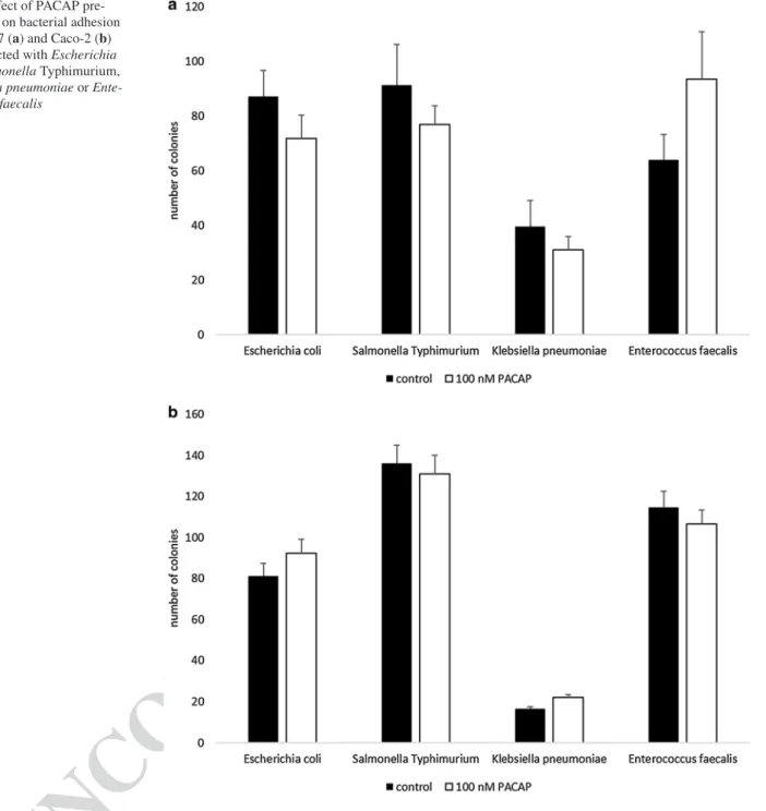
Escherichia coli ATCC 25922, Salmonella Typhimurium ATCC 14028, Klebsiella pneumoniae ATCC 13833 and Enterococcus faecalis (clinical isolate). The assay is based on studying the growth of bacteria being able to adhere to cells in vitro. In the current experiment, the influence of PACAP on the number of bacterial colonies adhered to small and large intestinal cells was investigated. PACAP pretreat- ment of INT407 cells was not able to influence the bacte- rial adherence (Fig. 1a). Furthermore, similarly to INT407 cells, infections of colon adenocarcinoma-derived Caco-2 cells could not be altered by PACAP-pretreatment (Fig. 1b).
Effect of PACAP on Cytokine Expression in INT407 Cells
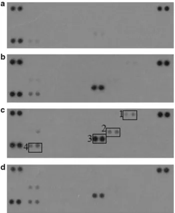
To elucidate whether PACAP has an effect on cytokine expression in INT407 cells, we used human cytokine array (Figs. 2, 3). PACAP alone significantly elevated the expres- sion of IL-8 and IL-18 changed slightly the expression of CXCL-1 (C-X-C motif ligand 1) and MIF (macrophage migration inhibitory factor). 100 ng/ml LPS exposure led to higher levels of CXCL-1, IL-8, IL-18 and MIF. These changes were significant in case of IL-8 and IL-18. PACAP- pretreatment was able to attenuate the LPS-induced elevated expression of IL-8 and CXCL-1. Both PACAP and LPS exerted a slight, but not significant, activating effect on MIF.
Discussion
In the present study we found that PACAP could alter the cytokine expression of INT407 small intestinal cells alone and especially after LPS exposure, indicating that the decreased cytokine levels after endotoxin insult can be an additional factor in its anti-inflammatory effect in several intestinal inflammatory conditions. However, we did not find any direct effect on bacterial adhesion, suggesting that PACAP does not affect bacterial adhesion on intestinal cells directly, but is rather involved in the inflammatory reactions induced by different pathogens.
Our finding, that PACAP did not directly influence bac- terial adhesion under the applied experimental conditions, is of importance in light of previous findings indicating that PACAP might have direct effects on bacteria and other
143 144 145 146 147 148 149 150 151 152 153 154 155 156 157 158 159 160 161 162 163 164 165
166
167 168 169 170 171 172 173 174 175 176 177 178 179 180 181 182 183 184 185 186 187
188
189
190 191 192 193 194 195 196 197 198 199 200 201 202 203
204 205
206 207 208 209 210 211 212 213 214 215 216
217
218 219 220 221 222 223 224 225 226 227 228 229 230 231
UN CORRECTED PR
OOF
pathogens. The first direct anti-microbial effects were proven in Tetrahymena thermophila, a protozoon, where PACAP acted as a chemorepellent (Mace et al. 2000), through the same receptor as lyzozyme (Hassenzahl et al. 2001). A sub- sequent study found antiparasitic activity against another parasite, Trypanosoma brucei (T. brucei). Both VIP and PACAP killed the infective bloodstream form but not the noninfective insect form of the parasite (Gonzalez-Rey et al.
2006). Parasite integrity was destroyed through a mecha- nism involving their entry and accumulation into the cyto- sol (Gonzalez-Rey et al. 2006). A recent study has proven that PACAP and its related peptides and analogs are able
to exert direct antibacterial effects (Starr et al. 2018). Both PACAP38 and 27, as well as related peptides, VIP and secre- tin, had antibacterial effects against Gram-negative bacteria, such as Escherichia coli. PACAP could act against the Gram positive Staphylococcus aureus. Another assay showed that PACAP had moderate sterilizing effect against Pseudomonas aeruginosa and Escherichia coli, an effect less pronounced by the other peptides. PACAP even had a moderate activity against Bacillus cereus (Starr et al. 2018). The mechanism of this effect was found to be a membrane permeabiliza- tion effect, without causing toxic side effects, as shown by the undisturbed hemolytic activity on red blood cells (Starr
Fig. 1 Effect of PACAP pre- treatment on bacterial adhesion in INT407 (a) and Caco-2 (b) cells infected with Escherichia coli, Salmonella Typhimurium, Klebsiella pneumoniae or Ente- rococcus faecalis
232 233 234 235 236 237 238 239 240 241 242 243
244 245 246 247 248 249 250 251 252 253 254 255
UN CORRECTED PR
OOF
et al. 2018). All these data point to the possibility of PACAP acting directly on bacteria. We hypothesized that PACAP might also influence the adhesion of bacteria to the intestinal wall, but we found no effect in the adhesion assay. There- fore, based on our currect knowledge, it seems that PACAP can exert a protective effect on bowel inflammatory condi- tions via both direct antibacterial as well as cytoprotective
actions, without influencing the adhesion of bacterial to the intestinal wall.
As a next step, therefore, we investigated the effects of PACAP on cytokine expression of INT407 cells. As PACAP is a known modulator of inflammatory cytokine and chemokine production in various cells, we aimed at testing this effect in small intestinal cells. We found that PACAP altered the expression of several cytokines. IL-8 is a member of the chemokine family identified as a strong chemotactic factor (Baggiolini et al. 1989). Interleukin-8 plays a crucial role in inflammatory, autoimmune and infectious diseases (Harada et al. 1994; Koch et al. 1992;
Smyth et al. 1991). In our present study, we detected ele- vated expressions of IL-8 upon exposure to LPS. PACAP was able to counteract the induction of IL-8 expression.
Our finding is in accordance with those of Zhang et al.
(2005), who found expression-decreasing effect of PACAP in ARPE cells stimulated with IL-1β. Besides IL-8, we found significantly elevated expression of IL-18 in LPS- induced samples, but in this case no effect of PACAP on it could be observed. Moreover, increase in expression of CXCL-1 could be measured upon LPS exposure. PACAP- pretreatment behaved in an opposite way, it was able to significantly decrease the activation of CXCL-1. Delgado et al. have previously decribed expression-decreasing effect of PACAP in case of LPS-stimulated peritoneal macrophages and microglial cells (2001, 2002). In sum- mary, PACAP is able to alter the expression of several cytokines. This has been demonstrated in many different cell and tissue types, such as lymphocytes (Wang et al.
1999), astrocytes and microglial cells (Gottschall et al.
1994; Delgado et al. 2002), in the retina in a chronic hypoperfusion model (Szabo et al. 2012) and in the kidney, in diabetic and ischemic nephropathy (Horvath et al. 2010;
Banki et al. 2013). The effects of PACAP on the cytokine expression varies between cells and also depends on the type of injury. In many cases, PACAP alone does not affect cytokine expression, but can counteract the injury- induced alterations (Szabo et al. 2012). Our observations indicate that while PACAP has no direct action on the bacterial adhesion to the intestinal wall, it can counteract the endotoxin-induced effects on cytokine expression, pos- sibly contributing to the well-known intestinal protective effects of the peptide.
Fig. 2 Representative human cytokine array showing the expression of various cytokines in control INT-407 cells (a), cells treated with 100 nM PACAP (b), cells exposed to 100 ng/ml LPS (c), treatment with 100 nM PACAP 2 h prior to 100 ng/ml LPS stimulation (d).
(1) Changes of CXCL-1 (1), IL-8 (2), IL-18 (3) and MIF (4) could be detected. LPS-induced changes of CXCL-1 (1) and IL-8 (2) were counteracted by PACAP-pretreatment. Other spots, where no sig- nificant changes were observed are (from upper left corner, without numbers): CCL-1, CCL-2, MIP-1α, RANTES, CD40 ligand, C5a, CXCL10, CXCL11, CXCL12, G-CSF, GM-CSF, ICAM-1, IFN-γ, IL-1a, IL-1β, IL-1ra, IL-2, IL-4, IL-5, IL-6, IL-8, IL-10, IL-12 p70, IL-13, IL-16, IL-17A, IL-17E, IL-18, IL-21, IL-27, IL-32α, MIF, Serpin E1, TNF-α, TREM-1
AQ3 256
257 258 259 260 261 262
263 264 265 266 267 268 269 270 271 272 273 274 275 276 277 278 279 280 281 282 283 284 285 286 287 288 289 290 291 292 293 294 295 296 297 298 299 300 301 302 303 304 305 306
307
UN CORRECTED PR
OOF
Acknowledgements EFOP-3.6.2-16-2017-00008 “The role of neuro- inflammation in neurodegeneration: from molecules to clinics”/Cen- tre or neuroscience, PTE AOK Research Grant KA-2017-17, National Research, Development and Innovation Office, Hungary (Grant Nos.
NKFIH119759, 2017-1.2.1-NKP-2017-00002, GINOP-2.3.2-15-2016- 00050 “PEPSYS,” MTA-TKI 14016). The study was financed by the Higher Education Institutional Excellence Programme of the Ministry of Human Capacities in Hungary, within the framework of the 20765- 3/2018/FEKUTSTRAT of the University of Pécs.
Compliance with Ethical Standards
Conflict of interest All authors declare that they have no conflict of interest.
Research Involving Human and Animal Rights There were neither human nor animal experiments in our studies. Every experiment was done using cell line purchased from ATCC.
Informed Consent Informed consent was not needed because of the in vitro nature of the investigations.
References
Al-Qudah M, Alkahtani R, Akbarali HI, Murthy KS, Grider JR (2015) Stimulation of synthesis and release of brain-derived neurotropic
factor from intestinal smooth muscle cells by substance P and pituitary adenylate cyclase-activating peptide. Neurogastroenterol Motil 27:1162–1174. https ://doi.org/10.1111/nmo.12604 Arciszewski MB, Mozel S, Sienkiewicz W (2015) Pituitary adenylate
cyclase-activating peptide-27 (PACAP-27) is co-stored with gala- nin, substance P and corticotropin releasing factor (CRF) in intra- pancreatic ganglia of the sheep. Pol J Vet Sci 18:343–350. https ://
doi.org/10.1515/pjvs-2015-0044
Azuma YT, Hagi K, Shintani N, Kuwamura M, Nakajima H, Hashi- moto H, Baba A, Takeuchi T (2008) PACAP provides colonic protection against dextran sodium sulfate induced colitis. J Cell Physiol 216:111–119. https ://doi.org/10.1002/jcp.21381 Baggiolini M, Walz A, Kunkel SL (1989) Neutrophil-activating pep-
tide-1/interleukin 8, a novel cytokine that activates neutrophils.
J Clin Invest 84:1045–1049. https ://doi.org/10.1172/JCI11 4265 Banki E, Degrell P, Kiss P, Kovacs K, Kemeny A, Csanaky K, Duh A,
Nagy D, Toth G, Tamas A, Reglodi D (2013) Effect of PACAP treatment on kidney morphology and cytokine expression in rat diabetic nephropathy. Peptides 42:125–130. https ://doi.
org/10.1016/j.pepti des.2013.02.002
Delgado M, Ganea D (2001) Inhibition of endotoxin-induced mac- rophage chemokine production by vasoactive intestinal peptide and pituitary adenylate cyclase-activating polypeptide in vitro and in vivo. J Immunol 167:966–975. https ://doi.org/10.4049/jimmu nol.167.2.966
Delgado M, Jonakait GM, Ganea D (2002) Vasoactive intestinal pep- tide and pituitary adenylate cyclase-activating polypeptide inhibit chemokine production in activated microglia. Glia 39:148–161 https ://doi.org/10.1002/glia.10098
Fig. 3 Quantification of cytokine array. Normalized data are expressed as mean of pixel intensity ± SEM. *p < 0.05 versus control group of cells,
#p < 0.05, ##p < 0.01 versus LPS-treated group
308 309 310 311 312 313 314 315 316
317
318 319 320 321 322 323 324
325
326 327
328 329 330 331 332 333 334 335 336 337 338 339 340 341 342 343 344 345 346 347 348 349 350 351 352 353 354 355 356
UN CORRECTED PR
OOF
Farre R, Auli M, Lecea B, Martinez E, Clave P (2006) Pharmacologic characterization of intrinsic mechanisms controlling tone and relaxation of porcine lower esophageal sphincter. J Pharmacol Exp Ther 316:1238–1248. https ://doi.org/10.1124/jpet.105.09448 2 Ferencz A, Racz B, Tamas A, Reglodi D, Lubics A, Nemeth J, Nedvig
K, Kalmar-Nagy K, Horvath OP, Weber G, Roth E (2009) Influ- ence of PACAP on oxidative stress and tissue injury following small-bowel autotransplantation. J Mol Neurosci 37:168–176.
https ://doi.org/10.1007/s1203 1-008-9132-0
Fujimiya M, Inui A (2000) Peptidergic regulation of gastrointestinal motility in rodents. Peptides 21:1565–1582
Gomariz RP, Juarranz Y, Abad C, Arranz A, Leceta J, Martinez C (2006) VIP-PACAP system in immunity: new insights for mul- titarget therapy. Ann NY Acad Sci 1070:51–74. https ://doi.
org/10.1196/annal s.1317.031
Gonzalez-Rey E, Chorny A, Delgado M (2006) VIP: an agent with license to kill infective parasites. Ann NY Acad Sci 1070:303–
308. https ://doi.org/10.1196/annal s.1317.032
Gottschall PE, Tatsuno I, Arimura A (1994) Regulation of interleu- kin-6 (IL-6) secretion in primary cultured rat astrocytes: syn- ergism of interleukin-1 (IL-1) and pituitary adenylate cyclase activating polypeptide (PACAP). Brain Res 637:197–203 Harada A, Sekido N, Akahoshi T, Wada T, Mukaida N, Matsushima
K (1994) Essential involvement of interleukin-8 (IL-8) in acute inflammation. J Leukoc Biol 56:559–564
Hassenzahl DL, Yorgey NK, Keedy MD, Price AR, Hall JA, Myzcka CC, Kuruvilla HG (2001) Chemorepellent signaling through the PACAP/lysozyme receptor is mediated through cAMP and PKC in Tetrahymena thermophila. J Comp Physiol A 187:171–176 Heimesaat MM, Dunay IR, Schulze S, Fischer A, Grundmann U,
Alutis M, Kühl AA, Tamas A, Toth G, Dunay MP, Göbel UB, Reglodi D, Bereswill S (2014) Pituitary adenylate cyclase-acti- vating polypeptide ameliorates experimental acute ileitis and extra-intestinal sequelae. PLoS ONE 9(9):e108389. https ://doi.
org/10.1371/journ al.pone.01083 89
Heimesaat MM, Reifenberger G, Vicena V, Illes A, Horvath G, Tamas A, Fulop BD, Bereswill S, Reglodi D (2017) Intestinal microbiota changes in mice lacking pituitary adenylate cyclase activating polypeptide (PACAP)—bifidobacteria make the difference. Eur J Microbiol Immunol 7:187–199. https ://doi.
org/10.1556/1886.2017.00021
Horvath G, Mark L, Brubel R, Szakaly P, Racz B, Kiss P, Tamas A, Helyes Z, Lubics A, Hashimoto H, Baba A, Shintani N, Furjes G, Nemeth J, Reglodi D (2010) Mice deficient in pituitary ade- nylate cyclase activating polypeptide display increased sensitiv- ity to renal oxidative stress in vitro. Neurosci Lett 469:70–74.
https ://doi.org/10.1016/j.neule t.2009.11.046
Horvath G, Illes A, Heimesaat MM, Bardosi A, Bardosi S, Tamas A, Fulop BD, Opper B, Nemeth J, Ferencz A, Reglodi D (2016) Protective intestinal effects of pituitary adenylate cyclase acti- vating polypeptide. In: Reglodi D, Tamas A (eds) Pituitary adenylate cyclase activating polypeptide—PACAP. Springer Nature, New York, pp 271–288
Ji H, Zhang Y, Shen XD, Gao F, Huang CY, Abad C, Busuttil RW, Waschek JA, Kupiec-Weglinski JW (2013) Neuropep- tide PACAP in mouse liver ischemia and reperfusion injury:
immunomodulation by the cAMP-PKA pathway. Hepatology 57:1225–1237. https ://doi.org/10.1002/hep.25802
Kaminska B, Landowski P, Gonkowski S, Majewski M, Renke J, Korzon M (2006) Changes in the number of neuroprotective transmitter containing mucosal nerve fibres in children with ulcerative colitis. Med Wieku Rozwoj 10:483–491
Kaminska B, Landowski P, Gonkowski S, Szlagatys-Sidorkiewitz A, Majewski M, Dobosz M, Ismail H, Korzon M (2007) Analysis of enteral nervous system in children with drug resistant ulcera- tive colitis. Med Wieku Rozwoj 11:117–122
Kaur CP, Vadivelu J, Chandramathi S (2018) Impact of Klebsiella pneumoniae in lower gastrointestinal tract diseases. J Dig Dis 19:262–271. https ://doi.org/10.1111/1751-2980.12595 Koch AE, Polverini PJ, Kunkel SL, Harlow LA, DiPietro LA,
Elner VM, Elner SG, Strieter RM (1992) Interleukin-8 as a macrophage-derived mediator of angiogenesis. Science 258:1798–1801
Koves K, Arimura A, Vigh S, Somogyvari-Vigh A, Miller J (1993) Immunohistochemical localization of PACAP in the ovine digestive system. Peptides 14:449–455
Mace SR, Dean JG, Murphy JR, Rhodes JL, Kuruvilla HG (2000) PACAP-38 is a chemorepellent and an agonist for the lysozyme receptor in tetrahymena thermophila. J Comp Physiol A 186:39–43
Miyata A, Arimura A, Dahl RR, Minamino N, Uehara A, Jiang L, Culler MD, Coy DH (1989) Isolation of a novel 38 residuehy- pothalamic polypeptide which stimulates adenylate cyclase in pituitary cells. Biochem Biophys Res Commun 164:567–574 Nemetz N, Abad C, Lawson G, Nobuta H, Chhith S, Duong L, Tse
G, Braun J, Waschek JA (2008) Induction of colitis and rapid development of colorectal tumors in mice deficient in the neu- ropeptide PACAP. Int J Cancer 122:1803–1809. https ://doi.
org/10.1002/ijc.23308
Oh DS, Lieu SN, Yamaguchi DJ, Tachiki K, Lambrecht N, Ohning GV, Sachs G, Germano PM, Pisegna JR (2005) PACAP regula- tion of secretion and proliferation of pure populations of gastric ECL cells. J Mol Neurosci 26:85–97. https ://doi.org/10.1385/
JMN:26:1:085
Reglodi D, Kiss P, Szabadfi K, Atlasz T, Gabriel R, Horvath G, Szakaly P, Sandor B, Lubics A, Laszlo E, Farkas J, Matkovits A, Brubel R, Hashimoto H, Ferencz A, Vincze A, Helyes Z, Welke L, Lakatos A, Tamas A (2012) PACAP is an endogenous protec- tive factor-insights from PACAP-deficient mice. J Mol Neurosci 48:482–492. https ://doi.org/10.1007/s1203 1-012-9762-0 Reglodi D, Illes A, Opper B, Schafer E, Tamas A, Horvath G (2018)
Presence and effects of pituitary adenylate cyclase activating polypeptide under physiological and pathological conditions in the stomach. Front Endocrinol 9:90. https ://doi.org/10.3389/
fendo .2018.00090
Simpson J, Sundler F, Humes DJ, Jenkins D, Scholefield JH, Spiller RC (2009) Post inflammatory damage to the enteric nervous system in diverticular disease and its relationship to symptoms.
Neurogastroenterol Motil 21:847–858. https ://doi.org/10.111 1/j.1365-2982.2009.01308 .x
Smyth MJ, Zachariae CO, Norihisa Y, Ortaldo JR, Hishinuma A, Matsushima K (1991) IL-8 gene expression and production in human peripheral blood lymphocyte subsets. J Immunol 146:3815–3823
Somogyvári-Vigh A, Reglodi D (2004) Pituitary adenylate cyclase activating polypeptide: a potential neuroprotective peptide. Curr Pharm Des 10:2861–2889
Starr CG, Maderdrut JL, He J, Coy DH, Wimley WC (2018) Pitui- tary adenylate cyclase-activating polypeptide is a potent broad- spectrum antimicrobial peptide: structure-activity relationships.
Peptides. https ://doi.org/10.1016/j.pepti des.2018.04.006 (Epub ahead of print)
Szabo A, Danyadi B, Bognar E, Szabadfi K, Fabian E, Kiss P, Mester L, Manavalan S, Atlasz T, Gabriel R, Toth G, Tamas A, Reglodi D, Kovacs K (2012) Effect of PACAP on MAP kinases, Akt and cytokine expressions in rat retinal hypoperfusion. Neurosci Lett 523:93–98. https ://doi.org/10.1016/j.neule t.2012.06.044 Szanto Z, Sarszegi Z, Reglodi D, Nemeth J, Szabadfi K, Kiss P,
Varga A, Banki E, Csanaky K, Gaszner B, Pinter O, Szalai Z, Tamas A (2012) PACAP immunoreactivity in human malignant tumor samples and cardiac diseases. J Mol Neurosci 48:667–
673. https ://doi.org/10.1007/s1203 1-012-9815-4 357
358 359 360 361 362 363 364 365 366 367 368 369 370 371 372 373 374 375 376 377 378 379 380 381 382 383 384 385 386 387 388 389 390 391 392 393 394 395 396 397 398 399 400 401 402 403 404 405 406 407 408 409 410 411 412 413 414 415 416 417 418 419 420 421 422
423 424 425 426 427 428 429 430 431 432 433 434 435 436 437 438 439 440 441 442 443 444 445 446 447 448 449 450 451 452 453 454 455 456 457 458 459 460 461 462 463 464 465 466 467 468 469 470 471 472 473 474 475 476 477 478 479 480 481 482 483 484 485 486 487 488
UN CORRECTED PR
OOF
Vaudry D, Falluel-Morel A, Bourgault S, Basille M, Burel D, Wurtz O, Fournier A, Chow BK, Hashimoto H, Galas L, Vaudry H (2009) Pituitary adenylate cyclase activating polypeptide and its receptors: 20 years after the discovery. Pharmacol Rev 61:283–
357. https ://doi.org/10.1124/pr.109.00137 0
Vu JP, Benhammou JN, Goyal D, Loung L, Oh S, Germano P, Pisegna JR (2016) PACAP regulation of gastrointestinal function and obesity. In: Reglodi D, Tamas A (eds) Pituitary adenylate cyclase activating polypeptide—PACAP. Springer Nature, New York, pp 266–271
Wang HY, Jiang X, Gozes I, Fridkin M, Brenneman DE, Ganea D (1999) Vasoactive intestinal peptide inhibits cytokine produc- tion in T lymphocytes through cAMP-dependent and cAMP- independent mechanisms. Regul Pept 84:55–67
Wu MJ, Kee KH, Na J, Kim SW, Bae Y, Shin DH, Choi S, Jun JY, Jeong HS, Park JS (2015) Pituitary adenylate cyclase-activating
polypeptide inhibits pacemaker activity of colonic interstitial cells of Cajal. Korean J Physiol Pharmacol 19:435–440. https ://doi.org/10.4196/kjpp.2015.19.5.435
Zhang XY, Hayasaka S, Chi ZL, Cui HS, Hayasaka Y (2005) Effect of pituitary adenylate cyclase-activating polypeptide (PACAP) on IL-6, IL-8, and MCP-1 expression in human retinal pigment epithelial cell line. Curr Eye Res 30:1105–1111. https ://doi.
org/10.1080/02713 68050 04214 44
Zhang ZH, Wu SD, Gao H, Shi G, Jin JZ, Kong J, Zhong T, Yang S (2006) Expression of pituitary adenylate cyclase-activating poly- peptide 1 and 2 receptor mRNA in gallbladder tissue of patients with gallstone or gallbladder polyps. World J Gastroenterol 12:1468–1471
489 490 491 492 493 494 495 496 497 498 499 500 501 502 503 504
505 506 507 508 509 510 511 512 513 514 515 516 517