A myriad of roles of miR-25 in health and disease
Márta Sárközy1, Zsuzsanna Kahán2 and Tamás Csont1
1Department of Biochemistry, Faculty of Medicine, University of Szeged, H-6720 Szeged, Hungary 2Department of Oncotherapy, Faculty of Medicine, University of Szeged, H-6720 Szeged, Hungary Correspondence to: Márta Sárközy, email: sarkozy.marta@med.u-szeged.hu
Keywords: cardiovascular diseases; oncology; p57; SERCA2a; TRAIL
Received: August 31, 2017 Accepted: January 30, 2018 Published: April 20, 2018
Copyright: Sárközy et al. This is an open-access article distributed under the terms of the Creative Commons Attribution License 3.0 (CC BY 3.0), which permits unrestricted use, distribution, and reproduction in any medium, provided the original author and source are credited.
ABSTRACT
Small non-coding RNAs including microRNAs (miRNAs) have been recently recognized as important regulators of gene expression. MicroRNAs play myriads of roles in physiological processes as well as in the pathogenesis of a number of diseases by translational repression or mRNA destabilization of numerous target genes. The miR-106b-25 cluster is highly conserved in vertebrates and consists of three members including miR-106b, miR-93 and miR-25. MiR-106b and miR-93 share the same seed sequences; however, miR-25 has only a similar seed sequence resulting in different predicted target mRNAs. In this review, we specifically focus on the role of miR- 25 in healthy and diseased conditions. Many of miR-25 target mRNAs are involved in biological processes such as cell proliferation, differentiation, and migration, apoptosis, oxidative stress, inflammation, calcium handling, etc. Therefore, it is no surprise that miR-25 has been reported as a key regulator of common cancerous and non-cancerous diseases. MiR-25 plays an important role in the pathogenesis of acute myocardial infarction, left ventricular hypertrophy, heart failure, diabetes mellitus, diabetic nephropathy, tubulointerstitial nephropathy, asthma bronchiale, cerebral ischemia/reperfusion injury, neurodegenerative diseases, schizophrenia, multiple sclerosis, etc. MiR-25 is also a well-described oncogenic miRNA playing a crucial role in the development of many tumor types including brain tumors, lung, breast, ovarian, prostate, thyroid, oesophageal, gastric, colorectal, hepatocellular cancers, etc. In this review, our aim is to discuss the translational therapeutic role of miR-25 in common diseased conditions based on relevant basic research and clinical studies.
INTRODUCTION
MicroRNAs and their biogenesis
Although almost 85% of the human genome is known to be transcribed [1, 2], only 2% of the genome codes for proteins. The vast majority of the genome yields non-coding RNAs including long and small non- coding RNAs [3, 4]. MicroRNAs (miRNAs, miRs) are a dominating class of small noncoding RNAs in most somatic tissues [3, 4]. They are approximately 21-25 nucleotide in length and their major function is to mediate post-transcriptional gene silencing [3, 4].
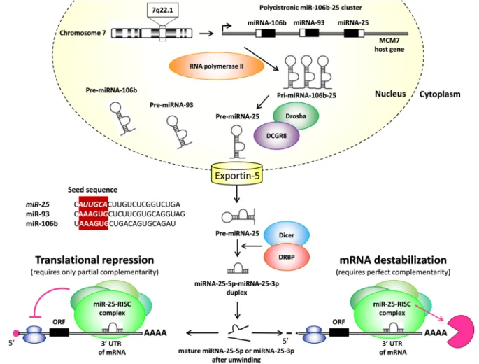
A detailed description of the miRNA biogenesis is beyond the scope of this review. Therefore, here we just briefly mention some important aspects and otherwise refer to excellent reviews for more details (Figure 1) [4–
6]. MicroRNA genes are processed either from introns of protein-coding genes or dedicated miRNA gene loci [7, 8]. An individual primary transcript can either produce a single miRNA or generate a miRNA cluster containing two or more miRNAs [7]. According to our current knowledge, microRNA genes are transcribed by RNA polymerase II as primary microRNAs (pri-microRNAs) (Figure 1). A typical pri-miRNA consists of a stem of 33- 35 bp, a terminal loop and single-stranded RNA segments
www.oncotarget.com Oncotarget, 2018, Vol. 9, (No. 30), pp: 21580-21612 Review
at both the 5’ and 3’ sites [5]. Pri-miRNAs are cleaved by the Microprocessor complex containing the RNase III enzyme Drosha and its essential cofactor DGCR8 and form precursor microRNAs (pre-miRNAs) in the nucleus (Figure 1) [5]. Drosha has tandem RNase III domains which dimerize to form one processing center [5]. The first RNase III domain cuts the 3’ strand of the stem of pri-miRNA and the second RNase III domain cuts the 5’
strand. The produced pre-microRNAs are double-stranded and approximately 70 nucleotide in length, and have a terminal loop [4–6] (Figure 1). These pre-miRNAs are subsequently transported to the cytoplasm by exportin-5 where their terminal loops are cleaved by another RNase III enzyme (DICER) to form a small RNA (miR-miR*) duplex [4–6] (Figure 1). DICER cleavage sites are located at a 21-25 nucleotides distance from the 3’ end and 22 nucleotides away from the 5’ end. This small RNA duplex is loaded onto an Argonaute (AGO) protein to form an effector complex called RNA-induced silencing complex (RISC) [4–6] (Figure 1). Then the two strands of the small RNA duplex are unwinded, and generally, only one strand will be the single-stranded mature microRNA (miR), and the other strand will be degraded (miR* or
passenger strand) [4–6] (Figure 1). The guide strand is determined during the AGO loading process and generally becomes later the mature miR [5]. The strand at 5’ side is typically selected as the guide strand (miR-5p) [5]. Strand selection is not completely strict. Therefore, the not- favored and less abundant passenger strand at the 3’ side could also be selected and act as mature miRNA (miR- 3p). Alternative strand selection (5p to 3p arm switching) could be tissue-specific [5]. Mature miRNAs can either inhibit the translation of target mRNAs or promote their destabilization and degradation through imperfect sequence-specific binding to the 3’ untranslated region (3’- UTR) of target mRNAs [5, 6] (Figure 1). Individual miRNAs may simultaneously target multiple mRNAs.
Moreover, the expression of individual mRNAs can be regulated by multiple miRNAs. Therefore, miRNAs may act as fine tuners or as on/off switches of gene expression [9, 10] (Figure 1).
Classification rules of miRNAs have not yet been unified. It is generally considered that miRNAs with identical seed sequences at nucleotides 2-8 of the mature miRNA belong to the same miRNA (“seed”) family [5]. The occurrence of miRNAs belonging to distinct
Figure 1: Maturation of miR-25. UTR: untranslated region, ORF: open reading frame.
“seed” families within the same cluster is also commonly observed [5]. The seed sequence corresponds to the 3’
UTR of the target mRNAs and determines the possible mRNA targets of the miRNA [5, 7].
Since the original discovery of the first miRNA, lin-4 in 1993 [11] and the second miRNA, let-7 in Caenorhabditis elegans [11–14], the exponentially increasing number of studies indicate that the functions of miRNAs are not limited to the regulation of developmental events. MicroRNAs also regulate many other aspects of biological processes in animals and plants including oxidative stress, cell death, cell proliferation, etc. in many tissue types [15–18]. Therefore, dysregulation of miRNAs in pathological conditions may alter gene networks. Consequently, miRNA replacement or anti- sense inhibition therapy offers a new approach to treat diseases by modulating gene pathways rather than single molecular targets [19].
MicroRNA-25
The miR-106b/25 cluster is highly conserved in vertebrates [20]. The three members of the cluster, miR-106b, miR-93, and miR-25 are located in a 515 bp region on chromosome 7q22 in intron 13 of the host gene minichromosome maintenance protein 7 (MCM7). These three miRNAs are co-transcribed with their host gene [6, 20, 21]. The host gene MCM7 is a component of the highly conserved MCM2-7 complex (MCM complex) [6, 20, 21]. The MCM complex is a member of DNA helicases which are essential in the initiation of DNA replication in eukaryotic cells [8].
MiR-106b and miR-93 share the same seed sequences;
however, miR-25 has only a similar seed sequence resulting in different predicted target mRNAs [20]. In this review, we focus on the role of miR-25 in healthy and diseased conditions. The mature miR-25 (miR-25- 3p) consists of 22 nucleotides (CAUU GCAC UUGU CUCG GUCU GA) (ww and www.miRbase.org) and has 1163 predicted target mRNA transcripts with conserved sites (TargetScanHuman version 7.1). Mature miR-25 belongs to the evolutionary broadly conserved miR- 25-3p/32-5p/92-3p/363-3p/367-3p seed family and has the same predicted mRNA targets as the other miRNA members of this seed family (TargetScanHuman version 7.1). The mature miR-25* (miR-25-5p) consists of 22 nucleotides (AGGC GGAG ACAC GGGC AAUU GC) (https://targetexplorer.ingenuity.com/index.htm and www.
miRbase.org) and has 1868 predicted mRNA transcripts;
however, these predicted targets are primarily false positives (TargetScanHuman version 7.1).
The role of miR-25 in health
It is very difficult to find literature about the function of miR-25 in healthy conditions in the PubMed database. Surprisingly, there is only a few
relevant research paper in the PubMed database using the following keywords: miR-25, health; miR-25, physiological conditions; miR-25, normal conditions;
miR-25, development [22–24]. Therefore, we can only deduce the possible roles of miR-25 in healthy conditions from research papers using miR-25 overexpressing or loss of function cells or animals. Under physiological conditions, mature miR-25 seems to play a crucial role in the regulation of developmental events [22, 23].
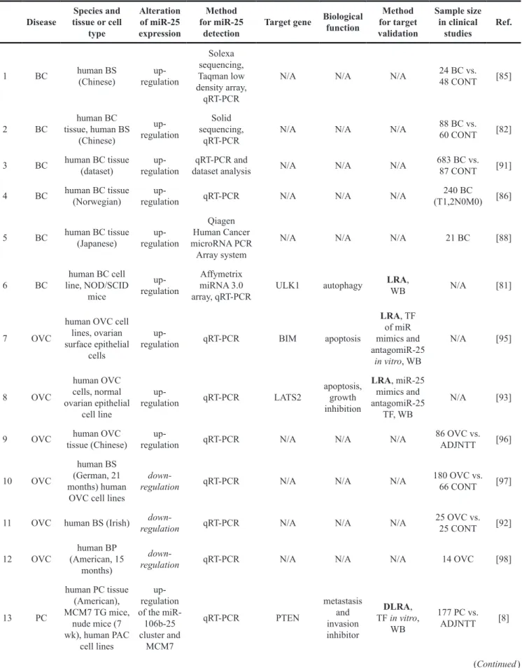
According to the AMIGO 2 gene ontology database, miR- 25 is a negative regulator of cardiac muscle growth and cardiac cell development (GO:0055022). Therefore, it is no surprise that miR-25 also play an important role in the development of cardiac hypertrophy and heart failure under pathophysiological conditions (Figure 2). Under normal conditions, many of miR-25 target mRNAs are involved in biological processes such as response to DNA damage, cell cycle regulation, cell proliferation, migration, and differentiation. In addition, many of miR- 25 target molecules can be found among extracellular matrix components and membrane receptors. Under pathophysiological conditions, miR-25 is also a well- described oncogenic miRNA. It plays a crucial role in the development and spread of many tumor types including brain tumors, lung, breast, ovarian, prostate, thyroid, esophageal, gastric, colorectal, hepatocellular cancers, etc.
Other groups of miR-25 target molecules are important regulators of apoptosis, autophagy, oxidative stress, inflammation, calcium handling, etc. These mechanisms could be key factors in the pathogenesis of acute myocardial infarction, heart failure, diabetes mellitus, diabetic nephropathy, tubulointerstitial nephropathy, asthma bronchiale, cerebral ischemia/reperfusion injury, neurodegenerative diseases, schizophrenia, multiple sclerosis, etc. In this review, we aim to provide an in- depth discussion of the translational therapeutic role of miR-25 in diseased conditions based on relevant basic research articles and clinical studies.
The role of miR-25 in diseased conditions Non-cancerous diseases
Cardiovascular diseases Acute coronary syndrome
The acute coronary syndrome (ACS) is the leading cause of morbidity and mortality in the industrialized countries. ACS represents a spectrum of coronary artery diseases including unstable angina (UA), non-ST- segment elevation myocardial infarction (NSTEMI) and ST-segment elevation myocardial infarction (STEMI) [25]. Acute myocardial infarction in patients is related to rupture-prone or vulnerable atherosclerotic plaques [26].
A cohort study recruiting 13 patients with non-cardiac chest pain and 13 patients with UA and angiographically- proven coronary artery disease reported that members of the miR-106b/25 cluster were significantly elevated in
plasma samples of UA patients [26] (Table 1). Another cohort study recruiting 13 NSTEMI and 13 STEMI patients showed that the expression of miR-25-3p was significantly increased in the blood of STEMI patients as compared to NSTEMI patients (Table 1). According to the aforementioned clinical studies, circulating miR- 25 seems to be a potential biomarker in ACS. However, both studies have many limitations including the small sample size, non-diverse genetic, social, and treatment characteristics of ACS patients. Moreover, investigation of the time dependency of miR-25 upregulation in ACS patients would be more informative. In contrast to these clinical observations, the expression of miR-25 failed to show any change in response to 30-min ischemia and 120- min reperfusion in isolated perfused hearts of male Wistar rats in our previous preclinical study [27]. Nevertheless, the event of 30-min coronary occlusion followed by 120- min reperfusion was considered an early phase of cell injury due to necrotic and/or apoptotic processes [27].
Moreover, since only a single time point was used upon ex vivo reperfusion for heart sample harvesting, the time course of miRNA expression changes remained unknown in heart tissue and blood [27].
Left ventricular hypertrophy and heart failure
Left ventricular hypertrophy (LVH) defined as increase in cardiomyocyte size secondary to i) increased
mechanical load (e.g. hypertension, valvular disease, etc.), ii) decreased mechanical performance (e.g. ischemic heart disease, myocarditis, contractile protein mutations, etc.), iii) increased neurohumoral activity, and iv) hereditary cardiomyopathies with seemingly normal performance and load [28–30]. During the development of LVH, a fetal cardiac gene program is activated by a defined set of transcription factors as an initially adaptive response to stress [29, 30]. The abnormal expression of fetal proteins includes contractile elements (e.g., alpha-MHC, beta- MHC), ECM matrix components (e.g., collagens), calcium handling (e.g., SERCA2a) and mitochondrial proteins (e.g., oxidized elements of the electron transport chain), etc. These changes lead to a maladaptive response to stress resulting in the development of fibrosis, heart failure (HF) and fatal arrhythmias [29, 30]. From a clinical point of view, heart failure could be classified as heart failure with preserved ejection fraction (HFpEF, i.e., left vetricular ejection fraction (LVEF)>50%), heart failure with reduced ejection fraction (HFrEF, LVEF <40%) and most recently heart failure with mid-range ejection fraction (HFmrEF, LVEF 40–49%) [31].
An experimental study proved that transforming growth factor-β1 (TGF-β1) signaling leads to cardiac hypertrophy and fibrosis through a Mothers Against Decapentaplegic Homolog 3 (SMAD3)-dependent manner 20 days after transverse aortic constriction (TAC)
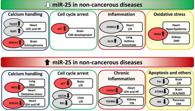
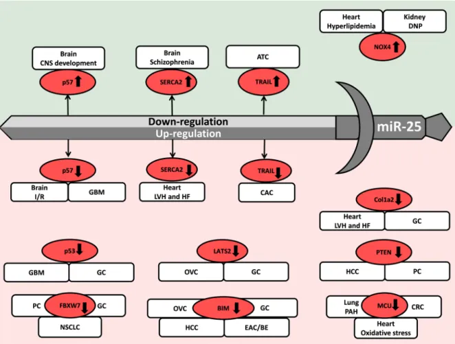
Figure 2: Repression and overexpression of miR-25 in non-cancerous diseases. CNS: central nervous system, DM: diabetes mellitus, DNP: diabetic nephropathy, LVH: left ventricular hypertrophy, HF: heart failure, I/R: ischemia/reperfusion, PAH: pulmonary arterial hypertension, SCA/MJD: Spinocerebellar ataxia type 3/Machado-Joseph Disease, TIN: tubulointerstitial nephropathy. Gene symbols in bubbles are targets of miR-25 in multiple organs/diseases. Genes in red bubbles are targets of miR-25 in multiple diseases.
in SMAD3-/- and littermate control mice [32] (Table 1).
This study also demonstrated that miR-25 transfection into isolated cardiac fibroblasts decreased collagen-1a2 expression [32] (Table 1). In another study, H9c2 cells subjected to hypoxia/reoxygenation showed decreased expression of miR-25 and increased level of its direct target high-mobility group box 1 (HMGB1). HMGB1 is known as a regulator of epithelial to mesenchymal transition and as an inflammatory as well as a pro-fibrotic factor [33] (Table 1). H9c2 cells showed increased levels of apoptosis (caspase-3) and fibrosis markers (collagen I, collagen III, fibronectin, TIMP1, and MM2) after hypoxia/
reoxygenation [33] (Table 1). The overexpression of miR- 25 resulted in inhibition of apoptosis and fibrosis through down-regulation of HMBG1 after hypoxia/reoxygenation in H9c2 cells [33] (Figure 2). These anti-apoptotic and anti-fibrotic effects of miR-25 could be further enhanced by a TGF-β1/SMAD3 inhibitor (SB431542) [33]. Therefore, miR-25 and HMGB1 seem to be early regulators of the TGF-β1/SMAD3 signaling pathway which is a key factor in the development of cardiac fibrosis and heart failure.
Dirkx et al. and Wahlquist et al. investigated the role of miR-25 in calcium handling and cardiac remodeling in TAC-induced LVH and HF models and also in human failing heart samples. However, their results are rather controversial [30, 34] (Table 1). Dirkx et al. found in a mouse model 20 days after TAC surgery that down-regulation of miR-25 and overactivation of the calcineurin/Nfat signaling resulted in overactivation of the Hand2 transcription factor and its target genes leading to cardiac fibrosis and heart failure [30] (Table 1) (Figure 2). In contrast, Wahlquist et al. reported that cardiac overexpression of miR-25 could lead to LVH and HF 5.5 months after TAC surgery in mice. Moreover, inhibition of miR-25 could ameliorate contractile dysfunction by improving sarcoendoplasmic reticulum calcium ATPase 2 (SERCA2a) activity and Ca2+ handling [34] (Table 1) (Figure 2). In response to these controversial results, Bush et al. wrote a commentary on the potential causes including differences in experiment durations and follow-up times after TAC surgery, as well as different antagomiR-25 chemistries and doses [35].
Interestingly, it was found in LVH and HF induced by uninephrectomy and high salt (10% w/w NaCl) diet for 20 weeks that cardiac miR-25 level was significantly up-regulated and its direct target SERCA2 level was significantly decreased [36] (Table 1) (Figure 2). In contrast, circulating miR-25 concentration was significantly decreased in LVH and HF rats as compared to controls [36] (Table 1).
These five articles showed that miR-25 expression is regulated in a time-dependent manner in cardiac hypertrophy, fibrosis, and heart failure. MiR-25 mimics could play a beneficial role in early phases of cardiac remodeling and heart failure. However, overexpression of
miR-25 could be detrimental due to maladaptive effects in chronic heart failure. Since miRNA levels in cardiac tissue and circulation are not always concordant, caution must be exercised during the utilization of miRs as diagnostic or prognostic biomarkers.
Arrhythmias
It is also well-known that increased sarcoplasmic reticulum (SR) Ca2+-leak via ryanodine receptor type- 2 (RyR2) contributes to the pathogenesis of atrial fibrillation (AF). Interestingly, the expression of miR-25 was decreased in atria of patients with paroxysmal atrial fibrillation (PAF) compared with patients with sinus rhythm [37] (Table 1). Moreover, this study showed that miR-106b-25−/− mice expressed increased atrial RyR2 protein levels as well as SR Ca2+-leak, and were more prone to atrial ectopy than wild-type littermates [37]
(Table 1) (Figure 2).
Hypercholesterolemia
Hypercholesterolemia is a well-known risk factor for cardiovascular diseases, and it leads to increased oxidative/nitrative stress in the myocardium [38].
Experimental data are very limited on the regulatory role of miRNAs in hypercholesterolemia-induced cardiac pathologies. We have previously shown that diet-induced hypercholesterolemia in male Wistar rats (2% cholesterol- and 0.25% sodium cholate-enriched diet for 12 weeks) leads to the down-regulation of miR-25 in the myocardium [39] (Table 1). Subsequently, the superoxide-generating NADPH oxidase 4 (NOX4) which is a direct target of miR-25, was up-regulated in the myocardium. In the same study, cardiac oxidative/nitrative stress was also increased leading to diastolic dysfunction in hypercholesterolemic rats [39] (Table 1) (Figure 2). Moreover, knock-down of miR-25 could significantly increase the oxidative stress and NOX4 protein levels in neonatal rat cardiomyocytes 24 hours after transfection of a miR-25 inhibitor proving the direct link between miR-25 and NOX4 expression [39]
(Table 1) (Figure 2). However, in another study it was reported that oxidative stress induced by 2 or 3 hours of H2O2-incubation (500 μM) could significantly up-regulate miR-25 expression in H9c2 embryonic rat ventricular myocytes [40]. Furthermore, the overexpression of miR-25 markedly reduced the oxidative stress-induced apoptosis in H9c2 cells by down-regulating mitochondrial calcium uniporter (MCU) which has been shown to control the Ca2+ flux through the inner mitochondrial membrane [40] (Figure 2).
Intimal hyperplasia
Thrombospondin-1 is known as a key factor in vascular smooth muscle cell (VSMC) migration after vascular injury. It has been reported that the expression of miR-25-5p beyond other miRNAs was significantly decreased in human VSMCs in response to a 6-hour thrombospondin-1 treatment [41] (Table 1). This study
Table 1: The role of miR-25 in cardiovascular and renal diseases
Disease Species and tissue
or cell type Alteration of miR- 25 expression
Method for miR-25
detection
Target
gene Biological function
Method for target validation
Sample size in clinical studies
Ref.
1 ACS - NSTEMI
vs. STEMI Human BP
(American) up-regulation Qiagen
miRNeasy kit N/A N/A N/A 9 STEMI
4 NSTEMIvs. [25]
2 ACS - UA Human BP
(Chinese) up-regulation (miR- 106b-25 cluster)
Taqman low density
miRNA array N/A N/A N/A 13 UA vs.
13 CONT [26]
3 H/R H9c2 cells
(24 h I/1 h R) down-regulation qRT-PCR HMGB1 inflammation DLRA, miR-
25 TF N/A [33]
4 LVH and HF heart of TAC mice (4,8,10 wk) and
human HF down-regulation
Affymetrix GeneChip Mouse Gene St 1.0 Array, Northern blot,
qRT-PCR
Hand2 hypertrophy and fibrosis
LRA, MHC-Hand2
TG mice, miR-25 TF, antagomiR-25
N/A [30]
5 LVH and HF
heart of SMAD3-/- TAC mice (10-20 days),
cardiac FB
up-regulation miRNA array,
qRT-PCR Col1a2 fibrosis miR-25 TF N/A [32]
6 LVH and HF
heart of TAC mice (22 wk), and human HF, HEK293 cells and
RCm
up-regulation miRNA array,
qRT-PCR SERCA2a calcium handling
LRA, miR-25 OE mice, SERCA2a KO mice, antagomiR-25
assay
N/A [34]
7 LVH and HF
uninephrectomized and salt-fed rat myocardium (20
wk) and plasma
up-regulation (myocardium)down-
regulation (plasma) qRT-PCR SERCA2a calcium
handling Western blot N/A [36]
8 Hyper-
cholesterolemia rat myocardium (12
wk) and RCm down-regulation
Agilent’s microRNA
complate labeling and hyb system, qRT-PCR
NOX4 oxidative
stress LRA N/A [39]
9 Paroxysmal atrial fibrillation
human RA (German), heart of miR-25/106b-/-
mice, H9c2 cells
down-regulation qRT-PCR RyR2 calcium
handling LRA 8 pAF vs.
10 CONT [37]
10 Atherosclerosis and vascular remodelling
human aortic
VSMCs down-regulation of miR-25-5p
Affymetrix GeneChip microRNA Array and qRT-PCR
N/A N/A N/A N/A [41]
11 Intracranial
aneurysm Human BP
(Chinese) up-regulation
Agilent’s microRNA
complate labeling and hyb system, qRT-PCR
N/A N/A N/A 40 IA vs.
20 CONT [42]
12 Pulmonary arterial hypertension
human PASMCs,
rat up-regulation
Affymetrix GeneChip
miRNA 4.0 Array, Northern blot,
qRT-PCR
MCU apoptosis
MCU OE, LRA, antagomiR-25
in rats
6 PAH vs.
3 CONT [43]
(Continued )
Disease Species and tissue
or cell type Alteration of miR- 25 expression
Method for miR-25
detection
Target
gene Biological function
Method for target validation
Sample size in clinical studies
Ref.
13 T1DM male Wistar rats (3 wk HFD+STZ),
INS-1 cells up-regulation
Exiqon miRCURY LNA array,
qRT-PCR
insulin glucose
homeostasis LRA, miR-25
TF N/A [45]
14 T1DM
human BS (European multicenter and Danish cohort)
up-regulation Illumina Solexa
Sequencing N/A N/A N/A
European 275 T1DM, 129 Danish T1DM and 151 CONT
[46]
15 TIN HK-2 cell line
(human)
down-regulation (miR-106b-25
cluster)
Exiqon miRCURY LNA array, qRT-PCR
TGFBR2 fibrosis WB, miR-106b
TF N/A [20]
16 DNP mesangial cells
from male SD rats
(12 wk, STZ) down-regulation qRT-PCR NOX4 oxidative
stress LRA N/A [48]
17 IgA NP human urine
(Chinese) up-regulation
Agilent human miRNA microarray
V19.0, qRT-PCR
N/A N/A N/A 93 IgA
NP vs.
82 CONT [49]
Abbreviations: ACS: acute coronary syndrome, BP: blood plasma, BS: blood serum, CONT: control, DLRA: dual luciferase reporter assay, DNP: diabetic nephropathy, FB: fibroblast, IF: immunofluorescence, HF: heart failure, HFD: high fat diet, H/R: hypoxia/reoxygenation, IA: intracranial aneurysm, IgA NP: IgA nephropathy, IH: immunohistochemistry, LRA: luciferase reporter assay, LVH: left ventricular hypertrophy, KO: knock out, NSTEMI: non ST-elevation myocardial infarction, OE: overexpression, pAF: paroxysmal atrial fibrillation, PAH: pulmonary arterial hypertension, PASMCs: pulmonary artery smooth muscle cells, RA: right atrium, RCm: rat cardiomyocytes, SD: Sprague Dawley, STEMI: ST-elevation myocardial infarction, STZ:
streptozotocin, T1DM: type 1 diabetes mellitus, TAC: transverse aortic constriction, TIN: tubulointerstitial nephropathy, TF: transfection, TG: transgene, UA: unstable angina, VSMC: vascular smooth muscle cell, WB: Western blot, wk: week. The abbreviations in bold are considered as gold standard methods for miRNA target validation.
may suggest that miR-25-5p and other altered microRNAs might contribute to the development of atherosclerosis and intimal hyperplasia.
Intracranial aneurysm
Interestingly, a clinical cohort study enrolling 40 patients with intracranial aneurysm (IA), 20 healthy volunteers and an independent validation cohort including 93 IA patients reported that increased plasma miR-25 level might be a potential biomarker for intracranial aneurysm [42] (Table 1).
Pulmonary arterial hypertension
Pulmonary arterial hypertension (PAH) is an obstructive, arterial vasculopathy characterized by excessive pulmonary artery smooth muscle cell (PASMC) proliferation and migration [43]. It leads to vascular stiffening, vasoconstriction, mitochondrial and metabolic dysfunction and finally right ventricular failure [43]. A study proved that overexpression of miR-25 in human PASMC samples resulted in decreased expressions of mitochondrial calcium uniporter (MCU) and cAMP response element binding protein (CREB1). These gene expression alterations resulted in mitochondrial and metabolic dysfunction and a cancer-like phenotype
with apoptosis resistance [43] (Table 1) (Figure 2). The downregulation of MCU by miR-25 has also been found in cardiomyocytes by Pan et al. [40] (Table 1) (Figure 2).
Diabetes mellitus
Diabetes mellitus is a heterogeneous chronic metabolic disorder characterized by hyperglycemia as a common feature resulting from impaired insulin secretion, insulin resistance, or both [44]. Type-2 diabetes mellitus (T2DM) accounts for more than 85% of all diabetes cases, and its incidence is continuously rising worldwide [44].
It has been reported that high-fat diet and streptozotocin- induced T2DM lead to increased pancreatic miR-25 expression and decreased mRNA expression of its direct target insulin stabilizing polypyrimidine tract binding protein 1 (PTBP1) which resulted in decreased insulin translation and secretion [45] (Table 1) (Figure 2).
Moreover, a clinical study enrolling two T1DM cohorts (n=275 for European T1DM children and 129 for Danish T1DM children) and one control group (n=151) reported that serum miR-25 level was significantly increased in T1DM children and was inversely correlated with residual beta cell function [46] (Table 1).
Chronic kidney disease
Chronic kidney disease is often associated with tubulointerstitial fibrosis leading to progressive functional deterioration [20]. It has been demonstrated that all members of the miR-106b-25 cluster including miR-106b, miR-93 and miR-25 were significantly down-regulated after 48 h TGF-β1 treatment (5 ng/ml) in human kidney proximal tubular epithelial cell line (HK-2) [20] (Table 1). Bioinformatics analysis identified the TGF-β type-II receptor as a potential target of the miR-106b-25 cluster.
Indeed, miR-106b transfection resulted in decreased expression of TGF-β type II receptor in HK-2 cells [20]
(Table 1). Hence, the repression of miR-25 and miR-106b may be a key factor in the development of TGF-β1-mediated fibrosis both in the kidney and the heart [32] (Table 1).
Diabetes mellitus is one of the leading causes of end-stage renal disease [47]. NADPH oxidase-derived superoxide seems to play a key role in hyperglycemia- induced oxidative stress in diabetic nephropathy [48].
MiR-25 expression was significantly reduced and NOX4 mRNA and protein levels were increased in the kidney of streptozotocin-induced diabetic rats and high glucose- treated mesangial cells [45] (Table 1). Furthermore, mesangial cells transfected with antagomiR-25 showed significantly increased NOX4 mRNA and protein levels [48] (Table 1) (Figure 2). These results are in line with our findings in the hearts of hypercholesterolemic rats with increased cardiac tissue oxidative stress and diastolic dysfunction in the presence of decreased miR-25 expression [39] (Table 1) (Figure 2). Therefore, the miR- 25-NOX4-oxidative stress axis seems to play a common role in kidney and heart disease.
IgA nephropathy is the most common primary glomerulonephritis leading to end-stage renal disease [49]. A clinical cohort study recruiting 3 control and 9 IgA nephropathy patients showed that miR-25 was significantly increased in the urinary sediment derived mainly from urinary erythrocytes of IgA nephropathy patients [49].
Non-cancerous nervous system diseases Cerebral ischemia/reperfusion injury
Cerebral ischemia is a condition when blood and oxygen supply to the brain tissues is insufficient [50, 51]. Rapid reperfusion is still the gold standard therapy for cerebral ischemia and other types of ischemic organ damages including e.g., ischemic stroke, myocardial infarction and organ transplantations [50]. However, reperfusion causes further tissue injuries due to increased oxidative/nitrative stress leading to mitochondrial dysfunction, lipid peroxidation, protein oxidation, DNA damage and finally cell death [38]. MiR-25 was repressed in a model of 48-h oxygen-glucose deprivation and 48-h reoxygenation in human neuroblastoma SH-SY5Y and
IMR-32 cells [50] (Table 2). Furthermore, overexpression of miR-25 protected cells against apoptosis induced by oxygen-glucose deprivation/reoxygenation possibly through the Fas/FasL pathway [50] (Table 2) (Figure 2). This protective effect of miR-25 against apoptosis in human neuroblastoma SH-SY5Y and IMR-32 cells are in concordance with the findings of Pan et al. reported in H9c2 embryonic rat ventricular myocytes [40, 50]
(Table 1-2) (Figure 2). Interestingly, 10-Hz repetitive transcranial magnetic stimulation was reported to improve proliferation of adult neural stem cells 7 days after focal cerebral ischemia in the subventricular zone in rats [52]
(Table 2) Moreover, the beneficial effect of repetitive transcranial magnetic stimulation was associated with the overexpression of miR-25 and repression of its direct target p57 (CDKN1C). It is a member of the cyclin- dependent kinase (CDK) inhibitors blocking the cell cycle in G1/S phase [49] (Table 2) (Figure 2). Interestingly, a study related to CNS development in zebrafish embryos has also shown that scratch2 transcription factor could block the cell cycle re-entry by maintaining high levels of CDKN1C (p57) via the repression of miR-25 in postmitotic primary neurons [53] (Table 2) (Figure 2).
Spinocerebellar ataxia type 3
Spinocerebellar ataxia type 3/Machado Joseph disease (SCA3/MJD) is the most common type of inherited spinocerebellar ataxia forms [48], and its symptoms include cerebellar ataxia, spasticity, parkinsonism, dystonia, eye movement disorders, sensory loss, muscle weakness, fasciculation, etc. [54]. SCA/MJD is also known as a polyglutamine (polyQ) disease caused by glutamine- encoding CAG nucleotide expansions within endogenous human genes resulting in an abnormal polyQ tract in the polyQ-expanded mutant ataxin-3 protein [48]. This abnormal ataxin-3 protein aggregates in the nucleus and adjacent areas of the affected neurons exacerbating cell death [51]. Overexpression of miR-25 was found to suppress apoptosis in SCA/MJD model cells possibly by the posttranscriptional reduction of polyQ-expanded ataxin-3 protein levels [51] (Table 2) (Figure 2). Indeed, the same research group reported in a clinical cohort study that serum level of miR-25 was significantly lower in SCA3/
MJD patients (n=35) as compared to healthy controls (n=25) [51] (Table 2). Moreover, serum miR-25 level was significantly decreased in SCA3/MJD patients with a course of disease more than 6 years as compared to those patients with shorter disease course [51].
Multiple sclerosis
Multiple sclerosis (MS) is an inflammatory and subsequently degenerative disease of the central nervous system (CNS). It is defined by focal demyelinated lesions in the white matter of the brain and spinal cord [55]. A clinical study enrolling 12 MS relapsing–remitting patients in stable condition and 14 healthy controls revealed that members of the miR-106b-25 cluster were down-regulated
in CD4+CD25highCD127dim/−T regulatory cells of MS patients [56] (Table 2).
Schizophrenia
Schizophrenia is a chronic severe neuropsychiatric disorder with strong genomic and environmental risk factors. The expression of miR-25, a transcriptional regulator of SERCA2 was downregulated in a mouse model of schizophrenia [57] (Table 2) (Figure 2). The 22q11 deletion syndrome (22q11DS) is one of the strongest known genetic risks for schizophrenia [54]. A mouse model of 22q11DS had an age-dependent increase in hippocampal long-term potentiation (LTP), a form of synaptic plasticity needed in learning and memory [57, 58]. In this mouse model of schizophrenia, the expression of SERCA2 is increased resulting in elevated loading of the endoplasmic reticulum with Ca2+ and enhanced neurotransmitter release as well as increased LTP [57, 58].
A study has found that haploinsufficiency of DGCR8; a miRNA biogenesis gene in the 22q11DS disease-critical region led to synaptic SERCA2 overexpression and increased LTP [57]. Moreover, SERCA2 was elevated in human brain samples with schizophrenia [57] (Table 2) (Figure 2).
Neurotoxicity by dioxins
Acetylcholinesterase (AChE) plays a central role in cholinergic neurotransmission in central and peripheral nervous systems by hydrolysation of the neurotransmitter, acetylcholine [56]. Dioxins were shown to decrease AChE expression directly in neuroblastoma cells and immune cells by transcriptional regulation via aryl hydrocarbon receptor and by post-translational regulation via microRNAs including miR-25 [59]. Proposed mechanisms of dioxin toxicity are reviewed in detail by Xie et al. [59].
Asthma bronchiale
A study using human tracheal smooth muscle cells revealed that miR-25 was significantly down-regulated after exposing cells to pro-inflammatory cytokines including IL-1β, TNF-α, and IFN-γ [60]. In this study, the repression of miR-25 resulted in the overexpression of its direct target Krüppel-like factor 4 (KLF4). KLF4 is known as an inhibitor of smooth muscle-specific gene expression and mediator of inflammation [60] (Table 2) (Figure 2).
Cancerous diseases
Cancerous nervous system diseases Glial tumors
Astrocytoma originating from astrocytic glial cells is the most common type of primary tumor type in the central nervous system [61]. Anaplastic astrocytoma (AA)
is a high-grade malignant glioma (grade III according to the WHO classification) developing from low-grade diffuse astrocytoma (DA; grade II) and progressing into glioblastoma of grade IV [61]. Glioblastoma multiforme is the most malignant and aggressive form of gliomas showing a median survival time of 15 months after standard therapy [62]. Few studies have investigated the role of miR-25 in the development of gliomas [62–
66] (Table 3). Most of them indicate that miR-25 is overexpressed, and behave as an oncomiR during the development of gliomas [62, 63, 65, 66] (Table 3). These results suggest that miR-25 has mRNA targets directly or indirectly regulating pathways related to cell cycle or cell death. Zhang et al. reported that miR-25 was overexpressed in more than 90% of human glioma tissues (grade II-IV) and 60% of human glioma cell lines [62].
Furthermore, miR-25 has been shown to increase glioma cell proliferation by directly targeting the CDK inhibitor type 1C (CDKN1C or p57) resulting in an increase of S/M phase cells and a decrease of G0/G1 phase cells [62] (Table 3) (Figure 3). The miR-25 and CDKN1C (p57) axis seem to play a central role in the regulation of cell cycle re-entry not only in glioblastoma and cancerous cell proliferation but also in healthy conditions and after ischemia/reperfusion injury as mentioned in the previous section [52, 53, 62]. In accordance with the findings of Zhang et al., another study has found that miR-25 was overexpressed in human astrocytoma samples and glioblastoma cell lines leading to tumor growth and invasion by directly targeting neurofilament light polypeptide (NEFL) which was an inhibitor of the mammalian target of rapamycin (mTOR) cell proliferation pathway [65] (Table 3) (Figure 3). Interestingly, a network analysis study investigating transcription factors, miRNAs and their target genes in human anaplastic astrocytoma reported that miR-25 might target tumor suppressor p53 and it could be regulated by another tumor suppressor, the phosphatase and tensin homolog (PTEN) [61] (Table 3) (Figure 3). In contrast, only one study has reported that overexpression of miR-25 could suppress glioblastoma growth in vivo and in vitro by a p53 tumor suppressor- dependent mechanism [64] (Table 3) (Figure 3). In this study, miR-25 was identified as a miRNA repressed indirectly by p53 through the transcriptional regulators of the P53 gene, E2F1 (also called retinoblastoma binding protein-3) and MYC [64] (Table 3) (Figure 3). In addition, overexpression of miR-25 resulted in the downregulation of its direct targets MDM2 and TSC1. Both targets are negative regulators of the p53 tumor suppressor and the mTOR cell proliferation pathway, respectively [64]. In that article, the authors speculated on that overexpression of miR-25 could stabilize p53 tumor suppressor expression through activation of mTOR pathway by targeting TSC1.
However, this finding is controversial to the results of Peng et al. [64, 65].
Retinoblastoma
Retinoblastoma (RB) is a typical malignant tumor appearing in children [67]. A clinical pilot study with a very limited sample size enrolling 3 healthy and 3 RB retina samples has reported that miR-25 was significantly overexpressed in RB [67] (Table 3) (Figure 3). After miRNA-target analysis using experimentally validated databases and pathway enrichment analysis, the apoptosis
regulator BCL2L1 seemed to be a potentially important target of miR-25 in RB [67] (Table 3) (Figure 3).
Lung cancer
Non-small cell lung cancer
Lung cancer is the leading cause of tumor-related mortality worldwide [68]. Non-small cell lung cancer Table 2: The role of miR-25 in non-cancerous nervous system diseases
Disease Species and tissue or cell
type
Alteration of miR-25 expression
Method for miR-25
detection
Target
gene Biological function
Method for target validation
Sample size in clinical
studies Ref.
1 Cerebral I/R
SH-SY5Y and IMR-32 cells
(48 h OGD/
48 h R)
regulationup- qRT-PCR Fas apoptosis DLRA, miR-25 TF,
WB N/A [50]
2 Cerebral I/R brain of male
SD rats (7 days) up-
regulation qRT-PCR p57 cell cycle
arrest antagomiR-25,
WB N/A [52]
3 CNS
development
postmitotic primary neurons
of zebrafish embryos
down-
regulation qRT-PCR p57 cell cycle
arrest DLRA N/A [53]
4 SCA3/MJD
SCA/MJD model cells,
293T and SH-SY5Y cells
regulationup- qRT-PCR ATXN3 ataxia DLRA N/A [51]
5 SCA3/MJD human BS
(Chinese) down-
regulation qRT-PCR N/A N/A N/A 35 SCA/MJD
vs. 25 CONT [54]
6 MS human BP,
T-cells (Italian)
down- regulation
(miR- 106b-25
cluster)
Agilent human miRNA microarray
V2.0, qRT-PCR
N/A N/A N/A 12 MS vs 14
CONT [56]
7 Schizophrenia
mouse hippocampus
(8-10, 16-20 wk) human brain tissues
down- regulation
Agilent mouse miRNA microarray,
qRT-PCR
SERCA2 calcium
handling miR-25 viral infection
schizophrenia 17 vs. 22 CONT [57]
8 Asthma
bronchiale human tracheal
SMCs down-
regulation miRNA
array KLF4 inflammation WB, anti-
miR-25 TF N/A [60]
9 CF human BS
(Australian) up- regulation
Qiagen miRNA PCR
array for serum and
plasma
N/A N/A N/A
52 CF with liver disease50 CF without liver
disease
[153]
10 Liver
regeneration male SD rats
(6, 12, 24, 36 h) up- regulation
Agilent rat miRNA array, V16,
8X15k
RB1,
KAT2B cell cycle
arrest LRA, IH, WB N/A [149]
Abbreviations: BP: blood plasma, BS: blood serum, CF: cystic fibrosis, CNS: central nervous system, CONT: control, DLRA: dual luciferase reporter assay, h: hour, IF: immunofluorescence, IH: immunohistochemistry, I/R: ischemia/reperfusion, LRA: luciferase reporter assay, KO: knock out, MS: multiple sclerosis, OE: overexpression, OGD: oxygen glucose deprivation, R: reoxygenation, SCA/
MJD: Spinocerebellar ataxia type 3/Machado-Joseph Disease, SD: Sprague Dawley, SMCs: smooth muscle cells, TF: transfection, TG:
transgene, WB: Western blot, wk: week. The abbreviations in bold are considered as gold standard methods for miRNA target validation.
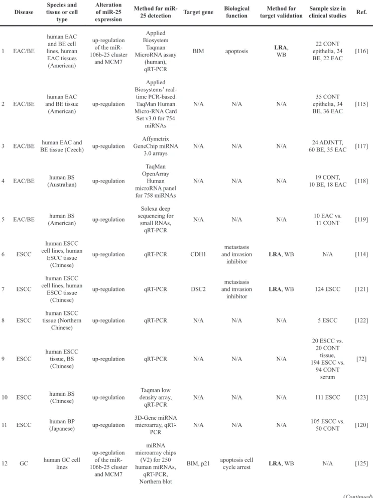
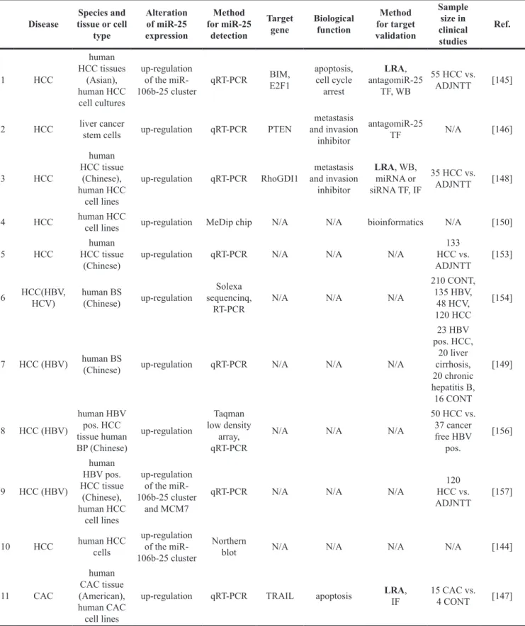
(NSCLC) accounts for 80-85% of all lung cancer cases, and its 5-year survival rate is about 15% [68]. Savita et al. reported that the overexpression of the miR-106b-25 cluster could directly suppress the ubiquitin ligase β-TRCP2 gene expression leading to decreased Snail degradation in H1299 non-small cell lung cancer cells [69]
(Table 4) (Figure 3). Snail has been reported to positively regulate cell adhesion and migration as well as invasion [70, 71]. Xiang et al. demonstrated that the oncogenic effect of miR-25 is partially due to direct targeting and repressing F-box and WD repeat domain-containing 7 (FBXW7 also known as FBW7) in human NSCLC tissue samples and cell lines [68] (Table 4) (Figure 3). FBXW7
is a putative tumor suppressor in human tumorigenesis due to its ability to recognize and bind target proteins for ubiquitination and degradation [68]. Other studies suggested that the overexpression of miR-25 could reduce apoptosis by different mechanisms in human NSCLC cells and cell lines [72, 73] (Table 4). Wu et al. reported that miR-25 directly targeted the modulator of the apoptosis 1 (MOAP1) gene which was a Bax-associated protein containing BH3-like motif and mediating caspase- dependent apoptosis [72] (Table 4) (Figure 3). Chen et al. found that overexpression of miR-25 directly repressed the regulator of G protein signaling 3 (RGS3) gene which potentially could play a role in apoptosis as Table 3: The role of miR-25 in cancerous nervous system diseases
Disease Species and tissue or cell type
Alteration of miR-25 expression
Method for miR-25
detection Target gene Biological function
Method for target validation
Sample size in clinical
studies Ref.
1 GBM
human brain tumor
tissue (American)
regulationup-
Agilent human miRNA microarray
version 1, qRT-PCR
N/A N/A N/A 24 CNS
tumors vs.
8 CONT [66]
2 GBM
human sample GBM (Chinese), GMB cell
lines
regulationup- qRT-PCR NEFL apoptosis LRA,
WB 44 GBM vs.
20 CONT [65]
3 GBM
human sample GBM (Chinese),
human GBM cell
lines
regulationup- qRT-PCR p57 cell cycle
arrest LRA,
miR-25 TF 35 GBM vs.
5 CONT [62]
4 GBM
human sample GBM (dataset)
regulationup- dataset
analysis p53 apoptosis,
cell cycle arrest
network
analysis N/A [61]
5 GBM
human GBM cell lines, mice
(35 days)
regulationup-
Nanostring assay, qRT-PCR
MDM2,
TSC1 apoptsosis LRA,
miR-25 TF,
WB N/A [64]
6 RB human RB
sample (Chinese)
regulationup-
Agilent human miRNA
microarray 2k Bcl2L1 apoptosis exp.
validated databases
3 RB vs.
3 CONT [67]
Abbreviations: CONT: control, DLRA: dual luciferase reporter assay, GBM: glioblastoma multiforme, IF:
immunofluorescence, IH: immunohistochemistry, LRA: luciferase reporter assay, KO: knock out, OE: overexpression, RB: retinoblastoma, TF: transfection, TG: transgene, WB: Western blot. The abbreviations in bold are considered as gold standard methods for miRNA target validation.
a cancer suppressor [73] (Table 4) (Figure 3). He et al.
reported that radiotherapy-resistant NSCLC human tissues overexpressed miR-25 as compared to radiosensitive or non-cancerous tissues [74] (Table 4). In addition, miR- 25 overexpression correlated negatively with its direct BTG anti-proliferation factor 2 (BTG2) expression (Table 4) (Figure 3). BTG2 has been shown to inhibit cell proliferation and invasion by repressing cyclin D1, matrix metalloproteinase-1 and metalloproteinase-2 in human lung cancer cells [74]. Interestingly, down-regulation of
miR-25 by antagomiR-25 treatment was shown to inhibit NSCLC cell proliferation and induce G1 cell cycle arrest possibly through indirect down-regulation of the cell division cycle 42 (CDC42) gene [75] (Table 4) (Figure 4).
REV3Lp is the catalytic subunit of DNA polymerase zeta playing a crucial role in genome stability in mammalian cells. A study investigating the polymorphism of REV3Lp in lung cancer susceptibility in Chinese Han population (n=500 lung cancer patients and 517 cancer- free control) revealed that (3’UTR) 460 T>C single
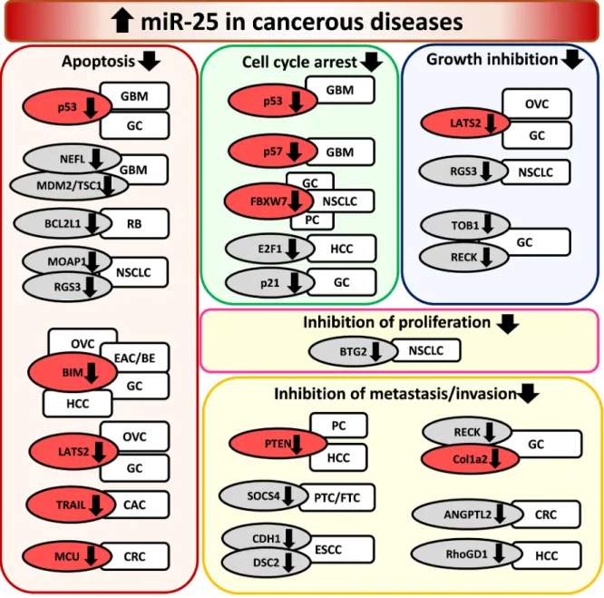
Figure 3: Overexpression of miR-25 in cancerous diseases. EAC/BE: esophageal adenocarcinoma/Barrett esophagus, ESCC:
esophageal squamous cell carcinoma, CAC: cholangiocarcinoma, CRC: colorectal cancer, GC: gastric cancer, GBM: glioblastoma multiforme, HCC: hepatocellular carcinoma, NSCLC: non-small cell lung carcinoma, OVC: ovarian cancer, PC: prostate cancer, PTC/FTC:
papillary thyroid carcinoma/follicular thyroid carcinoma, RB: retinoblastoma. Gene symbols in bubbles are targets of miR-25 in multiple organs/diseases. Genes in red bubbles are targets of miR-25 in multiple diseases.
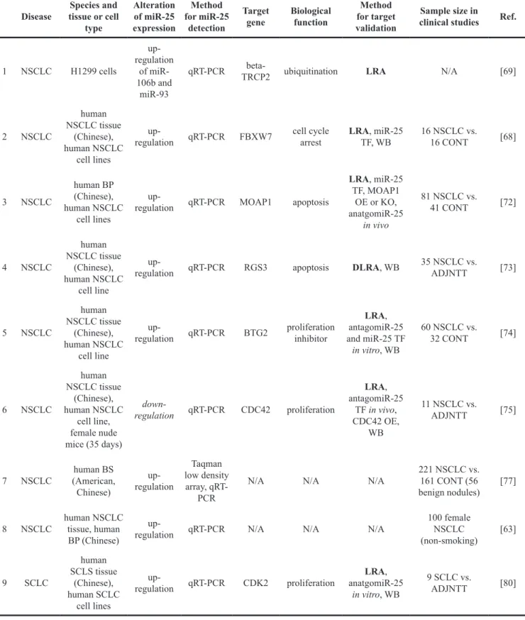
Table 4: The role of miR-25 in lung cancer Disease Species and
tissue or cell type
Alteration of miR-25 expression
Method for miR-25
detection
Target
gene Biological function
Method for target validation
Sample size in
clinical studies Ref.
1 NSCLC H1299 cells
regulation up- of miR- 106b and
miR-93
qRT-PCR beta-
TRCP2 ubiquitination LRA N/A [69]
2 NSCLC
human NSCLC tissue
(Chinese), human NSCLC
cell lines
regulationup- qRT-PCR FBXW7 cell cycle
arrest LRA, miR-25
TF, WB 16 NSCLC vs.
16 CONT [68]
3 NSCLC
human BP (Chinese), human NSCLC
cell lines
regulationup- qRT-PCR MOAP1 apoptosis
LRA, miR-25 TF, MOAP1
OE or KO, anatgomiR-25
in vivo
81 NSCLC vs.
41 CONT [72]
4 NSCLC
human NSCLC tissue
(Chinese), human NSCLC
cell line
regulationup- qRT-PCR RGS3 apoptosis DLRA, WB 35 NSCLC vs.
ADJNTT [73]
5 NSCLC
human NSCLC tissue
(Chinese), human NSCLC
cell line
regulationup- qRT-PCR BTG2 proliferation inhibitor
LRA, antagomiR-25 and miR-25 TF in vitro, WB
60 NSCLC vs.
32 CONT [74]
6 NSCLC
human NSCLC tissue
(Chinese), human NSCLC
cell line, female nude mice (35 days)
down-
regulation qRT-PCR CDC42 proliferation
LRA, antagomiR-25
TF in vivo, CDC42 OE,
WB
11 NSCLC vs.
ADJNTT [75]
7 NSCLC human BS (American, Chinese)
regulationup-
Taqman low density array, qRT-
PCR
N/A N/A N/A 221 NSCLC vs.
161 CONT (56 benign nodules) [77]
8 NSCLC human NSCLC tissue, human
BP (Chinese)
regulationup- qRT-PCR N/A N/A N/A 100 female
NSCLC
(non-smoking) [63]
9 SCLC
human SCLS tissue
(Chinese), human SCLC
cell lines
regulationup- qRT-PCR CDK2 proliferation LRA, anatgomiR-25
in vitro, WB
9 SCLC vs.
ADJNTT [80]
Abbreviations: ADJNTT: adjacent non-tumorous tissue, BP: blood plasma, CONT: control, DLRA: dual luciferase reporter assay, IF: immunofluorescence, IH: immunohistochemistry, LRA: luciferase reporter assay, KO: knock out, NSCLC: non-small cell lung carcinoma, OE: overexpression, SCLC: small cell lung carcinoma, TF: transfection, TG: transgene, WB: Western blot. The abbreviations in bold are considered as gold standard methods for miRNA target validation.
nucleotide polymorphism (rs465646) showed a strong association with lung cancer development [76]. The T allele demonstrated a stronger binding affinity for miR-25 and miR-32 which could down-regulate the endogenous tumor suppressor REV3L [76]. Furthermore, an ethnically diverse multicenter case-control study recruiting 221 NSCLC patients, 161 controls and 56 patients with benign nodules from China and America reported that serum levels of miR-25 and other four miRNAs (miR-483-5p, miR-193a-3p, miR-214 and miR-7) were significantly elevated irrespective of ethnicity groups [77] (Table 4). A clinical study enrolling 100 Chinese female non-smoking lung adenocarcinoma patients found that increased plasma miR-25 levels positively correlated with the mortality rate, advanced disease stage, regional and distant metastasis at diagnosis as well as epithelial growth factor receptor (EGFR) mutation status [63] (Table 4). Another clinical study investigating circulating miRNA levels in response to chemotherapy or operation in Russian lung cancer patients (n=23, T1-3N0-3M0) showed that the plasma concentration of miR-25 did not change 15 days after surgery or within 30 days after completing two courses of
paclitaxel-carboplatin chemotherapy as compared to that before the respective intervention [78]. In another clinical study enrolling 148 Chinese patients with histologically proven advanced or metastatic lung adenocarcinoma (stage IIIB or IV) and showing a complete or partial response to first-line therapy with the antifolate agent pemetrexed plus platinum of 4-6 cycles, were divided into an observation versus pemetrex maintenance group [79]. In the pemetrex maintenance group, significantly elevated serum miR-25 levels were negatively correlated with progression-free survival time [79]. In contrast, serum miR-25 levels failed to correlate with progression-free survival time in the observation group [79].
Small cell lung cancer
Small cell lung cancer (SCLC) accounts for approximately 15% of all new lung cancer cases and has unique clinical and histological characteristics [80]. Zhao et al., similarly to that in NSCLC samples, reported the overexpression of miR-25 in human SCLC tissue samples and cell lines [80]. Interestingly, down-regulation of miR- 25 by antagomiR-25 treatment was shown to inhibit SCLC
Figure 4: Repression of miR-25 in cancerous diseases. ATC: anaplastic thyroid cancer, CRC: colorectal cancer, NSCLC: non-small cell lung carcinoma, PC: prostate cancer. Gene symbols in bubbles are targets of miR-25 in multiple organs/diseases. Gene in red bubble is a target of miR-25 in multiple diseases.
cell proliferation and induced the cell cycle arrest in the G1 phase possibly through the down-regulation of the cell cycle-related proteins cyclin E2 and CDK2. Cyclin E2 was shown to be a direct target of miR-25 in this study [80]
(Table 4) (Figure 3).
Breast cancer
Breast cancer ranks the first among female cancers and is the second leading cause of death in women worldwide [81, 82]. Breast cancer is a heterogeneous disease entity, and numerous classification systems have been developed on the basis of histopathological and molecular genetic features or the appearance in imaging studies such as mammography or MRI [83].
From the point of view of therapy, both the stage/extent of the tumor and mentioned special features contribute to therapeutic decision. Breast cancer has always been managed more or less on an individual basis; the recent advances in precision medicine made a great impact on the management of breast cancer. Plenty of studies have examined the correlation between miRNA and mRNA expression in breast cancer and the role of miRNA expression profile in prognosis, but no clear conclusion has yet been reached [84]. The vast majority of studies investigating the role of miR-25 in the development of breast cancer have found that miR-25 was overexpressed in human breast cancer tissues and was elevated in the serum of patients [82, 84–90] (Table 5) (Figure 3). Hu et al. demonstrated in a two-stage case-control analysis that serum levels of miR-25, miR-16, miR-222 and miR- 324-3p were significantly upregulated as compared to those other miRNAs (miR-191 and miR-484) used as endogenous control in Chinese breast cancer patients [85] (Table 5). Wu reported that miR-25 expression was significantly higher in breast cancer patients based on data of 683 breast cancer tissues when compared to that in 87 normal breast tissues according to The Cancer Genome Atlas [83]. Interestingly, another clinical study enrolling 240 Norwegian early breast cancer patients showed strong and significant associations between the overexpression of miR-25, miR-18a/b, miR-106b, and miR-505 with high proliferation, estrogen receptor negativity and cytokeratin 5/6 positivity of cancer [86] (Table 5). Moreover, using dataset analysis Farazzi et al. reported that miRNA families might control subtype-specific pathways in breast cancer and miR-25 showed high miRNA regulatory activity in triple-negative and basal-like subtypes of breast cancer [84]. Another study using 21 surgical breast cancer specimens revealed that increased expression of miR-25 was associated with high Ki-67 (a marker of cell proliferation) expression and HER2, ER and PR positivity [88] (Table 5). Interestingly, a study enrolling 76 breast cancer patients reported that overexpression of miR-25*,
miR-142-3p, miR-505*, miR-1248, miR-181a-2*, and miR-340* could discriminate between tumor samples from BRCA1/2 mutation carriers and non-carriers [90].
A BRCA1/2A preclinical mechanistic study reported that down-regulation of miR-25 by isoliquiritigenin resulted in increased autophagic cell death by overexpression of ULK1 and induced chemosensitization in MCF7/ADR breast cancer cells [91] (Table 5) (Figure 3). Another study comprehensively analyzed tumor tissue miRNA expression and patient survival collecting data from the Cancer Genome Atlas (TCGA) which contains miRNA sequencing and overall survival datasets of 759 breast cancer patients [83, 89]. Surprisingly, this study found that increased expression of miR-25 predicted improved breast cancer survival [89].
Ovarian cancer
Ovarian cancer is the most common cause of gynecological malignancy-related mortality among women [92]. Epithelial ovarian cancer (EOC) accounts for approximately 90 % of ovarian tumors, including serous adenocarcinoma, endometrial adenocarcinoma and clear cell carcinoma [93]. Most cases are not diagnosed at an early stage [89]. Therefore, the 5-year survival rate is still poor despite advances in the diagnosis and chemotherapy [93, 94]. Studies investigating the expression of miR- 25 in epithelial ovarian cancer cell lines, tumorous tissues and serum from patients provided controversial findings. A preclinical study demonstrated that miR-25 was overexpressed in human ovarian cancer cells and cell lines (SKOV3, OVCAR3, OVCAR5, and A2780) and ovarian surface epithelial cells (OSE) [92] (Table 5) (Figure 3). In this study, repression of miR-25 in ovarian cancer cells enhanced apoptosis by directly targeting Bim (also known as BCL2L11) [95]. Bim is also known as a direct activator of Bax and neutralizer of Bcl2-like molecules [95] (Table 5) (Figure 3). Another preclinical study showed that expression of miR-25 was increased in human ovarian cancer cells (OVCAR3, SKOV3, ES-2) [90] (Table 5) (Figure 3). In this study, inhibition of miR- 25 could suppress proliferation, migration, and invasion of ovarian cancer cells by directly targeting large tumor suppressor 2 (LATS2) [93] (Table 5) (Figure 3). A clinical cohort study enrolling 86 ovarian cancer patients reported that the increased expression of miR-25 in EOC tissue was associated with advanced clinical stage, lymph node metastasis and shorter survival time indicating that miR- 25 might be involved in carcinogenesis and metastasis of EOC [96] (Table 5). Moreover, another clinical cohort study recruiting 180 treated EOC patients and 66 healthy women from Germany showed that serum levels of miR- 25 was down-regulated after a median follow-up time of 21 months [97] (Table 5). Interestingly, Langhe et al.