Petra Molnar1,2,†, Lıvia Marton1,†, Richard Izrael1,2, Hajnalka L. Palinkas1,3and Beata G. Vertessy1,2
1 Research Centre for Natural Sciences, Institute of Enzymology, BME-MTA Malaria Research Laboratory, Hungarian Academy of Sciences, Budapest, Hungary
2 Department of Applied Biotechnology and Food Science, Budapest University of Technology and Economics, Budapest, Hungary 3 Doctoral School of Multidisciplinary Medical Science, University of Szeged, Szeged, Hungary
Keywords
base-excision repair; DNA damage and repair; malaria, Uracil-DNA repair;
Plasmodium falciparum; Uracil-DNA detection
Correspondence
L. Marton and B. G. Vertessy, Hungarian Academy of Sciences, Research Centre for Natural Sciences, Institute of Enzymology, BME-MTA Malaria Research Laboratory, Budapest, Hungary
E-mails: marton.livia@ttk.mta.hu (LM);
vertessy@kutatok.org (BGV)
†Joint first authors
(Received 20 March 2018, revised 11 May 2018, accepted 21 May 2018)
doi:10.1002/2211-5463.12458
Plasmodium falciparum parasites undergo multiple genome duplication events during their development. Within the intraerythrocytic stages, para- sites encounter an oxidative environment and DNA synthesis necessarily proceeds under these circumstances. In addition to these conditions, the extreme AT bias of theP. falciparumgenome poses further constraints for DNA synthesis. Taken together, these circumstances may allow appearance of damaged bases in thePlasmodium DNA. Here, we focus on uracil that may arise in DNA either via oxidative deamination or thymine-replacing incorporation. We determine the level of uracil at the ring, trophozoite, and schizont intraerythrocytic stages and evaluate the base-excision repair potential ofP. falciparumto deal with uracil-DNA repair. We find approx- imately 7–10 uracil per million bases in the different parasite stages. This level is considerably higher than found in other wild-type organisms from bacteria to mammalian species. Based on a systematic assessment ofP. fal- ciparum genome and transcriptome databases, we conclude that uracil- DNA repair relies on one single uracil-DNA glycosylase and proceeds through the long-patch base-excision repair route. Although potentially efficient, the repair route still leaves considerable level of uracils in parasite DNA, which may contribute to mutation rates inP. falciparum.
Malaria is a major health threat affecting large regions globally, resulting in the death of ~450 000 people annually [1]. The parasite’s capability of adap- tation is a major hindering factor in the way of elimi- nating the disease, mostly represented by the growing resistance of parasites against antimalarials [1]. The causative agents of malaria belong to the Plasmodium genus. Among the five human parasites, Plasmodium
falciparum (P. falciparum) presents an exceptional biomedical challenge being responsible for the most serious infections and most of the lethal cases [2].
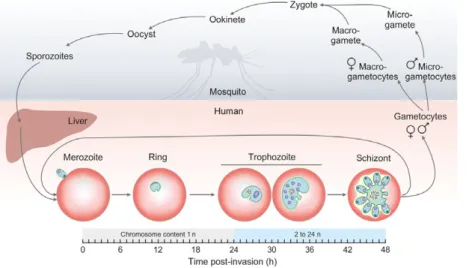
The life cycle ofP. falciparumis intriguingly complex (Fig.1). The parasites undergo multiple DNA replica- tions at several developmental stages in their vector (Anopheles mosquito) and host (human liver and bloodstream). The sexual phase of development occurs
Abbreviations
3meA, 3-methyladenine; 3meG, 3-methylguanine; 5-FU, 5-fluorouracil; 5-hC, 5-hydroxycytosine; 5-hmU, 5-hydroxymethyluracil; 5-hU, 5- hydroxyuracil; 7meG, 7-methylguanine; 8-oxoG, 7,8-dihydro-8-oxoguanine; AP lyase, apurinic/apyrimidinic lyase; APE1, apurinic/apyrimidinic endonuclease 1; BER, base-excision repair; DHU, dihydrouracil; dut- ung-, deoxyuridine-triphosphatase/uracil-DNA glycosylase double knockout; DUT, deoxyuridine-triphosphatase; FapyA or G, formamidopyrimidine lesions; FEN1, flap endonuclease 1; Hx, hypoxanthine; LIG 1/3, DNA ligase 1/3; MBMD 4, methyl–CpG-binding domain protein 4; MPG, DNA-3-methyladenine glycosylase; MUTYH, mutY homolog;
NEIL 1/2/3, endonuclease VIII like enzyme 1/2/3; NTHL1, endonuclease III-like protein 1; OGG 1, 8-oxoguanine glycosylase 1; PCNA, proliferating cell nuclear antigen; PNKP, polynucleotide kinase/phosphatase; Polb/d/e, DNA polymeraseb/d/e; RFC, replication factor C;
SMUG 1, single-strand selective monofunctional uracil-DNA glycosylase; TDG, thymine-DNA glycosylase; Tg, thymine glycol; UNG, uracil- DNA glycosylase; XRCC1, X-ray repair cross-complementing protein 1.
in the female Anopheles mosquito. The only meiotic division takes place when the zygote, originating from the fusion of microgametes and macrogametes inside the mosquito, evolves into ookinetes. These will then develop into multinuclear oocysts, wherein mitotic sporogenesis results in the formation of numerous sporozoites. After the mosquito bites a human host, sporozoites invade the liver and undergo at least a dozen rounds of mitosis to produce tens of thousands of haploid merozoites [3]. These start the intraerythro- cytic cycle by the invasion of red blood cells. The importance of this cycle is emphasized by the fact that about two-thirds of the genes of a murinePlasmodium have been shown to be necessary for the blood stage growth of parasites [4]. First, they develop into rings, followed by trophozoites. At this stage, parasites enter the G1 phase, and start to prepare for DNA replica- tion. The S phase starts around 30 h after erythrocyte invasion, when parasites are in the late trophozoite stage. The replication in the parasite is asynchronous and produces up to about 24n sister chromatids. Repli- cation ends around 44 h postinvasion, after which each genome is packed into daughter merozoites (schizont form) [3,5]. Merozoites may exit the continuous intraerythrocytic cycle by differentiating into male or female gametocytes. These sexual forms, consumed by the mosquito, evolve into micro- or macrogametocytes.
Microgametocytes undergo three mitotic cycles and form eight exflagellated microgametes [3].
Importantly, DNA replication cycles within the intraerythrocytic stages proceed in an environment rich in oxidative conditions. Especially, free heme and iron during the hemozoin formation process pose notable oxidative stress, which may result in DNA modifica- tions (e.g., oxidative cytosine deamination leading to uracil and other oxidative processes). The specific com- position of genomic DNA in Plasmodia may also
facilitate the appearance of uracil in the DNA.
Members of the Plasmodium genus possess the most AT-rich genome sequenced so far, including P. falci- parum, having namely~80% AT content in the exonic regions and ~90% in the intronic regions [6,7]. Com- paring to other organisms, like the host, Homo sapiens, where the average AT content is 58.9%, and to other eukaryotic pathogens, namely Toxoplasma gondii and Trypanosoma brucei, having 47.7% and 53.2% AT, respectively, the base composition of the P. falciparum genome is indeed extraordinary [8]. A mutation bias has recently been pointed out in P. falciparum, show- ing an increased occurrence of GC?AT substitutions, which could promote the AT-rich genome structure [8]. The AT richness of the genome may increase the possibility of uracil content, as huge amounts of thymidines are incorporated, giving more chance for the polymerase to mistake thymidines with uridines as compared to a genome with lower levels of AT con- tent. We therefore wished to determine uracil levels in genomic DNA of P. falciparum during the intraery- throcytic stages.
A dot-blot-based uracil detection method has recently been developed in our laboratory, which provides a robust and straightforward possibility for sensitive and quantitative detection of uracil-DNA levels [9]. The basis of detection is an engineered cat- alytically inactive uracil-DNA glycosylase (UNG), which is capable of recognizing and binding to uracils incorporated in DNA sequences. The uracil sensor UNG-construct can be equipped with diverse tags for ease of detection via antibodies using the dot-blot method. The sensitivity of the method is equivalent to that of MS-based methods, providing a limit in femto- molar concentrations [9].
In the present work, we analyzed the uracil content of genomic DNA from three different intraerythrocytic
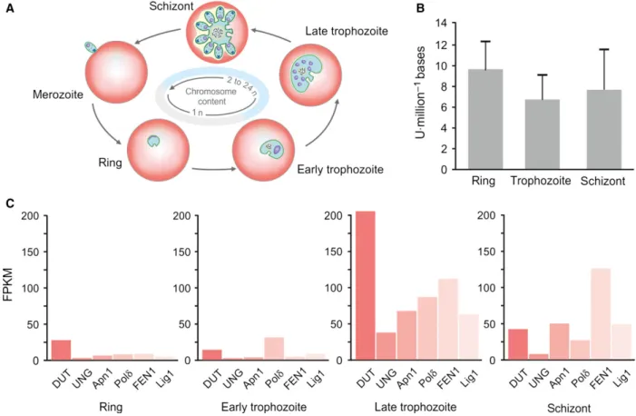
Fig. 1.The life cycle ofPlasmodium falciparum. Stages within the mosquito vector and inside the human host are on light gray or coral background,
respectively. Developmental stages of the intraerythrocytic cycle are represented by graphical illustrations, and changes in chromosome content in these stages are also indicated on a schematic horizontal axis.
developmental stages of P. falciparum 3D7 parasites, namely the ring, trophozoite, and schizont stages. The quantification of uracil moieties was performed by the aforementioned dot-blot-based uracil detection method [9]. To assess the potential efficiency of uracil-DNA repair, we compared the existing orthologues of mam- malian base-excision repair enzymes to those present in the parasite based on genome databases of H. sapiens andP. falciparum. We also analyzed transcriptome data- bases of the intraerythrocytic parasite stages with regard to expression level of base-excision repair enzymes.
Materials and methods
Maintenance of parasite cultures
Plasmodium falciparum 3D7 parasites were obtained from the University of Montpellier. Continuous cultures were maintained in human 0+erythrocytes. Parasites were grown at 5% hematocrit (HCT) in complete RPMI medium [incom- plete RPMI 1640 (w L-glutamine, w Hepes, w NaHCO3)]
(Lonza, Basel, Switzerland) supplemented with 50 mgL1 gentamicin (VWR Chemicals, Radnor, PA, USA), 37µM hypoxanthine (Alfa Aesar, Haverhill, MA, USA), and 1.25 gL1Albumax I (Gibco from Thermo Fisher Scientific, Waltham, MA, USA). Cultures were kept at 37°C in a labo- ratory incubator gassed with 5% O2, 5% CO2, and 90% N2. Cultures were synchronized by sorbitol at the young ring stage, and Percoll treatment at the schizont stage matured from sorbitol-treated rings, as described elsewhere [10,11].
Trophozoites were obtained from synchronized cultures, after parasites reached the early trophozoite stage.
Genomic DNA isolation
Synchronized parasites of different developmental stages were collected from two biologically independent cultures (i.e., biological replicates), and lysed as described elsewhere [12]. Briefly, red blood cells were lysed in 5% saponin
(Sigma-Aldrich, St. Louis, MO, USA) in PBS, then incu- bated at 37°C for 3 h for parasite lysis in a lysis solution (pH=7.5) of the following composition: 40 mMTris/HCl;
80 mMEDTA; 2% SDS; 0.1 mgmL1proteinase K. After treatment, genomic DNA was purified using the QuickDNA Miniprep Plus kit obtained from Zymo Research (Irvine, CA, USA).
Dot-blot measurement and analysis
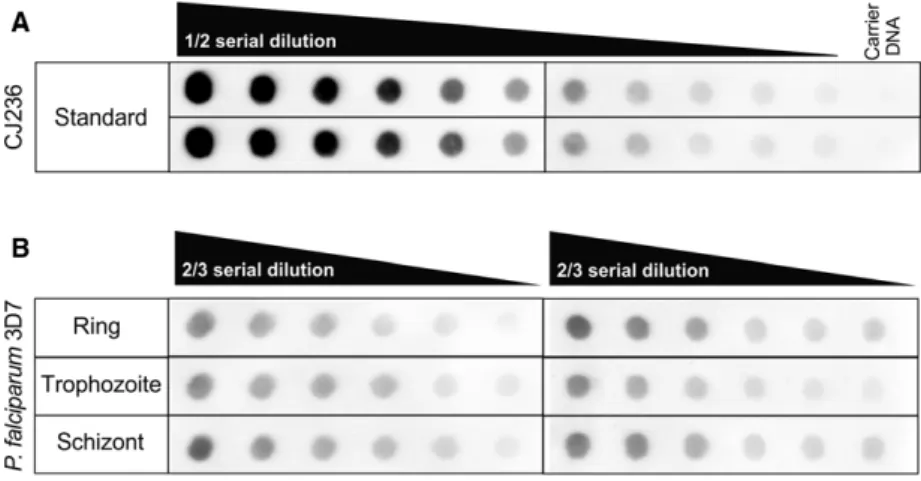
Dot-blot measurements were carried out in four indepen- dent replicates using samples from theP. falciparumdevel- opmental stages, namely rings, trophozoites, and schizonts, as described elsewhere [9]. Briefly, genomic DNA isolated from CJ236Escherichia colistrain [dut,ung] served as a uracil standard, applied in 15 ng diluted into 1lg of ultra- pure salmon sperm DNA. The standard was diluted in a ½ dilution series. The two-third serial dilutions for P. falci- parum samples started with 1lg of DNA. Samples were spotted onto a prewetted positively charged nylon mem- brane (Amersham Hybond-Ny+; GE Healthcare, Little Chalfont, UK) using a vacuum-driven microfiltration appa- ratus (Bio-Dot, Bio-Rad, Hercules, CA, USA). The DNA was immobilized, and the membrane was blocked and incu- bated with the detector construct of UNG. After several washing steps, first primary, then secondary antibodies were applied. Immunoreactive bands were visualized by enhanced chemiluminescence reagent (Western Chemilumi- nescent HRP substrate from Merck Millipore, Burlington, MA, USA), and images were captured by a Bio-Rad Che- miDocTM MPImaging system. Densitometry was per- formed usingIMAGEJ 1.48p software (National Institutes of Health, Bethesda, MD, USA). The number of deoxyuridine nucleotides was calculated as described elsewhere [9]. Cali- bration curve from the dilution of the standard was fitted with a polynomial with second order that provided a fit with R2≥0.98. The number of uracil per million bases in the ‘unknown’ genomic DNA was determined by interpo- lating their normalized intensities in the calibration plot based on the amount of DNA applied.
Fig. 2.Dot-blot assays for measuring genomic uracil levels of the different developmental stages ofPlasmodium falciparumparasites. (A) CJ236 [dut, ung]Escherichia coligenomic DNA was used as standard for the dot-blot assay.
(B) Representative dot-blot images of the measurement of the quantity of genomic uracil inP. falciparumring, trophozoite, and schizont samples.
Statistical analysis
Statistical analysis was carried out byORIGINPRO8.6 (Origi- nLab, Northampton, MA, USA) using one-way ANOVA test when samples passed homogeneity of variance test (Levene’s test) and normal distribution tests (Kolmogorov– Smirnov test). Differences were considered statistically sig- nificant atP <0.05.
Transcriptome analysis
Transcriptome analysis was carried out using the Transcrip- tomics function of the Plasmodb database. The gene expression level of each protein was estimated by RNA-Seq data for intraerythrocytic stages [13,14]. Raw data were plotted for four stages: ring, early and late trophozoite, and schizont.
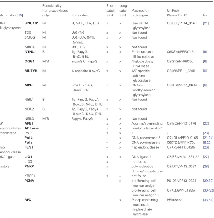
Table 1.Comparison of the mammalian andPlasmodium falciparum BER protein sets and their involvement in short-patch versus long- patch BER (cf ‘x’ marks). The functionality of DNA glycosylases is defined as mono- (M) or bifunctional (B). Question mark in case of DNA polymeraseb indicates that a polymerase b-like enzyme was reported in P. falciparum, with an activity related to mammalian Pol b; however, the respective gene is not annotated. All abbreviations are listed in the Abbreviations section of the article.
Mammalian [19]
Functionality (for glycosylases
only) Substrates
Short- patch BER
Long- patch BER
Plasmodium orthologue
UniProt/
PlasmoDB ID Ref.
DNA N-glycosylase
UNG1/2 M U, 5-FU, U:A, U:G x x Uracil-DNA
glycosylase
Q8ILU6/PF14_0148 [21]
TDG M U:G>T:G x x Not found
SMUG1 M U:G>U:A, 5-FU,
5-hmU
x x Not found
MBD4 M U:G, T:G x x Not found
NTHL1 B Tg, FapyG,
5-hC, 5-hU
x x Endonuclease
III homologue
C6KSY9/PFF0715c [6]
OGG1 M/B 8-oxoG:C, FapyG x x N-glycosylase/
DNA lyase
Q8I2Y2/PFI0835c [6]
MUTYH M A opposite 8-oxoG x x A/G-specific
adenine glycosylase
Q8II68/PF11_0306 [6]
MPG M 3meA, 7meG,
3meG, Hx
x x DNA-3-
methyladenine glycosylase
Q8IKG6/PF14_0639 [6]
NEIL1 B Tg, FapyG, FapyA,
8-oxoG, 5-hU, DHU
x x Not found
NEIL2 B Tg, FapyG, FapyA,
8-oxoG, 5-hU, DHU
x x Not found
NEIL3 M/B FapyA, FapyG x x Not found
AP
endonuclease
APE1 x x Apurinic/apyrimidinic
endonuclease Apn1
Q8IE02/PF13_0176 [22]
AP lyase x x
Polymerase Polb x x ? ? [23]
Pold – x DNA polymerased Q7KQL4/PF10_0165 [21,24]
Pole – x DNA polymerasee C6KTD8/PFF1470c [6,25]
Flap
endonuclease
FEN1 – x flap endonuclease 1 Q7K734/PFD0420c [26]
DNA ligase LIG1 x x DNA ligase I Q8IES4/MAL13P1.22 [27]
LIG3 x – not found
Factors PNKP x – polynucleotide
kinase/phosphatase
Q8ID74/PF13_0334 [28]
XRCC1 x – not found
PCNA – x proliferating cell
nuclear antigen
P61074/PF13_0328 [29,30]
proliferating cell nuclear antigen 2
Q7KQJ9/PFL1285c [30–32]
RFC – x P-loop containing
nucleoside triphosphate hydrolase
PFA0545c [33,34]
Enzymes written in bold designate the mammalian enzymes with Plasmodium orthologs.
Results and Discussion
We measured the amount of uracil moieties/million bases of DNA samples extracted from the three intraerythrocytic developmental stages of the P. falciparumparasites using a recently developed dot- blot-based detection method [9]. Parasite cultures were synchronized, and cultures of ring, trophozoite, and schizont were collected for the isolation of genomic DNA. Two representative dot-blot images are shown in Fig.2. The measured amount of uracil moieties in our samples could be fitted to the linear range of the standard dilution series (Fig.2).
The uracil content of ring, trophozoite, and schizont stage parasites were determined to be 9.62.8, 6.72.4, and 7.63.8 uracil per million bases, respectively. One-way ANOVA statistical analysis revealed that the uracil contents of the measured ery- throcytic stages under these experimental conditions do not differ significantly from each other (P =0.618).
It is of interest to note that these uracil-DNA levels are significantly higher than those observed in other samples from different wild-type organisms. The level of uracil moieties in genomic DNA has been assessed by various methods in numerous organisms so far [9,15,16] The general conclusion from these studies agrees that wild-type organisms from bacteria to mam- mals, as well as normal cell lines, show low levels of uracil in DNA in the range of 0.1–1 uracil per million bases, or even lower [16,17]. An interesting exception was found in Drosophila S2 cells, where the uracil- DNA content was reported to be around 15–16 uracil per million bases [9]. This considerable high genomic uracil level is, however, probably directly related to the lack of the most efficient uracil-DNA glycosylase enzyme (UNG protein) from the Drosophila genome [18]. Organisms under genotoxic stress or engineered to lack uracil-DNA glycosylases also present increased genomic uracil levels [9,15–17]. To discuss our results, it was therefore of immediate interest to investigate whether the approx. 7–10 uracil per million bases levels in theP. falciparum genomic DNA samples may be related to a limited set of repair enzymes in the par- asite.
The DNA repair route to remove uracil from DNA relies on the base-excision repair (BER) pathway [19].
We therefore systematically compared the relevant set of proteins encoded in mammalian species vs P. falci- parum. For the initial search of the related P. falci- parumBER enzyme set, the KEGG pathway database [20] was used with manual curation and verification of the hits. In each case, we also performed a sequence alignment to decide whether the orthologues are truly
relevant and include the functionally important resi- dues. Whenever available, published studies on the specific proteins were also consulted. Results are shown in Table1 and identify two interesting limita- tions of the BER protein set inP. falciparum.
These two limitations relate to, on the one hand, the set of enzymes capable of recognizing and cleaving uracil from DNA, and on the other hand, to the set of proteins required for the short-patch versus long-patch BER routes. Uracil-DNA glycosylases in diverse organisms include at least four enzyme families (UNG, TDG, SMUG, MBD4) [35,36]. The diversity in these enzymes defines their specific roles and different sub- strate specificities and underlies the high significance of uracil removal from DNA. InP. falciparum, however, only one uracil-DNA glycosylase gene is present: It encodes the archetypical UNG enzyme.
With regard to the second limitation, concerning short-patch vs long-patch BER pathways, it has been argued earlier that P. falciparum predominantly
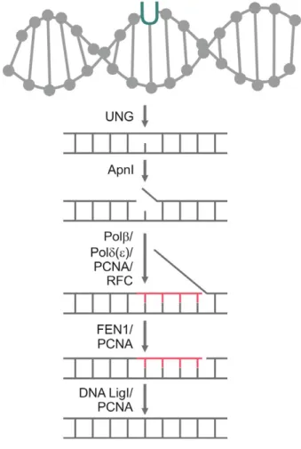
Fig. 3.A possible uracil-DNA repair mechanism of Plasmodium falciparumvia long-patch BER, based on the analysis of BER enzyme sets.
employs the long-patch pathway [37]. In agreement with this study, several proteins involved in short- patch BER was not identified in P. falciparum (e.g., polymeraseb, ligase 3) (cf Table1). It has to be men- tioned that although a protein with polymerase b-like enzyme activity has been reported in P. falciparum [23], its role in short-patch BER in P. falciparum has not been confirmed. As it is responsible for the synthe- sis of 3- to 5-bp oligonucleotides, it may be involved in long-patch repair [23]. Also, this ‘polymeraseb-like’
protein may have another role in an alternative end- joining pathway in the parasite [38]. No orthologues of the LIG3 and its stabilizing scaffold protein, XRCC1, have been found so far. LIG3 and XRCC1 are responsible for the ligation process in short-patch BER [19]. In summary, the protein set encoded in P. falciparum is deficient on short-patch BER, but all protein orthologues necessary for the long-patch BER pathway have been clearly identified in the parasite.
Based on these data, a possible route ofP. falciparum long-patch BER-based uracil-DNA repair mechanism is
shown in Fig. 3. The recognition and excision of uracil in the DNA of the parasites are performed by UNG. The next step is DNA strand cleavage by Apn1, which results in the formation of a nick in the DNA backbone. DNA polymerased(ore) binds to the DNA by the help of pro- liferating cell nuclear antigen (PCNA) and the replication factor C (RFC) orthologue to start the synthesis of ~10 new nucleotides, while removing the downstream 50 DNA end. The replaced section forms a so-called flap structure that is still connected to the DNA. It is removed by flap endonuclease 1 (FEN1). The leftover nick is ligated by DNA ligase I [39].
It was also of interest to look into the expression profiles of the key enzymes involved in uracil-DNA repair in the parasite, in relation to the uracil-DNA levels during the intraerythrocytic stages. In this analy- sis, we also considered the dUTPase enzyme, which is responsible for cleaving dUTP to prevent uracil incor- poration into DNA [36]. Based on the analysis of BER enzyme transcriptome levels of the different P. falciparum developmental stages, the pathway is
Fig. 4.Uracil-DNA and repair enzymes expression levels in intraerythrocyticPlasmodium falciparumstages. (A) Changes in chromosome content in the different stages. (B) The uracil-DNA levels in the ring, trophozoite, and schizont stage parasites are shown with error bars. (C) Analysis of transcriptomes of the long-patch BER enzyme set in P. falciparum intraerythrocytic developmental stages. FPKM is the transcript levels of fragments per kilobase of exon model per million mapped reads.
initiated in the late trophozoite stage (Fig.4). This is in good agreement with the DNA metabolism of the parasites, as DNA synthesis starts in the late tropho- zoite stage followed by DNA packaging into mero- zoites at the end of the intraerythrocytic cycle. In case of UNG and Pold, the expression level drops again in schizont stage. Apn1, FEN1, and DNA ligase I remain present after late trophozoite stage as well (Fig.4C).
The expression level of dUTPase, responsible for pre- venting uracil incorporation, is highly elevated in late trophozoite stage in parallel with the BER enzymes.
However, transcriptome analysis data should be evalu- ated with caution as they may not reflect the efficiency of the enzymes participating in uracil repair.
Conclusions
We have determined uracil-DNA levels in different intraerythrocytic stages of P. falciparum genomic DNA and found that approx. 7–10 uracil per million bases can be detected. To account for this level, which is significantly higher as compared to other normal wild-type organisms, the balance between processes
leading to uracil presence in DNA and its removal needs to be considered.
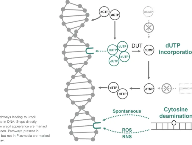
There are two mechanisms by which uracil can arise in DNA, as shown in Fig.5. On the one hand, if cellu- lar dUTP levels are high as compared to dTTP levels, polymerases can readily incorporate dUMP moieties into DNA. The enzyme family of dUTPases are responsible for keeping dUTP levels at a low value to prevent thymine-replacing incorporations. The signifi- cance of this DNA repair function of P. falciparum dUTPase is underlined by numerous studies that focus on plasmodial dUTPase inhibition as an important chemotherapeutic strategy against malaria [40–42].
Thymine-replacing uracil incorporation intoP. falci- parumgenomic DNA may be enhanced by the excep- tionally high AT content of the parasite genome. Also, the level of dUTPase expression as suggested by tran- scriptomic analysis is increased only in the late tropho- zoite stages, potentially allowing uracil incorporation at earlier stages where DNA replication is initiated.
This pathway of uracil incorporation does not result in a stable mutation, but is considered dangerous because high levels of uracil in the DNA can lead to
Fig. 5.Pathways leading to uracil appearance in DNA. Steps directly resulting in uracil appearance are marked by dark green. Pathways present in mammals but not in Plasmodia are marked by light gray.
the hyperactivity of the uracil-repair mechanism, resulting in thymine-less cell death [36].
Another possibility for uracil to appear in DNA is the oxidative deamination of cytosine, resulting in a G:U pair instead of G:C [36,43]. In the next round of replication, DNA polymerases will incorporate adeno- sine opposite of U. Without repair, this will lead to the formation of an AT pair, aka a GC?AT substitu- tion. The deamination of cytosine is considered one of the most frequent DNA mutations, with a rate of 100 to 500 Ucell1day1 [44]. In case of P. falciparum, the detoxifying process resulting in the formation of hemozoin crystals gives rise to the formation of oxida- tive agents. The presence of such reactive oxygen and nitrogen species in the parasitized erythrocytic environ- ment can cause the deamination of cytidine in increased frequency, possibly contributing to the GC?
AT substitutions [8].
Uracil removal from DNA requires the base-exci- sion repair process. In mammalian cells, both short- and long-patch BER pathways are present for the repair of base excisions, but the parasites rely only on the long-patch repair. Moreover, from the different families of uracil-DNA glycosylases, P. falciparum contains only the single UNG protein, further limiting the capacity of parasites to remove uracil from the DNA.
It has been discussed that genomic architecture of P. falciparum, containing low complexity regions and repetitive sequences as a consequence of the AT rich- ness, allows high indel mutation rates in coding and noncoding regions. Indel mutations occur 10-fold more frequently compared to base-pair substitutions, and this is probably the result of DNA polymerase slip- pages and unequal crossing over events [8]. Possible advantages of high mutation rates include an effect on gene regulation, an extended antigenic variance, a role in drug resistance, and an evolutionary benefit. The mutation of noncoding genes can have an effect on the gene expression, as these regions often have enhancer or repressor roles [8,45]. Probably, the high mutation rates combined with low complexity regions can facili- tate adaptive evolution in P. falciparum parasites [8].
The presence of uracil moieties in the parasite genome may also contribute to mutation rates.
Acknowledgements
The authors thank Dr. Kai Wengelnik form the University of Montpellier, France, for providing the P. falciparum3D7 strain. This work was supported by the National Research, Development and Innovation Office (K109486, K119493, NVKP_16-1-2016-0020,
2017-1.3.1-VKE-2017-00002, 2017-1.3.1-VKE-2017-00013, VEKOP-2.3.2-16-2017-00013, NKP-2018-1.2.1-NKP- 2018-00005 to BGV), the Hungarian Academy of Sciences MedInProt Program (to BGV), the BME-Bio- technology FIKP grant of EMMI (BME FIKP-BIO to BGV) and by ICGEB CRP/HUN14-01 (to BGV).
Author contributions
PM and LM participated in all preparations, experi- ments, data analysis, figure, and table preparation as well as in writing the study. RI analyzedP. falciparum BER enzyme orthologues and expression data. HLP participated in the dot-blot measurements and the related data analysis. BGV conceived and coordinated the study. The manuscript of this study was reviewed and approved by all authors.
Conflict of interest
The authors declare no conflict of interest.
References
1 WHO (2017) WHO|World malaria report 2017.World Malar Rep2017, 196.
2 Trampuz A, Jereb M, Muzlovic I and Prabhu RM (2003) Clinical review: severe malaria.Crit Care7, 315–323.
3 Arnot DE (2013) Cell cycle regulation in Plasmodium.
InEncyclopedia of Malaria(Kremsner PG and Krishna S, eds), pp. 1–9. Springer, New York, NY.
4 Bushell E, Gomes AR, Sanderson T, Anar B, Girling G, Herd C, Metcalf T, Modrzynska K, Schwach F, Martin REet al.(2017) Functional profiling of a Plasmodium genome reveals an abundance of essential genes.Cell170(260–272), e8.
5 Lee AH, Symington LS and Fidock DA (2014) DNA repair mechanisms and their biological roles in the malaria parasitePlasmodium falciparum.Microbiol Mol Biol Rev78, 469–486.
6 Gardner MJ, Hall N, Fung E, White O, Berriman M, Hyman RW, Carlton JM, Pain A, Nelson KE, Bowman Set al. (2002) Genome sequence of the human malaria parasite Plasmodium falciparum.Nature 419, 498–511.
7 Paila U, Kondam R and Ranjan A (2008) Genome bias influences amino acid choices: analysis of amino acid substitution and re-compilation of substitution matrices exclusive to an AT-biased genome.Nucleic Acids Res 36, 6664–6675.
8 Hamilton WL, Claessens A, Otto TD, Kekre M, Fairhurst RM, Rayner JC and Kwiatkowski D (2017) Extreme mutation bias and high AT content in
Plasmodium falciparum.Nucleic Acids Res45, 1889–1901.
9 Rona G, Scheer I, Nagy K, Palinkas HL, Tihanyi G, Borsos M, Bekesi A and Vertessy BG (2016) Detection of uracil within DNA using a sensitive labeling method for in vitro and cellular applications.Nucleic Acids Res 44, 1–13.
10 Lambros C and Vanderberg JP (1979) Synchronization ofPlasmodium falciparumerythrocytic stages in culture.
J Parasitol65, 418–420.
11 Rivadeneira EM, Wasserman M and Espinal CT (1983) Separation and concentration of schizonts ofPlasmodium falciparumby Percoll gradients.J Protozool30, 367–370.
12 Moll K, Kaneko A, Scherf A and Wahlgren M (2013) Methods in Malaria Research, 6th edn. www.evimalar.
org, www.mr4.org, www.beiresources.org, and www.ki.se.
13 Lopez-Barragan MJ, Lemieux J, Qui~nones M, Williamson KC, Molina-Cruz A, Cui K, Barillas-Mury C, Zhao K and Su X (2011) Directional gene expression and antisense transcripts in sexual and asexual stages ofPlasmodium falciparum.BMC Genom12, 587.
14 Lasonder E, Rijpma SR, van Schaijk BCL, Hoeijmakers WAM, Kensche PR, Gresnigt MS, Italiaander A, Vos MW, Woestenenk R, Bousema T et al.(2016) Integrated transcriptomic and proteomic analyses ofP. falciparumgametocytes: molecular insight into sex-specific processes and translational repression.Nucleic Acids Res44, 6087–6101.
15 Lari S-U, Chen C-Y, Vertessy BG, Morre J and Bennett SE (2006) Quantitative determination of uracil residues inEscherichia coliDNA: contribution of ung, dug, and dut genes to uracil avoidance.DNA Repair (Amst)5, 1407–1420.
16 Galashevskaya A, Sarno A, Vagbø CB, Aas PA, Hagen L, Slupphaug G and Krokan HE (2013) A robust, sensitive assay for genomic uracil determination by LC/
MS/MS reveals lower levels than previously reported.
DNA Repair (Amst)12, 699–706.
17 Shalhout S, Haddad D, Sosin A, Holland TC, Al-Katib A, Martin A and Bhagwat AS (2014) Genomic uracil homeostasis during normal B cell maturation and loss of this balance during B cell cancer development.Mol Cell Biol34, 4019–4032.
18 Muha V, Horvath A, Bekesi A, Pukancsik M, Hodoscsek B, Merenyi G, Rona G, Batki J, Kiss I, Jankovics F et al. (2012) Uracil-containing DNA in Drosophila: Stability, stage-specific accumulation, and developmental involvement. PLoS Genet 8, e1002738.
19 Krokan HE and Bjøras M (2013) Base excision repair.
Cold Spring Harb Perspect Biol5, a012583.
20 Kanehisa M and Goto S (2000) KEGG: kyoto encyclopedia of genes and genomes.Nucleic Acids Res 28, 27–30.
21 Suksangpleng T, Leartsakulpanich U, Moonsom S, Siribal S, Boonyuen U, Wright GE and
Chavalitshewinkoon-Petmitr P (2014) Molecular
characterization ofPlasmodium falciparumuracil-DNA glycosylase and its potential as a new anti-malarial drug target.Malar J13, 149.
22 Haltiwanger BM, Karpinich NO and Taraschi TF (2000) Characterization of class II apurinic/apyrimidinic endonuclease activities in the human malaria parasite, Plasmodium falciparum.Biochem J345(Pt 1), 85–89.
23 Nunthawarasilp P, Petmitr S and Chavalitshewinkoon- Petmitr P (2007) Partial purification and characterization of DNA polymeraseb-like enzyme fromPlasmodium falciparum.Mol Biochem Parasitol154, 141–147.
24 Ridley RG, White JH, McAleese SM, Goman M, Alano P, de Vries E and Kilbey BJ (1991) DNA polymerase delta: gene sequences fromPlasmodium falciparumindicate that this enzyme is more highly conserved than DNA polymerase alpha.Nucleic Acids Res19, 6731–6736.
25 LaCount DJ, Vignali M, Chettier R, Phansalkar A, Bell R, Hesselberth JR, Schoenfeld LW, Ota I, Sahasrabudhe S, Kurschner Cet al.(2005) A protein interaction network of the malaria parasitePlasmodium falciparum.Nature438, 103–107.
26 Falciparum P and Repair DNA (2009) Expression and biochemical characterization of thePlasmodium falciparumDNA repair enzyme, FLAP
ENDONUCLEASE-1 (PfFEN-1).DNA Repair (Amst) 157, 1–12.
27 Buguliskis JS, Casta LJ, Butz CE, Matsumoto Y and Taraschi TF (2007) Expression and biochemical characterization ofPlasmodium falciparumDNA ligase I.Mol Biochem Parasitol155, 128–137.
28 Siribal S, Weinfeld M, Karimi-Busheri F, Mark Glover JN, Bernstein NK, Aceytuno D and
Chavalitshewinkoon-Petmitr P (2011) Molecular characterization ofPlasmodium falciparumputative polynucleotide kinase/phosphatase.Mol Biochem Parasitol180, 1–7.
29 Kilbey BJ, Fraser I, McAleese S, Goman M and Ridley RG (1993) Molecular characterisation and stage-specific expression of proliferating cell nuclear antigen (PCNA) from the malarial parasite,Plasmodium falciparum.
Nucleic Acids Res21, 239–243.
30 Mitra P, Banu K, Deshmukh AS, Subbarao N and Dhar SK (2015) Functional dissection of proliferating- cell nuclear antigens (1 and 2) in human malarial parasitePlasmodium falciparum: possible involvement in DNA replication and DNA damage response.Biochem J470, 115–129.
31 Patterson S, Whittle C, Robert C and Chakrabarti D (2002) Molecular characterization and expression of an alternate proliferating cell nuclear antigen homologue, PfPCNA2, inPlasmodium falciparum.Biochem Biophys Res Commun298, 371–376.
32 Li J-L, Warren AV and Cox LS (2002) Identification of a second proliferating cell nuclear antigen in the human
malarial pathogenPlasmodium falciparum.Int J Parasitol32, 1683–1692.
33 Le Roch KG, Zhou Y, Blair PL, Grainger M, Moch JK, Haynes JD, De La Vega P, Holder AA, Batalov S, Carucci DJet al.(2003) Discovery of gene function by expression profiling of the malaria parasite life cycle.
Science (80-.)301, 1503–1508.
34 Jirage D, Chen Y, Caridha D, O’Neil MT, Eyase F, Witola WH, Ben Mamoun C and Waters NC (2010) The malarial CDK Pfmrk and its effector PfMAT1 phosphorylate DNA replication proteins and co-localize in the nucleus.Mol Biochem Parasitol172, 9–18.
35 Alsøe L, Sarno A, Carracedo S, Domanska D, Dingler F, Lirussi L, SenGupta T, Tekin NB, Jobert L, Alexandrov LBet al.(2017) Uracil accumulation and mutagenesis dominated by cytosine deamination in CpG dinucleotides in mice lacking UNG and SMUG1.
Sci Rep7, 7199.
36 Vertessy BG and Toth J (2009) Keeping uracil out of DNA: physiological role, structure and catalytic mechanism of dUTPases.Acc Chem Res42, 97–106.
37 Haltiwanger BM, Matsumoto Y, Nicolas E, Dianov GL, Bohr VA and Taraschi TF (2000) DNA base excision repair in human malaria parasites is
predominantly by a long-patch pathway.Biochemistry 39, 763–772.
38 Gupta DK, Patra AT, Zhu L, Gupta AP and Bozdech Z (2016) DNA damage regulation and its role in drug- related phenotypes in the malaria parasites.Sci Rep6, 23603.
39 Kim Y-J and Wilson DM III (2012) Overview of base excision repair biochemistry.Curr Mol Pharmacol5, 3–13.
40 Whittingham JL, Leal I, Nguyen C, Kasinathan G, Bell E, Jones AF, Berry C, Benito A, Turkenburg JP, Dodson EJet al.(2005) dUTPase as a platform for antimalarial drug design: structural basis for the selectivity of a class of nucleoside inhibitors.Structure 13, 329–338.
41 Nguyen C, Ruda GF, Schipani A, Kasinathan G, Leal I, Musso-Buendia A, Kaiser M, Brun R, Ruiz-Perez LM, Sahlberg B-Let al.(2006) Acyclic nucleoside analogues as inhibitors ofPlasmodium falciparum dUTPase.J Med Chem49, 4183–4195.
42 Hampton SE, Baraga~na B, Schipani A,
Bosch-Navarrete C, Musso-Buendıa JA, Recio E, Kaiser M, Whittingham JL, Roberts SM, Shevtsov M et al.(2011) Design, synthesis, and evaluation of 50- diphenyl nucleoside analogues as inhibitors of the Plasmodium falciparumdUTPase.ChemMedChem6, 1816–1831.
43 Krokan HE, Drabløs F and Slupphaug G (2002) Uracil in DNA–occurrence, consequences and repair.
Oncogene21, 8935–8948.
44 Olinski R, Jurgowiak M and Zaremba T (2010) Uracil in DNA-Its biological significance.Mutat Res–Rev Mutat Res705, 239–245.
45 Horrocks P, Wong E, Russell K and Emes RD (2009) Control of gene expression inPlasmodium falciparum– ten years on.Mol Biochem Parasitol164, 9–25.