Contents lists available atScienceDirect
Life Sciences
journal homepage:www.elsevier.com/locate/lifescie
Non-genomic actions of sex hormones on pregnant uterine contractility in rats: An in vitro study at term
Mohsen Mirdamadi
a, Anna Kothencz
a, Edina Szűcs
b, Sándor Benyhe
b, Mihály Szécsi
c, Róbert Gáspár
a,⁎aDepartment of Pharmacology and Pharmacotherapy, Interdisciplinary Excellence Centre, Faculty of Medicine, University of Szeged, Hungary
bInstitute of Biochemistry, Biological Research Center, Center of Excellence in European Union, Hungarian Academy of Sciences, Szeged, Hungary
cDepartment of Medicine, Faculty of Medicine, University of Szeged, Hungary
A R T I C L E I N F O Keywords:
Sex hormones Mifepristone Non-genomic pathway Myometrial contraction Pregnancy
Rat
A B S T R A C T
Aims: The non-genomic (prompt) actions of sex steroids on pregnant uterine contractility are not fully explored yet, the aim of our study was to clarify such effects of 17-β estradiol (E2), progesterone (P4) and testosterone (T) on late (22-day) pregnant uterine contractions together with the signaling pathways in rats in vitro.
Methods:The uterine effects of sex steroids on KCl-stimulated contractions were examined in the presence of genomic pathway blocker actinomycin D and cycloheximide, sex hormone receptor antagonists (flutamide, fulvestrant, mifepristone) and also after removing the endometrium. The modifications in uterine G-protein activation and cAMP levels were also detected.
Results:T and E2 both relaxed the uterine contractions in the concentration range of 10−8–10−3M with an increase in the activated G-protein and cAMP levels of the uterus, while P4 was ineffective. Cycloheximide, actinomycin D, antagonist for T and E2 were not able to modify the responses along with the endothelium removal. Mifepristone blocked the relaxing effects of T and E2 and reduced the activation of G-protein and the formation of cAMP.
Significance: T and E2 can inhibit KCl-stimulated contractions in the late pregnant uterus in high concentrations and in a non-genomic manner. Their actions are mediated by a G-protein coupled receptor that can be blocked by mifepristone. A single and high dose of T or E2 might be considered in premature contractions, however, further preclinical and clinical studies are required for the approval of such a therapeutic intervention.
1. Introduction
Sex hormones mediate a wide range of developmental processes and physiological functions, especially in reproductive organs. Sex hor- mones can even influence pregnant uterine contractility; their ratio may be an important key in the parturition process. Progesterone is well- known as a pro-gestational hormone reducing uterine contractility and maintaining pregnancy [1]. On the other hand, estrogens increase the contraction of the pregnant uterus and contribute to the parturition process [2]. However, the effect of testosterone (T) on pregnancy has not exactly been clarified yet, it is presumed to increase the rate of miscarriage [3].
The classical signaling pathway of steroids is the “genomic pathway”. Steroids first pass the membrane, bind to specific steroid receptors and make a ligand-receptor complex, which goes into the
nucleus; then, by binding to the hormone response element or func- tional proteins like nuclear factor kappa B, they alter gene transcription and protein synthesis [4]. This action has a significant gap time be- tween the drug administration or the secretion of hormones and the desired effect.
However, there is prompt action for all types of steroids which oc- curs immediately (without a significant gap time) called “non-genomic pathway”. One of the first studies about the non-genomic action of sex steroids demonstrated that immediately after the administration of 17-β estradiol (E2) to ovariectomized rat, the level of uterine cyclic adeno- sine monophosphate (cAMP) was doubled [5]. Several other studies showed that sex hormones exert a variety of prompt functional effects on different tissues, such as cancer cells in breast [6], pituitary glands [7], sperms [8], nerve cells [9] and many other targets.
It is known that the results of the non-genomic action can be the
https://doi.org/10.1016/j.lfs.2020.118584
Received 16 September 2020; Received in revised form 30 September 2020; Accepted 6 October 2020
⁎Corresponding author at: Department of Pharmacology and Pharmacotherapy, Interdisciplinary Excellence Centre, Faculty of Medicine, University of Szeged, Hungary, Szeged 6720, Hungary.
E-mail address:gaspar.robert@med.u-szeged.hu(R. Gáspár).
Life Sciences 263 (2020) 118584
Available online 13 October 2020
0024-3205/ © 2020 Elsevier Inc. All rights reserved.
T
same as or even different from the effects mediated through the genomic pathway. E.g. in the cardiovascular system and diabetes mel- litus, the outcome of both signaling pathways is the same [10–13], but in breast cancer cell lines, their actions can be the opposite [14].
Since the prompt actions of sex steroids on uterine contractility are not fully explored yet, the aim of our study was to clarify the non- genomic effects of E2, progesterone (P4) and T on late (22-day) preg- nant uterine contractions together with the signaling pathways in rats in vitro.
2. Materials and methods 2.1. Animals
Housing, handling, and mating of animals were performed as de- scribed previously [15]. In brief, 22-day-pregnant Sprague Dawley (SPRD) rats were chosen for the experiment, they were housed in the animal facility of the Department of Pharmacology and Pharma- cotherapy, Faculty of Medicine, University of Szeged under controlled temperature, humidity and light (20–23 °C and 40–60% and 12 h light/
dark regime, respectively). The animals were kept on a standard Al- tromin 1324 rodent pellet diet (Charles-River Laboratories, Sulzfeld, Germany), with tap water available ad libitum.
Mature female rats (180–200 g) in estrus cycle were chosen by va- ginal impedance with Estrus Cycle Monitor (Fine Science Tools, Foster City, CA, USA), the selected females and stud male rats (240–260 g) were placed separately in special breeding cages separated by an au- tomated movable gate. The gate was pulled up at 4 am and the mating was possible within 4–5 h. To confirm the intercourse, the native va- ginal smear or copulation plugs were evaluated. The positive cases were housed in separate cages and considered as on the first day of preg- nancy.
This project was approved by the Hungarian Ethical Committee for Animal Research (Permission number: IV./13071/2016). The animals were treated in accordance with the European Communities Council Directive (86/609/ECC) and the Hungarian Act for the Protection of Animals in Research (Article 32 of Act XXVIII).
2.2. Isolated organ bath contractility studies
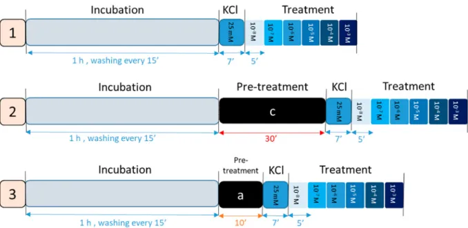
The experimental protocol is shown inFig. 1. The animals were terminated in a carbon dioxide chamber and the uterus samples were cut from both sides of the uterine horns. After cleaning from connective and adipose tissue, 3–4-mm dissected uterine tissues were tied with silk thread and mounted vertically in isolated organ bath filled with 10 ml de Jongh buffer consisting of 137 millimolar (mM) NaCl, 3 mM KCl, 1 mM CaCl2, 1 mM MgCl2, 12 mM NaHCO3, 4 mM NaH2PO4, 6 mM glucose, the pH was adjusted between 7.35 and 7.40 with constant temperature (37 °C) and with carbogen (95% O2+ 5% CO2) support.
Tissues were attached to a gauge transducer (SG-02; MDE GmBH., Heidelberg, Germany), with initial resting tension of 1.5 g, the con- tractions were measured, recorded and analyzed with a SPEL Advanced ISOSYS Data Acquisition System (MDE GmBH., Heidelberg, Germany).
The tissues were washed periodically every 15 min during the 1-h equilibrium incubation period.
To achieve a satisfactory rhythmic contraction response, KCl (25 mM) was added to each chamber for 7 min. Each steroid was added in a cumulative way (T, E2, P4) (10−8–10−3 M) every 5 min.
Concentration-response curves were plottedagainst the KCl-stimulated contraction response and the effects of steroids were expressed in per- centage change.
In another set of experiments, uterine tissues were pre-treated with cycloheximide (10−6M), a protein synthesis inhibitor and actinomycin D (10−6M), a transcriptional inhibitor for 2 steroids (E2 and testos- terone) separately for 30 min. Tissues were pre-treated with the fol- lowing steroid hormone receptor antagonists for 10 min before KCl stimulation: fulvestrant (10−6M) for E2, flutamide (10−6M) for T and mifepristone 10−6M for all types of steroids.
Finally, the endometrium of the uterine tissues was removed by scraping and the experiments were repeated to observe the effect of the steroids on the myometrium. The experimental protocol of the isolated organ bath study is shown inFig. 1.
The samples for each experiment were collected from both sides of the uterine horns of 2 animals (8 rings/experiment) and repeated at least 3 times for each individual set of experiments (n= 60).
Fig. 1.The isolated organ baths experimental protocol, the 1-h incubation period, 7-min KCl stimulation and cumulative dose treatment from 10−8- 10−3M in 5-min interval time were the same for all experiments; (1) intact or endometrium removed pregnant uterus samples treated with T, E2 and P4; (2) pregnant uterine samples with pre-treatment with actinomycin D and cycloheximide (c) for 30 min then treated with T and E2 treatment; (3) 10-min pre-treatment with steroid receptor antagonist (a): fulvestrant 10−6M for E2, flutamide 10−6M for T and mifepristone 10−6M for all steroid treatments, then treatment with T, E2 and P4.
2.3. [35S]GTPγS studies
The [35S]GTPγS binding experiments protocol was carried outby the previously described method [16] with modifications. Briefly, the pregnant uterine tissue samples from SPRD rats (n= 5) were collected and stored at −70 °C before preparation. The samples were ground, then homogenized with 20 volumes (W/V) of ice-cold Tris-EDTA buffer (composed of 10 mm Tris–HCl, 1 mM EDTA, 0.6 mM MgCl2, and 0.25 M sucrose, pH 7.4) with Ultra-Turrax® (IKA-Werke GmbH & Co. KG, Staufen in Breisgau, Germany) homogenizer in an ice bath for 2 × 30 s, after that suspended with 4-layer gauze filter, then centrifuged at 40000gfor 20 min at 4 °C. Later the pellets were suspended in 5 vo- lumes of buffer. The protein content was measured by a Nanodrop™
2000 spectrophotometer (Thermo Fisher Scientific, Wilmington, US) and diluted to 10 mg/ml sample.
Samples were pre-incubated in the final volume of 900 μl of Tris- EGTA buffer (pH 7.4) composed of 50 mM Tris-HC, 1 mM EGTA, 3 mM MgCl2, 100 mM NaCl, containing 20 mbq/0.05 cm3 [35S]GTPγS (0.05 nm) without or with mifepristone (10−6M) for 15 min in 30 °C.
After that, E2 and T were added separately to each tube in an increasing dose (10−8–10−4) for 20 min. The total binding was measured without
drugs. Nonspecific binding was evaluated by 10 μm unlabeled GTPγs and differences from total binding (basal activity).After the incubation time, by using vacuum filtration (through Whatman GF/B filters with Brandel M24R Cell harvester), the bound and free [35S]GTPγS were separated. The filters were washed 3 times in ice-cold buffer (pH 7.4), the radioactivity of the filters was measured in Ultimagold™ MV scin- tillation cocktail with Packard Tricarb 2300TR liquid scintillation counter. The experiment was arranged in triplicates and repeated three times.
2.4. Cyclic AMP studies
The cyclic AMP (cAMP) level in uterine tissues was measured by a commercial cAMP Enzyme Immunoassay Kit (Cayman Chemical, USA).
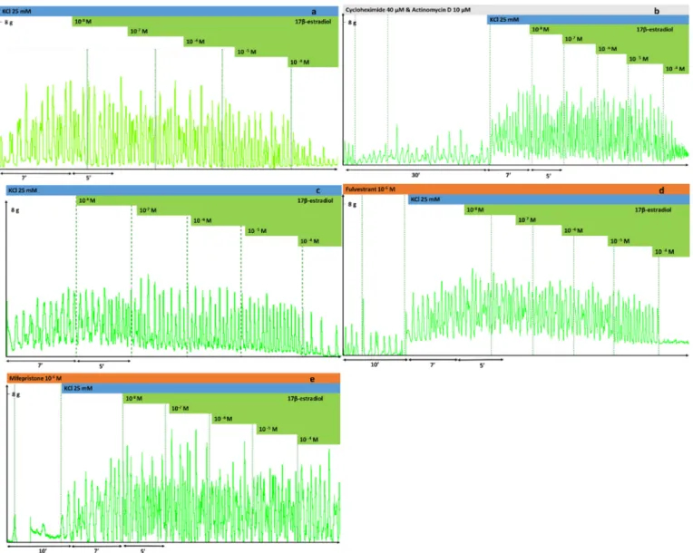
Uterine tissue samples of 22-day-pregnant SPRD rats (n= 8) were in- cubated in an organ bath filled with de Jongh buffer. The samples were incubated without or with mifepristone (10−6M) for 10 min, and KCl 25 mM was added for further 7 min. Then E2 and T or control (vehicle) were added in 2 different doses (10−4 and 10−6M) for 5 min, and forskolin (10−5M) was added for another 10 min. Finally, the samples were snap frozen by liquid nitrogen and stored at −70 °C for sample Fig. 2.Effects of testosterone (T) on KCl-induced (25 mM) uterine contractions at concentrations of 10−8–10−4M in a cumulative manner. Contraction was induced in the uterine rings prepared from rats on gestational day 22. Each figure is a representative record. (a) Effect of T after stimulation with KCl, (b) after 30 min of actinomycin D and cycloheximide pre-treatment, (c) after endometrium removal, (d) with pre-treatment with flutamide 10−6M and (e) with pre-treatment with mifepristone 10−6M.
preparation. During the preparation frozen tissues were weighed, pul- verized, mixed and homogenized with 10 volumes of 5% trichloroacetic acid (TCA) aqueous solution (0–4 °C), and centrifuged at 1500g for 15 min. The supernatants were mixed with 5 volumes of water-satu- rated ether and shaken in 10 s to extract TCA from it, the ether su- pernatant was removed, discarded and this process was repeated 3 times. Finally, after removing all the ether by heating, the liquid sam- ples were stored at −70 °C till the cAMP assay was carried out. The amounts of cAMP were expressed in nmol/mg tissue.
2.5. Drugs and chemicals
1,3,5-Estratriene-3,17β-diol (E2), 4-pregnene-3,20-dion (P4), 17β- Hydroxy-3-oxo-4-androstene (T), cycloheximide, actinomycin D, mife- pristone and flutamide were all purchased from Sigma-Aldrich, Budapest, Hungary. Fulvestrant (Falsoldex) 250 mg/ml injection was purchased from AstraZeneca Pharmaceutical, Budapest, Hungary.
Forskolin was purchased from Tocris, Norderstedt, Germany.
All the drugs were dissolved in ethanol 97%, the highest percentage of the solvent did not exceed 0.087%V/V.
2.6. Statistical analysis
Concentration-response curves were fitted by the analysis of the areas under curve (AUC) of contractility responses. Statistical analysis was carried out by the Prism 8.0 (GraphPad Software Inc. San Diego, CA, USA) computer program using ANOVA Dunnett's test. The Emaxand EC50values were calculated based on AUC. The results are presented as
-8 -7 -6 -5 -4 -3
-30 -10 10 30 50 70 90
log testosterone (M)
Relaxation(%)
a T
T+ ActD & Chx Endo removed
-8 -7 -6 -5 -4 -3
-30 -10 10 30 50 70 90
log testosterone (M)
Relaxation(%) T
T+ Flu T+ Mif
*** *** **
***
***
b
Fig. 3.Effects of T on pregnant uterine contractions stimu- lated with KCl (25 mM) and pre-treated with actinomycin D and cycloheximide, and after endometrium removal (a), and with pre-treatment with flutamide or mifepristone (b) pre- sented by percent of relaxation. **: p < 0.01; ***:
p< 0.001; ActD, actinomycin D; Chx, cycloheximide; Endo, endometrium; Flu, flutamide; Mif, mifepristone; T, testos- terone.
Table 1
Changes in the Emaxand EC50values of the uterine relaxing effect of T alone, with pre-treatment with actinomycin D and cycloheximide and mifepristone and also after the removal of the endometrium in the 22-day-pregnant rat. ActD, actinomycin D; Chx, cycloheximide; Endo, endometrium; Flu, flutamide; Mif, mifepristone; T, testosterone.
T T+ ActD+ Chx T+ FLU T+ Mif Endo removal
Emax(% ± S.E.M) 72.9 ± 7.8 77.4 ± 2.4 62.4 ± 8 41.9 ± 7.2 70.2 ± 2.7
EC50(M) 3.0e−005 3.8e−005 3.3e−005 5.8e−005⁎⁎⁎ 1.0e−005
⁎⁎⁎ p < 0.001.
-8 -7 -6 -5 -4
125 150 175 200 225 250
T T + Mif
Total
100 Level ofbasal activity
log testosterone (M) [35S]GTPS specificbinding(%)
**
Fig. 4.Effect of T (10−8–10−4M) on [35S]GTPγS binding with or without pre- treatment with mifepristone. Mifepristone reduced the T-induced increase in [35S]GTPγS binding. Basal activity (100%) refers to the level of [35S]GTPγS binding without any substances. **:p < 0.01; Mif, mifepristone; T, testos- terone.
Table 2
Changes in the [35S]GTPγS binding induced by T alone and with pre-incubation with mifepristone in the 22-day-pregnant rat uteri. Mif, mifepristone; T, tes- tosterone.
T T+ Mif
Emax(% ± S.E.M) 230 ± 9.3 166 ± 7.2⁎⁎
EC50(M) 7e-007 1.2e-006
⁎⁎ p < 0.01.
the mean ± SEM.
3. Results 3.1. Testosterone
3.1.1.1. Isolated organ bath study. T elicited a relaxing effect especially at high concentrations (10−5–10−3M) and reached 70% relaxation of the uterus (Figs. 2a,3a,Table 1). The presence of cycloheximide and actinomycin D (Figs. 2b,3b,Table 1), the removal of the endometrium (Figs. 2c,3b,Table 1) or even flutamide did not modify the relaxing effect of T. However, mifepristone shifted the T concentration-response curve to the right and reduced its maximal inhibitory effect. In the subsequent experiments investigating the signaling pathway, we measured the T action alone or in the presence of mifepristone. The interventions (endometrium removal) and drug treatments (actinomycin D, cycloheximide, flutamide) with non-significant modifications were omitted from further studies.
3.1.1.2. [35S]GTPγs binding assay studies. T increased the [35S]GTPγS Control
T 1-60M
T 10-6M+Mif
T 10-4M T 10
-4M+Mif 0
1 2 3 4 5
cAMPlevel ng/mgtissue
*** **
*
ns
***
ns
Fig. 5.Change in the level of uterine cAMP in the presence of T alone and after pre-treatment with mifepristone. The uterine cAMP level was expressed in ng/
mg tissue. *:p < 0.05 **: p < 0.01; ***:p < 0.001; Mif, mifepristone; T, testosterone.
Fig. 6.Effects of 17 β-estradiol (E2) on KCl-induced uterine contractions at concentrations of 10−8to 10−4M in a cumulative manner. Contraction was induced by KCl (25 mM) in the uterine rings prepared from rats on gestational day 22. Each figure is a representative record. (a) Effect of E2 after 7 min of stimulation with KCl, (b) after 30 min of actinomycin D and cycloheximide pre-treatment, (c) after endometrium removal, (d) with pre-treatment with fulvestrant 10−6M and (e) with pre- treatment with mifepristone 10−6M.
binding in a concentration dependent manner. The pre-treatment with mifepristone reduced specific binding and shifted the curve to the right, indicating less activation of G-proteins (Fig. 4andTable 2).
3.1.1.3. cAMP study. The pregnant uterus cAMP level was raised by T compared to the control at both low and high concentrations (10−6and 10−4(). Moreover, the pre-treatment with mifepristone significantly reduced the uterine cAMP levels by T (Fig. 5).
3.2. 17-β estradiol
Isolated organ bath study: E2 relaxed pregnant uterine contractions, 10-8 10-7 10-6 10-5 10-4 10-3
-30 -10 10 30 50 70 90
E2
log estradiol (M)
Relaxation(%) E2+ ActD & Chx
a
Endo removed
10-8 10-7 10-6 10-5 10-4 10-3 -30
-10 10 30 50 70 90
E2
E2+ Ful E2+ Mif
log estradiol (M)
Relaxation(%)
b
*
** ** ***
*** ***
Fig. 7.Effect of E2 on pregnant uterus tissue stimulated with KCl (25 mM) in the presence of actinomycin D and cyclo- heximide (a), and with pre-treatment with fulvestrant or mifepristone, and after endometrium removal (b) presented by percent of relaxation. *:p < 0.05 **:p < 0.01; ***:
p < 0.001; ActD, actinomycin D; Chx, cycloheximide; E2, 17-β estradiol; Mif, mifepristone; Ful, fulvestrant.
Table 3
Changes in the Emaxand EC50values of the uterine relaxing effect of E2 alone, with pre-treatment with actinomycin D and cycloheximide, fulvestrant and mife- pristone and after removing the endometrium. ActD, actinomycin D; Chx, cycloheximide; E2, 17-β estradiol; Ful, fulvestrant; Mif, mifepristone.
E2 E2+ ActD +Chx E2 + Ful E2+ Mif Endo removal
Emax(% ± S.E.M) 67.8 ± 2.8 71.7 ± 3.8 54.8 ± 2.9 15.4 ± 5.5⁎⁎⁎ 51.3 ± 4.6
EC50(M) 9.5e−006 1.3e−005 5.3e−006 6.7e−006 4.6e−006
⁎⁎⁎ p < 0.001.
-8 -7 -6 -5 -4
125 150 175 200 225 250
T T + Mif
Total
100 Level ofbasal activity
log testosterone (M) [35S]GTPS specificbinding(%)
**
Fig. 8.The effect of E2 (10−8–10−4M) on [35S]GTPγS binding with or without pre-treatment with mifepristone. Mifepristone reduced the E2-induced increase in [35S]GTPγS binding. Basal activity (100%) refers to the level of [35S]GTPγS binding without substance. **:p< 0.01; E2, 17-β estradiol; Mif, mifepristone.
Table 4
Changes in the [35S]GTPγS binding effect of E2 alone and with pre-incubation with mifepristone in the 22-day-pregnant rat. E2, 17-β estradiol; Mif, mife- pristone.
E2 E2+ Mif
Emax(% ± S.E.M) 196.3 ± 7.7 155 ± 4.5⁎⁎
EC50(M) 7.1e−007 8.5e−007
⁎⁎ p < 0.01.
Control
E210-6M
E210-6M+Mif
E210-4M
E210-4M+Mif 0
1 2 3 4 5
cAMPlevel ng/mgtissue ns
ns
*
**
ns
ns
Fig. 9.Changes in the level of cAMP in the presence of E2 alone (10−6and 10−4M) and with pre-treatment with mifepristone expressed in ng/mg tissue.
*:p < 0.05 **: p < 0.01; E2, 17-β estradiol; Mif, mifepristone.
at the highest concentration (10−4M), the inhibition was 70% (Figs. 6, 7a,Table 3). Actinomycin D and cycloheximide (Figs. 6b,7a,Table 3), the removal of the endometrium (Figs. 6c,7a,Table 3) or even pre- treatment with fulvestrant, (Figs. 6d,7b,Table 3) did not influence the effect of E2. In contrast, pre-treatment with mifepristone reduced the relaxing effect of E2, but did not modify the EC50value (Figs. 6e,7b, Table 3). In the subsequent experiments investigating the signaling pathway, we measured the E2 action alone or in the presence of mi- fepristone. The interventions (endometrium removal) and drug treat- ments (actinomycin D, cycloheximide, flutamide) with non-significant modifications were omitted from further studies.
3.2.1.1. [35S]GTPγS binding assay studies. The [35S]GTPγS binding was increased by E2 in a concentration dependent manner, which was reduced by mifepristone (Fig. 8andTable 4).
3.2.1.2. cAMP study. E2 increased the level of cAMP in the uterine tissue compared to the control in high concentrations, while it had no effect in a low dose. The pre-treatment with mifepristone reduced the high E2 concentration-induced cAMP increase (p < 0.01), but mifepristone had no action in the case of low E2 concentration (Fig. 9).
3.3. Progesterone
P4 had a negligible effect on KCl-stimulated uterine contractions.
The presence of mifepristone did not modify its action (Figs. 10, 11, Table 5). Since the relaxing effect of P4 was missing, we did not in- vestigate it further.
4. Discussion
The non-genomic action of sex steroids on different tissues, espe- cially on smooth muscles from different organs, has been investigated in several studies. It was proved that E2 and P4 had a vasorelaxant action through a non-genomic pathway. Studies on rat arterial beds showed that E2 induced vascular relaxation [17]. The same results were found on the arterial tissues in human [18,19] lamb [20], monkey [21] and mice [12]. On the contrary, the non-genomic action of E2 induced hyperreactivity and contraction on tracheal smooth muscles [22]. Ad- ditionally, E2 elicited non-genomic vasoconstriction in mice, which led to the reduction of skin cooling action [23]. The non-genomic smooth muscle relaxing effect of T was proved in human coronary arteries [24], umbilical arteries [25,26], peripheral vasculature of rats [27] and even in the trachea of guinea pigs [28,29]. However, a comparative in- vestigation of the non-genomic effect of sex hormones on pregnant uterine contractions has not been carried out yet.
Therefore, we aimed to investigate the effects of the 3 basic sex hormones (E2, P4 and T) on pregnant uterine contractions on the last day of pregnancy in rats in vitro. E2 and T elicited a significant re- duction in uterine contractions, while P4 was ineffective. The exposure time of the uterine tissues to the sex hormones was less than 30 min, which is considered to be too short to initiate the genomic response [30]. We also proved this, since the blockade of the genomic pathway by the RNA transcription inhibitor actinomycin D and the protein synthesis inhibitor cycloheximide did not modify the effects of E2 or T.
Subsequently, the removal of the endometrium did not modify the sex hormone effects either, so we also proved that the relaxation effects of E2 and T has a myometrial site of action. Surprisingly, both E2 and T had a remarkable relaxing effect (approximately 70% inhibition). Al- though such an action of T was described earlier on human and preg- nant rat uteri [31] [32], such a result about E2 has not been published yet. In contrast, E2, T and P4 were reported as ineffective on both pregnant rat and human myometrial contractions induced by oxytocin in vitro [33], but in that study the sex steroids were used in lower concentrations (below 10−6M), while we applied them in 10−4or 10−3M as the highest concentrations. Thus, the high concentrations can explain why we could detect quite a strong relaxing effect with T and E2. The other surprise was the ineffectiveness of P4 on pregnant contractions considering its clinical use against premature contractions Fig. 10.Effects of progesterone (P4) on KCl-induced (25 mM) uterine contractions at concentrations of 10−8to 10−4M in a cumulative manner. Uterine rings were gained from rats on gestational day 22. Each figure is a representative record. (a) Effect of progesterone after stimulation with KCl and (b) with pre-treatment with mifepristone 10−6M.
10-8 10-7 10-6 10-5 10-4 -30
-10 10 30 50 70 90
log P4. (M)
Relaxation(%)
P4 + Mif P4
Fig. 11.Effect of P4 on pregnant uterus smooth muscle contractions stimulated by KCl, alone and in the presence of mifepristone. Mif, mifepristone; P4, pro- gesterone.
Table 5
Changes in the uterine-contracting effect of P4 alone and in the presence of mifepristone. P4; progesterone. Mif, mifepristone;
P4 P4 + Mif
Emax(% ± S.E.M) 16.8 ± 6.9 20.6 ± 2.6
EC50(M) 1.2e007 1.2e−007
in threatening preterm birth [34], although in that case it is applied as a preventive agent. Our result suggests that there is no prompt relaxing action of P4 on pregnant uterine contractions. Similarly, earlier studies did not find any non-genomic relaxing effect for P4 either [35], al- though a synthetic P4 derivative, dydrogesterone was found to inhibit the pregnant myometrial contractions by the inhibition of voltage de- pendent Ca-channels [33]. Some other experiments found that P4 had an ability to relax human non-pregnant or pregnant uterine tissues in high dose [36–38] which findings are virtually in conflict of our results.
However, the findings may be a result of genomic feature of P4 since the P4 incubation period in both reported studies were more than 1 h.
The specific receptor antagonists of sex hormones (flutamide for T and fulvestrant for E2) did not reduce their actions, which is further evidence that the genomic pathway is not involved in the relaxing ef- fects of T and E2. Surprisingly, the actions of T and E2 were mifepris- tone sensitive, their maximum effects were reduced significantly by the compound. This suggests that mifepristone generally inhibits the non- genomic target of sex steroids for uterus relaxation, which is possibly independent of its progesterone and glucocorticoid receptor inhibitory action. The G protein coupled estrogen receptor (GPER or GPR30), which is coupled to Gs protein and enhances the intracellular cAMP level, has already been identified as a target of sex steroids in several tissues [39,40] as well as in human myometrium [41]. In our [35S]GTPγS binding and cAMP measurements we proved a significant increase in G-protein and cAMP levels after stimulation by T and E2, and their effects could be inhibited by mifepristone. The previously reported signaling pathway for putative sex steroid membrane re- ceptors involves phospholipase, kinase [42], calcium [43] and other second messengers such as IP3 or cAMP [44]. It is also possible that the activation of a G protein receptor by rising cAMP regulates the voltage- gated ion channels (e.g. BKCaand KV) [26] and the intracellular calcium regulation [11,22]. Our results suggest that T and E2 possibly activate GPR30 and mifepristone might be a competitive antagonist on this re- ceptor.
5. Conclusion
T and E2 can significantly inhibit KCl-stimulated contractions in the late pregnant uterus in high concentrations and in a non-genomic manner. Their actions are mediated by a G-protein coupled receptor (possibly GPR30) that can be blocked by mifepristone. However, P4 seems to be inefficient as a non-genomic relaxant of pregnant uteri.
Based on our results, a single and high dose of T or E2 might be con- sidered in premature contractions, however, further preclinical and clinical studies are required for the approval of such a therapeutic in- tervention.
Declaration of competing interest
No competing interest.
Acknowledgment
This work was supported by the Ministry of Human Capacities.
[Hungary grant 20391-3/2018/FEKUSTRAT] and the Stipendium Hungaricum Scholarship. Special thanks are due to Ágnes Csiszárné for technical assistance in the experiments.
References
[1] T. Sato, S. Miyagawa, T. Iguchi, Progesterone, in: Y. Takei, H. Ando, K.B.T.-H. of H. Tsutsui (Eds.), Handb. Horm, Elsevier, San Diego, 2016, ,https://doi.org/10.
1016/B978-0-12-801028-0.00220-8pp. 507-e94A-3.
[2] G.W. Corner, A. Csapo, Action of the ovarian hormones on uterine muscle, BMJ 1 (1953) 687–693,https://doi.org/10.1136/bmj.1.4812.687.
[3] M.A. Okon, S.M. Laird, E.M. Tuckerman, T.C. Li, Serum androgen levels in women who have recurrent miscarriages and their correlation with markers of endometrial
function, Fertil. Steril. 69 (1998),https://doi.org/10.1016/S0015-0282(98) 00007-7.
[4] M. Beato, S. Chávez, M. Truss, Transcriptional regulation by steroid hormones, Steroids, Elsevier Inc, 1996, pp. 240–251, ,https://doi.org/10.1016/0039-128X (96)00030-X.
[5] C.M. Szego, J.S. Davis, Adenosine 3′,5′-monophosphate in rat uterus: acute eleva- tion by estrogen, Proc. Natl. Acad. Sci. 58 (1967) 1711–1718,https://doi.org/10.
1073/pnas.58.4.1711.
[6] M. Nakashima, M. Suzuki, M. Saida, Y. Kamei, M.B. Hossain, T. Tokumoto, Cell- based assay of nongenomic actions of progestins revealed inhibitory G protein coupling to membrane progestin receptor α (mPRα), Steroids 100 (2015) 21–26, https://doi.org/10.1016/j.steroids.2015.04.002.
[7] T. Mitsui, M. Ishida, M. Izawa, J. Arita, Activation of G protein-coupled estrogen receptor 1 mimics, but does not mediate, the anti-proliferative action of estradiol on pituitary lactotrophs in primary culture, Endocr. J. 64 (2017) 103–115,https://doi.
org/10.1507/endocrj.EJ16-0079.
[8] A. Romarowski, C. Sánchez-Cárdenas, H.V. Ramírez-Gómez, L. del C. Puga Molina, C.L. Treviño, A. Hernández-Cruz, A. Darszon, M.G. Buffone, A specific transitory increase in intracellular calcium induced by progesterone promotes acrosomal exocytosis in mouse Sperm1, Biol. Reprod. 94 (2016) 136085,https://doi.org/10.
1095/biolreprod.115.136085.
[9] K. Barabás, S. Godó, F. Lengyel, D. Ernszt, J. Pál, I.M. Ábrahám, Rapid non-classical effects of steroids on the membrane receptor dynamics and downstream signaling in neurons, Horm. Behav. 104 (2018) 183–191,https://doi.org/10.1016/j.yhbeh.
2018.05.008.
[10] J. Kurokawa, M. Kodama, C.E. Clancy, T. Furukawa, Sex hormonal regulation of cardiac ion channels in drug-induced QT syndromes, Pharmacol. Ther. 168 (2016) 23–28,https://doi.org/10.1016/j.pharmthera.2016.09.004.
[11] M. Perusquía, N. Herrera, M. Ferrer, J.N. Stallone, Antihypertensive effects of an- drogens in conscious, spontaneously hypertensive rats, J. Steroid Biochem. Mol.
Biol. 167 (2017) 106–114,https://doi.org/10.1016/j.jsbmb.2016.11.016.
[12] M.R. Meyer, N.C. Fredette, T.A. Howard, C. Hu, C. Ramesh, C. Daniel, K. Amann, J.B. Arterburn, M. Barton, E.R. Prossnitz, G protein-coupled estrogen receptor protects from atherosclerosis, Sci. Rep. 4 (2015) 7564,https://doi.org/10.1038/
srep07564.
[13] M. Barton, E.R. Prossnitz, Emerging roles of GPER in diabetes and atherosclerosis, Trends Endocrinol. Metab. 26 (2015) 185–192,https://doi.org/10.1016/j.tem.
2015.02.003.
[14] S.R. Hammes, E.R. Levin, Extranuclear steroid receptors: nature and actions, Endocr. Rev. 28 (2007) 726–741,https://doi.org/10.1210/er.2007-0022.
[15] A. Kothencz, J. Hajagos-Tóth, K.F. Szűcs, A. Schaffer, R. Gáspár, α -Tocopherol potentiates the cervical resistance decreasing effects of COX inhibitors in pregnant rats: the putative role of cyclooxygenase-2 inhibition, J. Pharmacol. Exp. Ther. 368 (2019) 292–298,https://doi.org/10.1124/jpet.118.251850.
[16] E. Szucs, A. Büki, G. Kékesi, G. Horváth, S. Benyhe, Mu-opioid (MOP) receptor mediated G-protein signaling is impaired in specific brain regions in a rat model of schizophrenia, Neurosci. Lett. 619 (2016) 29–33,https://doi.org/10.1016/j.neulet.
2016.02.060.
[17] R.H.P. Hilgers, S. Oparil, W. Wouters, H.J.T. Coelingh Bennink, Vasorelaxing effects of estetrol in rat arteries, J. Endocrinol. 215 (2012) 97–106,https://doi.org/10.
1530/JOE-12-0009.
[18] E. Haas, I. Bhattacharya, E. Brailoiu, M. Damjanovic, G.C. Brailoiu, X. Gao, L. Mueller-Guerre, N.A. Marjon, A. Gut, R. Minotti, M.R. Meyer, K. Amann, E. Ammann, A. Perez-Dominguez, M. Genoni, D.J. Clegg, N.J. Dun, T.C. Resta, E.R. Prossnitz, M. Barton, Regulatory role of G protein-coupled estrogen receptor for vascular function and obesity, Circ. Res. 104 (2009) 288–291,https://doi.org/
10.1161/CIRCRESAHA.108.190892.
[19] K.S. Russell, M.P. Haynes, D. Sinha, E. Clerisme, J.R. Bender, Human vascular en- dothelial cells contain membrane binding sites for estradiol, which mediate rapid intracellular signaling, Proc. Natl. Acad. Sci. U. S. A. 97 (2000) 5930–5935,https://
doi.org/10.1073/pnas.97.11.5930.
[20] Z. Chen, I.S. Yuhanna, Z. Galcheva-Gargova, R.H. Karas, M.E. Mendelsohn, P.W. Shaul, Estrogen receptor mediates the nongenomic activation of endothelial nitric oxide synthase by estrogen, J. Clin. Invest. 103 (1999) 401–406,https://doi.
org/10.1172/JCI5347.
[21] R.D. Minshall, D. Pavcnik, D.L. Browne, K. Hermsmeyer, Nongenomic vasodilator action of progesterone on primate coronary arteries, J. Appl. Physiol. 92 (2002) 701–708,https://doi.org/10.1152/japplphysiol.00689.2001.
[22] E. Flores-Soto, I. Martínez-Villa, H. Solís-Chagoyán, B. Sommer, C. Lemini, L.M. Montaño, 17β-Aminoestrogens induce guinea pig airway smooth muscle hy- perresponsiveness through L-type Ca2+channels activation, Steroids 101 (2015) 64–70,https://doi.org/10.1016/j.steroids.2015.06.001.
[23] I. Serizawa, N. Iwasaki, H. Ishida, S. Saito, T. Ishikawa, G-protein coupled estrogen receptor-mediated non-genomic facilitatory effect of estrogen on cooling-induced reduction of skin blood flow in mice, Eur. J. Pharmacol. 797 (2017) 26–31,https://
doi.org/10.1016/j.ejphar.2017.01.013.
[24] K. Ruamyod, W.B. Watanapa, C. Shayakul, Testosterone rapidly increases Ca(2+)- activated K(+) currents causing hyperpolarization in human coronary artery en- dothelial cells, J. Steroid Biochem. Mol. Biol. 168 (2017) 118–126,https://doi.org/
10.1016/j.jsbmb.2017.02.014.
[25] P.A. Saldanha, E. Cairrão, C.J. Maia, I. Verde, Long- and short-term effects of an- drogens in human umbilical artery smooth muscle, Clin. Exp. Pharmacol. Physiol.
40 (2013) 181–189,https://doi.org/10.1111/1440-1681.12047.
[26] J. Feiteiro, A.J. Santos-Silva, I. Verde, E. Cairrão, Testosterone and atrial natriuretic peptide share the same pathway to induce vasorelaxation of human umbilical ar- tery, J. Cardiovasc. Pharmacol. 63 (2014) 461–465,https://doi.org/10.1097/FJC.
0000000000000060.
[27] M. Perusquía, C.D. Greenway, L.M. Perkins, J.N. Stallone, Systemic hypotensive effects of testosterone are androgen structure-specific and neuronal nitric oxide synthase-dependent, Am. J. Physiol. - Regul. Integr. Comp. Physiol. 309 (2015) R189–R195,https://doi.org/10.1152/ajpregu.00110.2015.
[28] E. Flores-Soto, J. Reyes-García, A. Carbajal-García, E. Campuzano-González, M. Perusquía, B. Sommer, L.M. Montaño, Sex steroids effects on guinea pig airway smooth muscle tone and intracellular Ca2+basal levels, Mol. Cell. Endocrinol. 439 (2017) 444–456,https://doi.org/10.1016/j.mce.2016.10.004.
[29] L.M. Montaño, E. Flores-Soto, J. Reyes-García, V. Díaz-Hernández, A. Carbajal- García, E. Campuzano-González, G.L. Ramírez-Salinas, M.A. Velasco-Velázquez, B. Sommer, Testosterone induces hyporesponsiveness by interfering with IP3 re- ceptors in guinea pig airway smooth muscle, Mol. Cell. Endocrinol. 473 (2018) 17–30,https://doi.org/10.1016/j.mce.2017.12.010.
[30] A.K.L. Herald, R. Alves-Lopes, A.C. Montezano, S.F. Ahmed, R.M. Touyz, Genomic and non-genomic effects of androgens in the cardiovascular system: clinical im- plications, Clin. Sci. 131 (2017) 1405–1418,https://doi.org/10.1042/CS20170090.
[31] M. Perusquía, E. Navarrete, J. Jasso-Kamel, L.M. Montaño, Androgens induce re- laxation of contractile activity in pregnant human myometrium at term: a non- genomic action on L-type calcium Channels1, Biol. Reprod. 73 (2005) 214–221, https://doi.org/10.1095/biolreprod.104.036954.
[32] Z. Yin, Y. Li, W. He, D. Li, H. Li, Y. Yang, B. Shen, X. Wang, Y. Cao, R.A. Khalil, Progesterone inhibits contraction and increases TREK-1 potassium channel ex- pression in late pregnant rat uterus, Oncotarget 9 (2018) 651–661,https://doi.org/
10.18632/oncotarget.23084.
[33] K. Yasuda, G. Sumi, H. Murata, N. Kida, T. Kido, H. Okada, The steroid hormone dydrogesterone inhibits myometrial contraction independently of the progesterone/
progesterone receptor pathway, Life Sci. 207 (2018) 508–515,https://doi.org/10.
1016/j.lfs.2018.07.004.
[34] V. Berghella, Progesterone and preterm birth prevention: translating clinical trials data into clinical practice, Am. J. Obstet. Gynecol. 206 (2012) 376–386,https://
doi.org/10.1016/j.ajog.2012.03.010.
[35] J. Baumbach, S.Q. Shi, L. Shi, J. Balducci, D.V. Coonrod, R.E. Garfield, Inhibition of uterine contractility with various tocolytics with and without progesterone: In vitro studies, Am. J. Obstet. Gynecol, Mosby Inc, 2012, pp. 254.e1–254.e5, ,https://doi.
org/10.1016/j.ajog.2011.12.011.
[36] T.A. Orth, S.Q. Shi, K. Williamson, L. Shi, L. Chambliss, D.V. Coonrod, J. Balducci, R.E. Garfield, Additive inhibitory effects of progesterone and sodium nitroprusside on uterine contractility during pregnancy, Reprod. Sci. 18 (2011) 868–875,https://
doi.org/10.1177/1933719111398141.
[37] M. Lucovnik, R.J. Kuon, L.R. Chambliss, W.L. Maner, S.-Q. Shi, L. Shi, J. Balducci, R.E. Garfield, Progestin treatment for the prevention of preterm birth, Acta Obstet.
Gynecol. Scand. 90 (2011) 1057–1069,https://doi.org/10.1111/j.1600-0412.
2011.01178.x.
[38] A. Kostrzewska, T. Laudánski, S. Batra, Effect of ovarian steroids and diethyl- stilbestrol on the contractile responses of the human myometrium and in- tramyometrial arteries, Eur. J. Pharmacol. 233 (1993) 127–134,https://doi.org/10.
1016/0014-2999(93)90358-o.
[39] M. Barton, E.J. Filardo, S.J. Lolait, P. Thomas, M. Maggiolini, E.R. Prossnitz, Twenty years of the G protein-coupled estrogen receptor GPER: historical and personal perspectives, J. Steroid Biochem. Mol. Biol. (2017) 1–12,https://doi.org/10.1016/
j.jsbmb.2017.03.021.
[40] S.H. Lindsey, K. a Carver, E.R. Prossnitz, M.C. Chappell, Vasodilation in response to the GPR30 agonist G-1 is not different from estradiol in the mRen2.Lewis female rat, J. Cardiovasc. Pharmacol. 57 (2011) 598–603,https://doi.org/10.1097/FJC.
0b013e3182135f1c.
[41] K. Maiti, J.W. Paul, M. Read, E.C. Chan, S.C. Riley, P. Nahar, R. Smith, G-1-acti- vated membrane estrogen receptors mediate increased contractility of the human myometrium, Endocrinology 152 (2011) 2448–2455,https://doi.org/10.1210/en.
2010-0979.
[42] N. Schwartz, A. Verma, C.B. Bivens, Z. Schwartz, B.D. Boyan, Rapid steroid hor- mone actions via membrane receptors, Biochim. Biophys. Acta - Mol. Cell Res. 1863 (2016) 2289–2298,https://doi.org/10.1016/j.bbamcr.2016.06.004.
[43] N.E. Vega-Vela, D. Osorio, M. Avila-Rodriguez, J. Gonzalez, L.M. García-Segura, V. Echeverria, G.E. Barreto, L-type calcium channels modulation by estradiol, Mol.
Neurobiol. 54 (2017) 4996–5007,https://doi.org/10.1007/s12035-016-0045-6.
[44] M.L. Oróstica, J. Lopez, I. Rojas, J. Rocco, P. Diáz, P. Reuqueń, H. Cardenas, A. Parada-Bustamante, P.A. Orihuela, Estradiol increases cAMP in the oviductal secretory cells through a nongenomic mechanism, Reproduction 148 (2014) 285–294,https://doi.org/10.1530/REP-14-0128.