Functional Analysis on a
Naturally Occurring Variant of the Staphylococcus Aureus Uracil DNA Glycosylase Inhibitor
Veronika Papp-Kádár
1*, Zoltán Balázs
1, Gergely N. Nagy
1,2, Tünde Juhász
2, Károly Liliom
2,3, Beáta G. Vértessy
1,2**Received 14 October 2016; accepted after revision 12 December 2016
Abstract
Repair of DNA damage relies on various pathways including the base excision repair (BER) which targets erroneous bases in the DNA. Here, Uracil-DNA glycosylases (UDGs) are respon- sible for recognition and removal of uracil base from the DNA.
Here, we characterize the interaction of Staphylococcus aureus UDG (SAUDG) with a naturally occurring variant of S. aureus uracil-DNA glycosylase inhibitor (SAUGI). This variant con- tains a histidine instead of a glutamate at the 24th position which affects the SAUDG:SAUGI interaction surface. We assessed the complex formation of SAUDG with these two SAUGI variants by independent biophysical methods. Our data reveal that the residue difference at the 24th position does not have a marked effect on the binding affinity, yet it confers alteration of the thermodynamics of the interaction. We propose that the E24H variant of SAUGI allows efficient complex formation, and con- sequently, inhibition of SAUDG.
Keywords
DNA repair, base excision repair, uracil-DNA glycosylase inhibitor
1 Introduction
Appearance of uracil in DNA, either as a result of cytosine deamination or erroneous nucleotide incorporation of DNA polymerases, usually a mistake that needs to be excised [1].
To avoid mutations, several DNA damage recondition and repair pathways have been developed during evolution. The base excision repair pathway (BER) corrects DNA base dam- ages arising e.g. from oxidation, deamination or alkylation.
The damage is generally the result of the spontaneous decay of DNA that occurs during physiological processes. However, similar base mismatch may be caused by radiation and envi- ronmental chemicals as well [2, 3].
As the initial step in BER, the damaged or mismatched base is recognized by a DNA glycosylase. The removal of the modi- fied base performed by these enzymes does not cause major dis- tortions to the DNA duplex. Uracil-DNA glycosylase (UDG) employs a nucleotide-flipping mechanism in which the target uracil is extruded out of the DNA double helix and recognized by the active site of the enzyme, where catalysis takes place.
Simultaneously, a bulky hydrophobic or aromatic residue is usually inserted into the DNA helix to take the position of the displaced base [4].The enzyme hydrolyses the N-C1’ glycosidic bond linking the uracil base to the deoxyribose sugar moiety.
Glycosylase action leaves an abasic site that is further processed by short-patch repair or long-patch repair mechanisms that use largely different enzymes [5, 6]. The abasic site that is removed by a 5’-acting apurinic/apyrimidinic (AP) endonuclease and a deoxyribophosphodiesterase (dRpase), leaving a gap that is filled by DNA polymerase and closed by DNA ligase [7].
Three structurally unrelated inhibitors of uracil-DNA gly- cosylase have been identified to date. These include the Uracil Glycosylase Inhibitor (UGI) of Bacillus subtilis bacteriophage PBS1, the p56 protein encoded by the B. subtilis phage ϕ29 and the Staphylococcus specific uracil-glycosylase inhibitor (SAUGI) [7-10]. The three dimensional structures have also been determined for all the three inhibitor proteins in complex with their respective binding partners. In all 3D structures, the negatively charged residues of the inhibitors follow a recogni- tion pattern that closely mimics the UDG-bound DNA [8-10].
1 Department of Applied Biotechnology and Food Science, Faculty of Chemical Engineering and Biochemical Engineering, Budapest University of Technology and Economics,
H-1111 Budapest, Műegyetem rkp. 3., Hungary
2 Institute of Enzymology, Research Centre for Natural Sciences, Hungarian Academy of Sciences,
H-1117 Budapest, Magyar tudósok krt. 2., Hungary
3 Department of Biophysics and Radiation Biology, Semmelweis University,
H-1094 Budapest, Tűzoltó u. 37-47., Hungary
* First corresponding author, e-mail: kadar.veronika@ttk.mta.hu
** Second corresponding author, e-mail: vertessy@mail.bme.hu
62(1), pp. 51-56, 2018 https://doi.org/10.3311/PPch.10163 Creative Commons Attribution b research article
PP Periodica Polytechnica
Chemical Engineering
In the UDG inhibitor research, the UGI protein was the first to be described and a straightforward physiological func- tion could also be defined in this case. Namely, the genome of B. subtilis bacteriophages PBS1 and PBS2 naturally contain uracil in place of thymines, thus it produces UGI proteins to protect their genomic DNA from host UDG repair enzymes [8-10]. The second reported inhibitor, termed as p56, is a small, 56-residue-long acidic polypeptide encoded by the B.
subtilis lytic phage ϕ29. Unlike PBS2 phage, the DNA genome of ϕ29 does not contain uracil residues. Protein p56 is syn- thesized upon ϕ 29 infection and knocks out a host-encoded BER system that could be harmful for viral replication if uracil residues arise in the replicative intermediates [9, 13-15]. Last, the S. aureus uracil-DNA glycosylase (SAUGI) have been identified in 2014 [10]. While within this study the structure of SAUGI: SAUDG complex has been solved, the biological role of SAUGI still remains unclear, with several hypotheses being proposed. SAUGI may facilitate increased incorpora- tion of uracil into the viral genome, and this replacement may efficiently block the replication of various DNA viruses [10].
According to another theory, the SAUGI protein has an inher- ently increased affinity towards viral UDGs and this may also be further tailored, e.g. inhibition of herpes viral UDG can be largely elevated [16].
In the present work, we focused on one of the naturally occurring residue variation of the SAUGI proteins encoded by different Staphylococcal strains. Namely we investigated the SAUGIE24H variant as compared to the SAUGI described in [10]
which is termed here as wild type (SAUGIWT). We employed two independent biophysical methods and also provided a structural modelling for comparisons. Our data suggest that the exchange of glutamate to histidine at this specific location does not result in heavy perturbation of the interaction.
2 Materials and Methods
2.1 In silico Blast search and alignments
Blast search for homologous sequences of SAUGI proteins, was performed using NBCI Blast and the wild type SAUGI sequence (UniProt accession code: Q936H5). The search was performed using translated nucleotide query (blastx), in the non-redundant protein sequences database, in S. aureus (taxid:
1280) organism. The alignment was done on sequences with the similarity higher than 90%, then adjusted manually. At the 24th residue of SAUGI, 70% of the aligned sequences contained glu- tamate whereas the remaining 30% histidine.
2.2 Cloning and mutagenesis
pET21b vectors encoding SAUDG and SAUGI (later termed as SAUGIWT) were kind gifts from Hao-Ching Wang, from Taipei Medical University [10]. We have re-cloned the DNA coding sequence of SAUGI into the pET15b vector that also includes an N-terminal His6 epitope tag. The SAUGI
mutant construct (SAUGIE24H) was engineered by site-directed mutagenesis from SAUGIWT [17] using the QuikChange method (Agilent). Primers used for mutagenesis (Table 1) were synthe- sized by Eurofins MWG GmbH. Constructs were verified by DNA sequencing at Eurofins MWG GmbH.
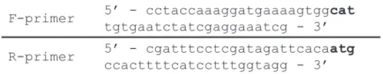
Table 1 Primers for constructing E24H point mutation F-primer 5’ - cctaccaaaggatgaaaagtggcat
tgtgaatctatcgaggaaatcg - 3’
R-primer 5’ - cgatttcctcgatagattcacaatg ccacttttcatcctttggtagg - 3’
2.3 SAUGI and SAUDG protein expression and purification
Plasmids for the production of SAUDG, SAUGIWT and SAUGIE24H were transformed into E. coli Rosetta BL21 (DE3) PlysS cells (Novagen). The cells were grown in LB medium at 37°C, and induced at OD600nm = 0.6 with 0.6 mM isopropyl-β-D-1- thiogalactopyranoside (IPTG), followed by overnight expression at 16 °C, then, harvested by centrifugation (4 °C, 4000 g, 20 min).
The pellet was lyzed in lysis buffer containing 50 mM Tris HCl, pH 8.0, 150 mM NaCl, 1 mM ethylenediaminetetraacetic acid (EDTA), 5 mM β-mercaptoethanol, 0.1 mM phenylmethylsulfo- nyl fluoride, 5 mM benzamidine, 0.1 mg/ml lysozyme, 0.1 mg/
ml DNase (Sigma, St. Louis, MO, USA) and 0.01 mg/ml RNase A (Invitrogen, Carlsbad, CA, USA). Cell extraction was achieved by Potter-Elvehjem homogenization and further assisted by soni- cation. Cell debris were pelleted by centrifugation at 20 000 × g for 30 min. Supernatant was applied onto a Ni-NTA column (Qiagen, Hilden, Germany) and washed with a set of washing buffers: low salt buffer (50mM HEPES, pH 7.5, 30 mM KCl, 5 mM β-mercaptoethanol), high salt buffer (50 mM HEPES, pH 7.5, 300 mM KCl, 5 mM β-mercaptoethanol) and wash buffer (50mM HEPES, pH 7.5, 30 mM KCl, 50 mM imidazole, 5 mM β-mercaptoethanol). Constructs were finally eluted with elution buffer (50 mM HEPES, pH 7.5, 30 mM KCl, 500 mM imidazole, 5 mM β-mercaptoethanol) and dialyzed against the following phosphate buffer: 137 mM NaCl, 2.7 mM KCl, 10 mM Na2HPO4, 2 mM KH2PO4, pH 7.5. Purity of SAUDG (Mw: 26032 Da), SAUGIWT (Mw: 15520 Da) and SAUGIE24H (Mw: 15528 Da) con- structs was assessed by SDS-PAGE (not shown).
2.4 Microscale thermophoresis (MST)
The interaction of SAUDG and SAUGI proteins was stud- ied using microscale thermophoresis (MST) as implemented in the NanoTemper Monolith NT.115 instrument (Nanotemper Technologies GmbH, Germany). We used Premium Nanotemper capillaries with 60% light emitting diode power and 95%
MST power for 40 s at 20 °C. SAUDG was labelled using the Monolith NT.115 NT-647 RED-NHS amine reactive protein labelling kit according to the NanoTemper protocol. Unlabelled SAUGIWT or SAUGIE24H was diluted in sixteen twofold dilution
n series starting from 2.5 mM. 5 nM of the labelled SAUDG was added to each dilution of SAUGI variants and the com- plex was incubated for 10 minutes at room temperature in the final buffer containing 50 mM Tris HCl pH 7.4, 150 mM NaCl, 10 mM MgCl2, 0.05% Tween 20. Data were analysed using the software provided by the manufacturer. Quadratic fit to the data according to mass law of action was used for apparent KD calcu- lation. Each experiment was done in triplicate.
2.5 Isothermal titration calorimetry (ITC)
Calorimetric measurements were performed on a MicroCal- ITC200 titration calorimeter (Malvern) at 20°C. The protein samples were co-dialysed overnight against phosphate buffered saline (PBS) pH 7.5 including 137 mM NaCl, 2.7 mM KCl, 10 mM Na2HPO4, 2 mM KH2PO4 and also supplemented with 0.5 mM Tris(2-carboxyethyl)phosphine (TCEP). In the experi- mental setup, the cell of the instrument was filled with 20 μM SAUGIWT or SAUGIE24H and the syringe with 200 μM SAUDG.
Each titration included 19 steps of injection with 2 μl of ligand per injection spaced 180 s apart from each other, with the injection syringe rotating at 750 r.p.m. The titration data were analysed using MICROCAL ORIGIN software, following the directions of the manufacturer, in order to calculate the thermo- dynamic parameters: dissociation constant (KD), stoichiometry (N), enthalpy (ΔH) and entropy (ΔS).
3 Results and Discussion
The complex formation of SAUDG with SAUGIWT and SAUGIE24H variants were first assessed by MST (Fig. 1).
The thermophoresis induced fluorescence change of NT-647 dye labelled SAUDG clearly indicated the binding interaction of SAUDG with both SAUGI variants. While the binding curves had similar appearance for both SAUGIs, the obtained apparent KD values of 0.01 ± 0.05 nM indicated that the qualitative deter- mination of the dissociation constants for these interactions can- not be accomplished under the experimental conditions due to the
relative high affinity of SaUGI-SAUDG interaction. Therefore, we investigated these interactions by ITC to account for the qual- itative characterization of binding affinity and energetics.
ITC is a highly relevant biophysical technique for biomo- lecular interaction analysis which in addition to binding affin- ity also simultaneously reports stoichiometry and the enthalpic component of complex formation. Titration of SAUDG to both SAUGI variants provided similar binding affinities and a one to one stoichiometry of interaction (Fig. 2). Importantly, Fig. 2 and Fig. 3 show a drastic change in the thermodynamic character of the interaction for the wild type and the mutant cases. In the complexation of SAUDG and SAUGIWT favourable enthalpic and entropic components could be detected, while in the case of SAUGIE24H, binding is governed by large favourable entropic component opposed by small unfavourable binding enthalpy.
We consider that our ITC experiments are reliable in terms of binding energetics characterization even despite the presence of the background noise. Yet the obtained KD values are far from the ones published before for SAUDG: SAUGIWT interaction [10]. Additional experiments, e.g. parallel measurements reverse ITC titration or a displacement titration, are required to better describe the binding affinity of the SAUDG: SAUGI interaction.
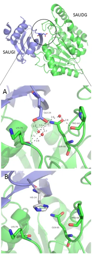
To further explore the effect of the mutation we also mod- elled this residue change into the experimentally determined 3D structure of the SAUDG: SAUGI complex [10], using the built-in Mutagenesis module of the PyMOL program (PyMOL Molecular Graphics System, Version 1.8 Schrödinger, LLC).
The most probable histidine conformer of the E24H mutation is visualized on Fig. 4.
In the wild type complex the Glu24 residue participates in sol- vent mediated polar contacts with SAUDG Lys79, Gln66 and Pro64 which may be affected by the replacement of this glutamate to histidine. Note that water-mediated polar interactions often play an instrumental role in stabilization of protein-protein interac- tions e.g. in case of [19, 20]. However, potential alterations of indirect interprotomer interactions due to the different variants
Fig. 1 SAUDG interaction with SAUGI variants measured by microscale thermophoresis (MST). The concentration of SAUDG was kept constant at 5 nM during the measurements. Data were fitted with quadratic equation with the software of MST to provide apparent dissociation constants. Mean and
SD of three replicates are shown.
cannot be faithfully interpreted on the sole basis of the in silico model. To account for the in-depth molecular basis of the E24H mutation, additional evidences from, e.g. molecular dynamics or high resolution x-ray crystal structures may be required.
4 Conclusions
The biochemical properties of glutamate and histidine resi- dues show clear differences with regard to electrostatic charge, aromaticity and involvement in hydrogen bond interactions as acceptor or donor. However, in our specific situation the per- formed biophysical measurements suggest that the affinity of the interaction between the SAUDG glycosylase and its SAUGI inhibitor was not largely altered by the replacement of SAUGI Glu24 to His at the interprotomer interaction surface. Despite the observed differences in binding energetics, our in vitro
characterization suggests that both SAUGIWT and SAUGIE24H variants have similar physiological role in SAUDG inhibition.
This finding potentially underlines the significance of this inter- action for the pathogenic Staphylococcus organism.
Acknowledgement
The project presented in this article is supported by the Hungarian Scientific Research Fund (OTKA 119493, OTKA K109486 to BGV, OTKA K82092 to KL, OTKA PD104344 to TJ), the Hungarian Academy of Sciences (TTK IF-28/2012 and MedinProt) and the European Commission FP7 BioStruct-X project (con-tract No. 283570), ICGEB CRP/HUN14-01, as well as the Core Technologies Centre of the Research Centre for Natural Sciences, Hungarian Academy of Sciences.
Fig. 3 Energetic components of SAUDG-SAUGI binding interactions revealed by ITC.
Fig. 2 Titration of SAUGI variants with SAUDG measured by isothermal titration calorimetry (ITC). For each measurements, the upper panel of the figures depicts raw titration data, whereas integrated binding heat corresponding to each titration point is shown in function of SAUDG: SAUGI molar
ratio in the lower panel.
References
[1] Vertessy, G. B., Toth, J. "Keeping uracil out of DNA: physiological role, structure and catalytic mechanism of dUTPases." Accounts of Chemical Research. 42(1), pp. 97–106. 2009. https://doi.org/10.1021/ar800114w [2] Krokan, H. E., Bjøra, M. "Base Excision Repair." Cold Spring Harbor
Perspectives in Biology. 5(4), pp. 1-22. 2013.
https://doi.org/10.1101/cshperspect.a012583
[3] Lindahl, T. "Instability and decay of the primary structure of DNA." Na- ture. 362, pp. 709–715. 1993. https://doi.org/10.1038/362709a0 [4] Wallace, S. S. "Base excision repair: a critical player in many games."
DNA Repair. 19, pp. 14-26. 2014.
https://doi.org/10.1016/j.dnarep.2014.03.030
[5] Krokan, H. E., Standal, R., Slupphaug, G. "DNA glycosylases in the base excision repair of DNA." The Biochemical Journal. 325, pp. 1-16. 1997.
https://doi.org/10.1042/bj3250001
[6] Nilsen, H., Krokan, H. E. "Base excision repair in a network of defence and tolerance." Carcinogenesis. 22(7), pp. 987–998. 2001.
[7] Wang, Z., Mosbaugh, D. W. "Uracil-DNA glycosylase inhibitor gene of bacteriophage PBSB encodes a binding protein specific for uracil-DNA glycosylase." The Journal of Biological Chemistry. 264(2), pp. 1163- 1171. 1989.
[8] Mol, C. D., Arvai, S. A., Sanderson, R. J., Slupphaug, G., Kavli, B., Krokan, H.E., Mosbaugh, D.W., Tainer, J. A. "Crystal structure of hu- man uracil-DNA glycosylase in complex with a protein inhibitor: protein mimicry of DNA." Cell. 82(5), pp. 701–708. 1995.
https://doi.org/10.1016/0092-8674(95)90467-0
[9] Serrano-Heras, G., Ruiz-Masó, J. A., Del Solar, G., Espinosa, M., Bravo, A., Salas, M. "Protein p56 from the Bacillus subtilis phage Φ29 inhib- its DNA-binding ability of uracil-DNA glycosylase." Nucleic Acids Re- search. 35(16), pp. 5393-5401. 2007.
https://doi.org/10.1093%2Fnar%2Fgkm584
[10] Wang, H. C., Hsu, K. C., Yang, J. M., Wu, M. L., Ko, T. P., Lin, S. R., Wang, A. H. J. "Staphylococcus aureus protein SAUGI acts as a uracil- DNA glycosylase inhibitor." Nucleic Acids Research. 42(2), pp. 1354- 1364. 2004. https://doi.org/10.1093/nar/gkt964
[11] Parikh, S. S., Putnam, C. D., Tainer, J. A. "Lessons learned from structur- al results on uracil-DNA glycosylase." Mutation Research/DNA Repair.
460(3-4), pp. 183-199. 2000.
https://doi.org/10.1016/S0921-8777(00)00026-4
[12] Putnam, C. D., Shroyer, M. J., Lundquist, A. J., Mol, C. D., Arvai, A. S., Mosbaugh, D. W., Tainer, J. A. "Protein mimicry of DNA from crystal structures of the uracil-DNA glycosylase inhibitor protein and its com- plex with Escherichia coli uracil-DNA glycosylase." Journal of Molecu- lar Biology. 287(2), pp. 331-346. 1999.
https://doi.org/10.1006/jmbi.1999.2605
[13] Serrano-Heras, G., Salas, M., Bravo, A. "A uracil-DNA glycosylase in- hibitor encoded by a non-uracil containing viral DNA." The Journal of Biological Chemistry. 281, pp. 7068-7074. 2006.
https://doi.org/10.1074/jbc.M511152200.
[14] Asensio, J.L., Pérez-Lago, L., Lázaro, J. M., González, C., Serrano- Heras, G., Salas, M. "Novel dimeric structure of phage φ29-encoded protein p56: Insights into uracil-DNA glycosylase inhibition." Nucleic Acids Research. 39(22), pp. 9779-9788. 2011.
https://doi.org/10.1093/nar/gkr667
[15] Baños-Sanz, J. I., Mojardín, L., Sanz-Aparicio, J., Lázaro, J. M., Villar, L., Serrano-Heras, G., González, B., Salas, M. "Crystal structure and functional insights into uracil-DNA glycosylase inhibition by phage ϕ29 DNA mimic protein p56." Nucleic Acids Research. 41(13), pp. 6761- 6773. 2013. https://doi.org/10.1093/nar/gkt395
Fig. 4 Structural representation of SAUDG: SAUGI complexes. Three dimensional structural model of the complex formed by SAUDG (green cartoon) and SAUGIWT (blue cartoon) (PDB: 3WDG). A) Close-up of the interaction surface display solvent mediated contacts of SAUGI E24 residue.
B) Proposed conformation of H24 residue of UGIE24H at the SAUDG: SAUGI interaction surface. The most probable H24 conformer generated by the SAUGI E24H exchange performed by the Pymol Mutagenesis tool is depicted.
[16] Wang, H. C., Ho, C. H., Chou, C. C., Ko, T. P., Huang, M. F., Hsu, K. C., Wang, A. H. J. "Using structural-based protein engineering to modulate the differential inhibition effects of SAUGI on human and HSV uracil DNA glycosylase." Nucleic Acids Research. 44(9), pp. 4440-4449. 2016.
https://doi.org/10.1093/nar/gkw185
[17] Landt, O., Grunert, H. P., Hahn, U. "A general method for rapid site- directed mutagenesis using the polymerase chain reaction." Gene. 96(1), pp. 125-128. 1990.
https://doi.org/10.1016/0378-1119(90)90351-Q
[18] Miles, L. A., Crespi, G. A N., Doughty, L., Parker, M. W. "Bapineuzumab captures the N-terminus of the Alzheimer’s disease amyloid-beta peptide in a helical conformation." Scientific Reports. 3, Article number: 1302.
https://doi.org/10.1038/srep01302
[19] Bahadur, R. P., Zacharias, M. "The interface of protein-protein com- plexes: analysis of contacts and prediction of interactions." Cellular and Molecular Life Sciences. 65(7-8), pp. 1059-1072. 2008.
https://doi.org/10.1007/s00018-007-7451-x
[20] Levy, Y., Onuchic, J. N. "Water and proteins: a love-hate relationship."
Proceeedings of the National Academy of the United States of America.
101(10), pp. 3325-3326. 2004.
https://doi.org/10.1073/pnas.0400157101