Functional analysis of the neonatal brain
PhD thesis
Zsófia Róna MD
Semmelweis University
Doctoral School of Clinical Medicine
Head of the Program and Supervisor: Zoltán Papp MD, DSc
Official reviewers: Ilona György MD, PhD
Miklós Szabó MD, PhD
Head of the Final Examination Committee: Fekete György MD,DSc.
Members of the Final Examination Committee: Lajos Kozák MD, PhD
Péter Mandl MD, PhD
Mónika Popper MD ,PhD
Budapest, 2017
1. Table of content
1. Table of content...1
2. List of Abbreviations...4
3. Introduction...6
3.1. Challenges of neonatal care...6
3.1.1. Development of perinatal care...6
3.1.2. Development of clinical practice at the neonatal ward...7
3.2. Brain development in the neonatal period...9
3.2.1. Structural Overview...9
3.2.2. Functional Overview and Plasticity...10
3.3. Neurophysiological methods in the neonatal period...11
3.3.1. The role of neurophysiological methods...12
3.3.2. Conventional EEG...13
3.3.3. Amplitude Integrated EEG...14
3.3.4. Visual Evoked Potentials...15
3.3.5. Event Related Potentials...16
3.4. Imaging of the neonatal brain...16
3.4.1. The role of imaging methods...16
3.4.2. Magnetic Resonance Imaging...17
3.4.3. Cranial Ultrasound...18
3.4.4. Computer Tomography...19
3.5. Common morbidities of the central nervous system during the neonatal period...19
3.5.1. Intraventricular haemorrhage...19
3.5.2. Posthemmorhagic ventricular dilatation...21
3.5.3. Asphyxia...21
3.5.4. Periventricular leukomalacia and diffuse white matter injury...22
3.6. Neurodevelopmental Outcomes...22
4. Objectives...24
4.1. Feasability of Functional Neurophysiological Methods and optimalization of neonatal care 24 4.1.1. Hydrocephalus Study...24
4.2. Optimalisation of Cerebral Imaging methods...24
4.2.1. MRI-compatible incubator Study...24
4.3. Use of functional neurophysiological or imaging methods and prediction of outcome...25
4.3.1. Asphyxia Study...25
4.3.2. Mismatch Negativity Study...25
4.3.3. Intraventricular Haemorrhage Study...25
5. Methods...27
5.1. Hydrocephalus Study...27
5.2. MRI-compatible Incubator Study...31
5.3. Asphyxia Study...33
5.4. Mismatch Negativity-Study...35
5.5. IVH Study...38
5.6. Neurodevelopmental Outcome Measurements...39
5.6.1. Bayley Scales of Infant Development...40
5.6.2. Kaufmann’s Assessment Battery for Children and Beery- Buktenica Developmental Test of Visual-Motor Integration...40
5.6.3. Gross Motor Function Classification System (GMFCS)...41
5.6.4. Scheffzek Neurological Classification...41
6. Results...43
6.1. Hydrocephalus Study...43
6.2. MRI-compatible Incubator Study...46
6.3. Asphyxia Study...51
6.4. Mismatch Negativity Study...54
6.5. IVH Study...57
7. Discussion...63
7.1. Hydrocephalus Study...63
7.2. MRI-compatible Incubator Study...66
7.3. Asphyxia Study...68
7.4. Mismatch Negativity-Study...71
7.5. IVH Study...73
8. Conclusions...76
9. Összefoglalás...79
10. Summary...81
11. Bibliography...82
12. Bibliography of the candidate’s publications...91
12.1. Related Publications...91
12.2. Unrelated Publications...91
13. Acknowledgments...92
2. List of Abbreviations
BAEP- Brainstem Auditory Evoked Potentials DTI – Diffusion Tensor Imaging
ERP – Event Related Potential GA-Gestational Age
INC - MRI Compatible Incubator IVH - Intraventricular Haemorrhage PHH - Posthaemorrhagic Hydrocephalus CNS - Central Nervous System
MRI - Magnetic Resonance Imaging
fcMRI – functional Magnetic Resonance Imaging PVL – Periventricular Leukomalacia
GMFCS- Gross Motor Function Classification System KABC-II - Kaufmann’s Assessment Battery for Children PDI - psychomotor developmental index
MDI - Mental developmental index MMN – Miss Match Negativity CP – Cerebral Palsy
SEP – Somatosensory Evoked Potentials VEP – Visual Evoked Potential
NICU – Neonatal Intensive Care Unit ROP – Retinopathy of prematurity
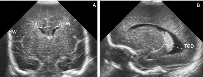
PDA – Persistent ductus arteriosus NEC – Necrotic Enterocolitis CLD – Chronic lung disease AIS – amniotic infect syndrome RDS – Respiratory distress syndrome TOD – Thalamo-occipital distance AHW – Anterior horn width
BPD – Bronchopulmonary Displasia
PHHN – Persistent Pulmonary Hypertension HIE – Hypoxic Ischaemic Encephalopathy
NIDCAP -Newborn Individualized Developmental Care and Assessment Program INC – MRI-compatible Incubator
3. Introduction
3.1. Challenges of neonatal care
Premature birth presents enormous complexities for all to consider, especially for expectant families. Survival rates of this population has dramatically increased in the last decades, although enthusiasm for this improvement is tempered by the long-term follow-up experience with these children. Despite a significant decline in mortality, neurodevelopmental injury rates remain high and do not seem to be consistently improving. [1, 2]
Follow-up examinations of surviving infants born at less than 27 weeks' of gestation from different institutions and populations demonstrate outcomes that seem to be remarkably consistent. Approximately 25% of infants suffer severe neurologic damage, while 25% has moderate impairment, and 50% are judged to be mildly impaired or normal.
[3] These investigations suggest the earlier the gestational age, the higher the disability rate. Furthermore, “normal” surviving infants are at considerable risk for a variety of neurobehavioral, social, and educational deficits that likely reflect altered neurobiology related to premature birth. [4] There is also a growing evidence that late preterm birth also carries a considerable risk for altered neurological outcome. [5]
The challenge of current neonatal care is to improve neurodevelopmental outcome.
To achieve this goal neuromonitoring and neuroimaging methods must be regularly used and further improved in order to optimise therapeutic efforts of this fragile population.
3.1.1. Development of perinatal care
In the last decades there is a growing effort that premature birth should take place at centralised level 4 perinatal centers all around the world. Obstetritians and neonatologist have worked out protocolls to protect the fetus and the mother in case of threatening preterm labour. Intrauterine transport, tocolisys with beta-mimetics, magnesium-sulphate or oxytocin-antagonists, cesarian section and antenatal steroids are routine protocolls of level 4 neonatal centers. [6]
In case of extreme premature birth a general agreement is also evident even for week 23 to 25 weeks of gestation: antenatal steroids are recommended, prenatal transport and cesarean section are also indicated to protect the fetus, and resuscitation is offered to all infants without fatal anomalies. In most guidelines for extremely premature birth, the gestational age is considered to be the best estimate of the infant's maturation, and consequently, his or her possibility of survival, although many other fetal/neonatal characteristics could play a role in the prognosis. [7]
A single course of antenatal corticosteroids given 24 hours to 7 days before birth to women in preterm labor at less than 34 weeks' gestation improves lung maturity and reduces neonatal problems, including respiratory distress syndrome, necrotizing enterocolitis, severe intraventricular hemorrhage, and death. [8] Exposure to antenatal corticosteroids was associated with lower mortality or neurodevelopmental impairment at 18 to 22 months in infants born at 23 to 25 weeks’ gestation. However, even though intact survival doubled with the administration of antenatal steroids in the entire cohort, it still remained relatively low (36%).
Regular ultrasound scans especially at 13 and at 20 weeks for genetic anomalies are very important in the management of pregnancies. For further analysis there are fetal MRI sequences that can further specify the fetal developmental problem and provide exquisite data for survival rates and parental counceling. [9]
Other Biomarkers such as Low levels of maternal serum PAPP-A, free β-hCG and increased fetal NT are associated with increased fetal death and genetic anomalies and are part of the routine or obtional screening in different countries. [10]
3.1.2. Development of clinical practice at the neonatal ward
There has been an immense effort to develop new strategies in the clinical care of extreme premature infants. New drugs have been tested such as sildenafil, new methods of ventillation are taking over, such as minimal invasive positive airway pressure and new ways of surfactant administration are introduced. [11] Trials that evaluate neurodevelopmental outcome are providing important data regarding safety and efficacy of NICU treatment strategies. Although hypotension is a risk factor, we do not know what
constitutes hypotension in extremely preterm infants and whether treatment with inotropes or hydrocortisone influences neurodevelopmental outcomes. [12] Different methods of ventilation, positive airway pressure or administration of surfactant via intratracheal tube with or without sedation have been shown to have an effect not only on mortality and morbidity such as the toccurenece of BPD or IVH, but also on long-term neurodevelopmental outcomes as well. [13] The successful usage of nitric oxide in respiratory failure and pulmonar hypertension is widely accepted in everyday clinical practice, but long term prospective randomised data suggest that it should be used only in specificly selected populations. [14] Becuase of some adverse effects with inhaled nitric oxide, prospective studies are needed to judge its effect, especially in combination with sildenafil (a phosphodiesterase (PDE) inhibitor with their potent vasodilator properties) as they might affect the diffusion and effectiveness of NO in persistent pulmonary hypertension (PPHN) [15]
Other nursing strategies are aviable, such as kangoroo mother care that proved to be safe and effective in improving weight gain, mother-infant bonding, reducing stress and improving neurocognitive outcome in low birth weight premature infants. [16]
The introduction of NIDCAP (Newborn Individualized Developmental Care and Assessment Program) has improved neurodevelopmental outcome in preterm infants with IUGR which was demonstrated in a randomized controlled trial. [17] Although Ohlsson et al.suggest in their recent metaanalysis that NIDCAP did improve long-term neurodevelopmental or short-term medical outcomes in the whole neonatal population.
[18]
The improvement of continuous bedside monitoring for physiological and neurological variables plays an essential role in everyday neonatal practice. Readily avaiable imaging methods from bedside sonography to MRI examinations are part of the daily routine for caregivers of premature infants in level 4 perinatal centers.
3.2. Brain development in the neonatal period
3.2.1. Structural Overview
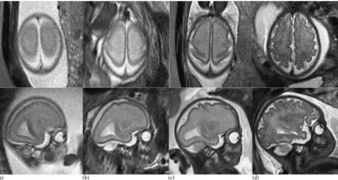
In the last two trimesters an immense macro- and microstructural change takes place in the neonatal brain. To visualise the macrostructural changes, the process of cortical folding can be displayed with fetal MRI. The following figure depicts the development of the primary sensory and motor cortex. [19]
Figure 3.2.1. Fetal development of primary sensory and motor cortex. Axial (upper row) and parasagittal (lower row) T2-weighted sequences of fetuses at 20 GW (a), 25 GW (b), 27 GW (c), and 32 GW (d). Beginning with 25 GW, the central sulcus can be delineated as a slight indentation of the posterolateral aspects of both hemispheres.
The microstructural changes during the second and third trimesters include the formation of synaptic connections and axonal ingrowth leading to cortical folding and grey matter formation. Altough most cortical neurons have migrated in the first trimester, neurogenesis continues in the superior ventricular zone and subplate neurons are at their peak of activity. Oligodendroglia in the subplate plays a role in myelin formation and produce guidance molecules for migrating neural cells. Preoligodendrocyte cells are susceptable to hypoxic injury, leading to maturation arrest of this cell population and
consequent myelination failure with accompanying white matter injury. [20] Finally in the third trimester maturation of oligodendroglial lineage and the initiation of myelination is completed and glial and neural cells migrate into their final position in the cortex. [21]
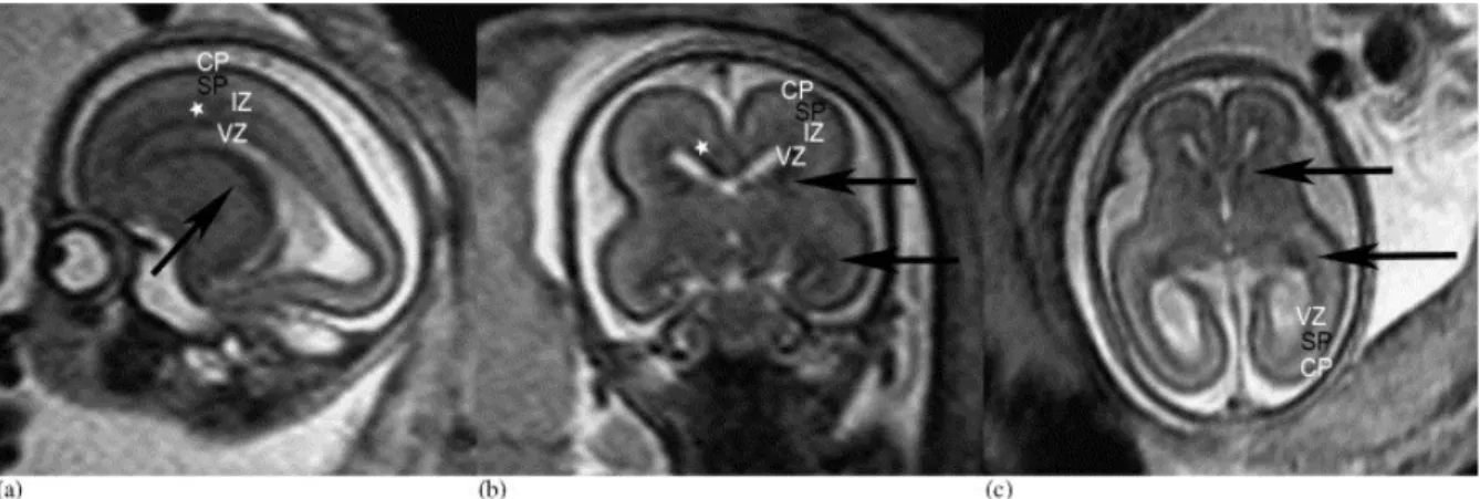
Figure 3.2.2. Sagittal (a), coronal (b), and axial (c) T2-weighted sections of a 20 GW fetus. The black arrows indicate the hypointense signal in the superior and inferior parts of the ganglionic eminence. The laminar organization of the fetal brain is visible—the hypointense cortical plate (CP), the hyperintense signal of the subplate (SP), the slim hypointense intermediate zone (IZ), and the hypointense ventricular zone (VZ). The subventricular zone can also be identified in the frontal regions (asterisks). [19]
3.2.2. Functional Overview and Plasticity
During the first trimester, sensory- and motor pathways fully develop in the fetus.
Complex functions such as circadian rhytmus and sleep organisation, start to function in the second trimester although responsible structures such as thalamus and brainstem are already functioning previously. [22] Resting state MRI examinations provide data that these networks are not only present at the second trimester, but also show different maturational patterns in different brain regions. Infant fMRI studies have shown that neural networks cover greater anatomical distances later in development and that the most developed functional connectivity appears to exist in regions related to sensation and action. [23] Other studies have demonstrated that preterm children and adolescents have decreased cortical gray matter, cortical white matter, deep gray matter, cerebellar and total brain volumes when compared to age-matched term control subjects. [24-28] Diffusion tensor imaging studies have demonstrated microstructural changes in both the corpus
callosum and those intra-hemispheric association fibers subserving language skills. . [29]
Interestingly in contrast to these data documenting aberrant brain development, behavioral studies suggest a pattern of recovery and preserved performance. [30]
Preterm children show a stronger neural circuit between Wernicke’s area and alternative language regions in passive language tasks using fMRI when compared to term controls at 8 years of age, suggesting alternative brain development and plasticity of neural processing. [31]
Figure 3.2.2.1. Premature Brain at Term. . Inder TE Pediatrics. 2005 Feb;115(2):286 As it is shown in the above figure, premature infants present smaller brain volumes, less cortical folding and narrower corpus callosum. [32]
The work of Staudt shows that sensorimotor reorganization with congenital hemiparesis is dependent on the time of injury during brain development. Ipsilateral corticospinal networks were further developed and the extent of motor dysfunction was smaller in case of early neonatal injury when compared to later injuries. This example demonstrates, that the efficacy of reorganization with ipsilateral corticospinal tracts indeed decreases during pregnancy. [33]
3.3. Neurophysiological methods in the neonatal period
Neonatal application of clinical neurophysiology has a steadily increasing role in the daily practice of neonatal intensive care units and follow up clinics. The importance of neurophysiological methods, mainly conventional EEG are recognised not only in epileptic syndromes and seizure diagnosis with treatment monitoring in intracranial pathologies and malformations, but also in the assessment of structural maturation and neural development.
In the assessment of the severity of neonatal encephalopathy and its prognosis conventional EEG and amplitude integrated EEG (aEEG) has proved to be an important an established tools for clinical decision makers.
Evoked potentials are electrical potential changes of sensory receptors, neural pathways and the brain following external or endogenous stimuli. Evoked potentials have attracted less interest in the neonatal population due to the lack of standardised maturational data in extreme premature infant and the difficulty of the measurement in the unstable patient treated mainly under intensive care. Nevertheless they offer precise answers to specific questions of neurological functions in the neonate. The following sensory evoked potentials are used occasionally: VEP-s in visual function analysis, AEP-s in auditory screening, SSEP in intraoperative monitoring. Event related potentials (ERP-s) like Miss Match Negativity (MMN) with P300 are used mainly in neuroscientific and cognitive developmental research. Brainstem auditory evoked potentials (BAEP) have shown a positive correlation with diffusion changes in MRI in the pons and impaired later neuromotor outcome. [34]
3.3.1. The role of neurophysiological methods
There is a growing need for continuous brain monitoring on the NICU, as adverse effects of everyday intensive therapy, infections, and immaturity present a risk for developing brain injuries and consequently influencing long term neurodevelopmental outcome.
Conventional EEG was the first method used successfully in the neonatal population. Due to increasing survival rates especially among the very premature population ( <28 weeks of gestation) the prevention from later neurological deficit currently becomes even more important. Despite recent advances in perinatal care the incidence of impaired outcome in preterm infants has not decreased. Rates of cerebral palsy and overt cerebral lesions (cystic periventricular leukomalacia and peri/intraventricular hemorrhage) are decreasing, but the incidence of neurodevelopmental impairment remains high in preterm infants. This is explained by the understanding of different mechanisms in brain injury (for example inflammation, oxidative stress, impaired connectivity) and result in mainly cognitive impairment.[35, 36] A quantitative analysis of studies published between 1998 and 2008 shows that very preterm babies have moderate to
severe deficits in academic achievement, attention problems, internalizing behavioral problems and poor executive function compared to controls.[37] These issues are strongly associated with cognitive impairment. Therefore greater attention needs to be directed toward preterm neonatal populations to better understand brain adaptation both with and without medical complications. Neurophysiologic surveillance is necessary in these infants to adequately asses cerebral function and is difficult within this population by clinical aspects only. Conventional EEG is today´s gold standard for neurophysiologic diagnosis.
Nevertheless it is not suitable for continuous recording since producing large data volumes which cannot be assessed directly at the bedside. In an effort to solve this problem, various methods of reducing and compressing the EEG signal have been developed, the amplitude- integrated EEG (aEEG), being one of them.
3.3.2. Conventional EEG
The electroencephalogram (EEGor conventional EEG) is a signal recorded from scalp electrodes and derived from the electrical activity of cortical neurons. The EEG signal represents the synchronious activity of neurons arranged at right angles to the surface, mainly the pyramidal neurons. The EEG changes through the neonatal period and childhood, thus it is essential to compare EEG measurements with normal values at the same maturational stage. EEG should be interpreted for background pattern, reaction to external stimuli, level of consciousness and for presenting pathological phenomena.
Conventional visual classification of the EEG signal of different brain regions has been the standard of analysis since the 1960s when first neonatal recordings were performed. Today more than 80% of extremely premature infants between 24-28 weeks of gestation survive. Therefore in the analysis of EEG signal there has been a growing need for more reliable automatic methods, being suitable for this specific population. There are several algorithms that have been developed in recent years, with different mathematical models to analyse not only the amplitude and frequency, but also the phase synchrony, coherence and temporal profiles of the EEG signal. [38, 39]
New nomenclature has emerged specifically for the premature population such as spontaneous activity transients (SATs), which constitute the most salient feature on EEG during the preterm period. This work has been based on animal models, showing that the characteristic discontinuous pattern found in premature infants is a universal phenomenon
during development in different species. These spontaneous bursts of activity, which are related to the excitatory role of GABAergic transmission during early development not only characterize the premature EEG, but also have been linked to the development of intracortical connections and neuronal wiring. SATs constitute of a very slow 0,1-0,5 Hz, with nesting activity at several higher frequencies. This activity represents the organization and development of thalamo-cortical connections, when neurons migrate from the subplate into the cortical plate in the primary sensory cortices. [40] SATs are shown to be useful in the everyday Neonatal Intensive Care Unit setting giving reliable information about the current clinical state and outcome of premature infants. [41] The development of sleep- wake-cycling is one of the ontogenetic oldest mechanisms of the developing brain and shows the integrity of a normal brain during development.
Pathological patterns should be differentiated as localised or generalised events of the recorded EEG. Seizure detection and localisation is the most common indication of neonatal EEGs, although research suggests that the electroclinical correlation of seizures are very poor in the neonatal population. Murray and Boyle suggest that only third of neonatal EEG seizures display clinical signs on video EEG recording and moreover only 60% of these are recognised at all. [42] They point out that unrecognised seizure burden is a serious problem, as untreated seizures can cause apoptosis of nerve cells and impaired neurogenesis suggested by animal research data.[43]
The seizures can be detected using conventional EEG in children presenting with IVH and the following hydrocephalus. [44] It is also usefull in case of subarachnoideal bleeding, metabolic diseases and most commonly in hypoxic ischaemic encephalopathy.
[45]
3.3.3. Amplitude Integrated EEG
For early identification of infants at high risk and to optimize treatment, it is mandatory to have access to a reliable validated diagnostic method with excellent predictive value for later neurodevelopmental outcome. The aEEG is a readily available, informative and reliable technique for continuous non-invasive monitoring of brain activity even in extremely premature infants. Amplitude-integrated EEG is a simple method for continuous bedside monitoring of neurophysiological parameters in the neonatal intensive care unit setting. As our group has recently shown, aEEG has a predictive value for later
outcome in preterm infants and can therefore be used as an early prognostic tool for neurodevelopmental outcome. [46]
We have found emerging sleep-wake cycles as early as 24-25 weeks of gestation in neurologically healthy premature infants. [47] On the contrary premature infants with intraventricular hemorrhage exhibited a significant delay in emergence of their sleep-wake cycles, on average with 32 weeks of gestation. [48] We know that at this early age the development of intercellular connections of the brain and synaptic branching is still in development and that these processes take place mainly during sleep. [49] Consequently, the early emergence of sleep-wake cycles is the sign of normal brain development at this early age. On the other hand the earlier regular sleep-wake rhythm would lead to a better synaptic development and wiring resulting in even better later neuro-developmental outcome. Kostovic et al. shows, that neurocognitive outcome depends on the active connection of different brain circuits during prenatal and early postnatal life. The period between 24 to 28 weeks of gestation in extremely premature infants is a very important time, because not only maturation but also the migration of the neural cells are still going on. [46]
3.3.4. Visual Evoked Potentials
Visual Evoked potentials can be evoked by brief changes either in luminance (flash visual evoked potentials fVEP) or in the contrast (pattern VEP, pVEP) within the visual field. As there is no need for fixation to evoke the fVEP and luminance changes are detected through the closed eyelids as well, it is an optimal method to use in the non- cooperative neonatal population. VEP waveforms are labeled according to their polarity and the mean latency. Adult pVEP have three peaks N70, P100 and N145. fVEPs have I- VI. peaks, where the most reliable components are peak III (corresponding to N70) and peak IV (corresponding to P100). Maturational data is aviable for premature infants and children for both VEPs. P100 a positive Wave at 100ms emerges soon after birth.
VEPs assist in the diagnosis of varios optic nerve disorders, such as optic neuritis, optic nerve atrophy, hypoplasia, tumors and compression and play an important part in the diagnosis of multiple sclerosis. They permit early identification of disfunction and can be used to monitor progress. VEPs were found to be useful in the neonatal population with regard to prediction of neurodevelopmental outcome. [50-52]
.They seem particularly helpful in patients with PHVD as the visual pathway is adjacent to the lateral ventricles. [53] Therefore, an increase in ventricular width seems to lead to an early compromise of fVEP values.
VEP have shown to be of predictive value in detecting increasing intracranial pressure in children as VEP latencies increased with the increase of intracranial pressure and normalised after neurosurgical intervention. [54]
3.3.5. Event Related Potentials
Event related potential (ERP) is a response to a stimulus, where from many trials the results are averaged together, causing random brain activity to be averaged out and the relevant waveform to remain. Currently, ERP is one of the most widely used methods in cognitive neuroscience research to study the physiological correlates of sensory, perceptual and cognitive activity associated with processing information. Event-related potentials are caused by the "higher" processes of deeper brain structures or associative cortical areas, that might involve memory, expectation, attention, or changes in the mental state, while evoked potentials are resposes of cortical areas to sensory stimuli.
Mismatch negativity (MMN) is an event-related potential (ERP) component that provides a good measure of auditory perception and function and is typically observed between 100 and 250 ms. [55, 56] MMN is generated by the automatic response of the brain to a mismatch in auditory stimulation. It is elicited when a deviant stimulus (e.g., with a probability of 15%) appears within a train of repeatedly presented standard stimuli (e.g., with a high probability of 85%). The MMN is observed irrespective of the subject's direction of attention and is a good measure of the auditory system's ability to detect differences between sounds. [57] It is often used in experimental psychology in non- cooperative subjects, such as infants and young children..
3.4. Imaging of the neonatal brain
3.4.1. The role of imaging methods
Preterm infants are at high risk of developing brain injury. Neuroimaging does not only play an important role in prognosticating later neurological problems, but provides also essential information and support for the neonatologist in clinical decision-making in
critically ill neonates. MR imaging of the premature infant has been proved to be superior to the widely used serial ultrasonographic examinations. [58, 59] In one study MRI has enabled a non-invasive high resolution evaluation of the developing brain, where several studies have shown delayed grey-white matter differentiation, and diffuse white matter signalintensities after premature birth. [60] They also present a smaller corpus callosum, less mielinated white matter, larger ventricles, altogether smaller global and local brain volumes, when compared to healthy controls. [21]
Sonography is the method of choice for prenatal and postnatal malformation screening but it does not always provide sufficient information for correct diagnosis, adequate abnormality evaluation or consequent outcome information. Fetal magnetic resonance imaging (MRI) is considered as a valuable second line imaging tool after sonography for confirmation, completion and correction of regular ultrasound findings.
[61] Fetal MRI has proven its value in the evaluation of central nervous system pathologies, especially of midline and posterior fossa malformations. Special sequences has been developed for this non-sedated, ever moving population. [19]
3.4.2. Magnetic Resonance Imaging
Specific sequences have been adapted for premature infants and newborns from research tools to routine imaging protocolls. Regular protocols including fast spin-echo T1/T2-weighted (w) sequences with long repetition-time and echo times in 3 section planes are part of routine protocolls of radiologists studying the neonatal brain and are useful mainly in the study of brain anatomy, malformations, bleeding and hydrocephalus.
Diffusion tensor imaging (DTI) enables the study of establishment of brain connectivity and plasticity. It is based on measures of water diffusion in biological tissues and is a powerfull tool to study white matter development and abnormalities. Fractional anisotropy (FA) can be quantified in different brain regions and show correlations with neurodevelopmental outcomes. FA values obtained in preterms at term-equivalent are lower in the right posterior limb of the internal capsule and at the splenum of the corpus callosum in case of developing cerebral palsy with two years of age. [62] Fibre tracking a voxel based analysis of DTI, enables the presentation of white matter tracts and their connectivity. A number of studies using DTI to visualize white matter tracts in neonates with white matter injuries and in older children with cerebral palsy have been published
with promising results. One study of 24 infants with birthweight below 1500 g who had DTI at 37 weeks postmenstrual age found a strong correlation between low fractional anisotropy values in the posterior limbs of the internal capsule and both diagnosis of cerebral palsy and severity of gait problems on outcome evaluations at 4 years. [63]
Functional MRI (fMRI) is a novel method in newborns, but is a promising research tool in neural processing and resting state connectivity. It refers to regional changes in signals that correlate with brain functional activity. It uses deoxygenated haemoglobin levels or otherwise known as BOLD (blood oxygenation level dependent) signals, which indirectly depicts regional activity.[64] Neonatal Current research concentrates on the development of neonatal brain networks with the use of resting state activity in order to understand maturation in a normaly developing fetus. [65]
Proton magnetic resonance spectroscopy of the brain is a non-invasive technique that supplies information about the presence and levels of metabolites, such as N- acetylaspartate (NAA), choline (Cho), creatinine (Cr) and other substances. They provide usefull information in metabolic diseases, neurodegenerative disorders and in neonatal encephalopathy as well.[66]
3.4.3. Cranial Ultrasound
Serial head ultrasounds are a valuable bedside tool for following brain development and and occuring intracranial pathologi in even the sickest preterm infants. Two studies have demonstrated that many preterm infants have a reduction in the size of the corpus callosum at term (compared with term controls); this is associated with lower gestational age at birth and with cerebral palsy and lower cognitive scores [67]
Blood velocities of cerebral arteries are especially usefull in hypoxic ischemic encephalopathy. Serial cranial sonographic examinations are part of the daily routine on the neonatal ward for the detection of common brain pathologies such as intracranial bleedings, PVL, PHVD and neonatal encephalopathy. IVH is classified according to Papile [68] PHVD is classified according to the ventricular index of Levene and recently an additional anterior horn width (AHW) and thalamo-occipital distance (TOD) helps better evaluation [69] Neonatal encephalopathy is classified according to the classification of Deeg et al.
Sonography has its limitations as well. De Vries et al. showed that the sensitivity of sequential ultrasound imaging for detecting abnormalities to predict later cerebral palsy was 76%, while Miller et al. described, that, although the positive predictive value of acute white matter injury was high, the sensitivity of these findings were low among premature infants.[70, 71] Further analysis with MRI is needed to clarify the extent of neuropathology found on ultrasound examinations.
3.4.4. Computer Tomography
Computer Tomography (CT) has a very limited role in neonatal brain imaging. As it is a major radiation burden for the developing brain, it is only used in emergency situations, when MR imaging would take too long to organize. Neonatal cranial CT examinations are still in use in case of subarachnoideal or subdural bleeding with progressive brain oedema or head trauma with skull injury and need for immediate neurosurgical intervention.
3.5. Common morbidities of the central nervous system during the neonatal period
3.5.1. Intraventricular haemorrhage
Intraventricular haemorrhage (IVH) is still the most common reason for brain injury in preterm infants. Additional progressive posthaemorrhagic ventricular dilatation (PHVD) is known to be associated with subsequent white matter damage and therefore increases the risk for neurodevelopmental disability furthermore. [72]
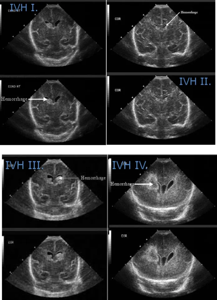
It is most commonly found in the preterm infants and the incidence is between 15- 20% under 32 weeks of gestation. In the rare cases in term infants IVH is related to birth trauma and originate from the choroid plexus, while in preterms the fragile involuting vessels of the subependymal germinal matrix and their rupture are responsible for the bleeding. Sudden changes in cerebral venous pressure, due to hypervolemia, hypoglycaemia, pneumothorax or seizures also contribute to IVH. [73] The following cranial ultrasound images of figure 3.5.1. show the different grades of IVH.
Figure 3.5.1.Cranial ultrasound images of different grades of IVH. White arrows show the heamorrhage at the pictures. IVH I-IV is presented from own patient population.
3.5.2. Posthemmorhagic ventricular dilatation
Hydrocephalus after intraventricular hemorrhage is as a major complication of preterm birth and is particulalry problematic to treat. The pathophysiology of hydrocephalus is usually ascribed to fibrosing arachnoiditis, meningeal fibrosis and subependymal gliosis, which impair flow and resorption of cerebrospinal fluid (CSF). IVH is associated with damage to periventricular white matter and the damage is exacerbated by the development of hydrocephalus; combinations of pressure, distortion, ischaemia, inflammation, and free radical-mediated injury are probably responsible. The damage to white matter accounts for the high frequency of cerebral palsy in this group of infants. 25%
of infants with IVH develop a progressive hydrocephalus and the incidence increases with severity. Clinical signs of hypertension are not always reliable defining severity of PHVD, imaging and neurophysiological examinations help to determine management and prognosis.
3.5.3. Asphyxia
Perinatal asphyxia is a condition of the neonatal brain due to hypoxemia and hypercarbia, due to impaired gas exchange. Characteristicly infants present with abnormal neurobehavior (defined by Sarnat and Sarnat), seizures, impaired vertebral blood flow.
Additional complications of neonatal encephalopathy include a multiorgan dysfunction with mostly renal, cardial, pulmonary and hepatic abnormalities. Despite joint efforts of neonatologists and obstetritians the incidence has been stable between 0,5-2%, with 10- 20% death and 25-50% neurodevelopmental disability in the surviving population.
Additional hematologic problems require a complex intensive management of these patients. Cranial ultrasound is an essential tool of bedside monitoring, but MRI has proved superior defining the extent of white matter abnormalities, restricted diffusion reflecting cytotoxic edema, or spectroscopy measuring metabolite ratios, such as Cho/Crea or NAA/Crea or lactate levels. aEEG has been proved to be a good outcome predictor when carried out within 48 hours after birth. [74]
3.5.4. Periventricular leukomalacia and diffuse white matter injury
Periventricular leukomalacia is a common injury of the preterm population with necrosis, gliosis and disruption of axons at border zones of vascular supply. Hypoxia- ischeamia is followed by cystic necrosis and diffuse white matter abnormalities with
hypertrophic astrocytes and loss of oligodendrocytes resulting in overall decrease of cerebral volume. The incidence of large cystic PVL decreases, but diffuse white matter injury remains significant, resulting in 10% CP and 50% mild disabilities. [75]
3.6. Neurodevelopmental Outcomes
Preterm children compared to term controls present with a variety of neurodevelopmental problems. At 6 years of age 30-40% have minor developmental impairment and 20% have major disabilities. Among these major disabilities 42–47% of children had cerebral palsy, while 27%, 37% and 23% of these children had a significant cognitive, visual and hearing impairments, respectively. [76] An increase in cerebral palsy with decreasing birthweight and gestational age category is a consistent finding in preterm outcome studies. This finding is not limited to extremely preterm infants. [75] EPIPAGE study reports an increase in cerebral palsy with each preterm week: 0.7%, 3.7%, 4.1%, 8.7% and 6.3% in children with gestational age 34, 33, 32, 31 and 30 weeks, respectively (P < 0.01).The intellectual deficit of preterm children present also in adolescent and young adulthood. [77] The study from Walther also shows that mortality and major handicap are relatively stable over time during follow up, but minor disabilities and developmental problems show a substantial increase over time.
Neurodevelopmental outcome measuremets have traditionally several subgroups, such as motor- and cognitiv outcomes, sensory impairment, -and language skills. Although many preterm infants demonstrate neuromotor abnormalities on examination, most do not develop cerebral palsy, but mild persistent neuromotor abnormalities (e.g. asymmetries, tight heel cords), motor planning problems and/or sensorimotor integration problems that lead to functional impairments (e.g. tying shoelaces), academic difficulties (e.g. writing).
[76]
Recent studies have not only confirmed that children born preterm have more cognitive impairments and academic difficulties than fullterm controls, but they also suggest that these are more common than motor, visual or hearing impairments, as they apper up to 55% of premature infants under 27th week of gestation. [78]
Difficulties with reading and spelling increased with decreasing gestational age (and birthweights) in a Danish study of 11–13 year olds. [79] They found significant differences between children born at 33–36 weeks (and at 37–38 weeks) compared with
children born at 39–40 weeks gestation, which points out the importance of risk factors for the late preterm population as well.
4. Objectives
The author of this paper would like to present through her research different aspects of neonatal neurophysiological examinations and neuroimaging methods, in order to better understand the development of neonatal brain pathologies and their effect on neurodevelopmental outcome.
4.1. Feasability of Functional Neurophysiological Methods and optimalization of neonatal care
4.1.1. Hydrocephalus Study
The aims of the present prospective study were firstly to evaluate the role of VEPs and aEEG in the monitoring of elevated intracranial pressure in congenital hydrocephalus and the development of PHVD in preterm infants and to define pattern changes with decompressing neurosurgical interventions. The second aim was to correlate our findings with the degree of ventricular dilatation and Doppler sonography. Thirdly, we wanted to compare late versus early intervention with the primary outcome, the need for long term ventriculoperitoneal shunt insertion. Our hypothesis was that both neurophysiological methods show changes in case of intracranial pressure elevation or normalisation.
4.2. Optimalisation of Cerebral Imaging methods
4.2.1.MRI-compatible incubator Study
Aim of our study was to assess our initial experience with an MRI-compatible incubator and analyse its impact on examination feasibility and further clinical management. In order to acheav this we first analyzed the use of MRI itself in unstable patients under intensive care management and secondly the usefulness of a special device (MRI-compatible incubator) making MRI examination in unstable patients more feasible.
Because the main advantage of the MRI-compatible incubator is optimizing thermoregulation during the MRI, we separately analyzed the usefulness in patients weighing under 2000g.
4.3. Use of functional neurophysiological or imaging methods and prediction of outcome
4.3.1. Asphyxia Study
The aim of our retrospective study was to analyse early versus late MRI and aEEG data separately and correlate its effect on the prognostic outcome of children with asphyxia at two years of age. Secondary aim was to combine aEEG and MRI data in order to develop a more exact prognostic value for this patient population. Our hypothesis was that neurodevelopmetal outcomes are predictable safely with the use of aEEG and there will be a difference between late versus eraly measurements. The second hypothesis regarding MRI was that it has a stable predicitve value independent of the timing of the measurement.
4.3.2. Mismatch Negativity Study
The aim of the Mismatch-Negativity (MMN) study was to test the maturation of phoneme and stress discrimination in case of natural speech in premature infants and healthy controls at 6 and 10 months of age. The main principle along which we planned our experiment is to investigate the typical stress information at the word level. Hence we used a complex pattern of acoustic cues while varying stress information using the miss match negativity paradigm of event related potentials. Our hypothesis was that we will find differences between the two age groups in case of both phoneme and stress detection, but preterm infants will show maturational lag only in case of stress processing.
4.3.3. Intraventricular Haemorrhage Study
The aim of this prospective study was to compare outcomes of preterm infants with different grades of IVH born below 32 weeks of gestational age (GA) with outcome of controls without IVH. Emphasis was on the comparison of the influence of low grade IVH on the neurodevelopmental outcome. Cranial ultrasound examinations were carried out on the 1st3rd, 5th, 7th, and 10th day of life and then once a week until discharge. Our first hypothesis was that gestational age has an effect on neurodeveopmental outcome in case of
IVH. The second hypothesis was, that neurodevelopmental outcome correlates with the severity of IVH in preterm infants.
5. Methods
5.1. Hydrocephalus Study
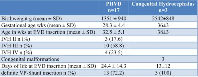
Patients who developed a posthaemorrhagic hydrocephalus and also required neurosurgical intervention were eligible for inclusion. Subjects with IVH received serial CUS scans every second day. PHVD was defined as the progressive increase of ventricular width following IVH as seen on CUS. In the case of PHVD, VEPs and aEEG examinations were performed at least once weekly before and after neurosurgical intervention in order to follow both the development of ventricular dilatation and the reduction of ventricular width after insertion of an external ventricluar drain (EVD) or implantation of a ventriculo- peritoneal shunt (VP-shunt). During the study period 17 patients met the inclusion criteria.
In all cases we were able to perform fVEPs and aEEGs prior to and after placement of CSF drainage systems.
In a further analysis the PHVD study population was compared to patients with congenital hydrocephalus. An additional 3 patients were included, and followed similarly with VEPs, aEEG and CUS until neurosurgical intervention. The underlying pathologies resulting in congenital hydrocephalus were, 1.rhombencephalosynapsis, 2.teratoid tumor of the neck compressing the fourth ventricle and 3.subcortical heterotropies with partial corpus callosum agenesis and aqueductal stenosis.
Cerebral ultrasound scans were performed on days 1, 3, 5, 7, and 10 of life and then once a week until discharge by using an Acuson 128XP (Mountainview, CA) with a 7.5-MHz transducer. IVH and periventricular leukomalacia were classified according to Papile and de Vries et al, respectively. [68, 71] PHVD was classified according to the criteria of the ventricular index to Levene (Ventricular width was measured in the coronal plane from the lateral wall of the body of the lateral ventricle to the falx), and a neurosurgical intervention of external ventricular drainage was latest performed if the ventricle was wider than 4mm above the 97th percentile. [80] Additionally, anterior horn width (AHW) and thalamo-occipital distance (TOD) (according to reference values by Brouwer et al. ) were evaluated.[81] [69] Using Doppler sonography, blood flow velocities and the resistance index were measured in the anterior cerebral artery. [82]
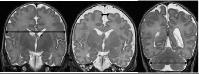
Figure 5.1.2.1. A) Measurements of the anterior horn width (AHW), the maximal diagonal width of the anterior horn, the ventricular index (VI), the distance between the falx and the lateral wall of the anterior horn and the frontal horn ratio, the ratio between the VI and corresponding hemispheric width, in the coronal plane at the level where the AHW appears maximal. (B) Measurement of the thalamo-occipital distance.
Congenital hydrocephalus was followed similarly and ventricular index, TOD and AHW was calculated. In case of these 3 patients an additional MRI has also taken place to define the extent of intracortical malformations.
Flash visual evoked potentials fVEP measurements were performed weekly in infants with developing PHVD and congenital ventricular dilatation. As soon as the ventricular index reached the 97th percentile recordings were performed twice weekly until neurosurgical intervention was performed. fVEPs were recorded using the Neuropack 8 (Nihon Kohden). The fVEP measurements were done in closed cots or open-air units, both covered with a blanket in order to create a semidark environment. The stimulating source was a red light emitting diode goggle held at a distance of 5 cm in front of the infant’s eyes. The evoked potentials were recorded using three cortical electrodes placed on the infants scalp (active electrodes at Oz and Fz, ground electrode at Cz according to the international 10/20-system). The stimulation frequency was 0.5 Hz, the electrical impedance below 5 kOhm and the emitted light energy was 0.4 Lux. Two courses aiming for 30 and 50 responses were averaged using a band pass filter of 1–100 Hz and a sweep time of 1 sec. Responses including excessive artefacts were automatically rejected and trials were performed together on both eyes (binocular). fVEP measurements were recorded during active sleep, determined using the simultaneously recorded aEEG
background pattern and the assessment of the behavioural state of the infant. [83]
Waveforms and latencies were then analysed off line for every measurement. Reproducible positive and negative waves were named according to the order of their appearance, N0, N1, P1, N2, P2 and N3 and were compared with the reference values published by Pike et al. [84]
Figure 5.1.2.2. The evolution of common fVEP waveform during maturation.
Results from Pike et al.
Figure 5.1.2.3. The light emitting diode goggle is held in front of the eyes of the sleeping infant for binocular stimulation. Photo of a participant in our hydrocephalus study.
Amplitude-integrated EEG At the same time as fVEP measurements were performed, aEEG was recorded as a single-channel EEG from biparietal surface disk electrodes using a cerebral function monitor (Olympic Cerebral Function Monitor 6000).
In brief, the obtained signal is filtered, rectified, smoothed and amplitude-integrated before it is written out at slow speed (6 cm/h) at the bedside. [85] Tracings were evaluated visually and classified according to the method previously described by Hellström-Westas et al. [86]
Descriptive analysis of the background activity of the aEEG tracings was done by dividing each trace in 10 min epochs and by calculating percentages of occurrence of the different patterns. Appearance of sleep-wake cycling (SWC) and seizure activity was noted within the entire recording. aEEG pattern was then scored according to the following:
1. Background activity (age-adequate distribution of pattern according to reference values previously published) [46, 69, 87]; a value within 25th and 75th percentiles for
every pattern was classified as ‘age-adequate’[69] 2. Appearance of SWC. 3. Presence or absence of seizure activity.
‘Normal aEEG-pattern’ (=score 0) was defined when all three categories were classified as normal, ‘moderately abnormal aEEG-pattern’ (=score 1) was defined when 1/3 categories were classified as abnormal and ‘severely abnormal aEEG-pattern’ (=score 2) was defined when 2 to 3/3 categories were classified as abnormal.
Conventional EEG with video was performed when seizure pattern was seen on aEEG. The aim of the EEG examination was to locate epileptogenic activity more precisely and to define seizure chracteristics, such as duration, generalisation, waveforms and electroclinical correlates. The System 98 software from Micromed was used, with using the neonatal version of 10/20 system with 8 electrodes for premature infants and 20 electrodes for term infants.
5.2. MRI-compatible Incubator Study
In a retrospective study we analysed the clinical and imaging data of neonates undergoing MR Imaging between 2003-2007. Our study population included all 129 premature and newborn infants during two consecutive time periods, undergoing MRI examinations at the Medical University Vienna, Austria. The first 18 months study period was between June 2003 and January 2005 and the second 18 months period with the MRI- compatible Incubator (INC) was between June 2005 and January 2007. The criteria of critically ill infants included one or more of the following: need for ventilation, the first day of life, unstable infant with bradycardias, desaturation and unstable blood pressure.
Subanalysis of data of infants with a weight below 2000g during MRI examination is given separately.
The premature infants were placed into the INC on the NICU (approximately 10-20 min before examination was scheduled), then transportation to the radiology department of our clinic, which is located in another building. Transport time is approximately 10-15 min and although all MRI examinations are previously arranged, waiting times vary between 0 and maximal 30 minutes. In the first time period the transport incubator was used with similar transport and waiting times, but in this case an extra 10-30 minutes were added to the whole process, while the infants had to be stabilized in the MRI when repositioned.
The criteria for change in management after MRI examination was: starting, or finishing a medical therapy due to the result (anticonvulsive drugs, metabolic supplementation or special diet, antithrombotic or antibiotic drugs), or changes in clinical practice after imaging, such as initiation of surgical intervention or postoperative decision- making.
The criteria for change in the ultrasound diagnosis was unconfirmed diagnosis or additional diagnostic information provided by the MRI examination. Infants needing ventilation were hand ventilated with an Ambu-balloon during the MRI examination in the first period, while in the second period the Pneupac babyPAC 1000 ventilator was used.
For monitoring the Invivo Precess (MeMed-Menges) was used in the first and the Invivo 4500 MRI Monitor in the second period. Both monitor and ventilator are integrated into the INC.
The same Philips Intera 1,5 Tesla (Philips-Best-The Netherlands) MRI System was available in both periods for the imaging of premature infants. Before the availability of the INC we used a birdcage knee coil; with the INC an in-built incubator head coil was used. For both coils the same sequences were used. Routine protocols including fast spin- echo T2-weighted (w) sequences with long repetition-time and echo times in 3 section planes, axial T1w spin-echo sequences, sagittal T1-w 3DGradient-echo sequences, diffusion w sequences. (standard newborn T2-weighted TSE sequence with TR 300ms, TE 140ms, duration 1.21 min., FOV 120, slice thickness 3mm, = gap maximum slices 28, Matrix 108x108, acquired voxel size 0.69/0.81/3mm, reconstructed voxel size=
0.43/0.4373mm). Angiography and spectroscopy were added in some cases. Diffusion- tensor imaging was done in 50% of the babies examined in the INC. Sequences, adapted from fetal protocols were done in cases of severe instability.7 Imaging time was calculated from the beginning of the first sequence until the end of the last sequence.
MRI-Compatible Incubator
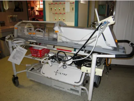
The MRI-Compatible Incubator from LMT MR Diagnostic Incubator Nomag IC 1.5 has been designed to provide a safe environment for the critically ill and very low weight premature infants with their special needs. (Figure 5.2.2.) The temperature and humidity regulators, the MRI compatible monitors and the ventilation support system are all necessary for the stability during transport and imaging of this patient group. The built
in head coils and auditory shielding have improved the imaging process for both these small patients and the radiologist. The advantage of the in-built head coil is that its surrounds the infants head completely, leading to a better signal in the parietal regions of the brain.
Figure 5.2.2. MRI Compatible Incubator used in our study. Ventillator and built in head coil are important part of the system.
5.3. Asphyxia Study
In a retrospective analysis we selected premature and term infants who developped a hypoxic ischeamic encephalopathie (HIE) between 2003 and 2006 and were admitted to the Neonatal Intensive Care Unit (NICU) at the Medical University of Vienna. Inclusion criteria included all infants with asphyxie, defined as 1) Apgar Score below 5 at one minute or 7 at 5 minutes, 2) cord pH below 7,0, Exclusion criteria were metabolic disorders, congenital malformations and genetic abnormalities. 142 participants met this criteria during the 4 year period. There was no hypothermia treatment avaiable at this time point on our neonatal ward. Neurodevelopmental outcome data at two years of age was collected at our follow up clinic. Further selection included only those patients who also underwent an MRI examination in the perinatal period (within 6 weeks after birth). Altogether 44 patients met this criteria.
Clinical and epidemiological data was collected. HIE was classified according to the clinical –neurological status by Sarnat. The severity of Sarnat stadiums range from light, to moderate, and severe. (I-III.) [88]
I. Mild HIE – Sarnat Stage I:Hyper-alert,Eyes wide open,Does not sleep,Irritable,No seizures Usually lasts < 24 hours
II.Moderate HIE – Sarnat Stage II: Lethargy (difficult to rouse), Reduced tone of the extremities and/or trunk, Diminished brainstem reflexes (pupil/gag/suck), Possible clinical seizures
III.Severe HIE – Sarnat Stage III: Coma (cannot be roused), Weak or absent respiratory drive, No response to stimuli (may have spinal reflex to painful stimuli), Flaccid tone of the extremities and trunk (floppy), Diminished or absent brainstem reflexes (pupil/gag/suck) Diminished tendon reflexes EEG severely abnormal (suppressed or flat EEG with or without seizures)
Neurophysiological examination
Routine neurophysiological monitoring included continuous aEEG data collection within 6 hours of birth until the third day of life, after this period routine aEEG examinations were on weekly basis, unless clinical status indicated otherwise. Epileptic activity on aEEG or clinical signs of seizures were followed through with a conventional EEG with video for further analysis. The aEEG was recorded as a single-channel EEG from biparietal surface disk electrodes using a cerebral function monitor (Olympic CFM 6000). In brief, the obtained signal is filtered, rectified, smoothed and amplitude-integrated before it is written out at slow speed (6 cm/h) at the bedside. [87] The appearance of sleep- wake cycling, the occurrence of seizure activity and the distribution of background pattern was analyzed according to the previously published protocol by Klebermass. [46] Tracings were evaluated visually and classified according to the method previously described by Hellström-Westas et al. [86] Tracing were classified as 1. normal, 2. light, 3. moderately or 4. severely abnormal. The presence or absence of seizure activity was additionaly evaluated.
Magnetic resonance imaging
The Philips Intera 1,5 Tesla (Philips-Best-The Netherlands) MRI System was used.
Routine protocols including fast spin-echo T2-weighted (w) sequences with long
repetition-time and echo times in 3 section planes, axial T1w spin-echo sequences, sagittal T1-w 3Dgradient-echo sequences, diffusion w sequences. (standard newborn T2-weighted TSE sequence with TR 300ms, TE 140ms, duration 1.21 min., FOV 120, slice thickness 3mm, = gap maximum slices 28, Matrix 108x108, acquired voxel size 0.69/0.81/3mm, reconstructed voxel size= 0.43/0.4373mm) were carried out. Diffusion tensor imaging was added in all but one case and spectroscopy were added in some cases. Sequences, adapted from fetal protocols were done in cases of severe instability[19] We grouped the time of the MRI Scans as early scans, within the first week of postnatal life and late scan between 1-6 weeks of postnatal life. We used the scoring system from Barkovich et al to identify changes in signal intensity of different regions. Regions of interest were basal ganglia/thalamus, posterior limb of the internal capsule, cortex and white matter were analysed (PLIC). [89] The maximum score was 5 when in all regions and the additional diffusion abnormalities were present, while 0 score represented a normal MRI.
Neurodevelopmental outcome
Outcome was assessed at two years of age using the Gross Motor Classification System and the Bayley Scales Psychomotor and Mental Developmental Index. The Bayley Scales of infant development were classified as normal when psychomotor (PDI) and mental developmental index (MDI) scores were >85 (± 1 SD of reference values). (Bayley N (1993) Bayley Scales of Infant Development II. Psychological Corp, San Antonio.
Scheffzek scores were calculated according to neuroclinical status, in case of missing BS.
[90]
5.4. Mismatch Negativity-Study
In a prospective study to analyse language development in the first year of life in term and preterm subjects we recruited eighty-nine infants to participate in the experiment.
Fifty-two of them were excluded either because they did not match the strict selection criteria concerning age, birth weight, and gestational age (GA) characteristics, aiming to promote group homogeneity (n=12), or because of a low percentage of the artifact-free trials in the electrophysiological data due to infants crying, and/or frequent and excessive head and body movements(n=40) All preterm infants were recruited by using the database of the Follow up Center for Developmental Neurology, I. Department of Obstetrics and Gynecology, Semmelweis University, Budapest, Hungary. Normal cerebral ultrasound and hearing (oto-acoustic emission test) was the inclusion criteria. According to the ethical
requirements set by the Ethical Board responsible for permission we conducted the experiment at the clinic applying a portable EEG/ERP recording system (BrainAmp from BrainProductsGmbh.) after having the parents’ written informed consent. The full-term infants were selected with help of pediatricians from the Health Care Centre, Vezérutca, Budapest, Hungary. The circumstances and conditions of the EEG recordings were similar to those of preterm infants.
We created 4 groups of infants based on age, and the term of birth. We had two age groups, 6-month-old infants with 19 participants and one of 10-month-old infants with 18 participants. Criteria used as preterm birth were the GA of weeks 37 and/or birth weight
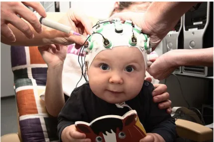
1500 gram. Consequently we had 21 preterm and 16 full term subjects. During the experiment babies sat on their parents lap, in a silent room. We used different toys as distractor stimuli in order to prevent the babies to pay attention to the acoustic stimuli. The experimental stimuli were presented via two loudspeakers placed in equal distance (40 cm) from the infants’head on the left and on the right side.
Figure 5.4.1. Young patients with an EEG cap before the experimental condition on the lap of his mother.
Stimuli and experimental conditions
We used a passive oddball paradigm with a standard Hungarian word `banán`
(`banana` in English) and two deviants: a voiceless phoneme deviant (`panán`, which is meaningless in Hungarian), and a stress deviant where the stress was on the second syllable, instead of the first which is a normal stress pattern in Hungarian (`ban:án`) (for all details see. [91] The experimental stimuli consisted of Hungarian words uttered by a native
female speaker and recorded and digitized by means of a personal computer with a sampling rate of 44100 Hz. The stimuli were presented in random order; the probability of the deviant stimuli was 25%, and the stimulus onset asymmetry (SOA) varied randomly between 730 and 830 ms.The two deviants were presented in separate series (150 standard and 50 deviant stimuli; the order of two series was counterbalanced)
Figure. 5.4.2. Derivation of MMN response. The evoked responses to the standard and deviant stimuli are extracted and the result is depicted as a negative curve called the MMN at two different electrode positions.
Data collection and measurement
The electroencephalogram was recorded from 16 scalp locations: F3, Fz, F4, C3, Cz, C4, P3, Pz, P4, O1, Oz, O2, T3, T4, M1, M2. The reference electrode was at point Fpz, and the ground was between Fz and Fpzon the midline. The offline data analysis was performed by using the BrainVision Analyzer software (BrainProductsGmbh.). The original EEG was algebraically re-referenced to the average activity of all electrodes.
Aband-pass filter of 0.3- 20Hz, 24 dB/octave was used.The raw EEG data was segmented into epochs of 800 ms, time-locked to the onset of the stimulus (-100 ms before onset to 700 ms after onset). Next, we applied an automatic artifact rejection method whereamplitudes above ±120µV were rejected. Participants whose recordings were below 20 artifact-free epochs were excluded from further analysis (see above).Then the segmented data was base-line corrected from -100 ms to the onset of the stimuli and finally, the remaining epochs were averaged.
5.5. IVH Study
The purpose of this prospective study was to compare outcome data of preterm infants with different grades of IVH with those of preterm infants without IVH in association to gestational age over a time period of twelve years. The main point of interest was the effect of low-grade IVH on neurodevelopmental outcome in this preterm cohort.
We included preterm infants with a gestational age below 32 weeks, who were admitted to the neonatal intensive care unit (Medical University of Vienna, Austria) between 1994 and 2005. Infants with additional periventricular leucomalacia and cerebellar lesions were excluded from the analysis. Neurodevelopmental assesment was carried out in our outpatient clinic. Demographic data include GA, birth weight, sex, antenatal steroids, multiple birth, mode of delivery and incidence of respiratory distress syndrome; chronic lung disease (defined as supplementary oxygen at corrected age of 36 weeks of gestation); patent ductus arteriosus (PDA) (defined as PDA needing treatment);
necrotising enterocolitis (NEC) (defined as any NEC≥Bell’s stage 1); amniotic infection syndrome or chorioamnionitis (defined as clinical and or histological signs of chorioamnionitis); posthemorrhagic hydrocephalus (defined as any ventricular dilatation after IVH); Shunt—necessity of implantation of ventriculoperitoneal shunt and retinopathy of prematurity (ROP) (defined as appearence of ROP of any grade). GA was determined from the date of the mother’s last menstrual period and according to antenatal ultrasound scans. For further statistical analysis, the study group was divided in two age groups (group I: patients born between 23+0 and 27+6 weeks of gestation; group II: patients born within 28+0 and 31+6 weeks of gestation.
Cerebral imaging to define the grade of intraventricular haemorrhage was conducted with a cerebral ultrasound (CUS) scan at the bedside by an experienced neonatologist. They were performed on days 1, 3, 5, 7, and 10 of life and then once a week until discharge, using an Acuson 128XP (Mountainview, California) with a 7.5-MHz transducer. Standard coronal and parasagittal transfontanellar images were performed and recorded. IVH was classified according to Papile et al. [68] and the most severe degree was taken into analysis. PVL was defined according to De Vries et al. and all infants with additional brain injury (PVL and/or cerebellar lesions) were excluded from analysis.[92]
Neurodevelopmental outcome was assessed at 1,2 and 3 years of age by assessment of the Bayley Scales of Infant Development II and at the age of 5 years by Kaufmann´s