Research Article
High-Grade Osteosarcoma of the Foot: Presentation, Treatment, Prognostic Factors, and Outcome of 23 Cooperative
Osteosarcoma Study Group COSS Patients
Anne J. Schuster ,
1Leo Kager,
2Peter Reichardt,
3Daniel Baumhoer,
4Monika Cs ´oka,
5Stefanie Hecker-Nolting,
1Susanna Lang,
6Sylvie Lorenzen,
7Regine Mayer-Steinacker,
8Thekla von Kalle,
9Matthias Kevric,
1Mathias Werner,
3Reinhard Windhager ,
10Thomas Wirth,
11and Stefan S. Bielack
1,121Center for Pediatric, Adolescent and Women’s Medicine, Olgahospital, Department of Pediatrics 5 (Oncology, Hematology, Immunology), Klinikum Stuttgart, Stuttgart, Germany
2St. Anna Children’s Hospital, Department of Paediatrics, Medical University of Vienna and Children’s Cancer Research Institute (CCRI), Vienna, Austria
3Department of Interdisciplinary Oncology, HELIOS Klinikum Berlin-Buch, Berlin, Germany
4Bone Tumour Reference Centre (BTRC), Institute of Pathology, University Hospital of Basel and University of Basel, Basel, Switzerland
52nd Department of Pediatrics, Semmelweis University, Budapest, Hungary
6Department of Pathology, Vienna General Hospital, Medical University of Vienna, Vienna, Austria
7Department of Hematology and Oncology, Klinikum rechts der Isar Technische Universit¨at M¨unchen, Munich, Germany
8Department of Internal Medicine III, University of Ulm, Ulm, Germany
9Center for Pediatric, Adolescent and Women’s Medicine, Olgahospital, Department of Pediatric Radiology, Klinikum Stuttgart, Stuttgart, Germany
10Department of Orthopaedics, Medical University of Vienna, Vienna, Austria
11Center for Pediatric, Adolescent and Women’s Medicine, Olgahospital, Department of Pediatrics, Division of Pediatric Orthopedics, Klinikum Stuttgart, Stuttgart, Germany
12Department of Pediatric Hematology and Oncology, University Children’s Hospital Muenster, Muenster, Germany Correspondence should be addressed to Anne J. Schuster; anne.schuster@klinikum-stuttgart.de
Academic Editor: Valerae O. Lewis
Copyright © 2018 Anne J. Schuster et al.This is an open access article distributed under the Creative Commons Attribution License, which permits unrestricted use, distribution, and reproduction in any medium, provided the original work is properly cited.
Osteosarcoma of the foot is a very rare presentation of a rare tumor entity. In a retrospective analysis, we investigated tumor- and treatment-related variables and outcome of patients registered in the Cooperative Osteosarcoma Study Group (COSS) database between January 1980 and April 2016 who suffered from primary high-grade osteosarcoma of the foot. Among the 23 eligible patients, median age was 32 years (range: 6–58 years), 10 were female, and 13 were male.The tarsus was the most commonly affected site (n!16).Three patients had primary metastases. All patients were operated: 5 underwent primary surgery and 18 received surgery following preoperative chemotherapy. In 21 of the 23 patients, complete surgical remission was achieved. In 4 of 17 patients, a poor response to neoadjuvant chemotherapy was observed in the resected primary tumors. Median follow-up was 4.2 years (range: 0.4–18.5). At the last follow-up, 15 of the 23 patients were alive and 8 had died. Five-year overall and event-free survival estimates were 64% (standard error (SE) 12%) and 54% (SE 13%), which is similar to that observed for osteosarcoma in general. Event-free and overall survival correlated with primary metastatic status and completeness of surgery. Ourfindings show that high-grade osteosarcoma in the foot has a similar outcome as osteosarcoma of other sites.
Volume 2018, Article ID 1632978, 11 pages https://doi.org/10.1155/2018/1632978
1. Introduction
Bone tumors of the foot have been reported to be rare, and reported studies are limited to case reports and very few small cohort studies [1–6]. Of these tumors, 23–26% are malignant and only 4% represent osteosarcomas, whereas only 1% of all osteosarcomas occur in the foot [6–8].
Tofill the current gap in literature, we evaluated in this study all patients with an osteosarcoma of the foot registered by the COSS to identify prognostic factors and to evaluate similarities and differences in outcome compared to other osteosarcoma sites.
2. Methods
2.1. Patient Eligibility. The analysis is based on all patients registered by the Cooperative German-Austrian-Swiss Os- teosarcoma Study Group (COSS) since 1980 [9–15]. The study group’s primary focus has generally been on patients with primary high-grade central osteosarcoma of bone under 40 years of age, but all other patients in a different age group or diagnosed with another type of osteosarcoma were also registered and followed.
All COSS studies were approved by the appropriate ethics and/or protocol review committee. Before registration and therapy, informed consent was obtained from all pa- tients and/or their legal guardians, depending on patients’
age.This study is based on all patients with a primary, previously untreated high-grade osteosarcoma of the foot registered between January 1980 and April 2016 with a follow-up of at least 3 months.
2.2. Diagnostics. Diagnostic procedures used to define the extension of the primary tumor included conventional ra- diography in all studies, whereas computed tomography (CT) scan and magnetic resonance imaging (MRI) varied over time. To exclude primary metastases, a chest X-ray and a 99mTc-methylene-diphosphonate bone scan were con- ducted, since 1991 a CT scan of the chest was mandatory as well. Follow-up analyses included X-rays of the chest and primary tumor site in intervals defined by the appropriate COSS protocol. In case of systemic metastases at any time after initial diagnosis, a complete restaging was performed.
2.3. Treatment. Treatment including preoperative (neo- adjuvant) and postoperative chemotherapy and surgery was to be performed according to the COSS protocols active at enrolment [10–13, 15–17]. In brief, all protocols included varying combinations of high-dose methotrexate with leu- covorin rescue, doxorubicin, cisplatin, and/or ifosfamide and sometimes others.
Local therapy was to be performed by surgery during weeks 9 to 11 of therapy, depending on the employed protocol. The type of resection was decided by the local surgeon but it was recommended to attempt wide or radical resections [18] and, if present, it was also recommended to completely resect all primary metastases [17].
2.4. Data Collection and Definition of Variables. All variables were collected prospectively and evaluated for distribution within the evaluated patient cohort and for possible cor- relations with outcome.
Patient age and sex: Self-explanatory.
Tumor site: Tumor site within the foot was classified by us into one of the three anatomic parts of the foot (phalanges, metatarsal bones, and tarsus) according to the specific bone involved.
Tumor size: Absolute tumor volume as measured by initial imaging.
Primary metastases: Primary systemic dissemination was assumed whenever metastases other than skip lesions were detected on initial staging, except when the suspicion was later excluded by surgery with negative histology. Patients with a radiologic diagnosis of primary metastases who never underwent surgery for the suspected metastases were in- cluded among those with primary dissemination.
Alkaline phosphatase (AP) and lactate dehydrogenase (LDH): Serum levels of AP and LDH were obtained at initial diagnosis. Levels were considered as elevated (E) if they exceeded the upper limit of normal (N) as stated by the local laboratory.
Symptoms and their duration: Most COSS protocols, except for those active between 1985 and 1990, included an assessment of symptom duration.The interval between the onset of pain and/or tumor-associated swelling and biopsy/primary operation was counted in days.
Delay of chemotherapy: The lag time from diagnostic procedure to the first day of chemotherapy. A treatment delay was arbitrarily defined as an interval of longer than 21 days.
Timing of surgery: Primary surgery was assumed whenever an attempt to remove the primary lesion had been performed before the initiation of chemotherapy, whether this had been done with or without the knowledge of the correct diagnosis, whereas primary chemotherapy was as- sumed if the start of chemotherapy had preceded surgery.
Type of local surgery: The surgical procedures were di- vided into amputation and foot-saving resections asfinal solution.
Complete surgical remission (CR): A complete surgical remission was assumed only when all detectable tumor foci were removed during first-line therapy. If no complete surgical remission could be achieved, the day after diagnostic biopsy was considered the day of thefirst event.
Tumor response: Response to preoperative chemotherapy was assessed histologically according to the six-grade scale of Salzer-Kuntschik et al. A good response was defined as less than 10% viable tumor residues (response grades 1–3), poor tumor response in case of more than 10% vital tumor cells (grade 4–6) [19].
2.5. Statistical Methods. All eligible patients were evaluated on an intent-to-treat basis. All parameters were investigated by univariate techniques. The Kaplan–Meier method [20]
was used for survival analysis, and for analysis of the sub- groups according to the defined variables, the log-rank test
(Mantel-Cox test) or, if appropriate, Breslow’s test (gener- alized Wilcoxon test) was used for comparisons [21–23].
Overall survival was calculated from the time of diagnostic biopsy until death. Event-free survival was calculated until death orfirst event, whatever occurred first. Patients who never achieved a complete surgical remission were assumed to have suffered an event on day one after diagnostic biopsy.
All P values are two-sided, and significant implies P<0.05. SPSS version 22.0 (SPSS Inc., Chicago, IL) was used for statistical calculations.
3. Results
We identified 30 patients registered as having osteosarcomas of the foot within the COSS database. Seven of these were excluded from further analyses:five low-grade osteosarco- mas (three low-grade central and two parosteal), one os- teosarcoma occurred as a secondary malignancy (following B-cell lymphoma), and one benign bone lesion originally misdiagnosed as osteosarcoma, leaving 23 patients with primary high-grade osteosarcomas for statistical analyses (Table 1). The diagnosis of osteosarcoma was made or confirmed by a member of the COSS reference pathology panel in 19 of 23 eligible patients, while four samples were seen by local pathologists only. Patients were registered by 18 institutions from three different European countries (Germany 14, Austria 3, and Switzerland 1).
There were 13 males and ten females, and median age was 32 years (range: 6–58 years). Among 21 of the 23 patients with information on prediagnostic symptoms, eight (38%) complained of pain only, two (10%) registered swelling only, and eleven (52%) reported both, resulting in a total of 19 (90%) patients with pain and 13 (62%) with swelling. In 20 of the 23 patients with relevant data available, the median duration betweenfirst symptoms and diagnostic biopsy was 154 days (range: 21–1940 days).The patient with the longest prediagnostic interval had received multiple previous biopsies, with diagnoses ranging from bone cyst to fibrous dysplasia, prior to the diagnosis of osteosarcoma.
Localization of the primary tumor was as follows: in two patients a phalanx (9%), infive patients a metatarsal bone (21%), and in 16 patients a tarsal bone (70%). Absolute tumor volume was documented for 10 of the 23 patients, the median being 31.5 cm3 (range: 3–54 cm3). All tumors were T1 tumors (<8 cm) according to AJCC staging system (Table 1).Three patients had evidence of primary metastases:
one had ipsilateral inguinal lymph node involvement and two suffered from pulmonary metastases.
Among 20 of the 23 patients with appropriate in- formation, serum alkaline phosphatase (AP) levels at di- agnosis were normal in 15 (75%) and elevated in five (25%). Among 19 out of 23 patients with available in- formation on lactate dehydrogenase (LDH) serum levels at diagnosis, these were normal in 16 (84%) and elevated in three (16%).
Eighteen of the 23 patients received preoperative che- motherapy, while five had primary surgery (three prior to receiving the correct diagnosis and two thereafter).
The median duration between diagnostic biopsy/primary sur- gery and start of chemotherapy was 28 days (range: 1–83 days).
Twenty-one of the 23 patients (87%) achieved a mac- roscopically complete surgical remission of all tumor sites (Tables 1 and 2).The remaining two were not operated for pulmonary metastases, one of these had progression of primary metastases and the other developed metastases during preoperative chemotherapy. Among the 23 patients with known surgery of their primary tumor, 19 (83%) re- ceived only one surgical procedure until obtaining their best total surgical outcome and 4 (17%) received two surgical procedure (three patients received amputation after in- complete primary resection and one patient received complete resection of pulmonary metastases). In total, 19 patients (83%) underwent amputations and four (17%) foot-saving resections (Tables 1 and 3). Among these four patients, three received a resection with wide margins and one with marginal margins.The patient receiving resection with marginal margins had primary pulmonary metastases, which were not operated, and developed a large local recurrence.
Four of 17 (25%) tumors which were resected following preoperative chemotherapy and for whom information on histological response was available responded well to pre- operative chemotherapy (<10% viable tumor), and thirteen (75%) responded poorly (Tables 1 and 4).
Twenty-two patients received systemic chemotherapy for their primary disease; information on the drugs used was available for 21. Among these, all 21 received doxorubicin, 21 received cisplatin (100%) (two additional carboplatin), 19 ifosfamide (90%), 16 high-dose methotrexate (76%), and five etoposide (24%) (Table 1).
After a median follow-up of 4.2 years (range: 0.4–18.5 years) for all 23 patients and 4.8 years (range: 0.4–18.45 years) for the 15 survivors, three- and five-year survival estimates were 84% (standard error (SE) 8.6%) and 64% (SE 12%), respectively (Figure 1). Among the 15 survivors, thirteen were in first complete remission, one was lost to follow-up while in first recurrence, and another one was alive with his third recurrence. Of the eight patients who died, six suffered from progressive disease (two without ever having achieved a complete remission, one infirst, one in second, and two after third recurrence), one of a secondary malignancy (Ewing sarcoma), and one of an unknown cause duringfirst recurrence (Tables 1 and 5).
Among 21 patients in whom complete surgical remission was achieved, thirteen remained event free and eight ex- perienced an event. Among these, five developed lung metastases (de novo, 1 following complete removal of pri- mary lung metastases), one de novo ipsilateral inguinal lymph node metastases, and one a recurrence in the ipsi- lateral proximal lower leg following complete surgery of both the primary tumor and primary (inguinal) lymph node metastases. In addition, one patient died from a secondary malignancy (Ewing sarcoma) (Table 1).Three- andfive-year event-free survival estimates were 62% (SE 12%) and 54%
(SE 13%), respectively.There was no significant difference in overall and event-free survival between thefirst 18 years of patient recruitment and the second 18 years.
Table1:Patientandtumorcharacteristics,treatment,andoutcome.
Pat. no.
AgeSexTumorsiteTumorsize (ml)
Primary mets.
APLDHPre-op chemotherapy
Surgical remission
Typeofsurgery
Tumor response Postoperative chemotherapy
Event
Further therapy EFS (years) OAS (years)
Status 110FTarsus (calcaneus)40.56max.; dimension: 6.5cmNoneNNA,M,I,PCRAmputation lowerlegbelow kneePoorA,M,I,ESMD(Ewing sarcoma)8.98.9
DOC- SMD
212FTarsus(os cuneiforme lateral)
5.5max.; dimension 2.5cmNoneNNA,M,I,PCR
Amputation forefoot
in ChopartjointGoodA,I,PNone1313
LFU- CR1
312MTarsus(talus: positionin theextensor aspect)
12.08max.; dimension 3cmNone——A,M,I,PCR
Resectionof talus,halfos naviculare,and distalpartof lateral calcaneusincl. sinustarsi;
reimplantation of
proximal talus:tumorin 1cmdistance
PoorA,M,I,PNone1111
LFU- CR1
413MTarsus(talus medialpart)31.2max.; dimension 5cm2–5lungEEA,M,I,PNoEnbloc resectiondistal partoftibia, fibulaandtalusPoorA,P,E,HD-INoCR progressive lungmets.,local rec.n.f.s.00.7 DOD- primary disease
513MTarsus (calcaneus)36.11max.; dimension 6.5cmNoneENA,M,I,PCR
Amputationof foot:
exarticulation upper
ankle joint
PoorA,M,I,PNone7.67.6
LFU- CR1
613MTarsus (calcaneus)54max.; dimension 8cmNoneENA,M,PCRAmputation lowerlegbelow kneePoorA,M,PNone0.860.86CR1 723MTarsus (calcaneus)—NoneNNA,M,PCRAmputation lowerlegbelow kneePoorA,M,PNone0.90.9
LFU- CR1
825FTarsus (calcaneus)—NoneNNCOSS96,n.f.sCRAmputation lowerlegbelow knee—n.f.s.None4.84.8 LFU- CR1
925FTarsus (calcaneus)—NoneNNA,M,I,PCR
Firstoperation:
excochleation ofcyst;second operation: amputationof lowerlegbelow knee
PoorA,M,I,PLymphnode metastases ipsilateralgroin
Second-and third-line chemotherapy0.81.4
DOD- Rec1
1029FTarsus (calcaneus)—NoneENA,M,PNo
Amputation lower
legn.f.sPoorA,M,I,P,E
NoCR: pulmonary filiaeduring
preoperative chemo,
mets. HWK4and BWK3
Corporectomyof HWK4andBWK3, mets.Ospubis, fourthrib02.1
DOD- primary disease
Table1:Continued.
Pat. no.
AgeSexTumorsiteTumorsize (ml)
Primary mets.
APLDHPre-op chemotherapy
Surgical remission
Typeofsurgery
Tumor response Postoperative chemotherapy
Event
Further therapy EFS (years) OAS (years)
Status 1132MTarsus(os naviculare+ cuneiforme III)—
Lymph node; left groin
NEA,M,I,PCR
Amputation: foot
through
calcaneus; resection
of groin metastases
PoorA,M,I,PRec.lowerleg andlungmets.Resectionofdistal fibula2.24.2 DOD- Rec2
1233FTarsus(os naviculare)—NoneEEA,M,I,PCRAmputationof forefootGoodA,I,P,E,MLungmets.High-dosechemo rejected4.95.0 LFU- Rec1
1338MTarsus (calcaneus)32 (5×5×2.5cm)NoneNNA,M,I,PCR
Firstoperation:
intralesional excochleation of
a“cyst,” second
operation: amputation
lowerlegbelow knee
N.A. (primary OP)A,M,I,PLungmets.3x2×metastasectomies2.24.1
DOD- Rec3
1438FTarsus (calcaneus)51max.; dimension 7cmNoneN—A,M,I,PCRAmputation lowerlegbelow kneePoorA,M,I,PNone1.11.1 LFU- CR1
1556M
Tarsus (talus)
—NoneNNA,P,ICR
Amputation distal
lowerlegGoodA,P,INone2.02.0
LFU- CR1
1658MTarsus (calcaneus)—NoneNN—CR
Firstoperation: resectionof calcanealcyst: second
operation: amputation
lowerlegbelow knee
N.A. (primary OP)A,P,INone0.40.4
LFU- CR1
1711F
Metatarsale IV:proximal—NoneNNA,P,MCR rightpart
Resectionray IV,metatarsale IIIandV
reconstruction ofaxisIIIwith fibula
PoorA,P,M,E,INone4.04.0
LFU- CR1
1839FMetatarsale IV: mediocranial
3.31max.; dimension 3.5cmNone———CR
Partialresection ofrayIII–V throughcuboid andcuneiforme III
N.A. (primary OP)A,I,P,MLungmets.Rec.I–III:3×wedge resectionofaffected pulmonarysite5.318.45CR3 1944MMetatarsale II,os
cuneiforme II
andIII—NoneNNA,I,PCR
Amputation forefoot
in ChopartjointPoorA,P,INone1.61.6CR1
Table1:Continued.
Pat. no.
AgeSexTumorsiteTumorsize (ml)
Primary mets.
APLDHPre-op chemotherapy
Surgical remission
Typeofsurgery
Tumor response Postoperative chemotherapy
Event
Further therapy EFS (years) OAS (years)
Status 2044MMetatarsale V—LungNNA,P,ICR
Firstoperation:
amputation atypical
in Chopartjoint; second operation: wedgeresection leftlowerlobe
GoodI,P,M,ALungmets. bilateralNofurthertherapy: patientwill1.54.72 DUC- Rec1
2145MMetatarsaleI—NoneNNA,P,ICRAmputationray IandIIandos navicularePoorA,P,INone2.02.0
LFU- CR1
2245FPhalanxI: proximal part—None——None;patient
disbelieved diagnosis
CRAmputationray IandIIrightN.A. (primary OP)None
Rec.1:lung mets.,Rec.2: lung+popliteal fossa,andRec. 3:lymphnode mets.:lung, mediastinal, andabdominal
2xwedgeresection, amputationrightleg1.56.6 DOD- Rec3
2357M
Phalanx distalis
I31max.; dimension 5cmNoneNN—CR
Amputation phalanx
IN.A. (primary OP)A,P,INone1.61.6
LFU- CR1
Pat.!patient;No.!number;status!statusatthelastavailablefollow-up;F!female;M!male;N!normal;E!elevated;N!normal;A!doxorubicin;M!methotrexate;I!ifosfamide;HD!highdose; P!cisplatin;E!etoposide;CR!completesurgicalremission(primarytumorandmetastases);good/poortumorresponse!</≥10%viabletumorfollowingpre-op.chemotherapy;SMD!secondarymalignant disease;n.f.s.!notfurtherspecified;mets.!metastases;OP!operation;N.A.!notapplicable;DOC!deathofothercause;LFU!losttofollow-up;DOD!deathofdisease;Rec.!recurrence.
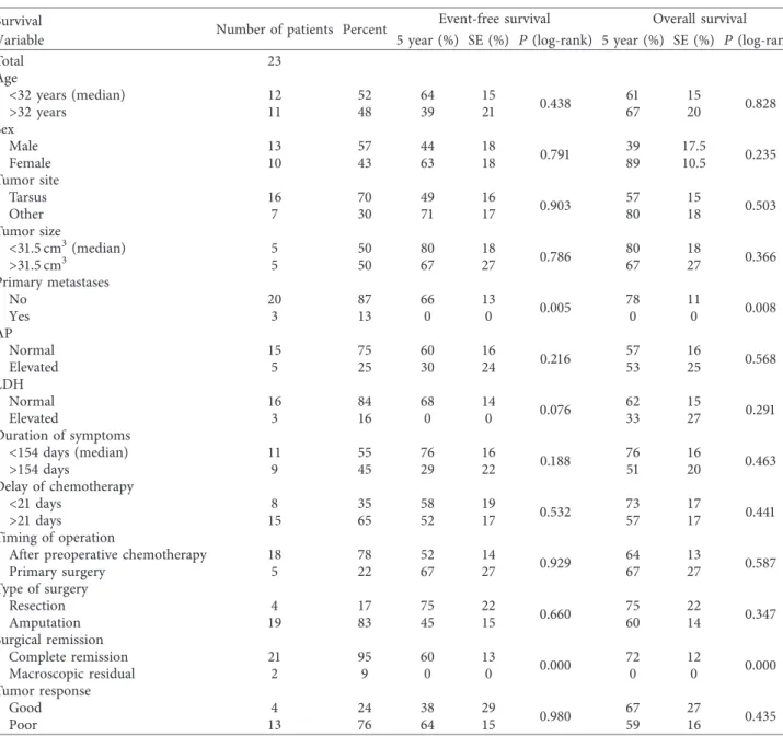
Event-free survival (EFS) and overall survival (OAS) correlated with primary metastatic status and best surgical remission status (Table 6).
4. Discussion
Osteosarcoma of the foot is exceedingly rare, and conse- quently the available information on patient and tumor characteristics, optimal management, and outcome is very limited. Therefore, we decided to investigate the greatest time span possible (36 years) using the data prospectively collected by the Cooperative German-Austrian-Swiss Os- teosarcoma Study Group. We were able to analyze 23 eligible patients with primary high-grade osteosarcoma of the foot, which represents one of the largest cohorts of such patients reported to date.
While recommended diagnostic and therapeutic pro- cedures have varied to some extent during this prolonged period, the overall results of osteosarcoma therapy have not [24–27], so we believe ourfindings hold true even for today.
Low-grade as well as secondary osteosarcomas were ex- cluded, as their biology and/or treatment differs from the more common primary high-grade central osteosarcomas.
Concerning patient-related variables, we observed the same slight male predominance as known for extremity osteosarcoma [28], but, similar to others [1, 2], a consider- ably older median age of 32 years. As in osteosarcoma, in general [13], pain was the most frequent presenting symptom.The median duration betweenfirst symptoms and
diagnostic biopsy was 154 days (range: 21–1940 days), which is shorter than that described in other series of osteosar- comas of the feet [2, 4] but longer than that we have observed for other extremity osteosarcomas (median: 69 days) [13].
Like others [1, 5], we observed the tarsal bones to be the most frequently affected site within the foot. In our 23 patient cohort, three had evidence of primary metastases Table2: Complete surgical remission in localized and metastatic disease.
Surgical remission Number of patients with localized
disease Number of patients with metastatic
disease Number of all
patients
Number of patients 20 3 23
Complete resection of primary tumor 20 3 23
Complete resection of metastases 0∗ 2 2
Complete surgical remission 19 2 21
One patient with localized disease developed pulmonary metastases during preoperative chemotherapy and did not receive metastasectomy because of progressive disease.
Table3: Type of surgery.
Type of surgery Number of patients with localized disease Number of patients with metastatic disease Number of all patients
Amputation 17 2 19
Resection 3 1 4
Table4: Tumor response to preoperative chemotherapy.
Tumor response Number of patients with localized
disease Number of patients with metastatic
disease Number of all
patients
Good (less than 10% viable tumor cells) 3 1 4
Poor (more than 10% viable tumor cells) 11 2 13
Not applicable (primary surgery) 5 0 5
Not documented 1 0 1
1.0 0.9 0.8 0.7 0.6 OAS versus EFS 0.5
0.4 0.3
Figure1: Overall survival ( ) (95% confidence interval: 0.0–10.8) and event-free survival ( ) (95% confidence interval: 3.6–14.3) of the 23 patients with high-grade osteosarcoma of the foot.
upon imaging, comparable to the situation in osteosarcoma of other sites [13, 29]. Two had lung metastases and one had lymph node metastases, the latter being rather unusual for osteosarcoma [30]. Compared to osteosarcoma in general
[31], fewer patients from our series presented with elevated alkaline phosphatase levels, probably correlating with their smaller tumor volumes, while the rate of elevated lactate dehydrogenase was similar [32].
Table5: Outcome at the last follow-up.
Outcome Number of patients with localized disease Number of patients with metastatic disease Number of all patients
Died 5 3 8
Alive 15 0 15
Alive CR1 13 0 13
Alive Rec1-LFU 1 0 1
Alive CR3 1 0 1
CR!complete surgical remission (primary tumor and metastases); CR1!first complete surgical remission; CR3!third complete surgical remission;
Rec. 1!first recurrence; LFU!lost to follow-up.
Table6: Univariate analysis of overall and event-free survival.
Survival Number of patients Percent Event-free survival Overall survival
Variable 5 year (%) SE (%) P(log-rank) 5 year (%) SE (%) P(log-rank)
Total 23
Age<32 years (median)>32 years 1211 5248 6439 1521 0.438 6167 1520 0.828
SexMaleFemale 1310 5743 4463 1818 0.791 3989 17.510.5 0.235
Tumor site
Tarsus 16 70 49 16 0.903 57 15 0.503
Other 7 30 71 17 80 18
Tumor size
<31.5 cm3(median) 5 50 80 18 0.786 80 18 0.366
>31.5 cm3 5 50 67 27 67 27
Primary metastases
No 20 87 66 13 0.005 78 11 0.008
Yes 3 13 0 0 0 0
APNormalElevated 155 7525 6030 1624 0.216 5753 1625 0.568
LDHNormalElevated 163 8416 680 140 0.076 6233 1527 0.291
Duration of symptoms
<154 days (median) 11 55 76 16 0.188 76 16 0.463
>154 days 9 45 29 22 51 20
Delay of chemotherapy
<21 days 8 35 58 19 0.532 73 17 0.441
>21 days 15 65 52 17 57 17
Timing of operation
After preoperative chemotherapy 18 78 52 14 0.929 64 13 0.587
Primary surgery 5 22 67 27 67 27
Type of surgery
Resection 4 17 75 22 0.660 75 22 0.347
Amputation 19 83 45 15 60 14
Surgical remission
Complete remission 21 95 60 13 0.000 72 12 0.000
Macroscopic residual 2 9 0 0 0 0
Tumor response
Good 4 24 38 29 0.980 67 27 0.435
Poor 13 76 64 15 59 16
5-year event-free and overall survival andPvalues in the log-rank test for all variables (see Data collection and Definition of Variables). SE!standard error;
P!two-sidedPvalues.
Our patients with osteosarcoma of the foot received the same multimodal therapy including chemotherapy and surgery as patients with osteosarcoma in general. While the more frequent osteosarcomas of long extremity bones have witnessed a major shift from amputation towards limb- saving surgery over the past several decades [14], we did not observe such a trend in this series, where three quarters of all affected feet were either completely or partially amputated.
Compared to osteosarcoma in general, where approxi- mately half of all tumors respond well to preoperative chemotherapy [13, 28, 33], only one quarter of 16 evaluable pedal osteosarcomas from our series did so. We were not able to extract information regarding response from other published series, so that this disparity must probably be considered a novelfinding for which there is no immediate explanation besides the small cohort size.The biology un- derlying this apparent difference remains to be elucidated.
Like in extremity osteosarcoma in general [28], most patients from our series achieved a first complete surgical remission.The recurrence rate and the time to recurrence were also similar to that which our group has observed for extremity osteosarcoma in general [13, 34]. Interestingly, there were no local recurrences asfirst event, a result which may have been favored by the aggressive, mostly ablative surgical approach employed. Given that wide margins may be difficult to achieve by foot-salvaging procedures, margins correlate with the local recurrence risk [35], prognosis following local osteosarcoma recurrence is very poor [36]
and gait performance is often quite good following partial or even complete amputation of the foot [37]; we believe that such an aggressive surgical approach is well justified.
The recurrences we observed were mostly lung metas- tases, again as well known for osteosarcoma in general [28, 34]. Two patients had either primary or secondary lymph node involvement, which is rather unusual for this particular malignancy [30]. However, the small numbers prohibit making definitive statements about whether the risk for lymphatic spread is truly higher than for osteosarcomas of other sites. Metastases in other published series were usually pulmonary [1]. Nevertheless, we would recommend careful assessment of the ipsilateral lymphatic drainage as part of staging and follow-up of patients with an osteo- sarcoma of the foot.
Given the very similar recurrence rates already discussed above, it comes as no surprise that the 5-year event-free and overall survival rates are also similar to those observed in other series which included both localized and primary metastatic osteosarcomas [14, 26, 38]. Even though tumor size is a very well-established prognostic factor and osteo- sarcomas of the foot are more likely to be detected at smaller size, the obtained results are certainly not superior to those our group has achieved in other long-bone extremity os- teosarcomas [13]. We can only assume that this may be due to a somewhat different tumor biology which also manifests in the low response rate to preoperative chemotherapy. As a note of caution, some papers on foot osteosarcomas have reported higher cure rates, albeit based upon even smaller patient numbers [4].
Patients with primary metastases are known to be as- sociated with inferior event-free and overall survival rates [13, 30, 39, 40], which was also seen in our cohort, where none of the three patients with primary metastases survived.
Complete surgical remission, mostly achieved by amputa- tion, was the strongest positive predictive factor for EFS and OS in our cohort. In this context, we have to emphasize that the subgroup of patients not receiving complete surgical remission consisted of only 2 of the 23 patients and these patients had inoperable primary metastases, respectively, progressive disease under chemotherapy. Nonetheless, our finding is in accordance with the general osteosarcoma literature [13, 28]. When investigating other factors for potential correlations with prognosis, such as tumor site within the foot, size, elevated serum LDH or AP levels, or response to preoperative chemotherapy [1, 13, 32, 41–43], we did not observe significant correlations with either event- free or overall survival, which may of course have been due to the limited number of patients included in our study.
5. Conclusion
Our study is one of the largest cohorts of patients with osteosarcoma of the foot reported to date despite the rela- tively small collection of only 23 patients. Using the same treatment strategy as employed in extremity osteosarcomas in general, we also achieved similar results. Primary meta- static status and surgical outcome correlated with prognosis.
These results argue in favor of treating osteosarcomas of the foot like other extremity osteosarcomas and further high- light the importance of achieving complete surgical re- mission, especially regarding the poor response of the tumors to neoadjuvant chemotherapy.
Disclosure
Peter Reichardt reports grants/personal fees from Novartis, Pfizer Bayer, PharmaMar, Amgen, AstraZeneca, Clinigen, Lilly, and Deciphera outside the submitted work; S. Bielack reports grants from Deutsche Krebshilfe, F¨orderkreis kreb- skranke Kinder Stuttgart e.V., and AXIS Forschungsstiftung during the conduct of the study and from Lilly, Bayer, Pfizer, Novartis, and Isofol outside the submitted work; Reinhard Windhager reports grants from Boehringer Ingelheim, Pfizer, Stryher, Taheda, and DePuy outside the submitted work. All other authors disclose that they have nofinancial or personal relationships with other people or organizations that could inappropriately influence (bias) their work.
Conflicts of Interest
The authors declare that they have no conflicts of interest.
Acknowledgments
The authors thank all patients who contributed to the COSS studies and acknowledge the physicians, nurses, data managers, and support staffof the collaborating centers for their active participation and Joachim Gerß for his support with the interpretation of survival analyses.Thanks are due
to German Cancer Aid (Deutsche Krebshilfe) and spon- sorship association for raising funds for children with cancer Stuttgart (F¨orderkreis krebskranke Kinder Stuttgart e.V.) for funding the work in question.
References
[1] J. K. Anninga, P. Picci, M. Fiocco et al., “Osteosarcoma of the hands and feet: a distinct clinico-pathological subgroup,”
Virchows Archiv, vol. 462, no. 1, pp. 109–120, 2013.
[2] R. Biscaglia, A. Gasbarrini, T. B¨ohling, P. Bacchini, F. Bertoni, and P. Picci, “Osteosarcoma of the bones of the foot–an easily misdiagnosed malignant tumor,” Mayo Clinic proceedings, vol. 73, no. 9, pp. 842–847, 1998.
[3] H. M. Ozdemir, Y. Yildiz, C. Yilmaz, and Y. Saglik, “Tumors of the foot and ankle: analysis of 196 cases,”Journal of Foot and Ankle Surgery, vol. 36, no. 6, pp. 403–408, 1997.
[4] M. Brotzmann, F. Hefti, D. Baumhoer, and A. H. Krieg, “Do malignant bone tumors of the foot have a different biological behavior than sarcomas at other skeletal sites?,” Sarcoma, vol. 2013, Article ID 767960, 8 pages, 2013.
[5] P. F. Choong, A. A. Qureshi, F. H. Sim, and K. K. Unni,
“Osteosarcoma of the foot: a review of 52 patients at the Mayo Clinic,” Acta Orthopaedica Scandinavica, vol. 70, no. 4, pp. 361–364, 1999.
[6] L. B. Chou, Y. Y. Ho, and M. M. Malawer, “Tumors of the foot and ankle: experience with 153 cases,” Foot & Ankle In- ternational, vol. 30, no. 9, pp. 836–841, 2009.
[7] R. Casadei, A. Ferraro, A. Ferruzzi, R. Biagini, and P. Ruggieri,
“Bone tumors of the foot: epidemiology and diagnosis,”La Chirurgia degli Organi di Movimento, vol. 76, no. 1, pp. 47–62, 1991.
[8] R. Eyre, R. G. Feltbower, P. W. James et al., “The epidemiology of bone cancer in 0-39 year olds in northern England, 1981–2002,”BMC Cancer, vol. 10, no. 1, p. 357, 2010.
[9] K. Winkler, G. Beron, G. Schellong et al., “Cooperative os- teosarcoma study COSS-77: results after 4 years,”Klinische Padiatrie, vol. 194, no. 4, pp. 251–256, 1982.
[10] K. Winkler, G. Beron, R. Kotz et al., “Neoadjuvant chemo- therapy for osteogenic sarcoma: results of a Cooperative German/Austrian study,”Journal of Clinical Oncology, vol. 2, no. 6, pp. 617–624, 1984.
[11] K. Winkler, G. Beron, G. Delling et al., “Neoadjuvant che- motherapy of osteosarcoma: results of a randomized co- operative trial (COSS-82) with salvage chemotherapy based on histological tumor response,”Journal of Clinical Oncology, vol. 6, no. 2, pp. 329–337, 1988.
[12] N. Fuchs, S. S. Bielack, D. Epler et al., “Long-term results of the co-operative German-Austrian-Swiss osteosarcoma study group’s protocol COSS-86 of intensive multidrug chemo- therapy and surgery for osteosarcoma of the limbs,”Annals of Oncology, vol. 9, no. 8, pp. 893–899, 1998.
[13] B. S. S. Bielack, B. Kempf-bielack, G. U. Exner et al.,
“Prognostic factors in high-grade osteosarcoma of the ex- tremities or trunk: an analysis of 1,702 patients treated on neoadjuvant cooperative osteosarcoma study group pro- tocols,”Journal of Clinical Oncology, vol. 20, no. 3, pp. 776–
790, 2002.
[14] S. Bielack, H. J¨urgens, G. Jundt et al., “Osteosarcoma: the COSS experience,”Cancer Treatment and Research, vol. 152, no. 7, pp. 289–308, 2009.
[15] J. S. Whelan, S. S. Bielack, N. Marina et al., “EURAMOS-1, an international randomised study for osteosarcoma: results
from pre-randomisation treatment,” Annals of Oncology, vol. 26, no. 2, pp. 407–414, 2015.
[16] S. Bielack, S. Flege, and B. Kempf-Bielack, “Behand- lungskonzept des osteosarkoms,”Der Onkologe, vol. 6, no. 8, pp. 747–758, 2000.
[17] D. Carrle and S. S. Bielack, “Current strategies of chemo- therapy in osteosarcoma,”International Orthopaedics, vol. 30, no. 6, pp. 445–451, 2006.
[18] W. F. Enneking, S. S. Spanier, and M. A. Goodman, “Current concepts review: the surgical staging of musculoskeletal sarcoma,”Journal of Bone and Joint Surgery, vol. 62, no. 6, pp. 1027–1030, 1980.
[19] M. Salzer-Kuntschik, G. Delling, G. Beron, and R. Sigmund,
“Morphological grades of regression in osteosarcoma after polychemotherapy? Study COSS 80,”Journal of Cancer Re- search and Clinical Oncology, vol. 106, no. S1, pp. 21–24, 1983.
[20] E. L. Kaplan and P. Meier, “Nonparametric estimation from incomplete observations,”Journal of the American Statistical Association, vol. 53, no. 282, pp. 457–481, 1958.
[21] N. Mantel, “Evaluation of survival data and two new rank order statistics arising in its consideration,”Cancer Chemo- therapy Reports, vol. 50, no. 3, pp. 163–170, 1966.
[22] D. R. Cox, “Regression models and life-tables,”Journal of the Royal Statistical Society: Series B (Methodological), vol. 34, no. 2, pp. 187–220, 1972.
[23] F. Wilcoxon, “Individual comparisons of grouped data by ranking methods,”Journal of Economic Entomology, vol. 39, no. 6, pp. 269-270, 1946.
[24] G. Gatta, R. Capocaccia, C. Stiller, P. Kaatsch, F. Berrino, and M. Terenziani, “Childhood cancer survival trends in Europe:
a EUROCARE working group study,” Journal of Clinical Oncology, vol. 23, no. 16, pp. 3742–3751, 2005.
[25] C. A. Stiller, S. S. Bielack, G. Jundt, and E. Steliarova-Foucher,
“Bone tumours in European children and adolescents, 1978–1997: report from the Automated Childhood Cancer Information System project,” European Journal of Cancer, vol. 42, no. 13, pp. 2124–2135, 2006.
[26] L. Mirabello, R. J. Troisi, and S. A. Savage, “Osteosarcoma incidence and survival rates from 1973 to 2004: data from the surveillance, epidemiology, and end results program,”Cancer, vol. 115, no. 7, pp. 1531–1543, 2009.
[27] G. Gatta, L. Botta, S. Rossi et al., “Childhood cancer survival in Europe 1999-2007: Results of EUROCARE-5-a population-based study,”The Lancet Oncology, vol. 15, no. 1, pp. 35–47, 2014.
[28] G. Bacci, A. Longhi, M. Versari, M. Mercuri, A. Briccoli, and P. Picci, “Prognostic factors for osteosarcoma of the extremity treated with neoadjuvant chemotherapy: 15-year experience in 789 patients treated at a single institution,”Cancer, vol. 106, no. 5, pp. 1154–1161, 2006.
[29] L. Kager, A. Zoubek, U. Kastner et al., “Skip metastases in osteosarcoma: experience of the Cooperative Osteosarcoma Study Group,”Journal of Clinical Oncology, vol. 24, no. 10, pp. 1535–1541, 2006.
[30] L. Kager, A. Zoubek, U. P¨otschger et al., “Primary metastatic osteosarcoma: presentation and outcome of patients treated on neoadjuvant Cooperative Osteosarcoma Study Group protocols,” Journal of Clinical Oncology, vol. 21, no. 10, pp. 2011–2018, 2003.
[31] H.-Y. Ren, L.-L. Sun, H.-Y. Li, and Z.-M. Ye, “Prognostic significance of serum alkaline phosphatase level in osteo- sarcoma: a meta-analysis of published data,”BioMed Research International, vol. 2015, Article ID 160835, 11 pages, 2015.
[32] G. Bacci, A. Longhi, S. Ferrari et al., “Prognostic significance of serum lactate dehydrogenase in osteosarcoma of the
extremity: experience at Rizzoli on 1421 patients treated over the last 30 years,”Tumori, vol. 90, no. 5, pp. 478–484, 2004.
[33] J. S. Whelan, R. C. Jinks, A. McTiernan et al., “Survival from high-grade localised extremity osteosarcoma: combined re- sults and prognostic factors from three European osteosar- coma intergroup randomised controlled trials,” Annals of Oncology, vol. 23, no. 6, pp. 1607–1616, 2012.
[34] B. Kempf-Bielack, S. S. Bielack, H. J¨urgens et al., “Osteo- sarcoma relapse after combined modality therapy: an analysis of unselected patients in the Cooperative Osteosarcoma Study Group (COSS),”Journal of Clinical Oncology, vol. 23, no. 3, pp. 559–568, 2005.
[35] P. Picci, S. Ferrari, G. Bacci, and F. Gherlinzoni, “Treatment recommendations for osteosarcoma and adult soft tissue sarcomas,”Drugs, vol. 47, no. 1, pp. 82–92, 1994.
[36] S. Weeden, R. J. Grimer, S. R. Cannon et al., “The effect of local recurrence on survival in resected osteosarcoma,”European Journal of Cancer, vol. 37, no. 1, pp. 39–46, 2001.
[37] R. Versluys, A. Desomer, G. Lenaerts et al., “From conven- tional prosthetic feet to bionic feet: a review study,” inPro- ceedings of the 2nd IEEE RAS & EMBS International Conference on Biomedical Robotics and Biomechatronics, pp. 49–54, Scottsdale, AZ, USA, October 2008.
[38] M. T. Harting, K. P. Lally, R. J. Andrassy et al., “Age as a prognostic factor for patients with osteosarcoma: an analysis of 438 patients,” Journal of Cancer Research and Clinical Oncology, vol. 136, no. 4, pp. 561–570, 2010.
[39] G. Bacci, M. Rocca, M. Salone et al., “High-grade osteosar- coma of the extremities with lung metastases at presentation:
treatment with neoadjuvant chemotherapy and simultaneous resection of primary and metastatic lesions,” Journal of Surgical Oncology, vol. 98, no. 6, pp. 415–420, 2008.
[40] M. San-Julian, P. Diaz-de-Rada, E. Noain, and L. Sierrasesumaga,
“Bone metastases from osteosarcoma,” International Ortho- paedics, vol. 27, no. 2, pp. 117–120, 2003.
[41] P. Bieling, N. Rehan, P. Winkler et al., “Tumor size and prognosis in aggressively treated osteosarcoma,” Journal of Clinical Oncology, vol. 14, no. 3, pp. 848–858, 1996.
[42] S. K. Min, S. Y. Lee, H. C. Wan et al., “Initial tumor size predicts histologic response and survival in localized osteo- sarcoma patients,”Journal of Surgical Oncology, vol. 97, no. 5, pp. 456–461, 2008.
[43] E. Gonz´alez-Billalabeitia, R. Hitt, J. Fern´andez et al., “Pre- treatment serum lactate dehydrogenase level is an important prognostic factor in high-grade extremity osteosarcoma,”
Clinical and Translational Oncology, vol. 11, no. 7, pp. 479–
483, 2009.
Stem Cells International
Hindawi
www.hindawi.com Volume 2018
Hindawi
www.hindawi.com Volume 2018
INFLAMMATION
Endocrinology
International Journal of
Hindawi
www.hindawi.com Volume 2018
Hindawi
www.hindawi.com Volume 2018
Disease Markers
Hindawi
www.hindawi.com Volume 2018
BioMed
Research International
Oncology
Journal ofHindawi
www.hindawi.com Volume 2013
Hindawi
www.hindawi.com Volume 2018
Oxidative Medicine and Cellular Longevity
Hindawi
www.hindawi.com Volume 2018
PPAR Research
Hindawi Publishing Corporation
http://www.hindawi.com Volume 2013
Hindawi www.hindawi.com
The Scientific World Journal
Volume 2018
Immunology Research
Hindawi
www.hindawi.com Volume 2018
Journal of
Obesity
Journal of
Hindawi
www.hindawi.com Volume 2018
Hindawi
www.hindawi.com Volume 2018
Computational and Mathematical Methods in Medicine
Hindawi
www.hindawi.com Volume 2018
Behavioural Neurology Ophthalmology
Journal ofHindawi
www.hindawi.com Volume 2018
Diabetes Research
Journal ofHindawi
www.hindawi.com Volume 2018
Hindawi
www.hindawi.com Volume 2018
Research and Treatment
AIDS
Hindawi
www.hindawi.com Volume 2018
Gastroenterology Research and Practice
Hindawi
www.hindawi.com Volume 2018
Parkinson’s Disease
Evidence-Based Complementary and Alternative Medicine
Volume 2018 Hindawi
www.hindawi.com