Full Terms & Conditions of access and use can be found at
https://www.tandfonline.com/action/journalInformation?journalCode=ijas20
ISSN: 0277-0903 (Print) 1532-4303 (Online) Journal homepage: https://www.tandfonline.com/loi/ijas20
A suitable protocol for measuring alveolar nitric oxide in asthma with differing severity to assess peripheral airways inflammation
Zsófia Lázár, Péter Horváth, Rita Puskás, Gabriella Gálffy, György Losonczy, Ildikó Horváth & András Bikov
To cite this article: Zsófia Lázár, Péter Horváth, Rita Puskás, Gabriella Gálffy, György Losonczy, Ildikó Horváth & András Bikov (2019) A suitable protocol for measuring alveolar nitric oxide in asthma with differing severity to assess peripheral airways inflammation , Journal of Asthma, 56:6, 584-593, DOI: 10.1080/02770903.2018.1477957
To link to this article: https://doi.org/10.1080/02770903.2018.1477957
Accepted author version posted online: 20 Jun 2018.
Published online: 30 Oct 2018.
Submit your article to this journal
Article views: 71
View Crossmark data
DIAGNOSIS
A suitable protocol for measuring alveolar nitric oxide in asthma with differing severity to assess peripheral airways inflammation
Zsofia Lazar,MD, PhDa, Peter Horvath,MD, PhDa, Rita Puskas,MDa, Gabriella Galffy,MD, PhDa, Gy€orgy Losonczy, MD,PhDa, Ildiko Horvath,MD,PhDb, and Andras Bikov,MD,PhDa
aDepartment of Pulmonology, Semmelweis University, 1/c Diosarok;bNational Koranyi Institute of Pulmonology, 1 Pihen}o Street, Budapest, Hungary
ABSTRACT
Objective:Extended nitric oxide (NO) analysis offers the partitioned monitoring of inflamma- tion in central and peripheral airways. Different mathematical models are used to estimate pulmonary NO dynamics in asthma with variable results and limitations. We aimed to estab- lish a protocol for extended NO analysis in patients with differing asthma severity.Methods:
Forty patients with stable asthma and 25 matched control subjects were recruited. Exhaled NO was measured at constant flow rates between 10 and 300 mL/s. Twelve controls per- formed NO measurements weekly for 4 weeks. Results: The proportions of patients with technically acceptable measurements at 10–30–50–100–150–200–250–300 mL/s exhalation flow rates were 8–58–100–98–98–95–90–80%, respectively. Alveolar NO (CANO) and total flux of NO in the conducting airways (JawNO) were calculated with the linear method from NO values measured at 100–150–200–250 mL/s exhalation flows. The mean intrasubject bias for JawNO and CANO in controls was 0.16 nL/s and 0.85 ppb, respectively. Both JawNO (1.31/0.83–2.97/vs. 0.70/0.54–0.87/nL/s, p<0.001) and CANO (4.08/2.63–7.16/vs. 2.42/
1.83–2.89/ppb, p<0.001) were increased in patients with asthma compared to controls. In patients, CANO correlated with RV/TLC (r¼0.58, p<0.001), FEF25-75% (p¼0.02, r¼–0.36) and DL,CO (r¼–0.46, p¼0.004). JawNO was not related to lung function parameters.
Conclusions: Calculation of alveolar NO concentration with the linear method from values obtained at medium flow rates (100–250 mL/s) is feasible even in asthmatic patients with severe airflow limitation and may provide information on small airways dysfunction in asthma.
ARTICLE HISTORY Received 23 February 2018 Revised 30 April 2018 Accepted 13 May 2018 KEYWORDS Airway inflammation;
exhaled biomarker;
extended nitric oxide analysis; disease
monitoring; severe asthma
Introduction
Bronchial asthma is a heterogeneous disease charac- terised by accumulation of inflammatory cells in the airways, hypertrophy and hyperplasia of airway smooth muscle layer, increased mucus production, and airway wall remodelling [1]. Inflammatory changes take place simultaneously in central and distal airways and in the alveoli [2–4]. The assessment of airway inflammation at these different localizations can aid better understanding of disease pathomechan- ism and facilitate the development of more tar- geted therapies.
Airway inflammation can be studied noninvasively by measuring the exhaled nitric oxide (NO) concen- tration. The two-compartment model allows the evaluation of NO dynamics in the large central or bronchial and in more distal airways (small airways
and the alveolar/acinar region) [5,6]. For this purpose, measurements are performed at multiple expiratory flow rates (10–500 mL/s) and mathematical models are applied [6]. The recent technical standard task force report of the European Respiratory Society rec- ommends the use of NO plateau values measured at least at three different exhalation flows and proposes several mathematical equations to calculate bronchial and alveolar NO parameters [7]. However, there are currently no standardised method that can reliably be applied to asthmatic patients with varying airflow limitation, smoking status and disease severity.
Therefore, in this study we aimed to establish a feasible method for the partitioned measurement of exhaled NO in asthma. Patients and control subjects carried out expiratory manoeuvres at a broad range of flows. We established a protocol for the calculation of
CONTACTZsofia Lazar, MD, PhD lazar.zsofia@med.semmelweis-univ.hu Department of Pulmonology, Semmelweis University, 1/c Diosarok, 1125- Budapest, Hungary.
ß2018 Taylor & Francis Group, LLC JOURNAL OF ASTHMA
2019, VOL. 56, NO. 6, 584–593
https://doi.org/10.1080/02770903.2018.1477957
central bronchial and peripheral airway NO parame- ters with low week-to-week variation. Furthermore, we compared central and peripheral airway inflamma- tion between patients and control subjects and corre- lated NO variables to clinical parameters in asthma.
Materials and methods Subjects
Patients were recruited at the Outpatient Clinic of Department of Pulmonology at Semmelweis University, Budapest, Hungary. They complained of symptoms consistent with the asthma diagnosis, they showed positivity for at least one airway allergen at skin prick testing or serum specific IgE testing.
Patients presented documented airflow limitation, and they had airway hyperresponsiveness or were positive for bronchodilator reversibility during prior testing [1]. A change in asthma therapy was not required in 4 weeks prior to the recruitment. Main exclusion crite- ria were other chronic respiratory diseases, asthma exacerbation in the previous 4 weeks, and signs of acute respiratory infections in the 2 weeks before recruitment. Healthy control subjects were recruited among employees working at the Department. Main exclusion criteria for controls were allergic airway dis- ease or chronic respiratory disease in history, systemic steroid or antibiotic treatment in the previous 4 weeks, and signs of acute respiratory infections in the previ- ous 2 weeks. Patient and control subjects were consid- ered ex-smokers if they had stopped smoking at least 6 months before inclusion. The study was approved by the ethics committee, and all procedures were in accordance with the 1964 Helsinki declaration and its later amendments or comparable ethical standards.
Informed consent was obtained from all individual participants included in the study. Measurements were performed between 10 am and 3 pm from 1 December 2015 until 30 November 2017.
Study design Clinical study
Twenty-five control subjects and 40 patients with asthma were recruited. Medical history was noted, blood leukocyte count and CRP concentration were measured, exhaled nitric oxide concentration was recorded and lung function tests were performed.
Exhaled nitric oxide measurements were performed before lung function tests in all cases. Furthermore, patients filled out the Asthma Control Test (ACT) [8].
Repeatability study
Exhaled NO measurements were repeated at a weekly basis for 4 weeks in 12 control subjects, who also par- ticipated in the clinical study.
Nitric oxide measurements at multiple constant exhalation flow rates
Subjects were asked not to use inhaled medication, refrain from eating, drinking and smoking 2 h prior to measurement. Exhaled NO concentration was meas- ured during a manoeuvre starting from total lung capacity at the expiratory flows of 10–30–50–100–150–200–250–300 mL/s with a chemi- luminescent analyser (Sievers Nitric Oxide Analyzer i280, GE Analytical Instruments, Boulder, CO).
Instrument calibration was performed daily according to the manufacturer’s instructions. The background NO concentration was <5 ppb. Restrictors, as pro- vided and calibrated by the manufacturer, were applied to generate the required expiratory flows and ensure the closure of the velum during expiration.
Manoeuvres at different flows with a duration of 20 s (10 mL/s exhalations flow), 10 s (30 mL/s), 6 s (50 and 100 mL/s) and 5 s (150, 200, 250 and 300 mL/s) were considered sufficient. Subjects received visual feedback of the expiratory flow during the entire manoeuvre. Plateau values of NO recordings were identified manually. Recordings corresponding to the initial expiratory volume of 150 mL air (i.e.
anatomic dead space) were disregarded. We consid- ered a recording technically acceptable and valid if the plateau NO concentration was in a 3-s window with minimal sloping [9] where the actual exhalation flow was ±10% of the target rate in compliance with the recommendations for FENO50 (fractional exhaled nitric oxide concentration at 50 mL/s exhalation flow) analysis. The mean values of two NO recordings with
<10% difference were used for further calculations.
Calculation of CANO and JawNO
Data were analysed based on the two-compartment model using the linear method of Tsoukias et al.
[5–7], which estimates acinar/alveolar NO (CANO) as the measure of the distal airways and total flux of NO in the conducting airway compartment (JawNO) as a marker of central airways.
Other variables
Leukocyte count (Sysmex XE-2100, Sysmex Corporation, Kobe, Japan) and serum CRP concentration (Beckman
Coulter AU680, Beckman Coulter Inc., Indianapolis, IN) were determined from venous blood samples (asthma: N¼38, control N¼25). Measurements for spirometry, body plethysmography and diffusion cap- acity were performed according to current guidelines (PDT-111, Piston, Budapest, Hungary) [10–12]. Two patients with asthma could not perform the man- oeuvre for diffusion capacity measurement. An ACT score 19 referred to uncontrolled asthma [8].
Statistical analysis
Demographic data were compared with unpaired t-test and expressed as mean ± standard deviation, categorical variables were compared with the Fisher’s exact test.
Inhaled corticosteroid (ICS) doses, blood eosinophil percentage, CRP value, FENO50, JawNO and CANO data did not show a normal distribution (D’Agostino- Pearson normality test), therefore, these variables were analysed with nonparametric tests (Mann–Whitney, Kruskal–Wallis with Dunn’s post hoc and Spearman tests) and expressed as median/interquartile range/.
Measurement repeatability was assessed using the Bland–Altman plot [13]. p<0.05 was considered sig- nificant (GraphPad Prism 5.0, GraphPad Software, San Diego, CA). Multiple regression analysis with smoking habits as covariates were used to assess relationship between CANO and lung function measures (Statistica 13.2, StatSoft, Tulsa, OK).
The sample size of the clinical study was calculated to reach a statistical power (1–b) of 0.80 and effect size of 0.75 with respect to the asymptotic relative effi- ciency of nonparametric tests. This effect size was based on the variability of JawNO andCANO data in the repeatability study.
Results
Subject characteristics
The main clinical characteristics of patients and con- trol subjects are shown in Table 1. Patients were treated at treatment steps of GINA 1 (steroid-naïve, n¼7), GINA 3–4 (moderate-severe, n¼16) and GINA 5 (severe on anti-IgE therapy, n¼17) [1].
Forty percent of patients had uncontrolled asthma according to the ACT scores.
Measurements of exhaled NO at different constant exhalation flow rates
Subjects performed exhalation manoeuvres at various constant flows (10–300 mL/s) both in the repeatability
and clinical studies (number of measurements/flow in controls subjects: 61, in patients with asthma: 40;
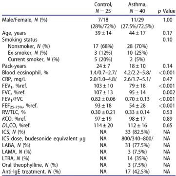
Table 2). Only a fraction of patients could perform a manoeuvre with a technically acceptable recording at very low flow rates (<50 mL/s), while the majority of manoeuvres were technically correct at higher flows (>50 mL/s). Therefore, for the extended NO analysis only the linear model could be applied [7], and NO values obtained at 100, 150, 200 and 250 mL/s expira- tory flow rates were used as inputs for the calculation of JawNO and CANO (asthma: r¼0.98 ± 0.03, con- trol: r¼0.98 ± 0.02). As four patients could not per- form the manoeuvre at 250 mL/s exhalation flow, values at 300 mL/s were included in the model in three cases. Using this strategy, 92.5% of all calcula- tions in asthma were executed on data at 4 flow Table 1. Patient characteristics.
Control, N¼25
Asthma,
N¼40 pValue
Male/Female,N(%) 7/18
(28%/72%)
11/29 (27.5%/72.5%)
1.00
Age, years 39 ± 14 44 ± 17 0.17
Smoking status 0.10
Nonsmoker,N(%) 17 (68%) 28 (70%)
Ex-smoker,N(%) 3 (12%) 10 (25%)
Current smoker,N(%) 5 (20%) 2 (5%)
Pack-years 24 ± 7 18 ± 10 0.14
Blood eosinophil, % 1.4/0.7–2.7/ 4.2/2.2–5.8/ <0.001
CRP, mg/L 2.0/1.0–4.8/ 2.6/1.7–5.1/ 0.47
FEV1, %ref. 103 ± 10 79 ± 18 <0.001
FVC, %ref. 107 ± 13 95 ± 14 0.002
FEV1/FVC 0.82 ± 0.06 0.70 ± 0.13 <0.001
FEF25-75%, %ref. 93 ± 18 54 ± 28 <0.001
RV/TLC, % 0.30 ± 0.21 0.33 ± 0.14 0.53
KCO, %ref. 97 ± 19 98 ± 17 0.89
DL,CO, %ref. 114 ± 20 112 ± 16 0.65
ICS,N(%) NA 33 (82.5%) NA
ICS dose, budesonide equivalentmg NA 800/340–800/ NA
LABA,N(%) NA 31 (77.5%) NA
LAMA,N(%) NA 3 (7.5%) NA
LTRA,N(%) NA 14 (35%) NA
Oral theophylline,N(%) NA 3 (7.5%) NA
Anti-IgE treatment,N(%) NA 17 (42.5%) NA
Data are presented as mean ± standard deviation or median/interquartile range/and compared with unpairedt-test, Mann–Whitney or chi-square tests (categorical variables).
CRP: C-reactive protein;DL,CO: diffusion capacity of the lung for carbon monoxide; ICS: inhaled corticosteroid; IgE: immunoglobulin E; FEF25–75%: forced expiratory flow at 25–75% of vital capacity; FEV1: forced expira- tory volume in 1 s; FVC: forced vital capacity;KCO: transfer coefficient of the lung for carbon monoxide; LABA: long-acting b2-agonist; LAMA:
long-acting muscarinic antagonist; LTRA: leukotriene receptor antagon- ist;N: number; NA: not applicable; ref.: reference; RV: residual volume;
TLC: total lung capacity.
Table 2. Percentage of technically acceptable exhaled NO manoeuvres with valid recordings at different exhalation flow rates.
Exhalation flow, mL/s 10 30 50 100 150 200 250 300 Control subjects 20% 79% 100% 100% 100% 100% 98% 98%
Patients with asthma 8% 58% 100% 98% 98% 95% 90% 80%
586 Z. LAZAR ET AL.
points (2.5% at 3 flow rates, 5% at 2 flow rates). Data obtained at 4 flow rates were used to calculateJawNO and CANO in each control volunteer. All subjects could perform valid manoeuvres forFENO50. Exhaled NO concentrations were elevated in asthma at all flow rates between 50 and 250 mL/s (p<0.001, Figure 1).
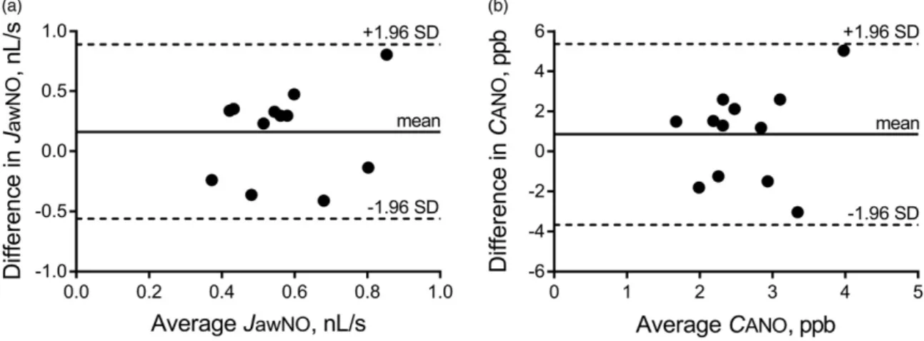
Intrasubject repeatability of JawNO and CANO Weekly JawNO values in control subjects were 0.73/
0.59–0.73/, 0.51/0.41–0.67/, 0.58/0.50–0.73/and 0.50/
0.39–0.68/nL/s. The Bland–Altman analysis for the lowest and highest individual values showed a mean difference of 0.16 nL/s (95% limits of agreement:
–0.56–0.89 nL/s; Figure 2a). CANO values at the weekly measurements were 2.64/2.25–3.08/, 3.19/
2.30–4.22/, 1.75/1.17–3.12/and 2.27/1.89–2.86/ppb.
The Bland–Altman graph for the lowest and highest individual values demonstrated a mean bias of 0.85 ppb (95% limits of agreement: –3.67–5.36 ppb;
Figure 2b).
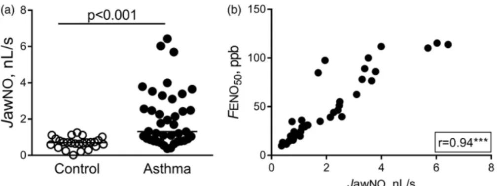
Increased JawNO and CANO in patients with asthma
JawNO was increased in patients with asthma com- pared to control subjects (1.31/0.83–2.97/nL/s vs. 0.70/
0.54–0.87/nL/s, p<0.001; Figure 3a). In asthma, JawNO showed a strong positive correlation with FENO50 (p<0.001, r¼0.94; Figure 3b) and blood eosinophil percentage (p¼0.001, r¼0.50), but not with lung function parameters, leukocyte count, CRP or age (p>0.05).
Alveolar NO concentration was higher in patients than in controls (4.08/2.63–7.16/ppb vs. 2.42/
1.83–2.89/ppb, p<0.001; Figure 4a). In asthma, CANO concentration positively correlated with blood eosinophil percentage (p¼0.002, r¼0.50), CRP con- centration (p¼0.001, r¼0.50), age (p¼0.003, r¼0.46), RV/TLC (p<0.001, r¼0.58; Figure 4c) and airway resistance (p¼0.02, r¼0.38). It inversely cor- related with FEV1% predicted (p¼0.03, r¼–0.34), FEF25–75% % reference (p¼0.02, r¼–0.36; Figure 4b) Figure 1. Exhaled NO concentrations at multiple flow rates.
Exhaled NO at different constant exhalation flows in controls (a) and patients with asthma (b). Lines show median values.
Mann–Whitney test:#p<0.001 compared to corresponding NO concentration in controls.
Figure 2. Intrasubject repeatability ofJawNO andCANO. The weekly intrasubject repeatability of total flux of NO in the conducting airway compartment (JawNO; a) and alveolar nitric oxide concentration (CANO; b) in control subjects as shown by the Bland–Altman plot with mean difference and limits of agreement (mean difference ±1.96 standard deviation).
and diffusion capacity of the lung for carbon monox- ide (p¼0.004, r¼–0.46; Figure 4d). We found a sig- nificant positive correlation between CANO and JawNO (p¼0.005, r¼0.44), and between CANO and FENO50 (p<0.001, r¼0.60).
The association of CANO to RV/TLC and DL,CO remained significant in patients with asthma when using multiple regression analysis with smoking status (beta ¼0.48, p¼0.003 and beta¼–0.45, p¼0.005) or packyears (beta ¼0.46, p¼0.007 and beta¼–0.43, p¼0.007) as a covariate. However, the relationship between CANO and FEF25–75% % reference became statistically insignificant in a model controlled for smoking status (p¼0.09) or packyears (p¼0.14).
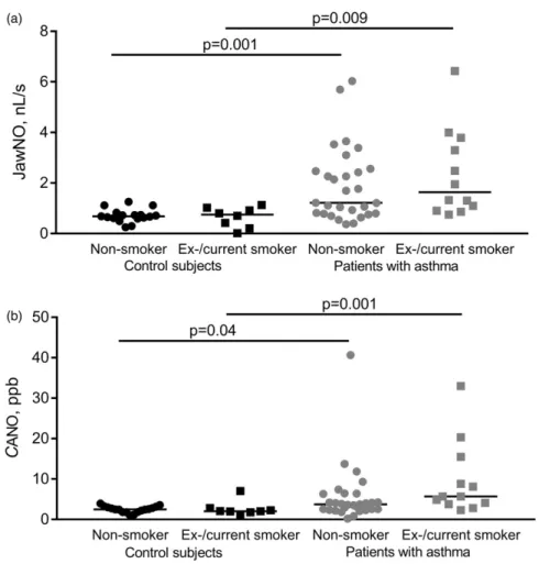
The effect of smoking status on NO parameters We also analysed nonsmoking and ex-/current smok- ing control subjects and patients in separate sub- groups. There was an increase in JawNO in patients compared to controls with relevant smoking status (control vs. asthma in nonsmokers: 0.68/0.61–0.78/nL/
s vs. 1.22/0.79–2.52/nL/s, p¼0.001 and in smok- ers: 0.75/0.26–1.00/nL/s vs. 1.64/0.95–0.3.67/nL/s, p¼0.009; Figure 5a). Likewise, CANO was higher in asthma than in control subgroups (control vs. asthma in nonsmokers: 2.48/1.83–3.04/ppb vs. 3.75/2.46-6.35/
ppb, p¼0.04 and in smokers: 2.04/1.82–2.66/nL/s vs.
5.70/3.86–13.81/ppb, p¼0.001; Figure 5b). We found Figure 3. JawNO in patients with asthma. JawNO was increased in asthma (Mann–Whitney test; a), and strongly correlated with FENO50(b). Spearman correlation:p<0.001.
Figure 4. CANO in patients with asthma.CANO was increased in asthma (Mann–Whitney test; a), and correlated with FEF25–75%% reference (b), RV/TLC (c) andDL,CO (d). Spearman correlation:p<0.05,p<0.01,p<0.001.
588 Z. LAZAR ET AL.
no difference in JawNO and CANO between non- smokers and smokers within the control (p¼0.99 and p¼0.99) and asthmatic (p¼0.99 and p¼0.23) groups.
Discussion
The inflammation and dysfunction of small airways are related to important clinical aspects of asthma such as airway hyper-responsiveness [14] or exacerba- tion risk [15], therefore, targeted anti-inflammatory treatment which can mitigate small airways inflamma- tion can convey clinical benefit. However, the nonin- vasive assessment of distal lung inflammation is an unmet need in clinical practice. Models using exhaled NO concentration measured at different flow rates allow the partitioned assessment of airway inflamma- tion in central and distal airways. The European Respiratory Society technical standard document pro- vides details for mathematical modelling of pulmonary NO dynamics and highlights the need for further studies [16]. In this study, we presented a feasible protocol for alveolar NO measurement and showed
that inflammation and dysfunction of small airways are related in asthma.
In our study, patients performed exhalation manoeuvres at constant flow rates between 10 and 300 mL/s, but we could measure exhaled NO concen- trations at low flow rates only in a minority of patients with asthma. Similarly, Gelb et al. also noted that measurements were not reproducible at low exhalation flows ( 50 mL/s) in asthmatics with FEV1<80% predicted [17], representing a significant number of treated patients in clinical settings. In add- ition, we also observed some failure in manoeuvre performance at 300 mL/s exhalation flow, which ques- tions the feasibility of applying very high flow rates in this population. Up to 30% of patients with severe asthma had to be excluded from previous studies due to negative CANO values, suggesting inadequate mod- els and the requirement of additional flow rates [18,19]. However, we established a linear model based on exhaled NO values at four flow points (100, 150, 200 and 250 mL/s), which could be successfully and reliably applied to measure alveolar NO concentra- tions in patients with differing asthma severity and smoking history.
Figure 5. NO parameters in smokers and nonsmokers. JawNO (a) and CANO (b) were increased in ex- and current smoking patients with asthma compared to corresponding control subjects (Kruskal–Walllis with Dunn’s post hoc test).
We confirm previous findings that alveolar NO concentration is increased in a mixed group of asth- matic patients with mild-to-severe disease compared to matched control subjects [17]. Other studies also showed that peripheral airway inflammation, as reflected by CANO, is elevated in clinically important phenotypes of asthma: in patients with refractory asthma on high ICS dose compared to mild-to-moder- ate asthma [20], in patients with steroid-dependent severe asthma compared to severe asthmatics on high dose ICS [19] and in subjects with nocturnal symp- toms [21]. Several authors observed that alveolar NO concentration could not be modified by initiating ICS therapy or increasing its dose [22,23]. While a decrease in CANO was reported after oral steroid therapy in some studies [17,20], interestingly, others found no treatment effect [23]. This suggests that des- pite ongoing anti-inflammatory treatment, increased inflammation in peripheral airways is a distinct dis- ease characteristic in certain asthma phenotypes, which is also steroid-resistant in some cases. Hence, the extended NO analysis might facilitate the identifi- cation and better understanding of asthma subgroups, and it can also aid monitoring of novel anti-inflam- matory therapies.
We analysed the relationship between alveolar NO concentration and physiological measures of distal lung dysfunction. We reported a moderate correlation between CANO and RV/TLC, which is a known marker of air trapping and hyperinflation in severe and nonsevere asthma [24]. This finding extends the results of a previous study that showed a similar, but stronger correlation between the two parameters in severe asthma [18]. In our study, there was a weak correlation between CANO and FEF25–75%, which was not present when the analysis was controlled for smoking. This lung function parameter is debated to truly reflect peripheral airways dysfunction, partly due to its high measurement variability [25]. Likewise, one study described a correlation with similar strength in mild-to-moderate asthma [26], but others found no relationship between alveolar NO concentration and FEF25–75% in mild-to-severe asthmatics [19].
Interestingly, in our cohort CANO moderately corre- lated to pulmonary diffusion capacity, as previously observed in alveolitis [27]. Besides the upregulation of the inducible NO synthase in alveolar epithelial cells, the decreased diffusion of NO might be another mechanism leading to increased CANO in asthma, nonetheless, it must be noted that pulmonary diffu- sion capacity was within the normal range in patients.
Despite of the exploratory nature of these results, they
imply that increased CANO can reflect distal airways dysfunction in asthma.
It was previously shown that alveolar NO concen- tration is strongly associated with eosinophil percent- age in bronchoalveolar lavage fluid in mild asthmatics [20]. However, the weak-to-moderate correlation between CANO and blood eosinophil percentage in our study suggests that inflammation in peripheral airways is not closely related to systemic eosinophilic inflammation, as already shown for FENO50 [28].
We reported a positive correlation between age and CANO, which was also observed in another cohort of asthmatic patients [29]. It is known that CANO increases with age in healthy subjects, which could be explained by the reduced pulmonary NO diffusion resulting from decreased capillary blood volume at an older age [30]. It can be speculated that this mechan- ism might also be present in older patients with asthma.
We found a close correlation between FENO50 and JawNO in asthma highlighting that these parameters assess inflammation at similar sites within the airways, and the additional information gained by the calcula- tion of JawNO might be limited as also suggested by others [31].
The weekly repeatability of CANO and JawNO in control subjects was assessed in a 1-month period, relevant to clinical settings for asthma follow-ups. The intrasubject repeatability of these parameters was somewhat better in a previous study using a day-to- day setup [19], nevertheless, the observed difference between controls and patients exceeded the mean intrasubject bias.
Some authors correct alveolar NO for trumpet model and axial NO back-diffusion [32,33]. However, these formulae disregard the effect of central airways constriction on axial back-diffusion, which potentially result in overcorrection [34,35]. According to the recent technical standard document the use of correc- tion factors for axial back diffusion is not recom- mended [7]. Therefore, we did not apply any correction in our data analysis.
This study has limitations. Cigarette smoking is known to interfere with exhaled nitric oxide concen- tration [36], and smoking had been previously shown to decrease CANO in asthma [37], which was not confirmed by our findings. Importantly, CANO was elevated in asthma, irrespective of the smoking status.
Our cohort reflects realistic asthmatic population in terms of smoking habits, as approximately one third of asthmatics were also reported to be current or for- mer smokers in larger cohorts [38,39]. Cigarette
590 Z. LAZAR ET AL.
smoking in asthma results in greater morbidity, uncontrolled and more severe disease, and accelerated decline in lung function [40]. In addition, a recent publication described that one third of patients with severe asthma were active or former smokers, who presented with fixed airflow limitation and clustered into clinical subgroups with either Th2-high or Th2- low signatures, underlining the potential therapeutic relevance of measuring inflammatory markers includ- ing exhaled NO in these populations [41].
Furthermore, this is a single-centre observational study, and our results should be validated by future investigations. The cross-sectional nature of the NO measurements does not allow to draw conclusions regarding the repeatability of CANO in asthma, or how it reflects disease course or therapeutic interventions.
Conclusions
The present study describes a feasible protocol for extended NO analysis to calculate alveolar nitric oxide concentration, a marker of distal lung inflammation.
Our method can successfully and reliably be applied to patients with asthma of differing severity including those with severe disease. Alveolar NO concentration shows a weak correlation to physiological measures of small airways dysfunction in asthma. The application of our protocol could facilitate understanding the role ofCANO in phenotyping asthma.
Declaration of Interest
The authors report no conflicts of interest. The authors alone are responsible for the content and writing of the paper. This study was funded by the Bolyai Research Gant of the Hungarian Academy of Sciences (BO/00559/16 to Dr. Zsofia Lazar, BO/00262/
15 to Dr. Andras Bikov), the Scientific Grant of the Hungarian Respiratory Society (MPA/2014 to Dr.
Zsofia Lazar) and the Hungarian National Research, Development and Innovation Fund’s (PuBioRep research project to Prof. Gy€orgy Losonczy).
Acknowledgements
We thank the kind participation of patients and control vol- unteers in this study. The authors are thankful to Mr.
Sandor Nyaguj for measuring lung function. We would like to acknowledge the assistance of Dr. Aniko Bohacs and Dr.
Ibolya Czaller in patient recruitment.
Funding
This study was funded by the Bolyai Research Grant of the Hungarian Academy of Sciences (BO/00559/16 to Dr.
Zsofia Lazar, BO/00262/15 to Dr. Andras Bikov), the Scientific Grant of the Hungarian Respiratory Society (MPA/2014 to Dr. Zsofia Lazar) and the Hungarian National Research, Development and Innovation Fund’s (PuBioRep research project to Prof. Gy€orgy Losonczy).
References
1. Global Strategy for Asthma Management and Prevention. Available from: http://www.ginaasthma.
org[last accessed 15 Dec 2016].
2. Hamid Q, Song Y, Kotsimbos TC, Minshall E, Bai TR, Hegele RG, Hogg JC. Inflammation of small air- ways in asthma. J Allergy Clin Immunol 1997;100:
44–51. doi:10.1016/S0091-6749(97)70193-3
3. Minshall EM, Hodd JC, Hamid QA. Cytokine mRNA expression in asthma is not restricted to the large air- ways. J Allergy Clin Immunol 1998;101:386–390. doi:
10.1016/S0091-6749(98)70252-0
4. Kraft M, Djukanovic R, Wilson S, Holgate ST, Martin RJ. Alveolar tissue inflammation in asthma. Am J Respir Crit Care Med 1996;154:1505–1510. doi:
10.1164/ajrccm.154.5.8912772
5. Tsoukias NM, George SC. A two-compartment model of pulmonary nitric oxide exchange dynamics. J Appl Physiol (1985) 1998;85:653–666. doi:10.1152/jappl.
1998.85.2.653
6. George SC, Hogman M, Permutt S, Silkoff PE.
Modeling pulmonary nitric oxide exchange. J Appl Physiol (1985) 2004;96:831–839. doi:10.1152/japplphy- siol. 00950.2003
7. Horvath I, Barnes PJ, Loukides S, Sterk PJ, Hogman M, Olin AC, Amann A, et al. A European Respiratory Society technical standard: exhaled biomarkers in lung disease. Eur Respir J 2017;49:1600965. doi:10.1183/
13993003.00965-2016
8. Nathan RA, Sorkness CA, Kosinski M, Schatz M, Li JT, Marcus P, Murray JJ, Pendergraft TB.
Development of the asthma control test: a survey for assessing asthma control. J Allergy Clin Immunol 2004;113:59–65. doi:10.1016/j.jaci.2003.09.008
9. American Thoracic Society; European Respiratory Society. ATS/ERS recommendations for standardized procedures for the online and offline measurement of exhaled lower respiratory nitric oxide and nasal nitric oxide, 2005. Am J Respir Crit Care Med 2005;171:
912–930. doi:10.1164/rccm.200406-710ST
10. Miller MR, Hankinson J, Brusasco V, Burgos F, Casaburi R, Coates A, Crapo R, et al. Standardisation of spirometry. Eur Respir J 2005;26:319–338. doi:
10.1183/09031936.05.00034805
11. Wanger J, Clausen JL, Coates A, Pedersen OF, Brusasco V, Burgos F, Casaburi R, et al. Standardisation of the measurement of lung volumes. Eur Respir J 2005;26:
511–522. doi:10.1183/09031936.05.00035005
12. Macintyre N, Crapo RO, Viegi G, Johnson DC, van der Grinten CP, Brusasco V, Burgos F, et al.
Standardisation of the single-breath determination of
carbon monoxide uptake in the lung. Eur Respir J 2005;26:720–735. doi:10.1183/09031936.05.00034905 13. Bland JM, Altman DG. Statistical methods for
assessing agreement between two methods of clinical measurement. Lancet 1986;327:307–310. doi:10.1016/
S0140-6736(86)90837-8
14. Zeidler MR, Goldin JG, Kleerup EC, Kim HJ, Truong DA, Gjertson DW, Kennedy NJ, et al. Small airways response to naturalistic cat allergen exposure in sub- jects with asthma. J Allergy Clin Immunol 2006;118:
1075–1081. doi:10.1016/j.jaci.2006.06.042
15. in ’t Veen JC, Beekman AJ, Bel EH, Sterk PJ.
Recurrent exacerbations in severe asthma are associ- ated with enhanced airway closure during stable epi- sodes. Am J Respir Crit Care Med 2000;161:
1902–1906. doi:10.1164/ajrccm.161.6.9906075
16. Roos AB, Mori M, Gronneberg R, Osterlund C, Claesson HE, Wahlstrom J, Grunewald J, et al.
Elevated exhaled nitric oxide in allergen-provoked asthma is associated with airway epithelial iNOS. PLoS One 2014;9:e90018. doi:10.1371/journal.pone.0090018 17. Gelb AF, Taylor CF, Nussbaum E, Gutierrez C,
Schein A, Shinar CM, Schein MJ, et al. Alveolar and airway sites of nitric oxide inflammation in treated asthma. Am J Respir Crit Care Med 2004;170:
737–741. doi:10.1164/rccm.200403-408OC
18. van Veen IH, Sterk PJ, Schot R, Gauw SA, Rabe KF, Bel EH. Alveolar nitric oxide versus measures of peripheral airway dysfunction in severe asthma. Eur Respir J 2006;
27:951–956. doi:10.1183/09031936.06.00087905
19. Brindicci C, Ito K, Barnes PJ, Kharitonov SA.
Differential flow analysis of exhaled nitric oxide in patients with asthma of differing severity. Chest 2007;
131:1353–1362. doi:10.1378/chest.06-2531
20. Berry M, Hargadon B, Morgan A, Shelley M, Richter J, Shaw D, Green RH, et al. Alveolar nitric oxide in adults with asthma: evidence of distal lung inflamma- tion in refractory asthma. Eur Respir J 2005;25:
986–991. doi:10.1183/09031936.05.00132404
21. Lehtimaki L, Kankaanranta H, Saarelainen S, Turjanmaa V, Moilanen E. Increased alveolar nitric oxide concentration in asthmatic patients with noctur- nal symptoms. Eur Respir J 2002;20:841–845. doi:
10.1183/09031936.02.00202002
22. Lehtimaki L, Kankaanranta H, Saarelainen S, Turjanmaa V, Moilanen E. Inhaled fluticasone decreases bronchial but not alveolar nitric oxide out- put in asthma. Eur Respir J 2001;18:635–639. doi:
10.1183/09031936.01.00000201
23. Williamson PA, Short PM, Vaidyanathan S, Lipworth BJ. Inhaled and systemic corticosteroid response in severe asthma assessed by alveolar nitric oxide: a randomized crossover pilot study of add-on therapy.
Br J Clin Pharmacol 2013;75:93–102. doi:10.1111/
j.1365-2125.2012.04319.x
24. Sorkness RL, Bleecker ER, Busse WW, Calhoun WJ, Castro M, Chung KF, Curran-Everett D, et al. Lung function in adults with stable but severe asthma: air trapping and incomplete reversal of obstruction with bronchodilation. J Appl Physiol 2008;104:394–403.
doi:10.1152/japplphysiol.00329.2007
25. Burgel PR. The role of small airways in obstructive airway diseases. Eur Respir Rev 2011;20:023–033. doi:
10.1183/09059180.00010410
26. Fujisawa T, Yasui H, Akamatsu T, Hashimoto D, Enomoto N, Inui N, Nakamura Y, et al. Alveolar nitric oxide concentration reflects peripheral airway obstruction in stable asthma. Respirology 2013;18:
522–527. doi:10.1111/resp.12031
27. Lehtimaki L, Kankaanranta H, Saarelainen S, Hahtola P, Jarvenpaa R, Koivula T, Turjanmaa V, Moilanen E.
Extended exhaled NO measurement differentiates between alveolar and bronchial inflammation. Am J Respir Crit Care Med 2001;163:1557–1561. doi:
10.1164/ajrccm.163.7.2010171
28. Malinovschi A, Janson C, Borres M, Alving K.
Simultaneously increased fraction of exhaled nitric oxide levels and blood eosinophil counts relate to increased asthma morbidity. J Allergy Clin Immunol 2016;138:1301–1308 e2.
29. Matsumoto H, Niimi A, Jinnai M, Nakaji H, Takeda T, Oguma T, Otsuka K, et al. Association of alveolar nitric oxide levels with pulmonary function and its reversibility in stable asthma. Respiration 2011;81:
311–317. doi:10.1159/000319566
30. Gelb AF, George SC, Camacho F, Fraser C, Flynn Taylor C, Shakkottai S. Increased nitric oxide concen- trations in the small airway of older normal subjects.
Increased nitric oxide concentrations in the small air- way of older normal subjects. Chest 2011;139:
368–375. doi:10.1378/chest.10-1157
31. Paraskakis E, Brindicci C, Fleming L, Krol R, Kharitonov SA, Wilson NM, Barnes PJ, Bush A.
Measurement of bronchial and alveolar nitric oxide production in normal children and children with asthma. Am J Respir Crit Care Med 2006;174:
260–267. doi:10.1164/rccm.200506-962OC
32. Kerckx Y, Michils A, Van Muylem A. Airway contri- bution to alveolar nitric oxide in healthy subjects and stable asthma patients. J Appl Physiol (1985) 2008;
104:918–924. doi:10.1152/japplphysiol.01032.2007 33. Gelb AF, George SC, Silkoff PE, Krishnan A, Fraser
C, Taylor CF, Shinar CM, Maginot T. Central and peripheral airway/alveolar sites of exhaled nitric oxide in acute asthma. Thorax 2010;65:619–625. doi:
10.1136/thx.2009.132696
34. Heijkenskjold-Rentzhog C, Nordvall L, Janson C, Borres MP, Alving K, Malinovschi A. Alveolar and exhaled NO in relation to asthma characteristics–ef- fects of correction for axial diffusion. Allergy 2014;69:
1102–1111. doi:10.1111/all.12430
35. Verbanck S, Malinovschi A, George S, Gelb AF, Vincken W, Van Muylem A. Bronchial and alveolar components of exhaled nitric oxide and their relation- ship. Eur Respir J 2012;39:1258–1261. doi:10.1183/
09031936.00105611
36. Giovannelli J, Cherot-Kornobis N, Hulo S, Ciuchete A, Clement G, Amouyel P, Matran R, Dauchet L.
Both exhaled nitric oxide and blood eosinophil count were associated with mild allergic asthma only in non-smokers. Clin Exp Allergy 2016;46:543–554. doi:
10.1111/cea.12669
592 Z. LAZAR ET AL.
37. Spears M, Weir CJ, Smith AD, McSharry C, Chaudhuri R, Johnson M, Cameron E, Thomson NC.
Bronchial nitric oxide flux (J’aw) is sensitive to oral corticosteroids in smokers with asthma. Respir Med 2011;105:1823–1830. doi:10.1016/j.rmed.2011.06.014 38. Orosz M, Tamasi L, Galffy G. Cigarette smoking and
asthma control in Hungarian asthmatic patients. Med Thor 2009;62:112–119.
39. Chen Y, Mai XM. Smoking and asthma in men and women with normal weight, overweight, and obesity.
J Asthma 2011;48:490–494. doi:10.3109/02770903.
2011.570404
40. Polosa R, Thomson NC. Smoking and asthma: dan- gerous liaisons. Eur Respir J 2013;41:716–726. doi:
10.1183/09031936.00073312
41. Konno S, Taniguchi N, Makita H, Nakamaru Y, Shimizu K, Shijubo N, Fuke S, et al. Distinct phenotypes of smok- ers with fixed airflow limitation identified by cluster ana- lysis of severe asthma. Ann Am Thorac Soc 2018;15:
33–41. doi:10.1513/AnnalsATS.201701-065OC