Fish Proteins with Special Reference to Freezing
W . J. DYER A N D J. R. DINGLE
Fisheries Research Board of C a n a d a , Technological Station, Halifax, N o v a Scotia, C a n a d a
I. Introduction 275 II. The Nature of Fish Muscle 276
A. The Structure 276 B. The Protein Content 276
III. Muscle Proteins 277 A. Concept of Muscle Proteins 277
B. Albumins 279 C. Proteins of the Myofibril 285
D. Other Muscle Proteins 300 IV. Changes in Frozen Fish Protein 301
A. Protein Denaturation 302 B. Changes in the Water-Protein-Salt System on Freezing 302
C. Water-extractable Proteins 307 D. The Actomyosin System 307 E. Interaction with Other Tissue Constituents 318
References 320
I. Introduction
This review has been restricted to the true fishes, in spite of much interesting work being carried out on other marine animals. The proteins from invertebrates like squid show differences from, as well as similari
ties to, those from fish. Much of the research work on animal proteins has been directed toward an understanding of the functioning of the living muscle, and a major part of this has been carried out on the striated muscle of rabbit. These basic studies have been summarized in an excellent review by Bailey (1954), and Hamoir (1955b) presented the work dealing primarily with fish muscle proteins. The investigations on fish muscle owe much to this work on other vertebrate muscle, al
though the objectives may differ.
275
276 W. J . DYER AND J . R. DINGLE II. The Nature of Fish Muscle
A. T H E STRUCTURE
The skeletal musculature of fish is divided into a number of segments called myotomes which are separated from one another by thin sheets of connective tissue, or myocommata. Each myotome is composed of a large number of parallel muscle fibers, terminating at the boundary myocommata, and generally lying parallel to the main axis of the muscle system. Usually, two types of skeletal muscle tissue, red and white, can be distinguished, but the proportions of these vary considerably from species to species. In cod, for example, the red muscle consists of a very thin layer just under the skin, whereas in tuna the amount of red muscle is much greater. Even so, the proportion is never more than 10% in fishes (Kishinouye, 1921-1923). The red color is due to the presence of the protein myoglobin, and the muscle is also richer than the white muscle in sarcoplasm, and in particulate bodies or granules.
The muscle fibers appear under the microscope to be striated, similar to skeletal muscle fibers of higher animals, and are generally supposed to have an analogous ultimate structure. In the higher animals, each fiber is composed of a number of fibrils, and these in turn are made up of filaments, the contractile units. The spaces between the fibrils are filled with an aqueous system called the sarcoplasm, containing various proteins as well as a number of substances of low molecular weight, and the whole is enclosed in a sheath or sarcolemma.
The muscle cell dimensions in cod (Gadus callarias) were recently studied by Love (1958a), who found that the number of segments was independent of the size of the fish, and that their widths varied in a characteristic way from head to tail. Furthermore, he found that the widths of the fibers followed the same pattern of variations as the fiber lengths, and that the fiber lengths and widths were proportional to the body length of the intact fish. It appeared, therefore, that the total number of fibers was constant for the species, and that body growth was the result of growth of existing fibers.
B . T H E PROTEIN CONTENT
According to the data collected by Shewan (1951), the protein nitrogen content varies from 2.10 to 2.96% of the fresh muscle weight, with the elasmobranchs and clupeids having the lower values. In most other fishes, the range is
2.3-2.6%.
In all cases, the proteins can be roughly classified according to the ease of their extraction with varioussolutions. Thus, 16-22% can be extracted with water or weak salt solutions. This "albumin" fraction is apparently derived almost entirely from the sarcoplasm and interstitial fluid (which, however, is considered to have a low protein content), and contains the enzymes of the res
piratory system. Approximately 7 5 % of the protein can be extracted only with solutions of electrolytes at ionic strengths greater than about 0.5, and even then, the tissue must be finely divided in order to obtain quanti
tative results. Such extracts contain the proteins comprising the con
tractile material of the muscle. The remaining material, about 3 % of the total protein in the case of teleosts, and up to 1 0 % in elasmobranchs, is insoluble even in dilute solutions of hydrochloric acid or sodium hydroxide, and has been called the stroma. It is derived largely from the connective tissue, and perhaps from the cell membranes and vas
cular tissue. The low content of stroma is one of the chief differences between fish and mammalian muscle, and is largely responsible for the characteristic tenderness of fish.
III. Muscle Proteins
A. CONCEPT OF MUSCLE PROTEINS 1. Early Investigations
Systematic research on muscle proteins began in the middle of the last century when Kühne (1859) prepared from frozen frog muscles a
"plasma" that coagulated within two or three hours at room temperature.
A theory which attributed the whole phenomenon of rigor mortis to the spontaneous clotting of muscle plasma was constructed by Kühne and others on this unsupported data. The whole subject became very confused, and has been reviewed by Bate-Smith (1930), who also found that he could not reproduce Kuhne's original results. Weber and Meyer (1933) then distinguished four muscle protein fractions; ( a ) water- soluble protein, or myogen; ( b ) globulin-X, extractable along with myo
gen by weak salt solutions but insoluble in water; ( c ) a globulin fraction extracted at elevated ionic strengths and called myosin; and ( d ) an insoluble fraction or stroma. Bate-Smith (1934, 1935, 1937) subdivided the water-soluble fraction by treatment with 0.01 Ν hydrochloric acid into a soluble myoalbumin, and a denatured, insoluble myogen. In the same period, 1930-1937, several muscle enzymes involved in glyco
g e n o s i s were identified and in some cases crystallized (Dubuisson, 1948). These proved to be components of Weber's myogen, which was thereby shown to be very complex. Meantime, Von Muralt and Edsall
278 W. J . DYER AND J . R. DINGLE
(1930a, b ) had applied physicochemical methods to the study of the salt-extractable protein myosin, and related the anisotropy of the particles in solution to the basic structural elements that play a part in the fundamental activity of the muscle. Then, Engelhardt and Ljubi- mova (1939) discovered that the myosin fraction could split off a phosphate group from adenosine triphosphate ( A T P ) , and thus estab
lished a link between the presumed basis of the contractile mechanism, and a principal source of muscle energy. Further discoveries have fol
lowed rapidly. Needham et al. (1941) found that the viscosity of myosin solutions fell in the presence of ATP. Szent-Györgyi and his co-workers (Banga and Szent-Györgyi, 1941-1942; Szent-Györgyi, 1947) found that
"myosin" was really composed of true myosin and another protein, acto- myosin. The latter was so named because it was found to be a complex of true myosin and actin, a protein first isolated by Straub (1942). Still other components of the salt-extractable protein mixture that have been described were tropomyosin (Bailey, 1946), and protein-Y (Dubuisson, 1950).
Practically all of the work just cited was related to mammalian, particularly rabbit, muscle. The earliest work on fish muscle proteins consisted of the extraction of muscle of the Atlantic haddock with solutions of sodium chloride and potassium phosphate (Logan, 1930).
The water-soluble and water-insoluble fractions were distinguished, but only about 1 0 % of the total protein was extracted. Finn (1932) studied the denaturation of proteins in muscle juice by freezing. Reay (1933, 1935) and Reay and Küchel (1937) applied the fractionation methods of Bate-Smith to haddock muscle, and found protein fractions similar to those in rabbit muscle. They also found that a part of the protein became unextractable after frozen storage of the muscle.
Subba Rao (1948) and Dyer et al. (1950) showed that this was due to a change in the globulin fraction, while the water-soluble fraction ap
parently remained unaffected. Modern methods of analysis, including electrophoresis and ultracentrifugation, have in recent years been ap
plied to fish muscle extracts, and several of the components have been isolated in reasonably pure form. Much of this recent work has been reviewed by Hamoir (1955a).
2. Recent Trends
In the past, most of the work on muscle, principally mammalian, was carried out with the object of understanding physiological phenom
ena such as contraction and rigor. This has resulted in studies of the prop-
erties of the isolated proteins, but recently there has been a return to histological techniques in an attempt to determine how these various con
stituents are arranged in the intact muscle (reviewed by Hamoir, 1956).
All of this work has served as a useful model for the investigation of fish muscle, and the tendency has been to look for analogous proteins in this material. The principal object, however, at most of the relatively few laboratories engaged in the study of fish muscle has been an under
standing of changes that occur in dead tissue, particularly during storage.
In both types of research, the advent of the new and relatively gentle physicochemical techniques such as electrophoresis and ultra- centrifugation has revolutionized these difficult investigations. The description of proteins is no longer limited largely to their solubility properties as in the past, and indeed all of the old fractions have been shown to be mixtures. This has stimulated an interest in the separation of their components, and progress in this field has been greatly aided by the same techniques. They are also proving invaluable in following the subtle changes that have been shown to occur in what must now apparently be called the actomyosin system of proteins. Besides the interest in the principal fish proteins, attention has been turned to some of the minor but important constituents such as the nucleic acids and the enzymes responsible for their degradation (Tarr, 1956, 1958), and to lipids (Lovern, 1955) and their possible involvement with the pro
teins (Dyer and Fraser, 1959). While the literature indicates that a good deal has already been done in muscle research, its modern study is still only about 20 years old, and we probably have not advanced much beyond the beginning.
B . ALBUMINS
1. Nomenclature
Originally, the term "myogen" was assigned by Von Fürth (1895) to that portion of muscle protein that was dissolved in the press juice.
Weber and Meyer (1933) used the term for the fraction which remained soluble after dialysis against water. Later, it was restricted to mean only a principal portion of the complex mixture so obtained (Jacob, 1947), and it now appears more convenient to apply the term "albumins"
to the whole of the original extract. This term will be used in the present discussion in spite of the fact that a part of the mixture can sometimes be precipitated by dialysis against water. This precipitate is usually found to be complex, and the rather useless term "globulin-X"
280 W. J . DYER AND J . R. DINGLE
has often been applied to it. It would seem preferable to distinguish the constituents of the albumin mixture by some other means, such as electrophoretic mobility or enzymic activity.
2. Extraction
It has been found advisable to break up the muscle structure in some way in order to obtain a satisfactory yield of albumins. Most often, the tissue is minced in a meat chopper, but Hamoir (1955b) has followed the practice of freezing the muscle and slicing it on a freezing microtome to a thickness of 40 microns. The tissue is then stirred with varying proportions of extracting solutions, for which water and dilute solutions of various salts and/or buffers (generally NaCl and phosphates) have been used. The total ionic strength of the mixture must be kept below about 0.3 to avoid extraction of the salt-soluble proteins, but it is customary to use extractants of μ = 0.15 or less. The contribution of the ions of the muscle itself to the total ionic strength of the extrac
tion mixture is frequently not considered, although it can be significant at high fish-to-extractant ratios. Α pH of 7.5 is common when leaching minced muscle, but Dyer et al. (1950) have reported that the pH must be kept below 7.0 when a Waring blendor is used, in order to avoid the formation of intractable gels. Quantitative data on the yields ob
tained are almost nonexistent in the literature. Dyer et al. (1950, 1956, 1957a, b ) have reported yields of "water-soluble protein" of the order of 22% of the total protein for cod and the Atlantic ocean perch rosefish and of 2 5 % for the ordinary Atlantic plaice. This refers, however, to the protein that remains unprecipitated when an extract made with 5 % NaCl is diluted tenfold. As will be seen later, the composition of this mix
ture may differ somewhat from that obtained by direct extraction at low ionic strength. Dingle (unpublished) has found that about 24 hours of gentle stirring is necessary to obtain maximum extraction of cod albumins at a fish-to-solution ratio of 1:8, while at higher ratios even longer times are required. The amount of protein extracted also depended on the total ionic strength of the mixture, the yield being 16.8% of the total protein of the muscle at μ = 0.03, and 22.8% at μ = 0.12 (see also Connell, 1953a, 1958a). High-speed blending yielded 22.5% of the total protein, but, as pointed out by Dingle et al. (1955), such extracts apparently contained particulate matter, and the procedure was not recommended for the preparation of albumin extracts.
Little attention appears to have been given to the composition of
the "drip" or the press-juice of the muscle. Love (1955a) has analyzed the expressible fluid of unfrozen cod muscle for deoxypentosenucleic acid, while Seagran (1958) has reported that the electrophoretic pat
tern of "drip" from thawed muscle of yellow-striped rockfish (Sebastodes nebulosus) was similar to that of extracts of fresh muscle made at low ionic strengths. Such solutions would, of course, be expected to contain the albumins, and, in the case of frozen samples, possibly other con
stituents as well, arising from changes occurring in the salt-soluble proteins of the muscle.
3. Analysis
The electrophoretic and ultracentrifugal techniques are complemen
tary methods for analyzing mixtures of protein since they are based upon different molecular properties; viz., mainly on the distribution and size of the electric charge in the former, and upon molecular size and shape in the latter. Sedimentation data relative to muscle albumins are, however, very scarce. Hamoir (1955a, b ) has reported the ultracentri- fuge pattern of an albumin extract of white muscle of the common carp, after precipitation of a portion of the extract by dialysis against
FIG. 1. Ultracentrifugal pattern of carp muscle extract made at low ionic strength, after dialysis of the extract against water. (After Hamoir, 1955a.)
water (see Fig. 1 ) . The intermediate peak had a corrected sedimentation constant (S2o,w) of about 4.8 S which agreed with that of a protein iso
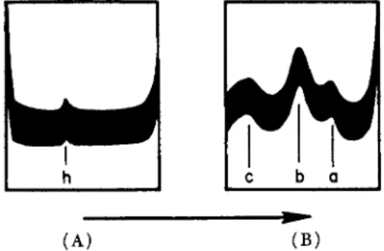
lated from a similar extract by Henrotte (1952, 1954). The latter had a molecular weight of 67,000, so that the slower ultracentrifugal peak, with S2o,w ^ 1-5 S, was probably due to particles with molecular weights of the order of 10,000 to 20,000 (Hamoir, 1955b). Recently, Connell (1958a) has reported the ultracentrifugal analysis of codling extracts (Fig 2 . ) . There was a small amount of a heavy component, h, (S2o = 93 S ) , while the sedimentation constants of the main components a, b, and c were 6.4 S, 4.4 S, and 1.3 S, respectively. Extracts made at higher ionic strengths showed, in addition to these, two fast components, and some very heavy, polydisperse material. The main components in the
282 W. J . DYER AND J . R. DINGLE
ultracentrifugal patterns of codling and carp, therefore, agreed very well both in number and in sedimentation constant, although there were differences in their relative amounts.
h
I
c b o(A) ( B ) FIG. 2. Ultracentrifugal pattern of a 5-min. extract of codling muscle at μ = 0.05, pH 7.5 (A) 480 and ( Β ) 7812 sec. after reaching full speed of 59,780 rev./min.
Temperature 2.3°C. (After Connell, 1958a.)
Electrophoretic analysis shows that albumin extracts of muscle are much more complex than is indicated by the ultracentrifugal patterns.
Connell (1953a, b ) has made such analyses on extracts of codling, cod, and 19 other species of fish, and Dingle et al. (1955) have also inves
tigated the albumin extracts of postrigor cod. A typical pattern of a cod
FIG. 3. Electrophoretic pattern (ascending) of an extract of cod muscle made at low ionic strength. Run made in 0.1 Μ KCl and Κ phosphate buffer, total μ = 0.15, pH 6.65. Numbering of peaks is arbitrary. (After Dingle et al., 1955.) extract is shown in Fig. 3, where, in addition to a rapid component not shown, at least six others can be distinguished. Further components were noted when the analysis was carried out at still lower pH values.
Connell (1953b) and also Nikkilä and Linko (1955) found that each species had a characteristic electrophoretic diagram, and that consider
able differences in mobility and composition existed between species, and even between closely related species. The peaks in the diagrams
could, however, be divided into definite mobility groups of approxi
mately consistent quantitative size. Thus, one group consisted of small amounts of ill-defined rapid components; a second group of intermediate speed comprised roughly 2 0 % of the extracts; while the third group contained most of the material as slow-moving components. The same rough division into mobility groups has been found with albumin ex
tracts of rabbit and other mammals, but it is noteworthy that the cor
responding mobilities are considerably higher and the isoelectric points lower in the cases of most fishes than for the warm-blooded animals.
(A) ( B ) FIG. 4. Ascending electrophoretic patterns of extracts of carp muscle made at
low ionic strength. Runs made at μ = 0.15, pH 7.2-7.3. (A) White muscle.
( B ) Red muscle (crosshatching indicates turbidity). Top patterns, unfractionated extracts; bottom patterns, after dialysis of extracts against water. Numbering of peaks is arbitrary. (After Hamoir, 1955a.)
Exceptions to this are the elasmobranchs, whose muscles contain pro
teins with isoelectric points higher than those in other fishes (Connell, 1953b; Dingle and Dyer, 1955).
Hamoir (1955a, b ) has investigated the electrophoretic patterns for both the white and red muscles of carp (Fig. 4 ) . A notable difference between the two was the appearance in the latter of a peak associated with the red color (myoglobin). When the extract of the white muscle was dialyzed against water, there was a considerable simplification of the pattern due to the precipitation of some of the protein (so-called globulin-X). Much less change was noted when the red-muscle extract
284 W. J . DYER AND J . R. DINGLE
was similarly treated, and Connell (1953a) found that with codling extract, only peak 2 (Fig. 3 ) was preferentially precipitated. The proteins involved in these changes appear to have low stability under the conditions of the experiments, and when precipitated can usually be only partly redissolved. They may be responsible for the rather poor reproducibility of extracts found to contain them. Peak 2 is also note
worthy in that any turbidity occurring in the extract always seems to coincide with this peak, and is also readily lost by dialysis against water, or by lowering the pH (Dingle et al, 1955).
4. Fractionation and Nature
Relatively little progress has been reported so far in separating and identifying the various components of the albumin extracts of fish muscle. On the other hand, there have been a number of reports of fractionation procedures applied to mammalian muscle extracts. Notable is the work of Disteche (1953), who used salting out with ammonium sulfate at carefully selected pH's, and of Askonas (1951), who employed precipitation with organic solvents. Some workers have applied similar procedures to fish muscle extracts. Henrotte (1952, 1954) succeeded in crystallizing a component of carp albumins that had an electro
phoretic mobility corresponding to the main peak of the water-dialyzed extract. Hamoir (1955a, b ) prepared crystals of the pigmented protein, myoglobin, from the red muscle of carp, while Huys (1954) achieved the complete purification of a similar protein from the bluefin tuna.
A protein of low molecular weight and unusual amino acid compo
sition has been isolated from the albumins of carp and also of plaice (Henrotte, 1955; Hamoir, 1957; Jebsen and Hamoir, 1958). Nikkilä and Linko (1955) have used paper electrophoresis to follow the frac
tionation of albumins of the Northern pike by ethanol at low tem
peratures, and apparently succeeded in separating the principal com
ponents. Dingle and Neelin (unpublished) have attempted fraction
ation of cod albumins by several methods. Precipitation with acetone at low temperature was apt to lead to considerable denaturation of the protein unless carefully controlled, but a fraction consisting mainly of component 5 (Fig. 3 ) was prepared by this method. ATP-creatine- transphosphorylase activity was found in certain fractions prepared by selective precipitation of the albumins with Z n + + ion at low concen
trations. These fractions contained components 5 and 6. It was further found that proteins corresponding to components 3 and 4 resisted pre-
cipitation with Z n+ +, and that component 4 could be removed by H g + + ion. The protein that remained in solution was rather heat- stable, and proved to have myokinase activity. Finally, by two frac
tionations of the crude muscle extract with neutral ammonium sulfate between 1.8 Μ and 2.0 M, it was easy to prepare a fraction of which 9 0 % consisted of a component corresponding to number 6 (Fig. 3 ) . This fraction was found to have aldolase activity, but in all likelihood will eventually be shown to be a mixture of a number of enzymes.
Several other enzymes of the glycolytic cycle have been detected in the unfractionated extract, and it is to be expected that most of the enzymes that have been found in mammalian muscle will eventually be isolated from fish muscle as well. Bailey (1954) gives a list of nearly 50 of these.
It is sometimes tacitly assumed that the electrophoretic components in muscle extracts are all proteins, but this is not necessarily the case.
Dingle and Dyer (unpublished), for example, have found that cod albumin extracts contain a nondialyzable substance with a nucleotide- like ultraviolet absorption, and that this material migrates with a high mobility in electrophoresis, free of protein. It is probably responsible for the rather rapidly diffusing fast peaks that have been noted in electro
phoresis of most muscle extracts.
C . PROTEINS OF THE MYOFIBRIL
1. Extraction
The yield and the nature of the constituents obtained when fish muscle is extracted with solutions of moderate ion strength depend upon the physiological state of the muscle, the ionic strength, the pH, the nature of the salt, and the degree of comminution of the tissue. Leaching of minced muscle with salt solutions leads to rather low yields, and grinding the tissue with sand is apt to cause denaturation of the protein.
When, however, cod muscle is comminuted for 3 to 5 min. with 20 volumes of normal salt solutions of pH 7.5 in a modified Waring blendor, maximal extraction of the proteins is obtained (Dyer et al., 1950). The yields vary between 77 and 9 1 % of the total protein. There is a possi
bility that some of this variation may be due to minor differences in technique, and Ellis and Winchester (1959), in an ultracentrifugal study of these extracts, found it necessary to control the operation very carefully. It is also apparent now that some care has to be exercised in the choice of salt for the extracting solution. Potassium iodide, for example, while a very efficient extractant, seems to alter the proteins in some way not yet fully understood (Dingle, 1958).
286 W. J . DYER AND J . R. DINGLE
Dyer et al. (1950) investigated the influence of pH on the extraction of cod muscle blended for 3 to 5 min. in 0.85 Μ NaCl adjusted to various pH values with several buffers. The solubilization increased sharply from zero to nearly maximum between pH 5 and 6. Hamoir (1955a, b ) obtained similar results when extracting microtome slices of frozen carp muscle. The effect of pH does not appear to depend upon the species of fish or the method of subdivision of the muscle, but is slightly influ
enced by the ionic strength.
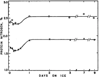
FIG. 5. The Deuticke effect in pollock. Ordinate: protein nitrogen (as per cent of sample weight) extracted by blending with 5% NaCl at pH 7. Abscissa: time of storage of fillets in ice before extraction. Top curve: total protein Ν extracted.
Lower curve: protein Ν precipitated by dilution to 0.5% NaCl. (After Dyer, unpublished.)
The amount of protein that can be extracted from Baltic herring (Clupea harengus var. membranus) by the blendor technique falls off by as much as 5 0 % when the fish enters rigor (Nikkilä and Linko, 1954).
Dyer (1951) has observed a similar Deuticke effect when pollock in the prerigor stage was stored in ice, and samples extracted by blending with 5 % NaCl after various times of storage (see Fig. 5 ) . Ellis (unpublished) obtained similar results with cod, and found also that extracts of prerigor and postrigor muscle showed no significant difference in the ultracentrifugal patterns provided that the temperature was kept near 0°C. during passage through rigor. It is not unlikely, however,
that the process of blending caused some changes to occur in the pro
teins, so that the prerigor extracts may not be truly representative of prerigor muscle. In view of the apparent importance of the physio
logical condition of the fish at the time of freezing to its keeping qual
ities in storage, there is need for further information about the changes in fish muscle associated with rigor. Many studies along these lines have been made on rabbit muscle, and have been reviewed by Bailey (1954).
Extraction of the salt-soluble proteins from cod begins at approxi
mately μ = 0.3 as shown by Dyer et al. (1950) and confirmed by Con- nell (1953a, 1958a). On the other hand, Hamoir (1955a, b ) found that the threshold in the case of carp was μ = 0.45 at pH 7.5, and the reason for this difference is not apparent. An upper limit of ionic strength for extraction is more difficult to fix. Dyer et al. (1950) showed that the efficiency of extraction fell off at ionic strengths greater than about 1.0, and Ellis and Winchester (1959) have found that certain of the com
ponents appeared to be dissociated under these conditions. It is probably advisable, therefore, to keep the ionic strength below this value.
If the residue remaining after the extraction of albumins from minced muscle is used for the extraction of the proteins soluble at higher ionic strengths, it is found that the amount of material remaining undissolved is greater than if the whole muscle had been used. It ap
pears, therefore, that the extraction of the albumins causes some changes in solubility of the other proteins. While the effect of this procedure on the ultracentrifugal and electrophoretic patterns of the salt extracts does not seem to have been determined for fish, it is felt by some that the residue from an albumin extraction is not a suitable starting ma
terial for the preparation of the other muscle proteins (Perry, 1953;
Bailey, 1954).
2. General Properties of the Extracts
Extracts of total soluble proteins made by blending fish muscle with KCl or NaCl at μ = 0.6-0.9, and pH 7.0-7.5 are turbid, exhibit bire
fringence of flow when freshly made from fresh muscle, and have a high, non-Newtonian viscosity. Such solutions probably contain very asymmetric particles of high particle weight. When ATP is added to these extracts, there is a dramatic fall in viscosity, followed by a slow recovery to approximately the original value. The time for recovery depends upon the amount of ATP added, and this reagent is dephos- phorylated in the process. When such extracts are diluted to μ = 0.2
288 W . J . DYER AND J . R. DINGLE
or less, a precipitate of protein is obtained that can be redissolved completely at μ = 0.6, provided it is not centrifuged at too high a speed or allowed to stand too long at the low ionic strength. The amount precipitated is about 7 5 % of the total protein in the extract.
Salt extracts made after prior removal of the albumins behave in a similar way, except that now about 9 5 % of the protein can be pre
cipitated by dilution. The viscosity of the extracts falls with increasing ionic strength, and also upon aging (Dingle, 1958). All of these prop
erties suggest that the salt extracts contain large proportions of acto- myosin, a protein first named by Szent-Györgyi (1947) in studies on rabbit muscle.
3. Analysis of the Extracts
The composition of the salt extracts, however, is not simple. In an ultracentrifugal study of total protein extracts of postrigor cod muscle made at μ = 0.60 and pH 7.0, Ellis and Winchester (1959) found that
(A) ( B ) FIG. 6. Ultracentrifugal patterns of extract of postrigor cod muscle made with 0.47 Μ NaCl and Na phosphate buffer, total ionic concentration 0.60, pH 7.0.
(A) Untreated extract; ( B ) solution of the precipitate obtained by tenfold dilution of the extract. Temperature 20°C. (After Ellis and Winchester, 1959.)
five constituents could be distinguished (Fig. 6 A ) . In addition to the four peaks indicated, a gel amounting to roughly 2 0 % of the total pro
tein was rapidly sedimented. The amount of component I was quite variable, and was present only in solutions containing more than 0.75 mg. protein nitrogen per milliliter. Component II was the major con
stituent. Its very sharp peak showed low spreading, and its sedimenta
tion constant was very dependent upon concentration, varying from 15 S (uncorr.) at moderate concentrations to 60 S when extrapolated to zero.
This behavior would be expected for very large or very asymmetric mole-
cules. Components I I I and IV were present in small amounts. They sedi- mented slowly, showed rapid diffusion, and little concentration de
pendence of the sedimentation constant. When the ionic strength of the extract was raised to μ = 1.2 or higher, an irreversible change in the pattern occurred. The amount of peak II, and also of peak I when present originally, decreased, while components I I I and IV increased, suggesting some kind of dissociation. When the protein precipitated by dilution of the extract to μ = 0.06 was redissolved, its ultracentrifuge pattern was similar to that of the whole extract, although an increase in the tendency of the peaks to spread indicated an increase in the hetero
geneity of the constituents (Fig. 6 B ) . Ellis (unpublished) has also found that similar patterns were obtained from prerigor fish, except that the relative amount of peak I was usually larger.
Connell (1958a) has carried out an ultracentrifugal study of extracts of minced prerigor codling muscle made with KCl at various ionic
LuLutU
A M A
(A) ( B ) ( C ) FIG. 7. Ultracentrifugal patterns of extracts of prerigor cod muscle. (A) Five- min. extract in KCl phosphate buffer, μ = 0.30, pH 7.5; temperature of run, 2.1° C.
(B) Solution of precipitate obtained by diluting extract of run (A) to μ = 0.05.
Run made in KCl phosphate buffer, μ = 0.40, pH 7.5. Temperature, 1.0°C. ( C ) Twenty-four-hr. extract in KCl phosphate buffer, μ = 0.50, pH 7.5. Temperature of run, 0.9° C. A denotes actomyosin, Μ is possibly myosin and its aggregates. (After Connell, 1958a.)
strengths. The extraction of the proteins was not complete; even after 24 hr. at μ = 0.5, only 5 3 % of the total protein was obtained in solution.
Nevertheless, the patterns revealed interesting selective extractions.
The extracts made at low ionic strengths contained mostly the albumins, but in that made at μ = 0.30 there was in addition a component ex
hibiting a sharp boundary with little spreading tendency, and a sedi
mentation constant of 4.5-6.0 S (Fig. 7 A ) . It could be precipitated by
290 W. J . DYER AND J . R. DINGLE
dilution to μ = 0.05 and redissolved, but the ultracentrifuge then showed three peaks as though polymerization may have occurred (Fig, 7 B ) . A rapid aggregation of this protein also seemed to occur even in the un
disturbed extract at 0°C. The viscosity was not sensitive to ATP, except when actin was added to the solution. These properties agreed with those assigned to the protein myosin, which had been isolated from rab
bit muscle. A 24-hr. extract at μ = 0.50 was similar to that made at μ = 0.30 except that it contained an additional extremely sharp peak, and had a high opacity and ATP sensitivity (Fig. 7 C ) . The sharp peak was, therefore, probably due to actomyosin. Connell also reported that
(A) ( B ) FIG. 8. Electrophoretic patterns of carp proteins. (A) Ten-min. extract of
carp muscle at μ = 1.0, pH 5.5. Run made at μ = 0.35, pH 7.1. ( Β ) Solution of precipitate obtained by dialysis of extract of (A) at μ = 0.25, pH 7.2. Condi
tions of run as before. (After Hamoir, 1955a.)
the peak apparently due to myosin could not be detected in extracts made from postrigor cod or from rapidly frozen prerigor cod that was extracted during the thawing process.
Hamoir (1955a, b ) has studied by electrophoresis a number of ex
tracts of microtome slices of frozen carp muscle made at relatively low pH (5.0-6.2) and μ — 0.5 and 1.0. The conditions chosen were close to the threshold of solubility of the structural proteins of this fish, but, as in Connell's work, they served to emphasize differences in solubility of the various components. The pattern of such an extract made at μ = 1.0 and pH 5.5 is shown in Fig. 8A. When the solution was dialyzed at μ = 0.25 and pH 7.2, the precipitated protein, when redissolved, exhibited a single sharp peak like number 2 of Fig. 8A, and this peak
also disappeared from the pattern of the supernate from the dialysis (Fig. 8 B ) . The solution of the peak 2 material had a strong flow bire
fringence and high viscosity characteristic of actomyosin. The shape and area of peak 1 changed after removal of peak 2, and this could be explained by the existence of some kind of interaction between the two proteins. When the supernate from the dialysis was diluted to μ = 0.025, the precipitated protein, after redissolving, usually showed two peaks corresponding to numbers 1 and 3 of Fig. 8A and B . A marked asym
metry of the ascending and descending patterns also indicated an inter
action to exist between these two proteins as well. They could, how
ever, be separated by ammonium sulfate fractionation. Component 1 corresponded to tropomyosin (which has been prepared from fish by a different method) while component 3 was analogous to the myosin found in and prepared from rabbit muscle extracts.
4. Myosin
The protein myosin was first prepared from minced prerigor rabbit muscle by a 10-min. extraction with 0.6 Μ KCl, but until recently all at
tempts to isolate it from fish muscle by a similar procedure seemed to fail. Nevertheless, it is known from the work of Hamoir and Connell just briefly described that a protein of analogous properties exists in certain extracts of fish muscle. Moreover, when ATP is added to a salt extract of carp or cod, the heavy components disappear, and are re
placed by a symmetrical peak with a sedimentation constant of approx
imately 6 S (Hamoir, 1951, 1955b; Dingle, 1958), in agreement with the behavior of rabbit muscle extracts. As already mentioned, the protein in question can be precipitated by selective dialysis, but when redissolved shows signs of having undergone some aggregation. Connell (1958b) has recently succeeded in isolating unaggregated cod myosin by ultracentrifuging a solution of the salt-soluble proteins in the presence of ATP to remove actin. The myosin remaining in the supernate was probably contaminated with a small amount of tropomyosin. Such preparations showed a single sharp boundary in the ultracentrifuge, but analyses of diffusion behavior showed that the material was slightly heterogenous. It was proved that cod myosin aggregates much more readily than the rabbit protein by working up preparations from the two animals exactly in parallel. The cod myosin could not be precipi
tated even from monodisperse solution without aggregating, and could not be kept longer than 10-12 hr. at 0°C. before the first signs of aggre-
292 W . J . DYER AND J . R. DINGLE
gates appeared. The principal physical constants for Connell's cod myosin are given in Table I.
TABLE I
PHYSICAL CONSTANTS OF COD MYOSIN0
Constant Value Sedimentation constant(Svedbergunits) Where S0 S = S0 — kc (c in g./ml.) 0 = 6.43 (standard error = 0.08)
Where k = 3.18 (standard deviation = 0.39) Temperature 0-5 °C.
Diffusion constant D = 1.10 X lO^cm^sec."1 (standard error 0.05 Χ 1 0- 7) (stationary boundary) Temperature 0.5°C.
Specific volume V = 0.735 cm.3g.-i a t 20°C. and mean c = 0.34 g./100 ml.
Molecular weight 530,000 (Svedberg equation)
(standard deviation ^ 30,000) 590,000 (Archibald procedure)
(standard deviation ^ 50,000) Intrinsic viscosity 1.80 cm.3g._1 (standard deviation = 0.05)
Temperature 0.5°C.
β Connell, 1958b.
6 For carp myosin at 2 0 ° C , S2 0 = 6.55 and k = 1.6 (Svedberg units) (Hamoir, 1951).
5. Actin
When rabbit muscle is dehydrated by acetone and then extracted with water, there is obtained a solution of a protein that is capable of forming some kind of a complex with myosin, and that can also undergo a reversible polymerization in the presence of salts. This protein has been called F-actin in the polymerized form, and G-actin in the un- polymerized form. Ellis and Winchester (1959) have speculated that the gel fraction observed in their extracts may have been composed largely of F-actin, but the presence of either form does not appear to have been detected in other studies of salt extracts of fish muscle.
Hamoir (1955a) did, however, observe a gradient in electrophoresis of a 1 Μ KCl extract of carp muscle that had been previously treated with ethanol, that he thought was due to F-actin.
Connell (1954) has described the preparation of actin from prerigor cod muscle according to a modification of the acetone procedure that had been used for rabbit actin. The product appeared to be nearly pure according to electrophoresis. The depolymerized or G-actin ob
tained could be polymerized upon addition of salts to a viscous solution
of F-actin that exhibited pronounced birefringence of flow. The F-actin could be irreversibly depolymerized again by dialysis against water or by treatment with 0.6 Μ KI, in agreement with the behavior of rabbit actin. A method due to Szent-Györgyi (1951) has also been applied to the preparation of cod actin (Dingle, 1959). The protein precipitated by dilution of a KCl extract of postrigor cod was dissolved in 0.6 Μ KI containing ATP and ascorbic acid, and then treated with 0.25 volume of ethanol. This procedure supposedly first dissociated the acto- myosin present to G-actin and myosin, and then denatured the myosin,
TABLE I I
PHYSICAL CONSTANTS OF FISH ACTINS
Constant G-Actin F-Actin
Electrophoretic mobility, ascending
( Χ 1 05 cm^volrisec.- 1) —3.9« — 6.5(?)«
—4.58* — 6.5*
Sedimentation constant (Svedberg units) 3.3<* 59.6*
Sedimentation constant (Svedberg units)
4.6& 28.0&
2.1& 4.3&
2.3*
Diffusion coefficient (cm.2sec.- 1) 2.3 χ 10-7« —
Molecular weight ^ 1 3 0 , 0 0 0 * —
Frictional ratio 2.7<* —
Axial ratio (assuming prolate ellipsoid 25<* — with 30% hydration)
β ConneU (1954).
b Dingle (unpublished).
ο Hamoir (1955a).
* Connell (1958b).
while the ATP was intended to ensure that the polymerizability of the actin was not destroyed by the KI. After dialysis in the presence of ATP to remove the KI, the addition of KCl caused an increase in vis
cosity and the appearance of strong flow birefringence. Electrophoresis of an irreversibly depolymerized sample indicated only a minor im
purity, while sedimentation suggested rather more. Tropomyosin was a likely impurity, and under the conditions of electrophoresis used would probably migrate with nearly the same mobility as actin.
The rather scanty physical data concerning fish actin are collected in Table II.
294 W. J. DYER AND J. R. DINGLE
6. Actomyosin
The term "actomyosin" is often applied to that portion of fish muscle protein that can be extracted with salt solutions of moderate strength, but which precipitates upon dilution to low ionic strength. In this respect, at least, the fraction does appear to behave as though it were a single substance, although, as we have seen, it is actually composed of several constituents. To increase the confusion, this material was once called "myosin," before it was realized that two myosins could be dis
tinguished: myosin-Α, or true myosin; and myosin-B, or actomyosin.
These could be obtained more or less separately by extracting prerigor rabbit muscle for 10 min. and 24 hr. respectively with suitable salt solutions. It is, therefore, necessary to determine whether the terms
"actomyosin" and "myosin," when they occur in the literature, are used as short forms for "salt-soluble muscle proteins," or whether they refer to actual homogeneous protein components. Perhaps it would be better to return to the still older term "muscle globulins" when referring to the unfractionated salt-soluble proteins.
The Szent-Györgyi group of investigators has shown that when myo
sin and actin prepared from rabbit muscle are mixed, the viscosity of the resulting solution is much greater than the sum of the viscosities of the two proteins separately. They also showed that the viscosity could be greatly reduced temporarily by the addition of ATP. It was con
cluded that the myosin and actin had combined to form a new protein that they called actomyosin. Because of the qualitative similarities in the behaviors of such mixtures and of extracts of fish muscle made at moderate ionic strength, it was natural to suppose that the fish extracts must contain such a complex protein. As we have seen, this has proved to be the case, and the protein has been isolated by a careful adjustment of ionic strength.
The electrophoretic pattern of actomyosin is characteristic (Fig.
8 B ) . While the descending peak is fairly symmetrical and spreads during the run, the ascending peak is extremely sharp and shows very little spreading tendency. Both peaks are usually partially obscured by the turbidity of the solution. The mobility appears to be independent of the mode of preparation and is close to that of rabbit actomyosin (Hamoir, 1955b). This author reports ascending and descending mo
bilities of —2.88 X 1 0 -5 and —2.60 X 1 0 "5 cm.2 volt"1 s e c . "1 re
spectively for carp actomyosin in NaCl-phosphate of μ = 0.35 and pH 7.1. The mobility of cod actomyosin is similar to this but appears to
depend upon whether Na+ or K + ions are present in the solutions (Dingle, 1958).
Carp actomyosin also exhibits a very sharp peak in the ultracentri- fuge except at low concentrations, and has a sedimentation constant very dependent upon concentration (Hamoir, 1955b). When extrapolated to zero concentration, the constant approaches 90 S.
The viscosity of fish actomyosin is also dependent upon concentration and is higher than that of rabbit, although the intrinsic viscosities ap
pear to be about the same, indicating similar axial ratios (Hamoir, 1955a; Dingle, 1958). The differences between these two actomyosins in the properties related to the size and shape of the molecules at mod- rate concentrations appear to be due to a more pronounced solution structure in the case of the fish actomyosin.
Fish actomyosin appears to be slightly but definitely more soluble than that of rabbit. The solubility threshold for the latter is about μ = 0.30 in neutral NaCl-phosphate, while it is approximately μ = 0.25 in the case of carp and cod (Hamoir, 1955a, b; Dingle, 1958). The molecular weight of actomyosin is extremely large, and the difficulty of measuring the size of such giant molecules is probably responsible, at least in part, for the lack of agreement in the values reported. In a recent investigation of cod actomyosin, turbidity measurements gave a molecular weight of 60-80 X 1 06 (Connell, 1958b). From the intrinsic viscosity of carp actomyosin Hamoir (1955b) calculated an ellipsoidal axial ratio of 137. Introduced into Perrin's equation, this gave a fric- tional ratio f/f0 of 4.73, and together with the classical sedimentation equation gave a molecular weight of 39.9 Χ 106. This would correspond to an ellipsoid with dimensions of 12,000 χ 88 A.
The effect of adenosine triphosphate upon actomyosin is of consider
able interest and importance. The characteristic decrease of viscosity and even the change in the ultracentrifuge pattern could, in the absence of other evidence, be interpreted either as a change of molecular shape to a more symmetrical one, or as a dissociation into smaller molecules.
Light-scattering measurements appeared to indicate the former (Blum and Morales, 1953), but the weight of evidence at present favors the latter conclusion. The reason for the discrepancy is still not known.
The most conclusive evidence for dissociation is probably the ultra
centrifugal separation of myosin from solutions of actomyosin containing ATP (Weber, 1956; Connell, 1958b), by which actin appears to be sedimented out, leaving an abnormally high concentration of myosin in the supernate.
296 W. J . DYER AND J . R. DINGLE
When relatively pure actin and myosin are mixed, the behavior of the resulting solution suggests that actomyosin is formed, but it cannot yet be said that this material is identical with the actomyosin extracted directly from the muscle. In fact, many conflicting reports on the ratio of myosin to actin in various actomyosins indicate that the natural and synthetic proteins are not identical.
Both myosin and actomyosin are capable of hydrolyzing one phos
phate group of ATP with the formation of adenosine diphosphate, which is incapable of affecting the properties of actomyosin as does ATP (Engelhardt and Ljubimova, 1939). This ATPase activity is an obvious explanation of the temporary nature of the ATP effect on actomyosin, and also suggests that the dissociation is at least partly reversible. There are many reports, however, indicating that after the ATP has been destroyed, the properties of the solution are not completely restored, so that here again the reformed actomyosin does not appear to be iden
tical with the "natural" actomyosin. The ATPase activity of the protein from fish does not seem to have been studied systematically as yet.
The cause of the changes in properties of solutions rich in acto
myosin that occur in the presence of salts at high ionic strengths is not clear. At first sight, they appear to be qualitatively similar to those brought about by ATP, but are permanent and irreversible (Ellis and Winchester, 1959). However, when ATP is added to a solution of acto
myosin at μ = 2.0, it seems to override the effect due to the high ionic strength, and gives rise to the characteristic pattern found in the presence of ATP at lower ionic strengths (Dingle, 1958). Barany et al (1951) have also reported that the two effects are different.
7. Tropomyosin
Tropomyosin was first described by Bailey (1946), who isolated it from rabbit muscle that had been dehydrated by organic solvents. By a similar procedure it has also been prepared from whiting (Bailey, 1948), carp (Hamoir, 1951), haddock (Kubo, 1957), and cod (Dingle and Odense, 1959). It is readily purified by precipitation from 1 Μ KCl solution at pH 4.3, and by fractionation with ammonium sulfate (1.7 to 2.8 Μ ) , or with ethanol, giving a final purity of 9 0 - 9 5 % . The yields of purified tropomyosin varied from 1.8 to 2.6% of the total muscle protein, but the extractions were probably not quantitative. It does not appear to arise from some degradation of the other constituents by the action of the ethanol and ether used in extraction, since attempts
to prepare it by treating actomyosin in the same way have failed (Bailey, 1948). It has also been obtained by extraction of carp muscle with salts of various ionic strengths and pH (Hamoir, 1955a, b ) , al
though the yields were small. This author also reported the occurrence of several tropomyosins, characterized by different contents of ribonu
cleic acid as well as different viscosity, electrophoretic and sedimentation behaviors. Perry (1953) has shown that tropomyosin is a component of the fibrils, since it was slowly leached from isolated rabbit fibrils by 0.078 Μ borate buffer at pH 7.1, without the intervention of organic solvents. He calculated a yield of 2.6% of the total muscle proteins, in agreement with the yield by Bailey's ethanol-ether treatment. Fur
thermore, the phosphorus content indicated that tropomyosin could contain not more than 2.5-3.0% nucleic acid. The origin of Hamoir's nucleotropomyosin is not clear.
Ordinary cod and haddock tropomyosins extracted from the dehy
drated muscle appear as well-defined and monodisperse components in electrophoresis and sedimentation. They can be readily crystallized in a characteristic form which is the same as that obtained from carp and rabbit. Once extracted, tropomyosin remains soluble in water at pH's above about 6, although under these conditions the viscosity increases considerably. Nevertheless, it does not seem to occur in the albumin extracts made at low ionic strength.
Some physical constants of several tropomyosins are collected in Table III.
8. An Unidentified Muscle Protein
In his work with isolated rabbit myofibrils, Perry (1953) found that besides tropomyosin, an unidentified protein amounting to about 8%
of the total muscle protein was extracted by the 0.078 Μ borate buffer.
It was not myosin, F-actin, or G-actin, and presumably not actomyosin.
This unknown protein seems to resemble the Y-protein of Dubuisson (1950), although the latter was extracted from frozen normal muscle only at high ionic strength. Similar work does not appear to have been carried out with fish muscle, but the occurrence of such a protein in fish extracts should be expected.
9. Amino Acid Composition of Structure Proteins
Almost no complete amino acid analyses of these proteins from fish have yet been published, and with the exception of tropomyosin, un
certainties about the purities of the fractions prepared up to the present
PHYSICAL CONSTANTS TABLE III OF FISH TROPOMYOSINS, PREPARED BY BAILEY'S PROCEDURE (1948) Constant Cod<* Carp0 Haddock0 Electrophoretic mobility, —7.28 (— -6.26) —7.11 (—6.40) — (—6.53) cm^volfisec.-1 Χ 105 (K+ ions, μ = 0.15, pH 7.0) (Na+ ions, μ = 0.15, pH 7.29) (Na+ ions, μ = 0.15, pH 7.4) (descending mobilities μ = 0.15, pH 7.0) (Na+ ions, μ = 0.15, pH 7.29) (Na+ ions, μ = 0.15, pH 7.4) in parentheses) Sedimentation constant 1.7-2.3 2.15-2.31 — (Svedberg units) (Na+ ions. , μ = 0.60, pH 7.0) (μ = 0.35, pH 7.1) Molecular weight 52,600
— —
Intrinsic viscosity 0.63 (μ = 0.10)— —
Intrinsic viscosity 0.44 (μ = 0.30) a Dingle and Odense (1959). ύ Hamoir (1955a). Data on the various salt-extracted tropomyosins of carp are also given in this paper. ο Kubo (1957).298 W. J. DYER AND J. R. DINGLE
would have made the value of such analyses questionable. Even the amino acid content of whole, unfractionated fish is unknown at present.
Kubo (1957) determined the amino acid composition of haddock tropo
myosin, and found that it agreed fairly well with that of rabbit. Hoog
land (unpublished) has obtained similar results with cod tropomyosin, and has estimated a minimum molecular weight of 52,600. Tropomyosin is noteworthy in having no proline or tryptophan. Connell (1959) has analyzed cod myosin, actin, and "natural" actomyosin, finding fair agree
ment with the rabbit proteins. From these results, he estimated a weighted average of about 7 3 % myosin in the actomyosin, agreeing with the myosin to actin ratio of 3 to 1 generally accepted for rabbit actomyosin.
The lack of tryptophan in tropomyosin is useful in estimating the purity of preparations, and the amounts of other acids may be simi
larly useful with the other proteins. Although it will be difficult to achieve, a knowledge of the distribution of the amino acids as well as the over-all composition would be very interesting. It would probably help explain the greater instability of fish myosins compared with myo
sins from warm-blooded animals, for instance, and it might also clarify the nature of the interactions between the proteins that eventually lead to their insolubilization in frozen muscle.
10. The Organization of the Myofibril
The presence of any particular protein in any of the various extracts of fish muscle does not necessarily mean that the protein occurs in the muscle in the same form as it occurs in the extract. This applies espe
cially to actomyosin, which could conceivably be formed from actin and myosin as they are extracted from the muscle. The albumins, how
ever, do not exhibit any clear evidence of interactions, so they are prob
ably extracted without change. Tropomyosin also probably exists as such in the fibrils, although its role is unknown.
Considerable advances in determining the fine structure of the fibrils of striated rabbit muscle have been made in recent years with the help of electronmicrography combined with various extractions of the fibrils themselves. Complete agreement has, however, not been attained in interpretation of the evidence. This work has been recently reviewed by Hamoir (1956). By means of density measurements by interference microscopy before and after extraction of isolated rabbit myofibrils for myosin, Huxley and Hanson (1957a, b ) have shown fairly conclusively
300 W. J . DYER AND J . R. DINGLE
that at least four-fifths of the myosin is concentrated in the nonisotropic or Α-bands of the fibrils. The extraction procedure also removed a small amount of an unidentified protein, but did not appear to affect any but the Α-bands of the fibrils. Electron micrographs of cross sec
tions of the fibrils made at different locations indicated the presence of two sets of filaments in hexagonal arrays. One set, consisting of heavy filaments, occurred only in the Α-bands and was probably myosin, since it disappeared after a myosin extraction. The second set, of finer fila
ments, which were probably composed of actin, occurred in the isotropic or I-bands, and also in part of the Α-bands. In the latter, the two sets intermeshed (Huxley, 1953). These observations have formed the basis of a model structure for muscle, in which the two sets of overlapping filaments slide past each other when the muscle changes length (Hanson and Huxley, 1955). Presumably, the amount of ATP present would govern the degree of interaction between the two sets of filaments.
While this particular model may not be universally accepted, it seems probable that some such ordered array of actin and myosin molecules exists in muscle. If the nature of this fine structure could be established, it would probably assist in understanding the changes that occur in frozen storage, which probably involve the establishment of some form of crosslinks between the filaments.
D . OTHER MUSCLE PROTEINS
That portion of the muscle that remains insoluble after extraction with salt solutions, called the "stroma," is made up of the tough myo
commata together with vascular tissue and probably fiber membranes or sarcolemma. It amounts to only 3 % of the total muscle protein in teleosts, and about 1 0 % in elasmobranchs, compared with about 17%
for mammals. Very little is known of the nature of fish stroma. The myocommata probably consist largely of collagen and elastin, and changes in these may be responsible for some of the soft fish sometimes encountered in the warmer months.
Muscle cells contain various particulate bodies including nuclei and granules or "sarcosomes," and these occur in the sarcoplasm. They are less numerous in white muscle than in red, and little work seems to have been done on these bodies in fish muscle. According to Perry (1952), the granules, like other mitochondria, consist of a phospholipid- protein-nucleic acid complex. Love (1955a, b ) was probably dealing with these bodies when he measured the nucleic acid content of ex-
pressible fluid of fish fillets as a possible index of damage to cell mem
branes. His failure to centrifuge down the nucleic acid even after 60 min. at 24,000 g may have been due to convection disturbances rather than to the acid being in true solution. The granules of pigeon breast muscle have been shown to respire, giving rise to energized phosphate bonds, and are hence responsible for the aerobic supply of ATP to the living muscle (Chappell and Perry, 1953). The granules also appear to contain a Mg-activated ATPase. A list of other mitochondrial enzymes is given by Bailey (1954).
The nucleic acids of fishes and the enzymes concerned in their post
mortem breakdown have been the subject of an interesting series of investigations. It was found that the muscles of certain fishes were capable of forming free ribose from added ribonucleic acid, ATP, ribonucleotides, ribosides, and ribose-5-phosphate (Tarr, 1953). The ribose is an important contributor to the browning (Maillard) reaction that occurs when such flesh is heated, and the enzymes responsible for this breakdown (and synthesis) of nucleic acids could be of considerable economic importance, apart from their contribution to browning re
actions. Tarr (1955a, b, 1957) has succeeded in isolating several of these enzymes, and in finding evidence for the existence of other similar en
zymes in ling cod and rock cod, but it is not clear from the isolation procedures whether these would be expected to occur in the sarcoplasm or within the fibrils. Crude phosphoribomutaste of ling cod has also been used to synthesize for the first time deoxyribose-l,5-diphosphate.
IV. Changes in Frozen Fish Protein
The occurrence of deterioration in frozen fish products has been known for many years (Huntsman, 1929). Frozen fish frequently was dry and tough and comparatively flavorless. Further, it occasionally developed "rust" on the surface.
Huntsman (1929), on the other hand, reports as early as 1924 that frozen haddock stored six months was practically indistinguishable from those freshly landed. Frozen haddock fillets were also put on the To
ronto market on a commercial basis (Huntsman, 1931).
Unfortunately, many of the shortcomings of frozen fish still remain.
To many basic research findings the trade has not paid attention. In some respects the specific protein changes are not well understood.
Several points have to be taken into consideration when analyzing these phenomena: first, the changes brought about by freezing in the
302 W. J . DYER AND J . R. DINGLE
milieu of the protein micelles and the effect of these changes on the muscle proteins; second, the various factors which affect the degree and rate of these changes during storage; and third, the influence of other tissue constituents.
A . PROTEIN DENATURATION
The term "denaturation" should be defined, since it has come to have a broad meaning. For our purposes it will suffice to define it as a change in the protein, such that it is no longer soluble or extractable by salt solutions under conditions in which the native protein is soluble or extractable.
In frozen fish it was noted by Reay (1933, 1934, 1935), following the work on salt extraction of muscle proteins by Bate-Smith (1934, 1937), that there was a diminution of the extractable protein on frozen storage. He also showed that this was greater at high storage temper
atures than at low, and that it increased with storage time. The major decrease was found to be in the globulin fraction of the protein. Since the extraction methods used were slow and inaccurate, Reay relied mainly on the development of a sheen after smoking as a convenient test. This test depended upon the swelling effect of the presmoking brine dip on the actomyosin, resulting in a swollen surface, slippery to the touch, which dried during the smoking to give a shiny appearance.
In contrast, products which had undergone denaturation exhibited a dull rough appearance.
Shortly afterward, a blending technique was developed (Dyer et al, 1950) which allowed almost complete extraction of the proteins from fresh fish and could be conveniently used to determine the loss in extractability on frozen storage.
By the use of this method it was shown that the actomyosin fraction became insoluble during storage. In some species, there appeared a general parallel between this insolubility and deterioration in quality as measured by tasting tests.
B . CHANGES IN THE WATER-PROTEIN-SALT SYSTEM ON FREEZING
1. Composition
The composition of fish muscle has been discussed in Chapter 6. It will be seen that in whitefish containing only 1% or less of lipid con
stituents, the moisture content is approximately 80-82%. The total protein content is about 16-18%. The remaining 1-3% is accounted
for by various inorganic and organic salts, and various nonionic organic compounds. Freezing curve data have shown that the freezing point depression of these salts is equivalent to a 1.4% solution of NaCl (Dyer et al., 1957a). Phosphate makes up a large proportion of the inorganic constituents of these salts, with much smaller amounts of K, Na, Ca, and Mg (see Vinogradov, 1953). The organic constituents include lactic acid, about 0.5% (Reay et al, 1943); trimethylamine oxide, 0.4r-1.0%
(Dyer, 1952); taurine, 0.3% (Shewan and Jones, 1957); other constitu
ents which are only partially ionized; and traces of amino acids, NH3, etc. It should be pointed out that no good analysis of the inorganic and organic constituents of the muscle tissue of various species of fish has been made (Love, 1957). In addition it must be recognized that there is considerable variation in some of the components with season and sexual maturity.
Thus in fresh fish muscle we have a water-salt-protein system with various inorganic and organic salts dissolved in a solution approximately equivalent to 1.4% NaCl, which is adsorbed more or less strongly by the protein micelles in the muscle fibrils.
2. Freezing-out of Water
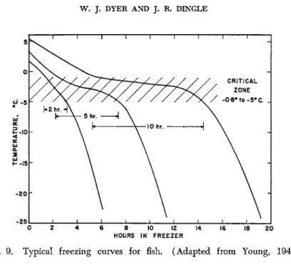
As fish muscle is cooled below 32 °F. there is often some supercooling, after which the temperature rapidly approaches the freezing point of 1.4% NaCl solution. At this point water freezes out as crystals of pure ice, and as this occurs, the salt becomes more concentrated in the liquid phase, thus further lowering the freezing point. Maximum ice crystal formation occurs between —0.8° and about —5°C. resulting in a very slow cooling rate in this zone. Below this temperature range rapid lowering of temperature can again occur (see Fig. 9 ) .
Early data on the amount of water frozen out at various temperatures were summarized by Birdseye (1929). Later data combining the results of at least three different methods are shown in Fig. 10 (Dyer et al., 1957a). Finn (1934) determined the amount of ice frozen out directly from muscle press-juice of halibut by separating through filtration the ice formed at successively lower temperatures. Mahadevan and Carter (1948) on the other hand measured the volume change on the basis of the ice formation in ling cod by means of a dilatometer. Dieuzeide and Novella (1950) show data for haddock using a method not stated. The fourth curve is based on the N a C l - H20 phase diagram with an initial NaCl concentration of 1.4%, which was found to have the same initial
304 W. J . DYER AND J . R. DINGLE
FIG. 10. Water frozen out, as per cent of total water content, at various temperatures; X, haddock; O, halibut juice; Δ, ling cod; —, 1.4% NaCl solution.
(After Dyer et al, 1957a.)
FIG. 9 . Typical freezing curves for fish. (Adapted from Young, 1 9 4 0 . )
freezing point as the muscle of the Atlantic cod. These curves obtained by different methods and on different species agree remarkably well.
They also agree with values for cod, haddock, and burbot as they were calculated by Mennie (1932), based on calorimetric measurements. The values for skates were considerably lower, but the elasmobranchs are known to have a much higher content of nonprotein nitrogenous con
stituents. The curves illustrate that about 8 0 % of the water in the tissue is frozen out between —0.8° and —5°C. About 9 0 % is frozen out at
—10 ° C , and below this the change becomes smaller and more difficult to measure.
During this freezing-out of water as ice, the moisture remaining associated with the protein micelles becomes more and more concen
trated in salts, the amount of moisture becomes very much less and considerable dehydration of the protein occurs. This becomes important when the effects of freezing rate, thawing rate, and of fluctuating storage temperatures are considered (see below). Unfrozen cod muscle, for instance, contains about 18 g. protein, and about 80 g. water which is mostly incorporated in the protein gel. At — 1 0 ° C , only about 8 g.
water will remain unfrozen, and at — 2 0 ° C , this will be reduced still further to about 3 g. Also, the salts present will have become quite concentrated. Since many of them are very hygroscopic, e.g., trimethyl
amine oxide and lactic acid, presumably some of the water will also be bound by these materials, thus further limiting the moisture available to the protein gel. However, not much is known of the processes occurring at these temperatures in such a complex system.
3. Theories of Freezing Damage
Most of the older theories are concerned with mechanical damage to the cell walls and structure by the formation of ice crystals of various sizes. The early literature was summarized by Zarotschenzeff (1930), Notevarp and Heen (1940), and Callow (1952).
MacKay and Weld (1926), Weld (1927; see also Zarotschenzeff, 1930) showed that the average size of the crystals formed during slow freezing was appreciably larger than those formed during rapid freezing. Further, in agreement with earlier German work, histological examination showed that large extracellular crystals were formed on very slow freezing, and many broken cells were present. Very fast freezing, on the other hand, resulted in large numbers of small crystals throughout the tissue, leaving the cells undamaged. This general picture has been amply confirmed by subsequent investigations. Weld also showed that during storage the