Current Medicinal Chemistry, 2020, 27, 1-21 1
REVIEW ARTICLE
0929-8673/20 $65.00+.00 © 2020 Bentham Science Publishers
The Nitric Oxide Pathway in Pulmonary Arterial Hypertension: Path- omechanism, Biomarkers and Drug Targets
Zsófia Lázár
1*, Martina Mészáros
1and Andras Bikov
1,2*1Department of Pulmonology, Semmelweis University, Budapest, Hungary; 2Manchester University NHS Foundation Trust, Manchester, United Kingdom
A R T I C L E H I S T O R Y
Received: November 08, 2019 Revised: January 03, 2020 Accepted: February 20, 2020
DOI:
10.2174/0929867327666200522215047
Abstract: The altered nitric oxide (NO) pathway in the pulmonary endothelium leads to in- creased vascular smooth muscle tone and vascular remodelling, and thus contributes to the development and progression of pulmonary arterial hypertension (PAH). The pulmonary NO signalling is abrogated by the decreased expression and dysfunction of the endothelial NO synthase (eNOS) and the accumulation of factors blocking eNOS functionality. The NO defi- ciency of the pulmonary vasculature can be assessed by detecting nitric oxide in the exhaled breath or measuring the degradation products of NO (nitrite, nitrate, S-nitrosothiol) in blood or urine. These non-invasive biomarkers might show the potential to correlate with changes in pulmonary haemodynamics and predict response to therapies. Current pharmacological thera- pies aim to stimulate pulmonary NO signalling by suppressing the degradation of NO (phos- phodiesterase-5 inhibitors) or increasing the formation of the endothelial cyclic guanosine monophosphate, which mediates the downstream effects of the pathway (soluble guanylate cyclase sensitizers). Recent data support that nitrite compounds and dietary supplements rich in nitrate might increase pulmonary NO availability and lessen vascular resistance. This re- view summarizes current knowledge on the involvement of the NO pathway in the pathome- chanism of PAH, explores novel and easy-to-detect biomarkers of the pulmonary NO.
Keywords: Pulmonary arterial hypertension, nitric oxide, nitric oxide synthase, FENO, biomarkers, systemic scle- rosis.
1. INTRODUCTION
Pulmonary hypertension (PH) is defined as an in- creased mean pulmonary arterial pressure (mPAP ≥ 25 mmHg) measured during right heart catheterization.
Patients with PH are classified into five groups based on clinical presentation, haemodynamic parameters, pathological alterations and treatment strategies (Table 1) [1]. These groups show different survival trajectories and aetiological backgrounds. Pulmonary arterial hy- pertension (PAH), group 1, is a rare and progressive form with currently no cure and an average survival rate of less than 3 years without treatment [2].
Various subgroups of PAH have been reported in- cluding the idiopathic, the hereditary, the drug- and
*Address correspondence to this author at the Manchester Univer- sity NHS Foundation Trust, Manchester, United Kingdom, M23 9LT; Tel/Fax: +36203141599, +441612915730;
E-mail: andras.bikov@gmail.com
toxin-induced variants and those associated with other diseases, most commonly with connective tissues dis- eases such as systemic sclerosis (SSc) and congenital heart disease [1] (Table 1).
In PAH, the pulmonary vascular resistance is ele- vated (PVR > 3 Wood units), and pathological altera- tions in the pre-capillary pulmonary vasculature are characteristic [1]. It is described by vascular remodel- ling, including medial hypertrophy and hyperplasia, intimal and adventitial fibrosis, plexiform and throm- botic lesions, which mainly affect the distal muscular type of pulmonary arteries [3]. These alterations result in gradually increasing PVR and right ventricular after- load leading to decreased cardiac output, which can be initially compensated by the adaptive processes of the right ventricle. The preservation of the right ventricular function is a major therapeutic goal in PAH, as right heart failure is a key determinant of survival [4].
Table 1. Clinical classification of pulmonary hypertension (simplified from Galie et al.[1]).
Group Number
Name of the Group Subgroups
1 Pulmonary arterial
hypertension 1.1. Idiopathic 1.2. Heritable
1.3. Drugs- and toxin-induced (e.g. anorexigens including aminorex, fenfluramine, dex- fenfluramine and benfluorex; toxic rapeseed oil; selective serotonine reuptake inhibitors) 1.4. Associated with
1.4.1. connective tissue disease (e.g. systemic sclerosis) 1.4.2. HIV infection
1.4.3. portal hypertension 1.4.4. congenital heart disease 1.4.5. schistosomiasis
2 PH due to left heart disease Most commonly patients with systolic or diastolic left ventricular dysfunction or valvular disease
3 PH due to lung diseases
and/or hypoxia Including patients with COPD, interstitial lung disease, sleep-disordered breathing, alveo- lar hypoventilation or chronic exposure to high altitude
4 Chronic thromboembolic PH and other pulmonary artery
obstructions
Other pulmonary artery obstructions include angiosarcoma, intravascular tumors and congenital pulmonary artery stenoses
5 PH with unclear and/or mul- tifactorial mechanisms
Such as patients with haematological, systemic or metabolic disorders COPD: chronic obstructive pulmonary disease, HIV: human immunodeficiency virus, PH: pulmonary hypertension.
All layers of distal pulmonary arteries undergo vas- cular remodelling in PAH manifested in structural changes in the endothelium (concentric intimal fibrosis, endothelial cell proliferation resulting in plexiform le- sions), medial layer (proliferation of smooth muscle cells) and adventitia (fibrous broadening, perivascular inflammation, appearance of tertiary lymphoid tissue).
Adapted from Dorfmüller et al.[5].
Genetic predispositions together with pathological triggers, i.e. inflammatory stimuli, oxidative stress, hy- poxia, mechanical strain and altered cellular metabo- lism, induce the remodelling and dysfunction of all layers of the pulmonary arteries [3], with endothelial injury being the main disease driver (Fig. 1). Endothe- lial dysfunction in PAH mostly refers to the imbalance between the production of endothelium-derived vaso- constrictive and vasodilatory molecules. Nonetheless, the dysfunction also denotes other metabolic changes in endothelial cells including reduced anticoagulant properties, altered proliferative capacity, sensitivity to apoptosis, the altered production of reactive oxygen species, cytokines and chemokines. Increased cytosolic calcium (Ca2+) concentration is an important factor in the pathomechanism of pulmonary vasoconstriction
and endothelial dysfunction in PAH. Calcium sensing receptor (CaSR) is expressed on pulmonary artery smooth cells and it regulates intracellular Ca2+ concen- tration due to monitoring extracellular Ca2+ levels [6].
Experimental models of PAH have demonstrated over- expression of CaSR contributing to extracellular Ca2+- induced elevation in intracellular Ca2+ concentration and proliferation of smooth vascular cells in the lung by regulating several signalling pathways [7]. How- ever, the exact causes and mechanisms of endothelial dysfunction in PAH are still not wholly understood.
The diagnosis of PAH is delayed in most cases due to the unspecific symptoms (most commonly dyspnoea, syncope and chest pain) and the lack of easy-to-use diagnostic measures [8]. Large cohort studies have proved that the survival of patients has considerably improved since the introduction of specific PAH thera- pies [9], however, the rate of disease progression shows higher individual differences and initiating the right treatment at the right time is crucial in disease man- agement. Current treatment modalities target the inhibi- tion of endothelial vasoconstrictive pathways (endo- thelin receptor antagonists e.g. bosentan, ambrisentan) or aim to induce vasodilatory signals via the nitric ox-
ide pathway (e.g. tadalafil, sildenafil) or prostacyclin signalling (e.g. epoprostenol, treprostinil) [1].
The dampened nitric oxide (NO) pathway is a major player in endothelial dysfunction, and drugs upregulat- ing this process are at the forefront of recommended therapies [1]. However, the involvement of NO signal- ling can be different in individual patients with PAH, and the therapeutic response cannot be fully predicted in advance besides some unspecific clinical factors such as age, sex, functional capacity and disease aetiol- ogy [10]. The treatment effect of drugs can be meas- ured by the change in haemodynamic parameters using the invasive right heart catheterization, which is bur- densome for patients and requires sophisticated meas- urement techniques.
Hence, there is an urgent need to find non-invasive biomarkers to aid diagnosis, predict therapeutic re- sponse and guide treatments. This review summarizes the current knowledge on the involvement of endothe- lial NO signalling in the pathomechanism of PAH and discusses the application of non-invasive markers to assess the endothelial NO pathway - with focus on ex- haled NO – with the aim to facilitate disease diagnosis and monitoring.
2. ALTERED NITRIC OXIDE SIGNALLING IN PAH
2.1. The NO Signalling Pathway in the Pulmonary Vasculature
The pulmonary circulation is the low resistance part of the circulatory system which carries deoxygenated, carbon dioxide-rich blood from the right heart to the lungs and returns oxygen-rich blood to the left heart
[11]. The vascular tone is maintained by the balance of vasoconstrictive (i.e. endothelin-1, serotonin, etc.) and vasodilator (prostacyclin and nitric oxide) mediators.
Nitric oxide (NO) is a highly soluble free radical gas which is produced from L-arginine during its con- version to L-citrulline by nitric oxide synthases (NOS, Fig. 2). Tetrahydrobiopterin (BH4) is an important cata- lysator for this reaction, and the lack of BH4 is associ- ated with uncoupling of NOS, decreased NO and in- creased superoxide production [11]. The three NOS isoenzymes include neuronal NOS (NOS1 or nNOS), inducible NOS (NOS2 or iNOS) and endothelial NOS (NOS3 or eNOS). eNOS is mainly expressed in the vascular endothelium and it is the main source for NO in the pulmonary circulation [11]. However, other cells in the lungs, such as alveolar type II cells [12] and al- veolar macrophages [13] express eNOS as well. nNOS is expressed by the endothelium, vascular smooth mus- cle cells and cholinergic neurons [11, 14]. Endothelial and neuronal isoforms of NOS are constitutively ex- pressed and their activation depends on cal- cium/calmodulin [15], however other factors, such as shear stress, hypoxia, inflammation, growth factors, hormones and lipoproteins may also modulate their expressions [16]. iNOS can be expressed by most cell types upon stimulation by hypoxia, inflammatory cyto- kines or bacterial lipopolysaccharide [17]. Nitric oxide acts through various pathways, such as inducing the soluble guanylate cyclase (sGC) enzyme, oxidation of NO to nitrite and nitrate or reactions with protein thiols to form S-nitrosothiols. NO exerts its well-known smooth muscle relaxation effect through sGC by cata- lysing the formation of cyclic guanosine monophos- phate (cGMP) from guanosine triphosphate (GTP).
Fig. (1). Main pathological alterations in pulmonary arteries in PAH. (A higher resolution / colour version of this figure is available in the electronic copy of the article).
cGMP acts through cGMP-dependent protein kinase, ion channels and phosphodiesterase, leading to relaxa- tion of the smooth muscles (Fig. 2). Nitrite, nitrate and nitrosothiols are longer half-life metabolites of NO which serve as potential NO donors but they may also induce vasodilation in pulmonary vessels [11]. The role of NO depends primarily on its concentration, as in low levels it stimulates sGC, while in higher levels, oxida- tion is the main pathway [18]. Further details on the regulation of vascular NO formation can be found in recently published reviews [19, 20].
2.2. Damage to eNOS Signalling in PAH
Several articles have described impaired NO pro- duction in various forms of pulmonary hypertension, however, with conflicting results [11, 18, 21-25]. Some of these discrepancies are attributed to methodological factors, such as interracial differences in animal stud- ies, various measurements of NOS activity and its products. Experimental models to investigate NO and NOS activity in pulmonary vessels were discussed in the review article of Hampl and Herget [18]. Most im- portantly, many studies used hypoxic challenge to in- duce pulmonary hypertension. Chronic hypoxia can lead to decreased eNOS expression in pulmonary ves- sels [26, 27], however the data are conflicting in other studies showing that chronic hypoxia induced the ex- pression of all NOS isoforms [28, 29]. Nevertheless, the hypoxic model more closely represents group 3 pulmonary hypertension than group 1 PAH (Table 1).
In contrast, although monocrotaline treatment induces PAH-like changes in the pulmonary vessels, the toxin damages the liver, kidneys and the heart as well [30].
In their elegant study, Fagan et al. selectively dis- rupted each NOS isoform in transgenic mice. Inhibition of eNOS induced pulmonary hypertension, iNOS blockage only very mildly elevated PAP, while nNOS disruption did not have any effect [31]. In contrast, de- letion of eNOS gene induced only mild pulmonary hy- pertension in rats suggesting other responsible mecha- nisms for vascular tone [25]. It seems that under physiological condition, nNOS and iNOS do not con- tribute to the control of vascular tone. However, vascu- lar inflammation noticed in PAH can induce iNOS [17, 18]. Interestingly, in eNOS knock out rats, an increased iNOS expression and exhaled nitric oxide levels were reported, suggesting compensatory mechanisms [32].
In their seminal study, Giaid and Saleh reported de- creased eNOS expression in patients with idiopathic pulmonary arterial hypertension (IPAH - group 1.1. as shown in Table 1). Moreover, eNOS expression was the lowest in plexiform lesions in the remodelled endo- thelium [21]. This has been challenged by Mason et al.
who found significantly increased eNOS expression in plexiform lesions [33], concluding considerable het- erogeneity in eNOS appearance in patients with PAH.
Interestingly, not only vascular, but platelet eNOS ex- pression is also decreased in IPAH [34]. In contrast, in PAH associated with congenital heart defect (subgroup
Fig. (2). The pharmacological modulation of NO signalling in the pulmonary vasculature. (A higher resolution / colour version of this figure is available in the electronic copy of the article).
1.4.4. in Table 1), NO concentrations in blood were higher in patients than in controls suggesting compen- satory pulmonary mechanisms [35, 36]. Tuder et al.
studied various subgroups of patients with PAH and found no difference in eNOS expression compared to controls [37]. Interestingly, eNOS expression in pul- monary arteries can be modulated by lifestyle factors such as smoking, as low eNOS has been found in pul- monary arteries of non-hypoxemic smokers as well [38, 39].
The discrepancies discussed above may be related to the simultaneous presence of factors that up- and downregulate eNOS expression in PAH. Moreover, endothelial cell injury and plexiform regions tend to be topical in pulmonary arteries in PAH, contributing to focal differences in eNOS expression.
Mechanisms upregulating eNOS in PAH include growth factors, endothelin-1 and serotonin [40-42], the levels of which are elevated in PAH [24]. In addition, pulmonary blood flow is accelerated in PAH resulting in sheer stress which is a potent inductor for eNOS [18]. Nitric oxide production can also be induced by the vascular endothelial growth factor (VEGF) [43]
which is mainly produced in plexiform lesions in PAH and contributes to vascular remodelling [24].
Factors that downregulate eNOS in PAH are more broadly studied. Around 70% of patients with heritable and idiopathic PAH have mutations in the bone morphogenetic protein receptor II (BMPR2) gene [44].
BMPR2 is recognised as an important element in eNOS phosphorylation and upregulation [45]. Endothelial NOS is downregulated in proliferating endothelial cells and after endothelial injury [46]. Inflammation can also decrease eNOS production in the pulmonary arteries [27]. Oxidative stress can be enhanced in PAH due to sheer stress and perivascular inflammation, and it is known to reduce eNOS function by the following mo- lecular mechanisms [47]. An important mechanism leading to decreased NO bioavailability is the altered function of eNOS. Under oxidative conditions, the ac- tivity of eNOS is characterized by a decreased NO generation with concomitant superoxide anion produc- tion. This mechanism is referred as “eNOS uncou- pling” [48]. The depletion of BH4 is the main cause of eNOS uncoupling [49]. BH4 deficiency due to oxida- tive stress was reported in several animal models of PAH [50, 51]. The limited availability of L-arginine, which is the main substrate of eNOS, is also responsi- ble for eNOS-uncoupling [52]. Free radicals such as peroxynitrite can increase arginase activity resulting in lower L-arginine levels and consequential eNOS un-
coupling [53]. Moreover, increased levels of the L- arginine analogue asymmetric dimethylarginine (ADMA) also lead to the loss of eNOS activity in PAH [54]. Another potential mechanism that decreases NO production is the posttranscriptional regulation of eNOS. Phosphorylation of eNOS at different specific sites is the main regulatory mechanism in stimulation or inhibition of NO production. Phosphorylation of ser- ine 1177 (Ser1177) by serine/threonine protein kinase Akt/PKB results in enhanced eNOS activity [55].
However, phosphorylation at threonine 495 (Thr495) may constrain eNOS activity [56]. The inhibition of eNOS by abnormal Thr496 phosphorylation was con- firmed in pulmonary artery endothelial cell cultures of PAH patients [57]. Increased cytosolic Ca2+ also modu- lates eNOS function mainly by phosphorylation of eNOS (at Ser1177) by calcium-calmodulin dependent protein kinases [58]. Nevertheless, a recent study has demonstrated that lower expression of mitochondrial voltage-dependent anion channel-1 may inhibit the ac- tivity of eNOS by regulating calcium ion transport in PAH patients [59].
Low NO levels contribute to increased vascular tone in PAH. However, it may also be responsible for other pathological processes of this disease. For instance, NO can induce VEGF formation [43] as low levels of this molecule were associated with poorer right heart func- tion [60]. Nitric oxide can also inhibit thrombus forma- tion [61], thus reduced levels contribute to the in- creased tendency of in situ thrombosis observed in PAH [24]. Nitric oxide can also inhibit vascular smooth muscle proliferation and migration via iNOS [62]. Finally, NO has anti-proliferative effect as well by blocking the transforming growth factor-β1 signal [63].
The dysregulation of NO signalling in PAH has been extensively discussed in other review publications [64, 65].
2.3. Exhaled NO in PAH
Endogenous nitric oxide production can be analysed in exhaled breath samples [66]. However, to what ex- tent the NO produced in the pulmonary vessels con- tributes to fractional exhaled nitric oxide (FENO) lev- els [67] as most of it rapidly reacts with haemoglobin and is transported in the circulation is discussed [68].
Nitric oxide is the most validated biomarker in exhaled breath, as it has been investigated in various pulmonary and non-pulmonary diseases. Apart from the diseased conditions, several factors affect its concentration in exhaled air [66, 69, 70].
Most importantly, as FENO levels are influenced by the expiratory flow rate [69], the European Respiratory Society/American Thoracic Society (ERS/ATS) rec- ommendations [70] and later the ERS Task Force tech- nical standard document [66] suggested measuring ex- haled nitric oxide at 50 ml/s during single expiratory manoeuvre. It has also been acknowledged that by measuring exhaled NO at different flows, bronchial NO production (JawNO) and alveolar NO concentration (CANO) can be calculated [77].
All three NOS isoforms can contribute to FENO values, albeit of different quantities. In healthy sub- jects, exhaled nitric oxide mainly originates from eNOS and nNOS, while higher levels observed in in- flammatory airway diseases, such as asthma or obstruc- tive sleep apnoea [78], are most likely due to increased iNOS activity [67]. FENO data must be interpreted carefully, as various physiological and lifestyle factors contribute to mild changes. For instance, FENO is af- fected by the clinical characteristics, such as age, gen- der, height and weight [79-81], physical exercise [82], diet [83] or menstrual cycle [84] and most importantly smoking [85]. An analytical variance within [86, 87]
and between [87, 88] different FENO devices should also be noted. These analytical and physiological vari- ances have to be acknowledged when interpreting FENO data in pulmonary hypertension.
Kaneko et al. recruited 8 patients with primary PAH (currently classified as idiopathic or heritable PAH) and 8 controls. They measured intrabronchial exhaled NO via a bronchoscope during tidal breathing and re- ported lower levels in patients [71]. Intrapulmonary NO levels did not correlate with disease onset or sever- ity [71].
Ozkan et al. did not find any difference in exhaled NO concentrations measured with the tidal breathing method among 21 patients with primary PAH, 11 pa- tients with secondary pulmonary hypertension (seven with PAH due to congenital heart disease, portal hyper- tension or scleroderma, three patients from group 4 and one patients with group 3 disease) and nine controls [72]. However, when patients receiving epoprostenol were excluded, exhaled NO levels were significantly lower in the primary PAH group.
Cremona et al. measured exhaled nitric oxide with the tidal breathing method in 8 patients with primary PAH and 20 controls [89]. Exhaled NO production rather than its concentration was estimated. Patients with PAH had lower rate of production (2.85±0.7 nmol/l/min) compared to controls (4.69±0.35 nmol/l/min) which were related to altered diffusion
capacity [89]. Similarly, Riley et al. estimated exhaled NO production in 9 patients with primary PAH and 20 controls [90]. In contrast to the previous findings, this study did not show any difference between the two groups [90]. Archer et al. investigated exhaled nitric oxide in nine patients with anorexigen-associated PAH, 8 subjects with primary PAH and 12 controls using the tidal breathing method [91]. Interestingly, FENO levels (not reported) and NO production was higher in pri- mary PAH (198±79 nL/min) than in controls (40±10 nL/min) or anorexigen-related PAH (61±16 nL/min) without a difference between the latter two groups [91].
Kharitonov et al. compared 67 control individuals with 23 patients with systemic sclerosis (6 of them had PAH) [73]. Exhaled NO levels were measured at 500 ml/min flow (8.3 ml/s). FENO levels were lower in patients with pulmonary hypertension compared to the other two groups, and correlated with diffusion capac- ity, but not with PAP or arterial oxygen levels [73].
Girgis et al. studied exhaled NO levels at various flow rates in 5 patients with primary PAH, 20 healthy subjects and 20 patients with scleroderma (5 of them had pulmonary hypertension) [75]. There was no dif- ference in FENO measured at any flow rate or JawNO among the groups. While there was no difference in CANO between the primary PAH and control groups, CANO in patients with scleroderma and pulmonary hypertension was elevated [75]. Interestingly, when all scleroderma patients were analysed together, CANO levels were higher than in controls suggesting that the increase in alveolar NO may be due to scleroderma and not pulmonary hypertension itself [75]. The same workgroup evaluated FENO at multiple flow rates in 10 patients with PAH (8 idiopathic, 2 anorexigen- associated) and 12 controls [76]. FENO concentrations were reduced in PAH compared to controls, while there was no difference in CANO [76].
Cao et al. studied FENO at various expiratory flow rates in 115 patients with SSc (25 with PAH) and 84 control subjects [74]. There was no difference in FENO between the groups at any flow rate or in JawNO, nor was there any correlation between FENO and the pul- monary arterial pressures. CANO in pulmonary hyper- tension was significantly higher compared to both con- trol groups [74].
Malerba et al. investigated 50 patients with systemic sclerosis (12 with PAH) and 40 control subjects [92].
FENO was significantly higher in patients (11.7±8.1 ppb vs 9.0±2.1 ppb in controls). Within the systemic sclerosis group, patients with PAH had lower FENO values (10.5±4.1 ppb) compared to those without PAH.
A significant inverse correlation between FENO and systolic PAP was noted [92]. Rolla et al. studied 47 patients with SSc and 30 controls [93]. Similarly to the study by Malerba et al. [92], patients with systemic sclerosis had higher FENO levels (16.6±9.1 ppb) than controls (9.9±2.9 ppb). In contrast, subjects with SSc- PAH (n=16) had lower FENO values (10.7±5.9 ppb) compared to those without PAH. FENO levels in- versely correlated with PAP [93]. For the latter two studies, no comparison was made between the PAH and control groups [92, 93].
Akbay et al. surveyed 19 patients with PAH (13 with IPAH), 12 patients with chronic thromboembolic pulmonary hypertension (CTEPH) and 80 healthy con- trols for 6 months [94]. When all patients with PH were analysed together (n=31), they had lower FENO (16.5±6.7 ppb) compared to controls (19.8±7.7 ppb). In the PH group, there was a significant correlation be- tween FENO and tricuspid annular plane systolic ex- cursion, a marker for right heart function. Interestingly when patients within the PAH group were divided into IPAH and associated PAH subgroups, FENO levels in the IPAH group were higher (18.5±6.7 ppb vs.
12.8±4.3 ppb). No comparison has been made between the IPAH and control groups [94]. There was no differ- ence between the baseline and the 6-month FENO val- ues in any group.
Malinovschi et al. studied FENO at multiple flow rates in 22 patients with PH (PAH: n=13, groups 2-4:
n=9) and 21 healthy controls [95]. There was no differ- ence in FENO (data were not presented), JawNO or CANO between patients and controls. When PAH was compared to other forms of PH, significantly lower FENO values were obtained, however, they were not different from the healthy controls. Interestingly, JawNO was lower, while CANO was higher in PAH compared to health [95].
Carpagnano et al. investigated 24 patients with PH (10 PAH, 11 PH associated with COPD and 3 patients with PH due to left heart disease) [96]. FENO was measured according to the ERS/ATS recommendations at 50 ml/s [70] and also at different flows to estimate alveolar NO concentration. The authors did not find a significant difference between the three groups of pa- tients for FENO values measured at 50 ml/s. Interest- ingly, CANO values were higher in PAH and PH asso- ciated with COPD than in patients with left heart dis- ease. FENO or CANO levels did not correlate with any of the clinical variables.
Machado et al. prospectively investigated 17 pa- tients with IPAH treated with appropriate medications
(prostacyclin analogues: n=16, calcium-channel blocker: n=1), 5 of whom died during the 2 years of follow-up [97]. There was no difference in FENO be- tween the survivors and those who died. Baseline FENO correlated with overtime PAP drop and some increase in FENO was noted in the surviving patients' overtime [97].
In summary of the exhaled nitric oxide studies, PAH was associated with lower [71, 73, 76, 89], simi- lar [72, 74, 75, 90, 95] or elevated FENO levels or pro- duction compared to health [91]. In the studies investi- gating systemic sclerosis, the number of patients with PAH was relatively low and they were rarely compared with control subjects. FENO levels in SSc patients with PAH tended to be lower than those without [92, 93].
When evaluating alveolar NO concentrations in PAH, higher [74, 75, 95] or similar [76] values were obtained compared to health.
These discrepancies can be explained by methodo- logical differences discussed above (i.e. tidal breathing vs. single breath, various expiratory flows) and the ae- tiological heterogeneity within the PAH group (i.e.
IPAH or systemic sclerosis-associated). Nevertheless, even in studies showing reduced FENO in PAH, the differences are small and cannot easily be reproduced.
Interestingly, while lower FENO levels are concluded as the reason for impaired eNOS activity, higher CANO values, which should more closely represent pulmonary vascular NO production than the FENO, are usually attributed to perivascular inflammation. Table 2 summarises the studies which compared FENO con- centrations between patients with PAH and controls.
Reviews for further reading in relation to FENO and patients with PAH can be suggested [67, 98].
There are limited data on the dynamics of FENO in animal models of PAH. It was clearly shown that only iNOS is responsible for the changes in FENO in mice without pulmonary disease or airway inflammation [99]. In rodent models of airway inflammation, ele- vated FENO levels correlated with inflammatory com- ponents of the airways [100, 101]. Le Pavec et al. were the first to determine FENO in monocrotaline-induced PAH model [102]. In their study, common bile duct ligation (CBDL) was also performed to induce hepatic cirrhosis in the rats after monocrotaline injection.
FENO levels were lower in MCT rats compared to the controls due to decreased pulmonary eNOS expression.
CBLD led to improved survival and decreased total pulmonary resistance in MCT rats, and it was associ- ated with elevated FENO levels as a result of upregu- lated eNOS and iNOS in the lungs. The authors con-
cluded that cirrhosis may be protective against PAH [102]. Strobl et al. focused on FENO levels in a pneu- monectomy-monocrotaline rat model of PAH using a new mathematical modification of FENO measurement to calculate exhaled NO output in time [103]. In con- trast to the previous study, they found no difference in FENO in this PAH model. However, low basal exhaled NO output (21.21 ± 8.27 ppb/h) increased twenty-eight days after monocrotaline injection (23.96 ± 7.60 ppb/h) when PAH was established. Interestingly, L-arginine and BH4 combination therapy led to decreased mPAP and elevated exhaled NO output, but it had no effect on FENO. A significant inverse correlation was detected between exhaled NO output and mPAP in animals with PAH before and also after therapy, suggesting a possi- ble role for this marker in PAH models [103]. Further investigations are needed to evaluate the utility of FENO measurements in animal models.
2.4. Other Biomarkers of the Altered NO Pathway in PAH
Asymmetric dimethyl-arginine (ADMA) is a com- petitive antagonist of L-arginine for NOS, and it is in- volved in the regulation of NO signalling in the cardio- vascular system [104]. The main source of circulating ADMA is through the metabolism of methylarginines in the lungs [105]. Serum ADMA concentration was increased in patients with untreated PAH (most patients had IPAH) compared to age- and gender-matched con- trol subjects [106]. Moreover, plasma ADMA concen- trations negatively correlated with measures of right ventricular function (including cardiac index and right atrial pressure) and survival in patients with PAH [107]. ADMA also proved to be an independent predic-
tor of survival. In line with this, the same study de- scribed that the intravenous administration of ADMA increased PVR and lowered stroke volume in healthy volunteers.
Similarly, circulating ADMA concentration in pa- tients with PAH associated with congenital heart dis- ease (CHD) was higher than in controls and in patients with no PAH, but no change was noted compared to patients with idiopathic PAH [108, 109]. Interestingly, ADMA levels in PAH showed a close positive correla- tion to mPAP and PVR in both subgroups of PAH sug- gesting a role for ADMA for risk assessment of pa- tients [106, 110]. Importantly, plasma ADMA is re- sponsive to specific treatment as biomarker levels were decreased six months of PDE-5 inhibitor (sildenafil) therapy of patients with PAH due to CHD [110]. Fur- thermore, ADMA might be useful to screen for PAH among subjects with SSc, as a circulating ADMA con- centration above 0.7 µM showed good sensitivity and specificity to identify PAH in this patient group [111].
In addition, an elevated plasma ADMA concentration was also associated with the development of PAH among patients with human immunodeficiency viral infection [112].
Although ADMA production is increased primarily by hypoxia [113], it is also generated in response to inflammation [104]. A potential explanation for raised ADMA levels in PAH could be the decreased expres- sion of dimethylaminohydrolases (DDAH), which are responsible for ADMA degradation. In support, re- duced DDAH levels were found in the lungs of patients with IPAH [114]. As mentioned before, BH4 deficiency can lead to NOS uncoupling. Supporting this, inhibi- Table 2. FENO concentration in patients with PAH and control subjects.
Patients with PAH Controls
N FENO, ppb N FENO, ppb
Direction of differ-
ences in PAH Comments Reference
7 2.8±0.9 8 8±1 ↓ Exhaled NO was meas-
ured intrabronchially Kaneko et al. [71]
21 10±1.3 9 6.6±0.6 ↔ Exhaled NO was meas-
ured with the tidal
breathing method Ozkan et al. [72]
6 20±6 67 80±7 ↓ FENO was measured at
8.3 ml/s Kharitonov et al. [73]
21 19±12 84 21±11 ↔ - Cao et al. [74]
5 20.2±6.5 20 26±3 ↔ - Girgis et al. [75]
10 11±2 12 17±2 ↓ - Girgis et al. [76]
Data are shown as mean±standard deviation. FENO: fractional exhaled nitric oxide, N: number of subjects included in the study, PAH: pul- monary arterial hypertension, ppb: particles per billion, ↔ refers to no difference.
tion of BH4 synthesis was associated with the devel- opment of pulmonary hypertension in mice [50].
The concentration of plasma L-arginine, a substrate for NOS, was lower in treatment naïve patients with PAH than in patients with PH due to left heart disease implying a more severe deficiency of NOS functional- ity in the former group [115]. Of note, L-arginine lev- els showed a positive correlation to 6-minute walking distance (6MWD) and a higher L-arginine/ADMA was related to better World Health Organization (WHO) functional class in PAH. In an earlier study, plasma levels of L-arginine also closely correlated to cardiac output, right atrial pressure and functional parameters [116].
The combined plasma levels of nitrate and nitrite (NOx) were decreased in 104 patients with treatment naïve IPAH compared to healthy controls [117]. Inter- estingly, biomarker concentration was even lower in the hereditary form (mutations in the BMPR2 gene), than in the idiopathic type. NOx levels correlated nega- tively with mPAP, PVR and cardiac output. Of signifi- cance, patients with plasma NOx concentration ≤ 10 µM presented with worse survival and this biomarker was an independent predictor of increased risk for mor- tality. In line with this, urinary NOx concentration was lower in patients with PAH compared to controls, and it was normalized by 3 months of treatment with bosentan [76]. These data further support that abro- gated NO signalling is a major step in the pathomecha- nism of PAH.
Nitric oxide in the circulation rapidly forms S- nitrosothiol (SNO) as it binds to the cysteine residue on the β globin chain of haemoglobin (Hb) in red blood cells. NO is released as a result of the conformational switch of haemoglobin during deoxygenation to modu- late local blood flow [118]. SNO-Hb ratio was consid- erably decreased in patients with moderate to severe untreated PAH having hypoxaemia and showed an in- verse relationship to mPAP. As a functional conse- quence, red blood cells from patients induced an at- tenuated vasodilatory response of the pulmonary artery in vitro [121].
Endothelial nitric oxide synthase (eNOS) generates NO and L-citrulline from L-arginine in the vascular endothelium. In the medial layer NO binds to a heme on the β-subunit of the soluble guanylyl cyclase (sGC), which produces cyclic guanylyl monophosphate (cGMP) from guanosine triphosphate (GTP). cGMP negatively regulates intracellular calcium stores through protein kinase G. Calcium binds calmodulin to phosphorylate the myosin light chain kinase (MLCK-
p), which subsequently phosphorylates the myosin light-chain (MLC-p) leading to smooth muscle contrac- tion. Phospodiesterase-5 (PDE-5) hydrolyses cGMP and shifts the balance to an increased vascular tone.
The inhibitors of PDE-5 (sildenafil, tadalafil) promotes cGMP signalling and vasorelaxation, but it requires the presence of NO-sGC [119]. The sGC stimulator rio- ciguat activates sGC both independently and in synergy with NO [120]. Adapted from Toshner et al. [119]
3. THE THERAPEUTIC MODULATION OF THE PULMONARY VASCULAR NO PATHWAY AND FENO IN PAH
3.1. Pharmacodynamics and Clinical Efficacy of PDE-5 Inhibitors
Phosphodiesterase enzymes are responsible for the cytosolic degradation of cyclic adenosine monophos- phate (cAMP) and cyclic guanosine monophosphate (cGMP). Several PDE isoforms have been described in a wide range of tissues [122]. Phosphodiesterase iso- type-5 (PDE-5) is expressed in vascular smooth muscle cells of the lung [123, 124] and it is the major regulator of pulmonary vascular tone. PDE consists of two major domains, catalytic C-domain and regulatory R-domain.
The binding of cGMP by the allosteric site of R- domain enhances the catalytic activity in C-domain resulting in increased cGMP degradation [125]. The upregulation of PDE-5 expression and activity has been identified in PAH generating low cGMP levels and consequential vasoconstriction in the pulmonary vascu- lature [126]. Competitive and reversible inhibition of catalytic C-domain by sildenafil and vardenafil is based on their similar molecular structures with cGMP [127].
Thus PDE-5 inhibitors preserve cGMP levels in the pulmonary arteries [128]. It is noteworthy that PDE-5 inhibitors act in a NO-dependent manner because the absence of endogenous NO impairs sGC function re- sulting in decreased cGMP levels [129] (Fig. 2). Silde- nafil and tadalafil are approved for the treatment of PAH by European Medicines Agency and US Food and Drug Administration and vardenafil is currently being studied for PAH therapy [1, 130]. Besides their pulmo- nary vasodilatory effects, PDE-5 inhibitors have been shown to modulate vascular remodelling as they exert antiproliferative and apoptotic effects on smooth mus- cle cells via cGMP signalling [131, 132].
The SUPER-1 (Sildenafil Use for Pulmonary Arte- rial Hypertension), a multicentre placebo-controlled double-blind study included 278 patients with PAH receiving sildenafil for 12 weeks [133]. Sildenafil treatment demonstrated a significant decrease in mPAP
and PVR and improved cardiac index and WHO func- tional class. However, the time to clinical worsening was not significantly affected by sildenafil compared to the placebo group [133]. During the 3-year follow-up of the same cohort, a considerable improvement in functional capacity was noted with a favourable overall survival rate of 79% [134]. The clinical efficacy of ta- dalafil was evaluated in PHIRST study (Pulmonary Arterial Hypertension and Response to Tadalafil) [135]. The functional capacity of patients receiving tadalafil, as measured by the 6MWD, improved in a clinically meaningful extent, and a longer time to clini- cal worsening and a better quality of life were found compared to placebo treatment. The favourable effect of tadalafil on the 6MWD was maintained by 52 weeks of therapy [136]. Furthermore, PDE-5 inhibitors were associated with a significant reduction in mortality in some [137], but not all meta-analyses [138, 139]. In addition, Coeytaux et al. found a significant decrease in the rate of hospitalization associated with PDE-5 in- hibitors [138]. PDE-5 inhibitors have a mild-to- moderate and dose-dependent side effect profile [128], with adverse effects mostly related to vasodilatation, such as visual disturbance, headache, flushing, dyspep- sia and limb pain [139].
3.2. Pharmacodynamics and Clinical Efficacy of sGC Stimulators
The soluble guanylate cyclase is a cytosolic en- zyme, which is responsible for the conversion of GTP to cGMP (Fig. 2). cGMP is an important second mes- senger of NO, thus its decreased level results in inap- propriate vasodilatation. Moreover, cGMP itself has vasodilator, anti-inflammatory and antithrombotic ef- fects and it also inhibits the proliferation and fibrosis of vascular smooth muscle cells [140]. Soluble guanylate cyclase has a heterodimeric structure with two subunits. Under the physiological state, the smaller β- subunit contains a reduced heme with a ferrous iron (Fe2+), where NO binds to and enhances sGC activity by 400 folds resulting in high intracellular cGMP levels [141]. However, under oxidative stress usually present in PAH, the heme iron is converted into an oxidized form, and NO binding is abrogated. Thus, the stimula- tion of sGC and the subsequent increase in cGMP con- centration is a major therapeutic target in PAH. The sGC stimulator riociguat has been approved for the treatment of PAH and CTEPH [1, 130, 142]. After binding the sGC-NO-heme complex, riociguat causes a dose-dependent increase in sGC activity in two ways.
On one hand, via synergy with NO, it potentiates sGC- NO signalling by stabilizing the sGC-NO-heme com-
plex. Thus, it sensitizes the enzyme to the low avail- ability of endogenous NO, which is an important com- ponent of PAH pathomechanism. On the other hand, it directly stimulates sGC through NO-independent mechanisms [143].
Preclinical animal studies reported that riociguat promotes vasorelaxation, moreover it has antifibrotic, antiproliferative and antithrombotic effects [120]. Rio- ciguat induced more potent vasodilatation than silde- nafil in human and rat pulmonary arteries in vitro [144]. Moreover, vasodilatation associated with rio- ciguat was 3-fold stronger in hypoxia than under nor- moxia [144]. In hypoxia-induced PAH combined with VEGF receptor blockade in a rat model, the improve- ment in haemodynamic parametres, attenuated right heart hypertrophy and the reversal of structural changes was described after riociguat therapy [120, 145].
The PATENT-I study (Pulmonary Arterial Hyper- tension Soluble Guanylate Cyclase–Stimulator Trial 1) included 443 patients with symptomatic PAH [146], who were treatment-naïve or treated with endothelin- receptor antagonists or prostanoids before. Compared to placebo, 12 weeks of therapy with riociguat resulted in a decrease in PVR and serum brain natriuretic pep- tide level, and an increase in 6MWD and functional class [146]. In the extended PATENT-II study, the im- provement in 6MWD further increased and functional class improved in 33% of patients [147]. The combina- tion of sildenafil and riociguat treatment did not yield an improvement in either pulmonary haemodynamics or clinical parameters, however, an increased incidence of adverse effects was noted. [148]. Due to its lack of lung specificity, the side effects of riociguat are attrib- uted to its systemic arterial vasodilator effects such as hypotension, dizziness, haemoptysis or syncope [146].
Recent publications give further details of clinical trials and efficacy of drugs modulating the NO pathway in PAH [149, 150].
3.3. PAH Therapy and FENO
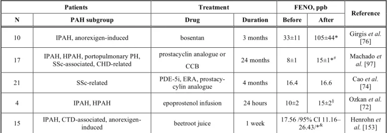
Effects of PAH therapies on FENO have been pre- viously investigated in a few studies (Table 3). In the study of Girgis et al., PAH patients showed lower lev- els of FENO compared to the control group. After hav- ing taken oral bosentan for 3 months FENO concentra- tions were increased and normalized to control levels [76]. The effects of parenteral epoprostenol or treprostinil on FENO levels were discussed by Machado et al. FENO was measured in 17 PAH pa- tients before and after treatment. After 24 months of therapy, FENO showed a 2-fold increase in survivors
Table 3. The effect of drugs with clinical benefit on FENO concentration in patients with PAH.
Patients Treatment FENO, ppb
N PAH subgroup Drug Duration Before After Reference
10 IPAH, anorexigen-induced bosentan 3 months 33±11 105±44* Girgis et al.
[76]
17 IPAH, HPAH, portopulmonary PH, SSc-associated, CHD-related
prostacyclin analogue or
CCB 24 months 8±1 15±1*# Machado et
al. [97]
21 SSc-related PDE-5i, ERA, prostacy-
cylin analogue 4 months 16.4 16.6 Cao et al.
[74]
4 IPAH, HPAH epoprostenol infusion 24 hours 10±2 15±2§ Ozkan et al.
[72]
15 IPAH, CTD-associated, anorexigen-
induced beetroot juice 1 week 17.56 /95% CI 11.16–
26.43/*& Henrohn et al. [153]
Data are shown as mean or mean±standard deviation. After: following the course of therapy. Before: prior to drug therapy. CCB: calcium- channel blocker, CI: confidence interval, CHD: congenital heart disease, CTD: connective tissue disease, ERA: endothelin receptor antago- nist, FENO: fractional exhaled nitric oxide, IPAH: idiopathic PAH, HPAH: heritable PAH, N: number of patients included in the study, PAH:
pulmonary arterial hypertension, PDE-5i: phosphodiesterase-5 inhibitor, ppb: particles per billion, SSc: systemic sclerosis. *p<0.001, §p<0.05 vs. before, &median of differences between placebo treatment and beetroot juice ingestion, #only in the 12 survivors.
and correlated with the decrease in systolic PAP [97].
In addition, NO concentration measured in breath col- lected with tidal breathing was increased 24 hours after epoprostenol infusion in patients with PAH [72].
On the contrary, therapy with PDE-5 inhibitors has not proven to influence FENO. Rothe et al. evaluated the changes in expiratory NO concentrations after treatment with sildenafil in 10 healthy volunteers. Par- ticipants received 50 mg sildenafil monotherapy or pla- cebo on the first and the seventh day. Exhaled NO was unchanged 1 h, 24 h and 72 h after sildenafil intake [151]. In a recent study by Cao et al., exhaled NO was measured in patients with SSc-PAH. Specific PAH therapies included monotherapies with oral sildenafil, tadalafil, ambrisentan, bosentan and inhaled treprostinil and the combination of tadalafil and ambrisentan. After 4 months of these therapies no significant change was detected either in CANO or JawNO [74]. In the future, further investigations are needed to clarify the role of PAH therapies in FENO changes.
3.3. Other Pharmacological Strategies
Besides the effective targeted therapeutic options, non-specific pharmacological strategies have also been applied to modulate the pulmonary NO pathway in pa- tients with PAH under experimental settings. These interventions aim to either induce the NOS-dependent or NOS-independent pulmonary NO generation. A short course of oral L-arginine supplementation in pa- tients with pre-capillary PH resulted in enhanced NO
production as demonstrated by the increased plasma levels of L-citrulline, the end-product of NOS activity.
Of note, L-arginine administration decreased mPAP and PVR, and improved exercise capacity without in- ducing clinically significant hypotension [152].
A strategy is to improve the NOS-independent pro- duction of pulmonary NO is through the induction of the nitrate-nitrite-NO pathway. On one hand, NO has a short half-life, and in aqueous solutions, it is decom- posed to nitrite, while in tissues NO and nitrite are en- zymatically oxidized to nitrate [154]. In addition, die- tary inorganic nitrate can be transformed into nitrite by commensal oral bacteria [155]. On the other hand, ni- trite can serve as a pool for NO as it can be converted into NO by deoxyhemoglobin-mediated reduction [156]. Furthermore, various human enzymes with ni- trite reductase activity under certain conditions such as hypoxia and acidosis have been proposed, including xanthine oxidoreductase, aldehyde oxidoreductase, cy- tochrome C, deoxymyoglobin, and carbonic anhydrase [157].
In line with this, patients with PAH received nitrate- rich beetroot juice as a dietary supplement for one week, which resulted in an increase in FENO, CANO and JawNO, and elevated levels of plasma and salivary nitrate and nitrite. Importantly, those patients with at least 30% rise in plasma nitrite presented with an im- provement in exercise capacity [153]. These positive effects were seen, although most patients received treatment with a PDE-5 inhibitor.
Furthermore, the inhalation of sodium nitrite re- sulted in a decrease in mPAP and right atrial pressure in patients with PAH, and the therapy was well- tolerated. Importantly, there was no change in cardiac output, but mean systemic arterial pressure decreased significantly [158]. Of note, nitrite inhalation was asso- ciated with an acute 4-5-fold elevation of FENO in healthy subjects [159], which was not apparent 30 minutes after the dose.
CONCLUSION
Several experimental and human studies prove the crucial involvement of the attenuated endothelial NO signalling of the pulmonary vasculature in the path- omechanism of PAH. This is also supported by circu- lating biomarkers such as the increased concentration of ADMA or the reduced levels of NO metabolites.
However, studies on exhaled NO (both bronchial and alveolar readouts), which have the potential to more directly assess pulmonary vascular processes, show ambiguous results for differentiating disease from health. These discrepancies can be due to unstandard- ized measurement protocols, the low number of pa- tients, various aetiologies of PAH in patient groups, and probably the ignorance of factors confounding NO readings. Studies on the potential of exhaled NO to predict survival are also lacking.
PDE-5 inhibitors and sGC stimulators are available at high costs to induce pulmonary NO signalling and confer clinical efficacy in PAH. Currently, therapeutic decisions are made based on clinical and some labora- tory findings. However, data exploring the capacity of exhaled NO or other markers of the NO pathway to predict response to these drugs are missing, and only a handful of studies have assessed the effects of PAH- specific therapy on FENO, in general.
In conclusion, although exhaled NO is easy to measure and it would be an attractive option for assess- ing disease process in PAH, its current role in aiding the diagnosis or guiding the treatment is unclear. Mul- ticentre studies recruiting a homogenous group with increasing number of patients are needed in the future to explore the applicability of bronchial and alveolar NO parameters in disease assessment.
LIST OF ABBREVIATIONS
6MWD = 6-minute walking distance
ADMA = Asymmetric dimethylargin-
ine
Akt/PKB = Protein kinase B
ATS = American Thoracic Society
BH4 = Tetrahydrobiopterin
BMPR2 = Bone morphogenetic protein receptor II
Ca2+ = Calcium
cAMP = Cyclic adenosine monophos-
phate
CANO = Alveolar NO concentration CaSR = Calcium sensing receptor CBDL = Common bile duct ligation
CCB = Calcium-channel blocker
cGMP = Cyclic guanosine monophos-
phate
CHD = Congenital heart disease CHD-related
PAH = Congenital heart disease re- lated pulmonary arterial hy- pertension
CI = Confidence interval
COPD = Chronic obstructive pulmo- nary disease
CTD = Connective tissue disease CTD-associated
PAH = Connective tissue disease associated pulmonary arterial hypertension
CTEPH = Chronic thromboembolic
pulmonary hypertension
DDAH = Dimetylaminohydrolase
ERA = Endothelin receptor antago- nist
ERS = European Respiratory Soci-
ety
Fe2+ = Ferrous ion
FENO = Fractional exhaled nitric ox- ide
GMP = Guanosine monophosphate
GTP = Guanosine triphosphate
Hb = Haemoglobin
HIV = Human immunodeficiency
virus
HPAH = Heritable pulmonary arterial hypertension
IPAH = Idiopathic pulmonary arterial hypertension
JawNO = Different flows bronchial NO production
MCT = Monocrotaline
MLC-p = Myosin light-chain
MLCK-p = Myosin light chain kinase mPAP = Mean pulmonary artery pres-
sure
NO = Nitric oxide
NOS = Nitric oxide synthase
NOS1=nNOS = Neuronal nitric oxide syn- thase
NOS2=iNOS = Inducible nitric oxide syn- thase
NOS3=eNOS = Endothelial nitric oxide syn- thase
NOx = Combined plasma levels of
nitrate and nitrite
PAH = Pulmonary arterial hyperten- sion
PAP = Pulmonary artery pressure PATENT-I = Pulmonary Arterial Hyper-
tension Soluble Guanylate Cyclase–Stimulator Trial 1 PATENT-II = Pulmonary Arterial Hyper-
tension Soluble Guanylate Cyclase–Stimulator Trial 2 PDE-5 = Phosphodiesterase isotype-5 PDE-5i = Phosphodiesterase-5 inhibitor
PH = Pulmonary hypertension
PHIRST = Pulmonary Arterial Hyper- tension and Response to Ta- dalafil
ppb = particles per billion
PVR = Pulmonary vascular resis-
tance
Ser1177 = Serine 1177
sGC = Soluble guanylate cyclase
SNO = S-nitrosothiol
SSc = Systemic sclerosis
SSc-PAH = Systemic sclerosis-related pulmonary arterial hyperten- sion
SUPER-1 = Sildenafil Use for Pulmonary Arterial Hypertension
Thr495 = Threonine 495
VEGF = Vascular endothelial growth factor
WHO = World Health Organization
CONSENT FOR PUBLICATION Not applicable.
FUNDING
Andras Bikov has been supported by the NIHR Manchester BRC. This publication was supported by the Janos Bolyai Research Scholarship of the Hungar- ian Academy of Sciences to Zsofia Lazar (BO/00559/16).
CONFLICT OF INTEREST
The authors declare no conflict of interest, financial or otherwise.
ACKNOWLEDGEMENTS Declared None.
REFERENCES
[1] Galiè, N.; Humbert, M.; Vachiery, J.L.; Gibbs, S.; Lang, I.;
Torbicki, A.; Simonneau, G.; Peacock, A.; Vonk Noorde- graaf, A.; Beghetti, M.; Ghofrani, A.; Gomez Sanchez, M.A.; Hansmann, G.; Klepetko, W.; Lancellotti, P.;
Matucci, M.; McDonagh, T.; Pierard, L.A.; Trindade, P.T.;
Zompatori, M.; Hoeper, M. 2015 ESC/ERS Guidelines for the diagnosis and treatment of pulmonary hypertension: The Joint Task Force for the Diagnosis and Treatment of Pulmo- nary Hypertension of the European Society of Cardiology (ESC) and the European Respiratory Society (ERS): En- dorsed by: Association for European Paediatric and Con- genital Cardiology (AEPC), International Society for Heart and Lung Transplantation (ISHLT). Eur. Respir. J., 2015, 46(4), 903-975.
http://dx.doi.org/10.1183/13993003.01032-2015 PMID:
26318161
[2] D’Alonzo, G.E.; Barst, R.J.; Ayres, S.M.; Bergofsky, E.H.;
Brundage, B.H.; Detre, K.M.; Fishman, A.P.; Goldring, R.M.; Groves, B.M.; Kernis, J.T. Survival in patients with primary pulmonary hypertension. Results from a national prospective registry. Ann. Intern. Med., 1991, 115(5), 343- 349.
http://dx.doi.org/10.7326/0003-4819-115-5-343 PMID:
1863023
[3] Humbert, M.; Guignabert, C.; Bonnet, S.; Dorfmüller, P.;
Klinger, J.R.; Nicolls, M.R.; Olschewski, A.J.; Pullamsetti, S.S.; Schermuly, R.T.; Stenmark, K.R.; Rabinovitch, M. Pa- thology and pathobiology of pulmonary hypertension: state of the art and research perspectives. Eur. Respir. J., 2019, 53(1)1801887
http://dx.doi.org/10.1183/13993003.01887-2018 PMID:
30545970
[4] Vonk Noordegraaf, A.; Chin, K.M.; Haddad, F.; Hassoun, P.M.; Hemnes, A.R.; Hopkins, S.R.; Kawut, S.M.; Langle- ben, D.; Lumens, J.; Naeije, R. Pathophysiology of the right ventricle and of the pulmonary circulation in pulmonary hy- pertension: an update. Eur. Respir. J., 2019, 53(1)1801900 http://dx.doi.org/10.1183/13993003.01900-2018 PMID:
30545976
[5] Dorfmüller, P. Pathology of pulmonary vascualr dis- eases.Pulmonary circulation: diseases and their treatment;
Peacock, A.; Naeije, R.; Rubin, L.J., Eds.; Taylor & Francis Group: Boca Raton, FL, 2016, Vol. 4, pp. 61-78.
[6] Li, G.W.; Wang, Q.S.; Hao, J.H.; Xing, W.J.; Guo, J.; Li, H.Z.; Bai, S.Z.; Li, H.X.; Zhang, W.H.; Yang, B.F.; Yang, G.D.; Wu, L.Y.; Wang, R.; Xu, C.Q. The functional expres- sion of extracellular calcium-sensing receptor in rat pulmo- nary artery smooth muscle cells. J. Biomed. Sci., 2011, 18(1), 16-16.
http://dx.doi.org/10.1186/1423-0127-18-16 PMID:
21314926
[7] Yamamura, A.; Guo, Q.; Yamamura, H.; Zimnicka, A.M.;
Pohl, N.M.; Smith, K.A.; Fernandez, R.A.; Zeifman, A.;
Makino, A.; Dong, H.; Yuan, J.X.J. Enhanced Ca(2+)- sensing receptor function in idiopathic pulmonary arterial hypertension. Circ. Res., 2012, 111(4), 469-481.
http://dx.doi.org/10.1161/CIRCRESAHA.112.266361 PMID: 22730443
[8] Rich, S.; Dantzker, D.R.; Ayres, S.M.; Bergofsky, E.H.;
Brundage, B.H.; Detre, K.M.; Fishman, A.P.; Goldring, R.M.; Groves, B.M.; Koerner, S.K. Primary pulmonary hy- pertension. A national prospective study. Ann. Intern. Med., 1987, 107(2), 216-223.
http://dx.doi.org/10.7326/0003-4819-107-2-216 PMID:
3605900
[9] McGoon, M.D.; Benza, R.L.; Escribano-Subias, P.; Jiang, X.; Miller, D.P.; Peacock, A.J.; Pepke-Zaba, J.; Pulido, T.;
Rich, S.; Rosenkranz, S.; Suissa, S.; Humbert, M. Pulmo- nary arterial hypertension: epidemiology and registries. J.
Am. Coll. Cardiol., 2013, 62(25)(Suppl.), D51-D59.
http://dx.doi.org/10.1016/j.jacc.2013.10.023 PMID:
24355642
[10] Mathai, S.C.; Hassoun, P.M.; Puhan, M.A.; Zhou, Y.; Wise, R.A. Sex differences in response to tadalafil in pulmonary arterial hypertension. Chest, 2015, 147(1), 188-197.
http://dx.doi.org/10.1378/chest.14-0263 PMID: 25122150 [11] Coggins, M.P.; Bloch, K.D. Nitric oxide in the pulmonary
vasculature. Arterioscler. Thromb. Vasc. Biol., 2007, 27(9), 1877-1885.
http://dx.doi.org/10.1161/ATVBAHA.107.142943 PMID:
17541026
[12] Miles, P.R.; Bowman, L.; Rengasamy, A.; Huffman, L.
Alveolar type II cell cNOS activity and ATP levels are in- creased by lung surfactant or DPPC vesicles. Am. J.
Physiol., 1997, 273(2 Pt 1), L339-L346.
PMID: 9277445
[13] Miles, P.R.; Bowman, L.; Rengasamy, A.; Huffman, L.
Constitutive nitric oxide production by rat alveolar macro- phages. Am. J. Physiol., 1998, 274(3), L360-L368.
PMID: 9530171
[14] Ward, J.K.; Belvisi, M.G.; Fox, A.J.; Miura, M.; Tadjkarimi, S.; Yacoub, M.H.; Barnes, P.J. Modulation of cholinergic neural bronchoconstriction by endogenous nitric oxide and vasoactive intestinal peptide in human airways in vitro. J.
Clin. Invest., 1993, 92(2), 736-742.
http://dx.doi.org/10.1172/JCI116644 PMID: 8349813 [15] Zeidler, P.C.; Castranova, V. Role of nitric oxide in patho-
logical responses of the lung to exposure to environ- mental/occupational agents. Redox Rep., 2004, 9(1), 7-18.
http://dx.doi.org/10.1179/135100004225003879 PMID:
15035823
[16] Förstermann, U.; Boissel, J.P.; Kleinert, H. Expressional control of the ‘constitutive’ isoforms of nitric oxide synthase (NOS I and NOS III). FASEB J., 1998, 12(10), 773-790.
http://dx.doi.org/10.1096/fasebj.12.10.773 PMID: 9657518 [17] Barnes, P.J.; Belvisi, M.G. Nitric oxide and lung disease.
Thorax, 1993, 48(10), 1034-1043.
http://dx.doi.org/10.1136/thx.48.10.1034 PMID: 7903007 [18] Hampl, V.; Herget, J. Role of nitric oxide in the pathogene-
sis of chronic pulmonary hypertension. Physiol. Rev., 2000, 80(4), 1337-1372.
http://dx.doi.org/10.1152/physrev.2000.80.4.1337 PMID:
11015616
[19] Tejero, J.; Shiva, S.; Gladwin, M.T. Sources of Vascular Nitric Oxide and Reactive Oxygen Species and Their Regu- lation. Physiol. Rev., 2019, 99(1), 311-379.
http://dx.doi.org/10.1152/physrev.00036.2017 PMID:
30379623
[20] Klinger, J.R.; Kadowitz, P.J. The Nitric Oxide Pathway in Pulmonary Vascular Disease. Am. J. Cardiol., 2017, 120(8S), S71-S79.
http://dx.doi.org/10.1016/j.amjcard.2017.06.012 PMID:
29025573
[21] Giaid, A.; Saleh, D. Reduced expression of endothelial nitric oxide synthase in the lungs of patients with pulmonary hy- pertension. N. Engl. J. Med., 1995, 333(4), 214-221.
http://dx.doi.org/10.1056/NEJM199507273330403 PMID:
7540722
[22] Adnot, S.; Raffestin, B.; Eddahibi, S.; Braquet, P.; Chabrier, P.E. Loss of endothelium-dependent relaxant activity in the pulmonary circulation of rats exposed to chronic hypoxia. J.
Clin. Invest., 1991, 87(1), 155-162.
http://dx.doi.org/10.1172/JCI114965 PMID: 1985092 [23] Dinh-Xuan, A.T.; Pepke-Zaba, J.; Butt, A.Y.; Cremona, G.;
Higenbottam, T.W. Impairment of pulmonary-artery endo- thelium-dependent relaxation in chronic obstructive lung disease is not due to dysfunction of endothelial cell mem- brane receptors nor to L-arginine deficiency. Br. J. Pharma- col., 1993, 109(2), 587-591.
http://dx.doi.org/10.1111/j.1476-5381.1993.tb13611.x PMID: 7689396
[24] Schermuly, R.T.; Ghofrani, H.A.; Wilkins, M.R.; Grim- minger, F. Mechanisms of disease: pulmonary arterial hy- pertension. Nat. Rev. Cardiol., 2011, 8(8), 443-455.
http://dx.doi.org/10.1038/nrcardio.2011.87 PMID:
21691314
[25] Steudel, W.; Ichinose, F.; Huang, P.L.; Hurford, W.E.;
Jones, R.C.; Bevan, J.A.; Fishman, M.C.; Zapol, W.M. Pul- monary vasoconstriction and hypertension in mice with tar- geted disruption of the endothelial nitric oxide synthase (NOS 3) gene. Circ. Res., 1997, 81(1), 34-41.
http://dx.doi.org/10.1161/01.RES.81.1.34 PMID: 9201025 [26] Østergaard, L.; Stankevicius, E.; Andersen, M.R.; Eskild-
sen-Helmond, Y.; Ledet, T.; Mulvany, M.J.; Simonsen, U.
Diminished NO release in chronic hypoxic human endothe-
![Table 1. Clinical classification of pulmonary hypertension (simplified from Galie et al.[1])](https://thumb-eu.123doks.com/thumbv2/9dokorg/849538.44685/2.918.102.826.120.598/table-clinical-classification-pulmonary-hypertension-simplified-galie-et.webp)