Non-invasive measurements of oral mucosa blood flow in patients with various clinical conditions
PhD Thesis
Eszter Molnár
Doctoral School of Clinical Medicine Semmelweis University
Supervisor: János Vág, DMD, PhD
Official reviewers: Szabolcs Várbíró, MD, PhD Márk Fráter DMD, PhD
Head of the Final Examination Committee: Tamás Divinyi, DMD, PhD
Members of the Final Examination Committee: Tivadar Zelles, DMD, DSc Edina Lempel, DMD, PhD
Budapest
2019
2
Table of contents
1 List of Abbreviations ... 5
2 Preamble ... 7
3 INTRODUCTION ... 8
3.1 The significance of the vasculature in periodontal tissues ... 8
3.2 The framework of wound healing ... 9
3.3 Wound healing in the oral mucosa ... 12
3.4 The effect of gender on wound healing ... 12
3.5 Vascular changes in periodontitis ... 14
3.5.1 Changes in morphology and the number of vessels ... 14
3.5.2 Change in blood flow ... 16
3.6 Vascular changes in the periodontal flap ... 18
3.6.1 Changes in morphology and the number of vessels ... 18
3.6.2 Changes in blood flow ... 19
3.7 Examination methods of gingival microcirculation ... 21
3.7.1 Invasive methods ... 22
3.7.2 Non-invasive methods ... 23
3.7.3 Other factors that may affect oral mucosal blood flow... 29
3.8 Significance of the measurement of gingival crevicular fluid (GCF) ... 30
4 OBJECTIVES ... 33
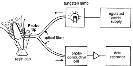
5 METHODS ... 35
5.1 Applied experimental methods ... 35
5.1.1 Laser Doppler blood flow measurement ... 35
5.1.2 Laser Speckle Contrast Imaging ... 36
5.1.3 Crevicular fluid measurement ... 38
5.1.4 Systemic Blood Pressure measurement ... 38
5.2 Subjects ... 38
5.3 Studies ... 39
5.3.1 I. The effect of warm saline on GBF in the healthy gingiva ... 39
5.3.2 II. The effect of light-induced heat on GBF in the healthy gingiva ... 40
5.3.3 III. The effect of periodontal inflammation on heat-induced hyperemia in non-smokers and smokers ... 40
5.3.4 IV. The effect of the incidence angle on reliability ... 41
3
5.3.5 V. The effect of retraction on reliability and the assessment of inter-day
reproducibility ... 41
5.3.6 VI. The effect of mirrors on reliability ... 42
5.3.7 VII. The long-term reliability of repeated measurements ... 42
5.3.8 VIII. Periodontal plastic surgery for root coverage ... 44
5.4 Statistics ... 46
6 RESULTS... 49
6.1 The effect of warm saline on GBF in the healthy gingiva (exp. I) ... 49
6.2 The effect of light-induced heat on GBF in healthy gingiva (exp. II)... 51
6.3 The effect of periodontal inflammation and smoking on heat-induced hyperemia (exp. III) ... 52
6.4 The effect of the incidence angle on reliability (exp. IV)... 54
6.4.1 The effect of the incidence angle on reliability in Zone A ... 54
6.4.2 The effect of the incidence angle on reliability in Zone B ... 56
6.4.3 The effect of the incidence angle on reliability in Zone C ... 56
6.5 The effect of retraction on intra- and inter-day reliability (exp. V) ... 57
6.5.1 The effect of retraction on intra- and inter-day reliability in Zone A ... 57
6.5.2 The effect of retraction on intra- and inter-day reliability in Zone B ... 59
6.5.3 The effect of retraction on intra- and inter-day reliability in Zone C ... 59
6.6 The effect of using a mirror on reliability (exp. VI) ... 60
6.7 The reliability of repeated measurements in a clinical surgical trial (exp. VII) . 61 6.8 Assessment of oral mucosal blood flow following periodontal plastic surgery (exp. VIII) ... 62
6.8.1 Blood flow at the treated sites on the days following the surgery in Zone A 62 6.8.2 Blood flow at the treated sites on the days following the surgery in Zone B 64 6.8.3 Blood flow at the treated sites on the days following the surgery in Zone C 66 6.8.4 The kinetics of wound fluid production ... 67
6.8.5 The correlation between WF and blood flow ... 69
6.8.6 Clinical parameters ... 69
7 DISCUSSION ... 71
7.1 The relationship between periodontal inflammation, smoking and blood flow, measured by LDF (exp. I, II, III) ... 71
4
7.2 Utilizing LSCI methods to measure blood flow in the human oral mucosa
(exp. IV, V, VI, VIII) ... 76
7.3 Blood flow and wound fluid measurements on a healing periodontal flap after root coverage surgery (exp. VII) ... 79
8 CONCLUSIONS ... 86
9 SUMMARY ... 88
10 Összefoglalás ... 89
11 BIBLIOGRAPHY ... 90
12 BIBLIOGRAPHY OF THE CANDIATE’S PUBLICATIONS ... 110
12.1 Related publications ... 110
12.2 Not related publications ... 110
13 ACKNOWLEDGEMENTS ... 112
14 Appendix ... 113
14.1 List of Tables ... 113
14.2 List of Figures ... 113
5
1 List of Abbreviations
BF – Blood Flow
CAF – Coronally Advanced Flap
CMBC – Concentration of Moving Red Blood Cells CR – Coefficient of Repeatability
CTG – Connective Tissue Graft CV – Coefficient of Variation
DALY – Disability Adjusted Life Years DM – Diabetes Mellitus
DPU – Doppler Perfusion Unit ECM – Extracellular Matrix EXP. – Experiment
GBF – Gingival Blood Flow GCF – Gingival Crevicular Fluid GFPA – Gingival Flux Pulse Amplitude GRD – Gingival Recession Depth GRW – Gingival Recession Width ICC – Intraclass Correlation Coefficient JE – Junctional Epithelium
KT – Keratinized Tissue
LDF – Laser Doppler Flowmetry LDI – Laser Doppler Imager
LSCI – Laser Speckle Contrast Imaging LSPU – Laser Speckle Perfusion Unit MAP – Mean Arterial Pressure
MARTD – Multiple Adjacent Recession Type Defects MAX – Maximum Absolute Change
MCAT – Modified Coronally Advanced Tunnel OPS – Orthogonal Polarized Spectral
PS – Periotron Scores PRP – Platelet-rich Plasma
6 RBC – Red Blood Cells
REC – Recession Depth Reduction RT – Recovery Time
RW – Recession Width Reduction SDF – Sidestream Darkfield SE – Standard Error
VEGF – Vascular Endothelial Growth Factor
7
2 Preamble
Microcirculation studies have had a long tradition at the Department of Conservative Dentistry, established by Professor Árpád Fazekas. This provided an opportunity to gain further knowledge in this field of science and planted the seed for further research. At the same time, new techniques in periodontology and oral surgery have developed, enabling graft materials and implant placement to become widespread in dentistry. The predictability of outcomes following surgical procedures has a fundamental importance in medicine, therefore, success can only be achieved if biological aspects and regeneration are taken into consideration. It has become inevitable to be familiar with the oral aspects of wound healing to be able to intervene in the process if difficulties occur. This requirement resulted in a demand for monitoring wound healing in an objective manner in addition to subjective methods. Because of the widespread application of surgical interventions, the observations were necessarily extended to various diseases and harmful behaviors, e.g. Diabetes Mellitus (DM), bisphosphonate-related osteonecrosis of the jaw (BRONJ) or smoking.
In the third year of my residency status at the Department of Conservative Dentistry, János Vág PhD offered me the opportunity to become a member of an emerging working group. As part of this group, I developed a particular interest in wound healing and microcirculation. Having been accepted to the Dental Research Program led by Professor Gábor Varga of the Semmelweis University School of Clinical Medicine, the initial objective of my PhD research was to investigate a new non-invasive method for measuring microcirculation in the oral cavity. After setting up a physiological test with Laser Doppler Flowmetry (exp. I–III) and getting familiar with the novel LSCI method for blood flow measurements (exp. IV–VII), I started to work with some of the best clinicians at Semmelweis University, including Bálint Molnár PhD from the Periodontal Department (exp. VIII).
8
3 INTRODUCTION
3.1 The significance of the vasculature in periodontal tissues
Periodontitis is a ubiquitous chronic inflammatory disease, initiated by the accumulation of pathogenic dental plaque biofilm above and below the gingival (gum) margin, and by microbial dysbiosis leading to a chronic nonresolving and destructive inflammatory response (1, 2). 75% of the population is affected based on a survey under the Global Burden of Disease 2010 study, which was compiled by the Institute for Health Metrics and Evaluation on a sample of 11% of the Earth’s population (3). The frequency of severe periodontitis increases with age, and among 35–59 year-olds, it is the world’s leading contributor to disability adjusted life years (DALY). It is also the major cause of tooth loss, nutritional compromise, altered speech or low self-esteem (4). Twenty years ago, 95% of the Hungarian population suffered from some type of periodontal disease (5) and that ratio was still 88% in 2009 (6). In addition to the local effects of periodontitis, it involves a significant risk factor for systemic diseases such as coronary heart disease, stroke, diabetes and osteoporosis (7, 8). In view of this, better understanding of the pathomechanisms of periodontitis and the development of treatment options are indispensable.
During inflammation, the bacterial biofilm adhering to the teeth produces bioactive substances (9), which induce a local immune response in the gingiva. The inflammatory reaction also involves local microvascular changes (10, 11), which also play a role in the progression of periodontitis according to some studies (12, 13). Several epidemiological studies confirm that there is a two-way relationship between periodontitis and diabetes mellitus (DM) (14-18). The pathomechanism in this interplay is not known. Considering the presence of severe microvascular damage in DM, it may be possible that the progression of periodontal disease is promoted by vascular effects (15, 19). There is very little data available on microcirculatory changes in periodontitis in diabetic patients, but animal experiments indicate a significant relationship (20).
Periodontal therapy frequently involves surgery. Taking the stages of wound healing into consideration, it is clear that blood flow is essential in this process. In the case of surgery, during the early-healing period, the transport of cellular and humoral factors, such as platelets, immune cells, oxygen and nutrients are critical to achieving an optimal process.
9
Reperfusion of the flap is a determinant of avoiding partial flap necrosis, especially in areas located distally from the flap basis (21). Postoperative reperfusion of the flap ultimately depends on the preservation of microcirculatory inflow (22). Nutrition is supplied to the distal part of the mucosal flap through extravasation in the early ischemic period of healing (23). Newly formed blood vessels in the provisional granulation tissue re-establish the microvascular network in the connective tissue and provide supply for healing (24).
3.2 The framework of wound healing
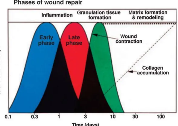
Wounds can be described as tissue disruption of normal anatomic structure with consecutive loss of function (25). Wound healing is a highly regulated process of cellular, humoral and molecular mechanisms which begins directly after wounding and might last for years. This complex process has been studied in great detail (26) and can be theoretically divided into three phases which overlap in time and space (Figure 1):
inflammation (early and late phase), granulation tissue formation, and matrix formation and remodeling (27).
10
Figure 1: Phases of wound healing.
Model made for epidermal incisional wounds. The inflammatory phase (including an early and a late phase), granulation tissue formation and matrix formation &
remodeling over time (27).
If an injury causes capillary damage and hemorrhage, blood clot is formed as the first step. This clot has two functions: it temporarily protects the denuded tissues and it serves as a provisional matrix for cell migration (28). Blood clot consists of all the cellular components of blood: fibrin molecules, fibronectin, vitronectin and thrombospondin, serving as a transitional matrix for the arriving leukocytes, keratinocytes, fibroblasts, endothelial cells and reservoirs for growth factors (29). Transient vasoconstriction is followed by vasodilation, with platelets overflowing the transitional matrix (30). Platelets produce cytokines and growth factors and attract leukocytes. Following this, leukocytes play a role in cytokine production as well, thereby stimulating collagen synthesis (FGF- 2, IGF-1), the transformation of fibroblasts into myofibroblasts (TGF-β), the initiation of angiogenesis (FGF-2, VEGF-S, HIF-1α, TGF-β) and support for epithelialization (EGF, FGF-2, IGF-1, TGF-α) (31). This inflammatory phase can be divided into two phases:
early neutrophil invasion and a late phase (31). Neutrophils are present after 2–5 days
11
of injury. Their phagocytic ability and protease secretion enables the destruction of the bacteria present and the breakdown of necrotized tissue. Protease secretion also acts as a chemoattractant for other inflammatory cells (32). About 3 days after injury (in the late phase), macrophages appear and they further support the process with phagocytosis, wound purification, growth factors and cytokines production (33).
The proliferation phase, i.e. granulation tissue formation, starts 3–10 days after the damage, when reepithelialization, angiogenesis and scar tissue formation begins.
Reepithelialization is started at the wound edges by keratinocytes (34, 35, 30). Collagen synthesis increases on the entire surface of the wound (36). Molecular mediators of angiogenesis are VEGF, PDGF, FGF and serine protease thrombin, which bind to endothelial cells. The activated endothelial cells produce proteolytic enzymes to dissolve the basal lamina, which enables them to proliferate and migrate. The final step of the proliferation phase is scar tissue formation. Scar tissue is characterized by the accumulation of fibroblasts, granulocytes, macrophages, capillaries and collagen bundles.
Fibroblasts have a decisive role in the formation of the extracellular matrix (ECM) (37, 38).
The last phase of wound healing is the restorative phase, i.e. matrix formation and remodeling. It begins around the 21st day and may take up to 1 year. Granulation tissue is eliminated by apoptosis. Components of the ECM undergo changes. Stronger collagen is formed. Once the collagen matrix has been synthesized, some fibroblasts undergo transformation into myofibroblasts and express α-smooth muscle actin. This transformation and synthesis is responsible for wound contraction (39, 33, 40). The time of angiogenesis is reduced and epithelialization is complete (31). In the skin, the tensile strength of the formed scar reaches 70% of the original tissue (26).
The healing of a wound serves the purpose of replacing it with similar, physiologically and functionally identical tissue. This process is called regeneration. Ideally, tissue deficiency is replaced by parenchymal cells; however, if total regeneration cannot be achieved for some reason, the parenchyma site becomes connective tissue scar, a process called reparation. The type of the damaged tissue and the nature of the injury determine which of the two processes occurs (41).
12 3.3 Wound healing in the oral mucosa
There are several types of tissues in the oral cavity, including soft tissues, muscles, bones, mucosa and teeth. In this milieu, wound healing and tissue regeneration require great coordination, since they involve structures that are close to each other in space, but are physiologically different (38). The general principles of healing and the associated cellular and molecular events were observed in non-oral sites, also applicable to the healing processes that take place following mucosal healing in the oral cavity. However, human oral mucosal wounds heal fast and with minimal scar formation as compared with the skin (42). This was demonstrated on experimental wounds created in the tongue and the buccal mucosa of rodents (41). Surgical wounds, especially in the oral keratinized attached gingiva and the palatal mucosa, heal with very little scar formation (43). Yet, there is a common belief (44) among clinicians alongside sparse documentation (45, 46) that improper incision and flap design may cause hypertrophic scar formation in the oral mucosa. However, this has not been systematically investigated.
The difference in the healing process is characterized by a lower inflammatory response in the mucosa. Studies have shown that in comparison to skin wounds, oral wounds exhibit lower neutrophil, macrophage and T-cell infiltration (47). Oral and skin wounds also exhibit differences in the expression of TGF-ß1, a pro-inflammatory, pro-fibrotic cytokine which has been implicated in the etiology of hypertrophic scars (48).
Angiogenesis in oral wounds is less robust than in the skin (49) and the dominant mediator of wound angiogenesis, the production of Vascular Endothelial Growth Factor (VEGF), is significantly less pronounced in oral than in skin wounds (50).
Primary and secondary forms of wound healing are fundamentally different. Primary wound healing is usually observed in the oral cavity, with sharp, non-inflamed, clean wound edges. The epidermis perfectly covers the affected area, without reparation tissue.
In secondary intention healing, wound edges do not meet and an inflammatory zone and tissue deficiency are found, with reparation scar tissue (51).
3.4 The effect of gender on wound healing
Clinical observations (52-55) and findings in an experimental excisional palatal wound model (56) suggest that mucosal wound healing is faster in males than in females. They
13
studied 3.5 mm circular wounds on the oral hard palate of 212 volunteers. Wound videographs were taken daily for 7 days after wounding to assess wound closure.
Although all wounds were initially the same size, 24 hours after wounding, men had significantly smaller wounds. This difference was apparent until day 5 (Figure 2). In addition, the proportion of individuals considered healed was significantly higher among men than among women on days 5 and 6 (Figure 2). These results suggest that oral mucosal wounds heal more slowly in women than in men, regardless of age. Such gender differences in wound healing may be explained by sex hormones.
Figure 2: Gender-related changes in wound healing parameters.
Less wounds were considered healed in women on days 5 and 6 compared with men (56).
However, female hormones such as estrogens seem to have a favorable effect on acute wound healing in rodents and in the human skin (57, 58). In contrast to estrogens, testosterone delays wound healing in the skin (59-61). Just as in the case of cutaneous wound healing, testosterone levels negatively correlate with mucosal wound healing in adults, regardless of gender (62). In an ischemia reperfusion murine model, permeability and leukocyte infiltration in the intestinal mucosa increased more in males than in females (63). Overall, the aforementioned evidence suggests gender difference in oral mucosal wound healing, but the mechanism has not been clarified yet. Possible differences in the regulation of blood flow during healing cannot be excluded either.
14 3.5 Vascular changes in periodontitis
3.5.1 Changes in morphology and the number of vessels
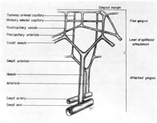
Nuki and Hock studied the structure and organization of vessels in gingiva with no previous history of inflammation. They described a microvascular bed around the teeth, where capillaries predominated within the crestal gingiva and within the superficial buccal and crevicular networks. Precapillary arterioles and postcapillary venules were most common in the mid-gingival region. Small arterioles and venules were present in the apical gingiva (Figure 3).
Figure 3: Drawing of the framework of the microvasculature.
Data were obtained from microfilm perfusion and serial tissue sections (64) (Nuki and Hock 1974).
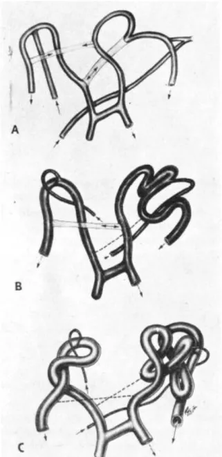
Morphologic changes in the capillary units of the network were seen as plaque increased, but prior to clinical signs of inflammation. The width and the length of the vessels changed and also their morphology has become different. Loop formation was observed on the vessels (Figure 4). With continuing inflammation, certain connecting vessels were lost while other vessels became spatially rearranged (64).
15
Figure 4: Illustration of the alteration of the vessels from a regular network to loops.
A: regular network, B: intermediate state, C: loops (64).
Also, in 1974, Kennedy examined the effect of inflammation on the collateral circulation of the gingiva and the periodontal ligament on monkeys. In clinically healthy tissues, a vascular connection between gingival and periodontal vessels was seldom observed.
Nevertheless, when inflammation occurred, vascularity and the number of connecting vessels increased (65).
The number of vessels connecting vessels of the periodontal ligament with supracrestal vessels was also significantly higher. There was a tendency for the number of vessels
16
perforating either the alveolar bone proper or the alveolar crest to increase when inflammation was induced. The results of this study demonstrate the development of collateral circulation from the periodontal ligament to the gingiva in response to inflammation and help to explain the extension of inflammation into the deeper structures of the periodontium, including the periodontal ligament. Vascular changes and remodeling in periodontitis were reported to affect the progression of the disease (12, 13).
Some vascular changes including the dilation of vessels, the formation of tortuous looping structures and the development of columnar endothelial cells may promote the defense mechanism against bacteria, however, the formation of perivascular hyaline material and the accumulation of basement membrane rests may assist the progression of periodontitis (12).
Interestingly, the thickening of the basal lamina around microvessels occurs just as in the case of diabetes mellitus (DM) (66, 67). In DM patients, microvascular complications in other organs, e.g. neuropathy, nephropathy and retinopathy, were found to be associated with the presence of a more severe inflammatory pathology of periodontal tissues (68). A recent study (20) found decreased post-occlusive reactive hyperemia in the gingiva of a diabetic rat compared to a healthy one and this response was further reduced by experimentally induced periodontitis. However, there is no direct evidence for the impairment of vascular reactivity in the human gingiva in periodontitis and/or DM.
3.5.2 Change in blood flow
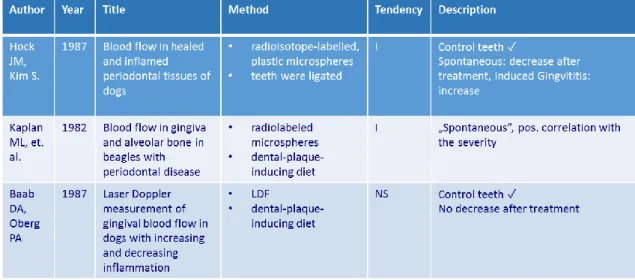
Studies investigating the effect of periodontal inflammation on basal gingival blood flow found conflicting results (Table 1).
17
Table 1: Studies investigating the effect of periodontal inflammation on basal gingival blood flow.
Dog studies (69, 70) demonstrated increased blood flow in the inflamed gingiva, involving bone loss (a combination of gingivitis and chronic periodontitis). However, in the same species, Baab and Öberg (71) found no significant correlation between the gingival index, GCF and blood flow, and the elimination of the inflammation did not result in a decrease in blood flow either. In humans, experimentally induced gingivitis resulted in decreased blood flow to the gingiva (72, 73) whereas naturally occurring gingivitis resulted in increased blood flow (72). GBF at rest was found to be smaller in periodontitis patients compared to the reference subjects (74) and the treatment of gingivitis (75) or periodontitis (76) reduced blood flow. One possible explanation for conflicting results is variations in gingival blood flow as a function of time and the location of the laser Doppler probe. Temporal variation related to biological variation may be influenced by many physiological factors in addition to the inflammation, such as circadian rhythm (77), blood pressure (78), temperature (79) or tooth brushing (72, 80, 81). Furthermore, although no data are available about the effects of disinfectant mouth rinses, eating and drinking on GBF, they may also influence the recordings. That is why it is important to standardize these factors before and during the measurements. To better control the temporal and spatial variation of blood flow, it is useful to implement a provocation test on the gingiva, which is a relative functional measurement, instead of an absolute measurement.
18 3.6 Vascular changes in the periodontal flap
3.6.1 Changes in morphology and the number of vessels
Histological observations suggested that tissue revascularization begins in mucosal flaps as early as 2–3 days postoperatively (82, 83).
Mormann et al. (84) demonstrated by fluorescence angiography that mucoperiosteal flap circulation dropped by 50% 24 hours after elevation. It was also observed that various incisions influence the circulation differently.
A study on four dogs (23) showed that in the case of simple elevation of mucoperiosteal flaps with immediate repositioning, the number of vessels dropped significantly, as assessed by fluorescence angiography, and it returned to the baseline level after 3 days at the interproximal site and after 10 days at the mid-buccal site. The application of either horizontal mattress or direct interrupted sutures did not influence revascularization.
Using orthogonal polarization spectral (OPS) imaging to capture and count the gingival vessels, Lindeboom et al. (85) investigated the revascularization of a mucoperiosteal flap elevated palatal to the top of the alveolar process with two vertical releasing incisions, followed by horizontal osteotomy and sinus elevation. The lateral wall osteotomy was covered with a resorbable collagen membrane. They found that vascularity regained its normal level in 14 days when only bone chips were applied but it took only 7 days when bone chips were combined with platelet-rich plasma (PRP).
The vascularity of a palatal flap, measured with a similar technique as OPS (sidestream dark field method), regained its normal level in 11 days after surgery in rabbits if there was no significant interface (i.e. any grafting material) between the bone and the flap and the flap was repositioned after 30 min suspension (86).
Another study (87) with OPS investigated the revascularization of a mucoperiosteal flap in patients receiving immediate dental implants, elevated by intraoral sulcular incision with two buccal releasing incisions. The bone defect at the buccal aspect was subsequently grafted with autogenous bone chips and covered with a native collagen membrane. This resulted in the separation of the flap from the bone. Immediately after surgery, the number of vessels dropped to 36% of the preoperative level. It took 3 weeks to be normalized, which highlights the role of re-uniting the alveolar and periodontal plexi to the mucosal one.
19
In conclusion, vascularity regains its normal level between 3 days to 3 weeks depending on flap and incision design, graft application and localization. However, this range seems to be considerable wide. Possible reasons for this may be the limited number of time points and the inaccuracy of some of the abovementioned methods (see later in Section 7.3). Furthermore, most of these methods are lacking in spatial resolution.
Consequently, this wide range may also be explained by the various localization and different spanning area of the measurement points in the various studies.
3.6.2 Changes in blood flow
Only a few studies measured the blood flow of the healing flap directly in the oral mucosa.
In cats, a 4.5-fold elevation of blood flow, measured by radiolabeled microspheres, was observed 2 hours after elevation of a mucoperiosteal flap, prepared by sulcular and vertical incisions (88). This suggests that the drop in the number of vessels after flap elevation mentioned in the previous section could be compensated by vasodilation.
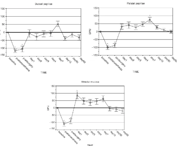
In a human study (89), blood flow was measured by non-invasive Laser Doppler Flowmetry (LDF) after periodontal surgery. Blood flow was measured at the alveolar mucosa, and the buccal and palatal papillae of the flap on postoperative days 1, 2, 3, 4, 7, 15, 30 and 60. A full-thickness flap was formed by intrasulcular incision without vertical releasing incisions. No ischemia was observed, but hyperemia was detected from day 1 to day 7 at the alveolar mucosa and the palatal papillae (Figure 5). Blood perfusion returned to the baseline on day 15. No change was observed in the buccal papillae.
20
Figure 5: Blood flow change values (Doppler Perfusion Units =DPUs) using the Laser Doppler method, in the alveolar mucosa and the flap over time.
Axis x shows measurement times; blood flow values on axis y are expressed in artificial DPU units (90).
After socket preservation, the flap – measured by LDF – was hyperemic for one month and returned to the control level at 4 months (91). Blood flow was measured only at one site, 1 mm buccally from the gingival margin.
Further evidence suggests that surgical factors, such as flap design or the application of graft material, significantly influence the blood flow.
In a study, blood flow was measured by fluorescent angiograms after the coverage of localized gingival recessions with two different surgical techniques (92). The microsurgical approach applied resulted in a higher rate of vascularization on day 3 (53%) and day 7 (85%) than the macrosurgical technique.
A clinical trial using LDF showed that the simplified papilla preservation flap may be associated with faster recovery of the gingival blood flow post-operatively compared with the modified Widman flap after pocket surgery (89).
21
In another study (93), the blood flow of the healing flap was measured at the buccal papillary base by LDF. In the control group, a mucoperiosteal flap was reflected by beveled internal and sulcular incisions for surgical crown lengthening and the bone was exposed without vertical releasing incisions. In the test group, the simplified papilla preservation flap or the modified papilla preservation flap techniques were used without vertical releasing incisions for regenerative periodontal therapy, combined with Emdogain and a granular bone substitute. In all groups, blood flow reached an ischemic level on day 1. On day 3, 7 and 14, it was not different from the baseline or the non- treated site, i.e. no hyperemic response was observed. No significant difference with regard to reduction in blood flow was found between the surgical groups.
Blood flow changes in a trapezoid full-thickness flap used for root coverage procedures were measured by LDF in a recent study (94). The flap was mobilized by two vertical incisions and a periosteal releasing incision. A xenogenic collagen matrix or an autogenous subepithelial connective tissue graft was applied on the exposed root surface before repositioning of the flap on the root surface. Blood flow was measured at two sites, at the gingiva and at the alveolar mucosa of the flap. No ischemia was observed during the healing period. At the gingival site, the microcirculation of the flap combined with the autogenous graft showed a more homogeneous curve and overall lower mean values – with the exception of the perioperative period and day 3 – compared with the flap with xenogenic matrix. At the alveolar mucosa, the autogenous graft had consistently lower mean blood flow values than the ginigva.
In conclusion, it seems that despite the fact that angiogenesis begins only 2–3 days after injury and the number of vessels returns to normality after 1–3 weeks, blood flow could be compensated in very early stages in most surgical conditions due to vasodilation.
To fully reveal the regulatory mechanism of flap circulation, we definitely need a non- invasive method with high spatial resolution.
3.7 Examination methods of gingival microcirculation
Blood flow can be tested from a morphological and also from a functional point of view.
The methods of measurement can be divided into invasive and non-invasive methods.
22 3.7.1 Invasive methods
Several methods can be used in small tissue quantities with exact results in ml, min or 100 g. However, their disadvantage is that they are invasive and cannot be used in human studies as they are harmful to health (e.g. radioactive materials are used in some methods) or lead to the death of the experimental animal, so it is difficult to perform reproducible measurements.
1. Fluorescence measurement:
a. The fluorescence principle is based on the fact that a material absorbs electromagnetic radiation of a certain wavelength and as a result emits light of a wavelength that is different from incoming radiation. Ohba et al. (95) used this method for mapping the arteries supplying head-to-neck tumors with indocyanine green.
b. Fluorescein angiography is a direct measurement of the number of vessels, however, its non-invasiveness strongly limits the amount of measurement time points. These measurements have no absolute value, but because of the distribution of fluorescein some spatial information is provided. The high reliability of these measurements allows us to use only a few animals per group in animal studies. The use of fluorescein angiography to observe blood circulation in the healthy and inflamed gingiva in man was described by Mormann & Lutz in 1974 (96). Employing antecubital venipuncture, 2 ml of a 20% sodium fluorescein solution was administered in these studies. After 15–
20 s, the Na-fluorescein entered the gingival capillary system, and a photographic sequence showed the intracapillary phase of fluorescein labeling.
The studies covered free gingival autografts (97), surgical procedures (84) and experimental wounds in man (98).
2. Radioactive microspheres: Kaplan et al. used microspheres to measure blood flow in beagle dogs with gingivitis and periodontal lesions. Microspheres were labeled with cobalt 57 isotopes. From the 9x10⁶ microspheres suspension, 3 ml was injected into the left ventricle of the dogs. Isotope activity was measured using a gamma scintillation counter in the removed gingival set and the removed piece of alveolar bone. Blood flow rates were set for 100 grams / tissue (70).
23
3. Fluorescence microspheres: The microspheres can be tested after fluorescence labeling. In Söderhol and Attström’s experiment, blood flow was examined in 4 beagle dogs suffering from gingivitis caused by a modified diet. Microspheres made of methyl methacrylate were used to which Fluorescent green HW 185 was added. After the study, the biopsy samples were evaluated by microscopy (99).
4. 86RB Isotope Assay: Fazekas et al. studied the blood flow of the lower jaw’s gingiva in rats with the 86RB isotope. Based on its physico-chemical properties, 86RB is an analogue of K. However, while the half-life of potassium is 12 hours, the rubidium isotope has a half-life of 18 days. The more increased the circulation is, the more isotopes the tissue can absorb (100).
5. H2 clearance: This method, which is based on the inhalation of H gas, was used for measuring the blood flow of the submandibular salivary gland. It provides accurate, absolute value measurements in ml/100 g/min. During the measurement, a platinum electrode and a reference electrode are used. The concentration of inhaled H2 gas is determined by a polarographic method. The development of the polarographic method is attributed to Aukland (101). Fazekas et al. used it on rabbit salivary glands (102), while Sasano et al. (103) employed it to study gingival blood flow in cats.
6. 133Xe clearance: It is a highly invasive method. Therefore, limited case numbers and time points are available. It does not provide regional information, but gives an absolute value. High variance requires a relatively larger sample size; 11–34 patients were involved in previous studies with 133Xe clearance (104, 105).
7. Histology: Invasive methods also include histology. It allows for the in-depth study of morphology during microscopic tissue examination. Histology is non- quantitative and does not furnish information on the blood flow. It only serves to make indirect inferences regarding angiogenesis and limited time points can be used for sampling. Studies often include 4–8 animals per group (106, 82, 107-111).
3.7.2 Non-invasive methods
For human studies, the most ideal method is a non-invasive, quick, easy to carry out and reproducible test. Many methods of analysis are known in the literature. The most appropriate methods in terms of the listed parameters, which are also used in human
24
studies are the Laser Doppler Flowmetry, Laser Doppler Imager and Laser Speckle Contrast Imager techniques.
1. Laser Doppler Flowmetry (LDF): It allows for the measurement of microcirculation in tissues of humans and animals, and measures blood flow in about 1 mm3 of tissue. Direct measurements of microcirculation can be made with low reliability, no spatial information and a bit larger sample size is required. This approach was first used in the 1980s and continues to be applied, because it is non-invasive, easy to use after training and provides a continuous or near-continuous record (112). The theory is based on the Doppler effect (113) (Figure 6). The main disadvantage of LDF is that it does not accurately measure blood flow, so it cannot be used to calculate absolute blood flow (e.g. in units of ml/min/100 g tissue) (114), i.e. LDF produces a relative value of blood flow. Furthermore, it has some additional drawbacks, namely that it only measures a small surface, it is motion-sensitive and it is hard to measure non- homogeneous tissues with it. In case of wound healing, we cannot place the probe on the same point every day. By contrast, it has the advantage of quick sampling, enabling non- invasive, self-controlled comparisons and being applicable in humans. It has been widely used in the field of plastic surgery for monitoring microvascular blood flow in skin transplants and flaps, in order to detect early signs of impaired circulation and thus predict and possibly prevent surgical complications (115). In the field of dentistry, LDF has been used, among other applications, in order to evaluate gingival blood flow variations related to periodontal disease (116) and smoking (117) or following periosteal stimulation (118) and LeFort I osteotomy (119). This technique has been used repeatedly for monitoring blood flow during periodontal surgical interventions. Donos et al. treated five chronic generalized periodontitis patients with the modified Widman flap technique (120). Red or near-infrared light (780 nm) from a low-power solid-state diode laser (1.6 mW) was directed via an optical fiber of 1.5 mm in diameter to the tissue and the laser light scattered back from the tissue. Within the tissue, light which is scattered from moving blood cells undergoes Doppler shifts in frequency, the magnitude of which depends upon the velocity of the cells. Wavelength shifts do not occur in the case of light reflected from non-moving cells. The light is collected by one or more optical fibers and analyzed. All the fibers are arranged in parallel within a single probe. The machine determines the relative amount of light beams affected by Doppler shifts. This provides information about the amount of
25
red blood cells (RBC) in the tissue unit, while the magnitude of frequency shifts depends upon the velocity of the cells. By multiplying the concentration of RBC and their mean velocity the FLUX unit is obtained, which correlates well with blood flow. This is a relative, arbitrary unit.
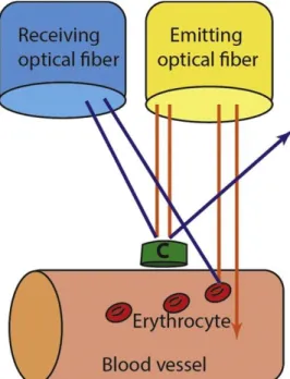
Figure 6: Effect of Doppler applied to laser radiation; c: immobile cell (A.A. Kouadio et. al.).
2. Laser Doppler Imager (LDI): For LDI, the laser beam is moved over a larger surface with a moving mirror. There is no direct contact with the test tissue. It can map the blood stream in large and small areas and assign color-coded images to it. Its advantage is that it provides information on the entire surgical area, but scanning one image takes more than a minute even with the latest devices. Bay et al. studied histamine- sensitized neurons in 13 healthy individuals and in 6 chronic oral disease patients. Blood flow in the palate, tongue and face was measured by the LDI method. Initial scanning was followed by 15 scans after histamine iontophoresis. After histamine administration, blood flow increased in all areas. Significantly higher values were obtained in the skin than in the oral regions. There was no significant difference between the blood flow of the examined groups (121).
3. Laser Speckle Contrast Imager (LSCI): LSCI is characterized by much higher reliability than LDF (122, 123). It has the unique advantage of providing regional
26
information. A small sample (4–7) is sufficient to distinguish minor differences in gingival blood flow between the groups (124, 125). The principle of the method is that illumination of the tissue surface with coherent monochromatic laser light creates an interference pattern on the surface of the tissue. This speckle pattern is captured by a camera, digitalized and transmitted to a computer, where the information is processed and an image is constructed based on the blood stream. If the illuminated particle is static, the speckle pattern is static. In the case of moving particles, the pattern fluctuates. Spatial resolution is determined by the camera used. The camera has a resolution of 1386x1034 pixels. 3x3 pixels represent a measurement unit for the purpose of contrast analysis. The more static the image – the smaller the red blood cell movement – the more contrast the image of the measurement unit will have. If blood flow increases, the image of the measurement units becomes blurred and contrast decreases. The software assigns a color code to the contrast value of the measurement pixels. The lower the contrast, the cooler the color of the pixel will be, while warmer colors are assigned in the case of higher blood flow. The measured pixels compose encoded, full-frame images. The recommended minimum measurement distance is 10 cm. The size of the measurement area is determined by the distance. For a 10 cm measurement distance it is 5.9x5.9 cm. The LSCI method has many benefits, such as quick sampling and the possibility to measure microcirculation on a large surface. Due to its high resolution, it is also suitable for evaluating functional tests. Furthermore, it has good reproducibility, as there is no contact and no effect on the tissue to be measured. Since the LSCI signal is based on the rate and concentration of red blood cells, the measurement results cannot be expressed in absolute values (ml / min / 100 g) (PeriCam PSI System Extended User Manual), (126) (Figure 7).
Clinical studies are suggesting that this technique may be a useful tool for assessing proper circulation during surgical intervention (127, 128) and evaluating wound healing (129, 130).
27
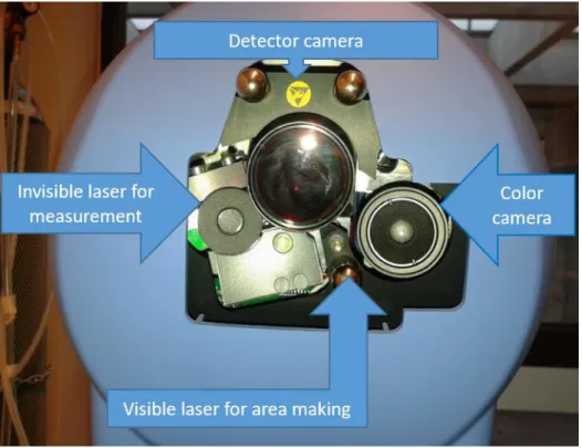
Figure 7: Perimed PeriCam PSI HR System Design.
Visible parts are marked with arrows (own photo).
4. Videomicroscopy: Videomicroscopy techniques are suitable for the direct visualization of microcirculation. In the case of the orthogonal polarized spectral (OPS) technique, the examined tissue is illuminated with polarized light. This method can measure the number of capillaries formed during healing with high reliability, but does not provide information on blood flow and spatial/regional relations. The penetrating light depolarizes in the tissue and the reflected beams enter back to the polarizer. The collected light forms an image of the illuminated area taken by the camera. 548 nm light is used for visualization, which is absorbed by hemoglobin, thereby allowing any structure containing hemoglobin to be seen (131, 132). It is possible to examine vessel density, flow and perfusion. Lindeboom et al. used the OPS method to study the capillary density of mucous membrane during healing after sinus-lift surgery. The maxillary reconstruction was performed with a hip bone by the addition of PRP bioactive material on one side and with placebo on the contralateral side. They found that PRP significantly enhanced mucosal revascularization (85). In another clinical trial, a mucoperiosteal flap was prepared to insert a dental implant (87). The OPS technique has shown that initial capillary density returns about 3 weeks later in the flap. The disadvantage of OPS is that
28
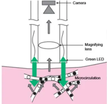
it needs a very high-intensity light source and visibility is limited due to blurred capillaries. This is why the SDF (sidestream darkfield) method has been developed, where the light source consists of concentrically placed diodes, emitting light around the optics.
The diodes emit pulsating green light, synchronized with the camera’s frequency, eliminating blurs caused by moving red blood cells (Figure 8). Because of the dark field of vision, red blood cells appear dark. Figure 8 shows the SDF imaging technique. The SDF method was used to monitor blood flow in rabbits after palatal flap formation and it was found that the flap was able to reach initial capillary density by day 11 (86).
Figure 8: The SDF technique: the light source consists of diodes emitting light, placed concentrically around the optics.
The diodes emit pulsating green light, synchronized with the camera’s frequency (126).
5. Photoplethysmography: Photoplethysmography is a non-invasive optical method that is suitable for pulse amplitude testing. RR intervals, i.e. the time elapsed between heart beats, carry a lot of information, among others regarding breathing and the funczion of the autonomic nervous system (133, 134). Using this method, Ikawa et al. compared blood flow changes in gingivitis and in the healthy gingiva after thermal (cold water, warm water) and mechanical (brushing) stimulation (Figure 9). It has been found that in
29
inflamed tissues, the pulse amplitude rise caused by warmth and mechanical stimuli decreases significantly (79).
Figure 9: Schematic drawing of photoplethysmography on the labial gingiva of an upper incisor (79).
3.7.3 Other factors that may affect oral mucosal blood flow
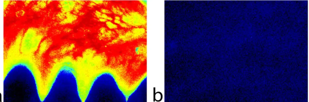
Spatial variation
Blood circulation in the gingiva is unique compared to the skin. The speckle image of the gums is more heterogeneous (Figure 10). Due to this, as the laser Doppler probe is too small, it may give confusing results measured on a small surface. It is accepted in the literature that the papilla is vascularized also by vessels originating in the crest of the interdental septa and in the palatal collaterals crossing the alveolar crest. These pathways of gingival blood supply are densely interconnected. The supraperiosteal plexus issues communicating branches to the plexus of the lamina propria and the periosteum. The multiple interconnections between the different plexuses through numerous anastomoses and native collateral pathways of circulation establish an adequate blood supply in the gingiva; however, the extent of the contribution of the various collaterals in maintaining resting blood flow has not been evaluated yet (124).
30
Figure 10: Color-coded LSCI images of healthy untreated gums (a) and forearm skin (b) with the same setup parameters.
Blood circulation is highly inhomogeneous in the human mucosa compared to the skin.
Temporal variation
Gingival blood flow has a high temporal variation as well. This could be related to many physiological factors which continuously occur during everyday life, such as gingival inflammation (72, 76, 135), circadian rhythm (77), blood pressure (78), temperature (79, 135), mechanical pressure (116, 136, 137, 124), tooth brushing (72, 80, 81) or orthodontic force (138). Therefore, the standardization and stabilization of these factors are obligatory for successful follow-up measurements.
3.8 Significance of the measurement of gingival crevicular fluid (GCF)
The junctional epithelium (JE) is part of the dento-gingival unit in relation with the teeth surfaces. GCF reaches the sulcus gingivalis between junctional epithelial cells through intracellular gaps, such as desmosomes and gap junctions (139-142). GCF plays an important role in the homeostasis of periodontal tissues, flushing the pocket, and gives passage for immune proteins (143), thereby contributing to microbial defense (144). Its antibacterial role is manifest both directly (physical barrier) and indirectly (e.g.
differentiation of molecules that promote the migration of polymorphonuclear cells) (145). There are a large number of lysosomes in JE cells that contain enzymes fighting various bacteria (146). Moreover, many other factors can increase the production of GCF,
31
such as smoking (147), oral contraceptives and pregnancy (148), or orthodontic treatment (149).
The initial fluid represents interstitial fluid which is a result of an osmotic gradient (150, 151). The pre-inflammatory fluid is considered to be a transudate, which changes to become an inflammatory exudate upon chemical or mechanical irritation. In the case of inflamed periodontal tissues, GCF is similar to serum. GCF flow appears to be directly related to the severity of the periodontal inflammation, and flow increase depends on greater vascular permeability and ulceration of the epithelium at inflamed sites (152).
The diagnostic assessment of periodontal disease by clinical methods is essential, but the currently available instruments and techniques are not always sufficient to locate the sites where the inflammation is progressing (153). In the 1960s, it was suggested that gingival crevicular fluid could be analyzed to assess quantitatively the site-specific inflammatory status of the periodontal tissues (152). Periotron (OraFlow Inc., NY, USA), a digital device, is suitable for the accurate determination of GCF volume and also for sample collecting to determine the composition of the fluid by laboratory tests. The instrument measures the effect on the electrical current flow of the wetted paper strips. The technique is rapid, very sensitive (can measure as low a volume as 0.05 µl) and has no discernible effect on the GCF sample (143). Therefore, it is also suitable for measuring postoperative wound fluid through several sessions in human subjects.
The following table (Table 2) shows the degree of gingivitis and the corresponding gingival index value that can be assigned to the values measured by the device. The values increase from 0 (calibration), indicating the severity of the inflammation. A value of 0 to 20 indicates that there is no or very slight inflammation. A value of 21 to 40 refers to mild inflammation. Values between 41 and 80 represent a moderate state, while values over 81 indicate severe inflammation. The sampling time was 5 s (143).
32
Table 2: Translation of Periotron values into clinical conditions and the Gingival Index with which they may be associated (143).
The inflammatory phase is an important part of wound healing and during inflammation vascular leakage increases. The measurement of crevicular fluid production to indirectly assess vascular permeability could be a useful tool for evaluating wound healing (91, 154, 93).
Periotron reading Level of gingival inflammation Gingival Index
0-20 healthy 0
21-40 mild 1
41-80 moderate 2
81-200 severe 3
33
4 OBJECTIVES
The goal of my PhD dissertation was to develop a practical method for investigating the microcirculation of the human gingiva. This was not so easy to implement, because standardization required great attention due to the varied tissue structure and function of the oral cavity. Wide variations take place in the blood flow of gingival tissues to adapt to the physiological stimuli. For a reliable method, resting and stimulated blood flow data had to be collected. Local tests had to be developed and adapted to the oral cavity. For the investigation of the marginal gingiva, laser Doppler was a more appropriate method, because it was easier to apply and fix the position of the probe. After the laser Doppler experiments, LSCI seemed more suitable for observing the healing of surgical flaps where two dimensional measurements of blood flow are indispensable. However, little data is available in the literature on the oral application of LSCI and there is a lack of relevant experience. My focus was on establishing a methodological basis for the development of future imaging applications for clinical use. During my PhD research, I have conducted clinical studies to find answers to the following main question: how non-invasive microcirculation measurements can be used in dentistry beyond scientific experimental approaches?
The aim of our studies were as follows:
I. Develop a heat provocation test in clinical practice. Test the effect of warm saline on GBF as a function of time in the healthy gingiva with LDF.
II. To investigate the effect of light-induced heat on GBF in the healthy gingiva with LDF.
III. To compare the effect of periodontal inflammation on heat-induced hyperemia between non-smokers and smokers.
IV. To evaluate the intraday reliability of LSCI in oral mucosa measurement and investigate the effect of a change of the incidence angle.
V. To evaluate the effect of retraction on intraday reliability and the assessment of inter- day reproducibility.
VI. To investigate the effect of measurement based on reflected images on reliability.
34
VII. To evaluate the test-retest reliability of repeated LSCI measurements at the contralateral side of the oral cavity of patients involved in a surgical clinical trial described in experiment (exp.) VIII.
VIII. To evaluate the capacity of LSCI to characterize the kinetics of blood flow after periodontal plastic surgery. As a further objective, comparison was made between the blood flow of Modified Coronally Advanced Tunnel (MCAT) flaps combined either with xenogenic (Geistlich Mucograft®) collagen graft material or the gold standard autogenic collagen tissue graft (CTG) harvested from the palate.
35
5 METHODS
5.1 Applied experimental methods
5.1.1 Laser Doppler blood flow measurement
Blood flow to the gingival margin (GBF) was measured by LDF (780 nm; MoorLAB;
Moor Instruments Ltd, Devon, UK). Laser Doppler instruments measure net red blood cell flux (Flux) as the product of the average speed of the blood cells (Speed) and the concentration of moving red blood cells (CMBC). Blood perfusion readings were made at rest in a room of a steady ambient temperature (26 °C). Subjects were forbidden to brush their teeth, gargle or eat and drink anything for 30 minutes prior to the measurements. Each patient was placed comfortably in supine position in a dental chair and was left undisturbed for a minimum of 15 minutes before any measurements were taken. The lips were retracted with a set of cheek retractors. Care was taken to ensure that the mucosal surface adjacent to the site of recording remained unstrained. A straight laser Doppler probe (outer diameter: 1.5 mm; Moor Instruments Ltd, UK) was attached to the flowmeter and directed 1 mm apical to the mid-buccal gingival margin at a perpendicular angle, without touching the gingiva. The probe was positioned using a steel manipulator anchored in a custom-made silicone occlusion block (Figure 11). The laser Doppler flowmeter was connected to a computer and the readings were recorded by a data acquisition software (MoorSoftMoorLab v2.01, Moor Instruments Ltd, Devon, UK).
Blood perfusion was recorded with a sampling rate of 40 measurements per second and averaged by seconds. LDF was used in exp. I, II, III.
36
Figure 11: Laser Doppler probe, lip retractor, silicone block in situ.
The probe is perpendicular to the gingival surface, and it is positioned as close as possible, but avoiding contact (own photo).
5.1.2 Laser Speckle Contrast Imaging
Blood flow was measured by a LSCI device (785 nm PeriCam PSI HR System, Perimed AB, Stockholm, Sweden) with a focal distance of 10 cm. The resolution was set to 60 µm/pixel. The measured values were displayed and recorded by a software (PimSoft, Perimed AB, Stockholm, Sweden). Non-smoker, healthy subjects were forbidden to brush their teeth, gargle and rinse, or eat and drink anything for 30 minutes prior to the measurements. Each patient was placed comfortably in supine position in a dental chair and a vacuum pillow was used for fixing their head. The patient was left undisturbed for 15 minutes before any measurements were taken. All measurements were carried out at a constant room temperature of 26 °C.
Our preliminary observation of GBF with rapid sampling (20 image/sec) revealed a clear and significant microcirculatory pulse (Figure 12). Therefore, to average out pulsatile variation, one snapshot included 20 consecutive images within a two-second time interval. Such rapid imaging facilitated multiple measurement at each session and reduced
37
the risk of movement artefacts (155). GBF was expressed in Laser Speckle Perfusion Units (LSPUs).
Figure 12: A representative recording of GBF in the selected region (blue curve) shows that gingival microcirculation has a pulsatile feature (a).
A single image is shown in panel (b). Twenty such images were averaged and the resulting smoothed image is shown in panel (c).
Regions of interest (ROIs) were defined on all snapshots using the rulers in the PimSoft software. Zone A is a 2 mm high ROI with a width equal to the distance between the tip of the two interdental papillae next to the selected tooth. The coronal margin of Zone A was determined by the contour of the marginal gingiva. Two more rectangular ROIs, Zone B and Zone C of the same height and width were drawn above Zone A (Figure 13).
LSCI was used in exp. IV, V, VI, VII, VIII.
Figure 13: Regions of interest (ROIs) around the teeth investigated.
38 5.1.3 Crevicular fluid measurement
Gingival crevice fluid (GCF) emerges between the epithelium and the surface of the tooth (143). As the MCAT involves an intrasulcular incision just at this site, we can regard GCF as a wound fluid (WF) after surgery. The relative volume of GCF and WF were assessed by Periotron 8000 (OraFlow Inc., NY, USA) with a filter paper (Periopaper, OraFlow Inc., NY, USA). Periopapers are 1.4 cm long paper strips that can be soaked to measure up to 1.2 µl fluid. They are composed of an absorbent part that has to be inserted into the measured area and the other extremity is coated with plastic for better handling with forceps. The Periotron device allows for the measurement of fluid volume by detecting conductivity changes between a dry control Periopaper and a test strip that has been dipped in fluid. The area was isolated with cotton rolls and the teeth were gently air-dried from saliva. The tip of the Periopaper was placed close to the orifice of the gingival sulcus of the teeth investigated for 10 s (156). Trauma was carefully avoided during the insertion of the strip in order to minimize the mechanical irritation of the healing sulcus. The Periotron instrument was operating at a room temperature of 26 °C and was ‘zeroed’
before each measurement. The samples were valued as fast as possible to avoid evaporation. The values are shown in Periotron Scores (PS) (157).
5.1.4 Systemic Blood Pressure measurement
Systemic blood pressure (systolic and diastolic) and the heart rate were recorded by an automated blood pressure monitor (Omron M4; Omron Healthcare Inc., Kyoto, Japan) on the left upper arm. In all the experiments, blood pressure was measured before and after the blood flow recordings and mean arterial blood pressure (MAP) was calculated.
5.2 Subjects
All participants were systemically healthy. The exclusion criteria were pregnancy, smoking (except in exp. III), general diseases; furthermore, the subjects were not allowed to take any antibiotics before the investigation, anti-inflammatory drugs, systemic
39
steroids, bisphosphonates and any other medicine possibly influencing mucosal wound healing, or any other products (except for contraceptives) in the preceding three months.
The studies were carried out in accordance with the Declaration of Helsinki. Ethical approval was granted on October 29, 2014 by the Hungarian authority called Committee of the Health Registration and Training Center (approval number: 034310/2014/OTIG).
5.3 Studies
5.3.1 I. The effect of warm saline on GBF in the healthy gingiva
This experiment was performed on nine non-smoking volunteers with a healthy gingiva.
Blood flow was recorded before, during (30 s) and after dropping 2 ml of pre-warmed (44 °C) sterile saline solution on the marginal gingiva right next to the laser Doppler probe. Baseline GBF was recorded for 1 minute. The recording of gingival perfusion was continued for an additional 5 minutes after carrying out the test. General measurement procedure of exp. I, II, III are shown in Figure 14.
Figure 14: General measurement procedure of exp. I, II, III.
40
5.3.2 II. The effect of light-induced heat on GBF in the healthy gingiva
This experiment was performed on twelve non-smoking volunteers with a healthy gingiva. Heat was generated on the gingiva using a dental curing light (Ivoclar Vivadent AG, Liechtenstein, 35W) from which the light filter was removed. The light guide was directed to the marginal gingiva at a distance of 1.5 cm. GBF was recorded before and 5 minutes after heat provocation, which was applied for 80 s on the marginal gingiva around the Laser Doppler Flowmeter probe. GBF and blood pressure were recorded in a similar way as in the first set of experiments, with the exception that GBF could not be recorded during the application of light as the high-intensity light characterized by a broad spectral feature interfered with the LDF signal.
5.3.3 III. The effect of periodontal inflammation on heat-induced hyperemia in non- smokers and smokers
This group was composed of twenty-nine volunteers with a periodontal condition of varying severity, from healthy to suffering from a moderately severe inflammation, assessed by a GCF reading (0–71). These patients were also systemically healthy based on the same exclusion criteria as above and were separated into two groups: smokers (n
= 11) and non-smokers (n = 18). Prior to blood flow measurements, GCF production and blood pressure were measured as described above. As in the previous set of experiments, GBF was recorded for at least 1 minute before and 5 minutes after the application of heat induced by light. The following circulatory parameters were calculated to characterize the individual heat response curve: maximum absolute change (MAX), maximum percentage change from the baseline (MAX%), the time to decrease to one third of the MAX% corresponding to the speed of recovery after hyperemia (RT, chosen due to differences in the point where the end of the curve returns to the baseline) and the area under the curve from the start of recording after heat stimulation to the point of RT (Area).
Average gingival flux pulse amplitude (GFPA) was also calculated at baseline (GFPA- bsl) and the first 15 seconds after heat provocation had been completed (GFPA-heat).
41
5.3.4 IV. The effect of the incidence angle on reliability
Twenty-two participants (6 males, 16 females) were involved in this series. Their mean age was 30 years (23–58). The lips were retracted by a lip retractor (Spandex®, Hager &
Werken, Germany) (Figure 15a). The LSCI device was centered perpendicular to the keratinized gingiva above tooth 12 for the first snapshot. Then the subject’s head was turned right as much as possible for tooth 12 to be seen on the side of the 2x3 cm wide snapshot picture. The incidence angle was recorded by a protractor. After GBF measurement, the same procedure was performed with a turn to the left and then all three types of measurements were repeated.
Figure 15: Three different methods of retraction.
The lip retractor (a) was not removed during the consecutive measurements. The dental mirror (b) and the photographic mirror (c) were removed between two readings.
5.3.5 V. The effect of retraction on reliability and the assessment of inter-day reproducibility
Twenty-two participants (6 males, 16 females) were involved in this series. Their mean age was 28 years (21–48). Participants’ upper lips were carefully retracted by two dental mirrors (Figure 15b). The LSCI device was centered perpendicular to the keratinized gingiva above tooth 12 for the first snapshot. The procedure was repeated twice more. In- between, the patients closed their mouth. This protocol was suitable to assess intra-day reliability, i.e. repeatability within one session. After one week, the whole experiment was repeated in order to assess inter-day reliability, i.e. reproducibility (158).
42 5.3.6 VI. The effect of mirrors on reliability
Twenty-five patients (11 males and 14 females) were recruited. Their mean age was 31 years (21–41). The LSCI device was centered perpendicular to the keratinized gingiva below the mandibular central incisors. Six snapshots of GBF were alternately taken either directly using a dental mirror for retraction of the lips or a silhouette-free dental photographic mirror, placed in the mandibular vestibulum to reflect the same region of interest (Figure 15c). The distance measurement of the LSCI was set to manual in PimSoft. ROIs were defined around tooth 31. Since the mirror interfered with the visibility of the other two zones, the data were evaluated in Zone A only.
5.3.7 VII. The long-term reliability of repeated measurements
This analysis was done on data from exp. VIII. Eight subjects (4 women and 4 men) exhibiting multiple Miller Class I and II gingival recessions had undergone periodontal plastic surgery in order to cover the exposed tooth surface. During this trial, the gingiva of 2–4 teeth in the non-operated area were selected as reference sites in each subject in order to control the possible systemic variation of GBF during the six-month follow-up (Figure 16).
Measurements were taken twice preoperatively and on the following days postoperatively: 1, 2, 3, 4, 5, 6, 7, 8, 10, 12, 14, 17, 30, 60, 90, 120, 150 and 180 by LSCI.
On each day, the measurements on each site were repeated 2–4 times in a randomized manner by retracting the lips carefully by dental mirrors. Zone A, B and C were defined on the keratinized gingiva at each reference site.
43
Figure 16: Treated, measured, control teeth in experiment VII, VIII.