Examination of the vascularization of fetal kidney with three-dimensional power Doppler technique in pregnancies complicated by increased maternal blood pressure
--Manuscript Draft--
Manuscript Number:
Full Title: Examination of the vascularization of fetal kidney with three-dimensional power Doppler technique in pregnancies complicated by increased maternal blood pressure Short Title: Fetal kidney circulation in maternal hypertension
Article Type: Original Paper
Keywords: fetus; kidney; power Doppler technique; prenatal diagnosis; renal function; 3- dimensional ultrasound
Corresponding Author: Andrea Suranyi, PhD, MD University of Szeged Szeged, HUNGARY Corresponding Author Secondary
Information:
Corresponding Author's Institution: University of Szeged Corresponding Author's Secondary
Institution:
First Author: Andrea Suranyi, PhD, MD
First Author Secondary Information:
Order of Authors: Andrea Suranyi, PhD, MD
Order of Authors Secondary Information:
Manuscript Region of Origin: HUNGARY
Abstract: The goal of this study was to investigate the fetal renal vascularization during the third trimester of gestation and the perinatal outcome in pregnancies diagnosed with hypertension.
Depending on the medical history, the cases were divided into two groups: chronic hypertension group and gestational hypertension group. The vascularization and the volume of kidneys were observed in prenatal period by three-dimensional ultrasound.
We monitored gestations and perinatal complications. Renal volume and
vascularization were detected in 45 cases complicated by gestational hypertension and 21 cases with chronic hypertension during the 20-month study period.
The alteration in fetal renal volume and vascularization may be an in utero cause of subsequent intrauterine and neonatal complications, such as Cesarean section because of fetal distress (36%), perinatal infection (24%), treatment in neonatal intensive care unit (39%), or increased perinatal mortality (1%) in affected cases. The results demonstrate that fetuses with depressed vascularization of medullae had 1.5 times the risk of an abnormal outcome compared to the control group. The volume of kidneys had a strong correlation with their vascularization.
Detailed ultrasound examinations of renal parenchyma appear to be useful for the prenatal diagnosis of intrauterine hypoxia, allowing the detection of potential pathological fetal conditions in utero.
Suggested Reviewers: Zoltan Toth, DSc
Professor, Debreceni Egyetem ztoth@med.unideb.hu
expert in ultrasound investigations Jean Michel Foidart, PhD MD
Professor, CHU de Liege - Hopital du Sart Tilman jmfoidart@ulg.ac.be
Expert in penatal diagnosis
Powered by Editorial Manager® and ProduXion Manager® from Aries Systems Corporation
Powered by Editorial Manager® and ProduXion Manager® from Aries Systems Corporation
1
Examination of the vascularization of fetal kidney with three-dimensional
power Doppler technique in pregnancies complicated by increased maternal blood pressure
Short running title: Fetal kidney circulation in maternal hypertension
Manuscript word: 1674 Table count: 2 Figure count: 3
Andrea Suranyi1, Miklos Nogrady1, Abel Altorjay1, Tibor Nyari 2, Gabor Nemeth1
1: Department of Obstetrics and Gynecology, University of Szeged, Szeged, Hungary (address: 1, Semmelweis, Szeged, Hungary, H-6725)
2: Department of Medical Physics and Informatics, University of Szeged, Szeged, Hungary (address: 9, Korányi fasor, Szeged, Hungary, H-6725)
Andrea Suranyi (ORCID ID: 0000-0002-1168-7261 ) email: gaspar-suranyi.andrea@med.u-szeged.hu Miklos Nogrady (ORCID ID: 0000-0002-9203-8203) email:nogradymiklos@gmail.com
Abel Altorjay (ORCID ID: 0000-0002-9052-8671) email: abel.tamas.altorjay@med.u-szeged.hu Tibor Nyari (ORCID ID: 0000-0001-8900-6641) email: nyari.tibor@med.u-szeged.hu
Gabor Nemeth (ORCID ID: 0000-0002-4870-9479) email: nemeth.gabor@med.u-szeged.hu
Corresponding author:
Andrea Suranyi
ORCID ID: 0000-0002-1168-7261 1, Semmelweis, Szeged, Hungary, H-6725 Tel.:+3662-545499
FAX:+3662-545711
email: gaspar-suranyi.andrea@med.u-szeged.hu
Funding:
This study was supported by the Hungarian EFOP-3.6.1- 16-2016- 00008 grant.
Abstract
The goal of this study was to investigate the fetal renal vascularization during the third trimester of gestation and the perinatal outcome in pregnancies diagnosed with hypertension.
Depending on the medical history, the cases were divided into two groups: chronic hypertension group and gestational hypertension group. The vascularization and the volume of kidneys were observed in prenatal period by three-dimensional ultrasound. We monitored gestations and perinatal complications. Renal volume and vascularization were detected in 45 cases complicated by gestational hypertension and 21 cases with chronic hypertension during the 20-month study period.
The alteration in fetal renal volume and vascularization may be an in utero cause of subsequent intrauterine and neonatal complications, such as Cesarean section because of fetal distress (36%), perinatal infection (24%), treatment in neonatal intensive care unit (39%), or increased perinatal mortality (1%) in affected cases. The results demonstrate that fetuses with depressed vascularization of medullae had 1.5 times the risk of an abnormal outcome compared to the control group. The volume of kidneys had a strong correlation with their
vascularization.
Detailed ultrasound examinations of renal parenchyma appear to be useful for the prenatal diagnosis of intrauterine hypoxia, allowing the detection of potential pathological fetal conditions in utero.
Keywords :
● fetus;
● kidney;
● power Doppler technique;
● prenatal diagnosis;
● renal function;
● 3-dimensional ultrasound
List of abbrevations
3D three-dimensional
3-DPD three-dimensional power Doppler
CHT chronic hypertension
FI flow index
GHT gestational hypertension
HDP hypertensive disorders of pregnancy
NICU neonatal intensive care unit
PE pre-eclampsia
VOCAL virtual organ computer-aided analysis program
VFI vascularization flow index
VI vascularization index
Introduction
Several ultrasound parameters have been proposed as predictors of long-term renal function, such as amniotic fluid index and evaluation of renal parenchyma. However, studies assessing these parameters have not yielded sufficiently high sensitivities and specificities to ensure accurate counseling using ultrasound alone [1-4].
Three-dimensional (3D) quantification of blood flow with the help of power Doppler and virtual organ computer-aided analysis (VOCAL) program provides an estimation of three-dimensional power Doppler (3- DPD) vascular indices: vascularization index (VI), flow index (FI), and vascularization flow index (VFI) [5).3- DPD indices reflect not only the blood flow, but the vascularization of the organ as well. A recent study revealed [6] a linear correlation between vascular indices and real blood flow in placental cotyledons in an animal model.
Furthermore, these indices have also been studied in some fetal organs and particularly in placenta to predict postnatal prognosis in fetuses displaying intrauterine growth restriction [7].
The screening of the urogenital tract is a very important investigation during pregnancy. The
pathological kidney parameters refer to pathological renal development and function. As it is widely known that fetuses with renal failure have poor perinatal outcome, the current study is aimed at evaluation of renal
vascularization using renal 3D power Doppler technique to verify possible correlation of vascular indices to postnatal prognosis.
Hypertensive disorders in pregnancy (HDP) are a major cause of lethal complications in pregnant women [8]. Hypertensive disorders are present in approximately 5 to 10% of pregnancies and pre-eclampsia (PE) complicates about 3% of pregnancies [9]. A World Health Organization review identified hypertension as the single leading cause of maternal mortality in industrialized countries, accounting for 16% of maternal deaths [10]. HDP causes also increased fetal and neonatal death [9, 11–13]. Since 2014 there is a new classification for hypertensive disorders in pregnancy, which includes chronic hypertension (CHT), gestational hypertension (GHT), pre-eclampsia (PE), de novo or superimposed on chronic hypertension and white coat hypertension [14].
GHT is represented by the disease of placenta with deteriorated function. The CHT is classified as primary or secondary high blood pressure. More than 90% of cases of CHT belong to primary hypertension, where underlying primary disease is not found in the background. The remaining 10% pertain to secondary hypertension caused by the illness of other organ systems (kidneys, heart, arteries or endocrine system).
HDP might decrease the exchange of oxygen between the pregnant woman and fetus and results in chronic hypoxia. Acute and chronic hypoxia also induce circulatory centralization and priority of blood flow to vital organs such as brain, heart and adrenal glands [15–17]. During this compensatory mechanism, spasms evolve in the blood vessels of the skin, muscles, kidneys and the abdominal cavity. The ultrasound examination
of the fetal kidneys is a non-invasive procedure and is easy to carry out. We hypothesized that these vascular changes in the kidneys might be a good indicator for fetal well-being in HDP. Although HDP occur very frequently nowadays, limited data are available about the vascularization and volume changes of fetal kidney in maternal hypertension. Our first objective was to survey the vascularization and the volume of fetal kidney in third trimester of gestation as well as the perinatal outcome in chronic and gestational hypertension.
Materials and Methods
A prospective case-control study was conducted at the Department of Obstetrics and Gynecology of the University of Szeged (Szeged, Hungary) during a 20 month long period between 2013 and 2015. The study group consisted of 107 patients: 41 persons with normal parameters (Q), 45 patients with GHT and 21 patients with CHT.
Inclusion criteria comprised singleton pregnancy complicated by gestational or chronic hypertension, maternal disease based on ISSHP [14] and gestational age between 20 and 40 weeks. The GHT and PE are characterized by the new onset of hypertension. The chronic CHT refers to high blood pressure preceding the pregnancy [14]. Controls were fetuses with normal evaluation of morphology at the second-trimester scan followed at our department. The patient’s withdrawal from the study and/or unavailability for follow-up belonged to exclusion criteria.
Each fetus underwent 3D evaluation of both kidneys to study renal vascularization and volume using a Voluson 730-expert ultrasound machine with 4-8 MHz multifrequency transducer (Kretztechnik, Zipf, Austria).
For all cases, power-Doppler settings were pre-established before volumetric acquisition of the image: mode cent; smooth: 4/5; FRQ: low; quality: 16; density: 6; enhance: 16; balance: GO150; filter: 2; actual power: 2 dB;
pulse repetition frequency: 0.9.
3D acquisition was performed for each kidney separately. Kidney imaging was set up longitudinally with the point of entry of renal artery visualized by power Doppler. Fetal renal volumes were measured
separately for each fetal kidney using the VOCAL programme pertaining to the computer software 4D View (GE Medical Systems, Austria, version 10.4). Subsequently, 3-DPD (Figure 1) was introduced to calculate the vascular indices: VI, FI and VFI (Figure 2). The VI, which corresponds to the colour voxel/total voxel ratio, measures the quantity of colour voxels in the volume of interest, refers to the blood vessels within the volume under examination, and appears as a percentage (vascularity). The FI is the mean colour value of all colour voxels and represents the average blood flow intensity from 0 to 100 (blood flow). The VFI demonstrating the weighted colour voxel/total voxel ratio, integrates the data on vessel presence (vascularity) and the number of blood cells transferred (blood flow). The value range extends from 0 to 100 as well.
All cases were followed up in the prenatal period by the staff of our department and in the postnatal period by neonatologists. Two acquisitions per patient were performed by the same operator.
All procedures performed in studies involving human participants were in accordance with the ethical standards of the institutional research committee (ethical approval No.: 32/2014) and with the 1964 Helsinki declaration
and its later amendments or comparable ethical standards. Informed consent was obtained from all individual participants included in the study.
Statistical analysis
Statistical analyses of the differences between normal and HDP groups were accomplished. The average and standard deviation (±SD) for FI, VI and VFI were compared between normal (Q) and GHT, CHT groups.
Was also calculated the mean and standard deviation of gestational age at birth, the weight and length of the newborn and the APGAR score in the first, fifth and tenth minute. Logistic regression analysis was used to identify the correlation between 3DPD indices and the fetal outcome.
The p values less than 0.05 were considered statistically significant.
Results
The vascular indices of the fetal kidney (VI, FI and VFI ) were significantly decreased (Figure 3,) in the GHT group compared to the control and CHT group (VI: GHT vs Q p=0,000, GHT vs CHT p=0,001; FI: GHT vs Q p=0,000, GHT vs CHT p=0,033; VFI: GHT vs Q p=0,000, GHT vs CHT p=0,021). There were no significant differences in the volume of the kidneys between the three groups.
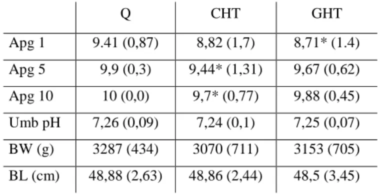
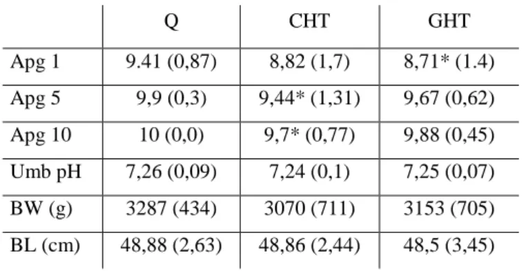
Differences between the Apgar scores (Table 1): the 1 minute Apgar scores were significantly decreased in the GHT group compared to the Q group (p=0.025). In the CHT group the 5 minute (p=0.042) and 10 minute (p=0.032) Apgar scores were significantly decreased compared to the Q group. Between the rest of the values no significant difference could be found. After the birth we measured the pH level from the umbilical vein, the birth weight (BW) and the birth length (BL). There were no significant differences between the three groups (Table 1).
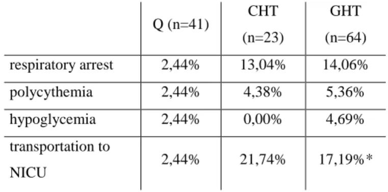
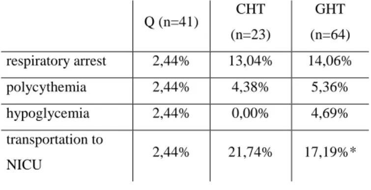
We observed the newborns in neonatal intensive care unit (NICU), if there were respiratory arrest, polycythemia, hypoglycemia or need for therapy at intensive care unit (Table 2). From the CHT group 21.7%
GHT group 17.2 % control group 2.4% of the newborns needed transportation to the NICU and it made a significant difference (p=0.021) among the groups.
Discussion
It is the first study, in which the vascularization of the fetal kidney was investigated in HDP. The monitoring of depression of renal vascularization can be quantified and it is significantly lower in fetuses with gestational hypertension that may evolve renal circulatory insufficiency, and may provide us with valuable new information helping the maternal care. Contrary to the GHT, in CHT the renal vascularization was not reduced.
Chronic hypertension does not cause reduction of the vascularization in the placenta. In these cases the human body can compensate the consequences of the hypertension.
The number of the newborns were significantly higher in the GHT group requiring neonatal intensive care. This also points to the fact how important it would be to identify patients as soon as possible with gestational hypertension, and check up this high risk group.
The power Doppler shows a high sensitivity to depict a vascular tree, because it is based on Doppler signal amplitude rather than average frequency values, unlike the color Doppler. Moreover, it does not generate the effects of aliasing and color maps are independent of the angle of insonation. The 3-DPD allows not only a full and adequate study of the vascular placental tree by identifying the different branches of villi [18,19], but also produces a quantitative assessment of the number of vessels through the vasculature and blood flow index, by applying flow rates and vascularization. By the 3D method vascular villi of the first, second and third order can be seen and thus a higher percentage is achieved in comparison with 2D Doppler technique.
Low VI values are interpreted as a decrease in the number of vessels within the placenta. The decreased FI values can be viewed as a reduction in placental blood flow due to increasing placental resistance. Low VFI values manifest a decrease in the number of vessels in the placenta, as well as a reduction of blood flow. If reduced the three indices reveal that there is a decrease in speed, intensity and placental blood perfusion.
Fetal downregulated vascularization had influence on fetal renal development [18,20]. This is an important conclusion, because it suggests that early identification of women at low risk of HPD can be improved to obtain prophylactic treatment and intense surveillance can be initiated at appropriate time.
Funding:
This study was supported by the Hungarian EFOP-3.6.1- 16-2016- 00008 grant.
Conflict of Interest Statement:
The authors declare that they have no conflict of interest
Author’ contribution:
AS did the majority of the literature research, prepared the manuscript, prepared the study design, analyzed the 3D images MN performed obstetrical 2D and 3D ultrasound, and compiled data, did some literature research, supervised the manuscript ÁTA performed obstetrical 2D and 3D ultrasound, and compiled data, did some literature research, supervised the
manuscript
TNy did the statistical analyses, and supervised the manuscript GN supervised the studyí design and manuscript
Acknowledgements
We are thank to Cedars Sinai Medical Center’s International Research and Innovation in Medicine Program, the Association for Regional Cooperation in the Fields of Health, Science and Technology (RECOOP HST Association) for their support of our organization as participating Cedars – Sinai Medical Center - RECOOP Research Centers (CRRC).
References
1. Qureshi F, Jacques SM, Seifman B, Quintero R, Evans MI, Smith C, Johnson MP: In utero fetal urine analysis and renal histology correlate with the outcome in fetal obstructive uropathies. Fetal Diagn Ther 11:306–312 (1996)
2. Mussap M, Fanos V, Pizzini C, Marcolongo A, Chiaffoni G, Plebani M: Predictive value of amniotic fluid cystatin C levels for the early identification of fetuses with obstructive uropathies. BJOG Int J Obstet Gynaecol 109:778–783 (2002)
3. Surányi A, Pál A, Streitman K, Pintér S, Kovács L: Fetal renal hyperechogenicity in pathological pregnancies. J Perinat Med 25:274–279 (1997)
4. Surányi A, Retz C, Rigo J, Schaaps JP, Foidart JM: Fetal renal hyperechogenicity in intrauterine growth retardation:
importance and outcome. Pediatr Nephrol 16:575–580 (2001)
5. Pairleitner H, Steiner H, Hasenoehrl G, Staudach A: Three-dimensional power Doppler sonography: imaging and quantifying blood flow and vascularization. Ultrasound Obstet Gynecol Off J Int Soc Ultrasound Obstet Gynecol 14:139–143 (1999)
6. Morel O, Pachy F, Chavatte-Palmer P, Bonneau M, Gayat E, Laigre P, Evain-Brion D, Tsatsaris V: Correlation between uteroplacental three-dimensional power Doppler indices and true uterine blood flow: evaluation in a pregnant sheep model. Ultrasound Obstet Gynecol Off J Int Soc Ultrasound Obstet Gynecol 36:635–640 (2010)
7. Molnár A, Surányi A, Nyári T, Németh G, Pál A: Examination of placental three-dimensional power Doppler indices and perinatal outcome in pregnancies complicated by intrauterine growth restriction. Int J Gynaecol Obstet Off Organ Int Fed Gynaecol Obstet 129:5–8 (2015)
8. Hwang J-W, Park S-J, Oh S-Y, Chang SA, Lee SC, Park SW, Kim DK: The Risk Factors That Predict Chronic Hypertension After Delivery in Women With a History of Hypertensive Disorders of Pregnancy. Medicine (Baltimore) 94:e1747 (2015)
9. Hutcheon JA, Lisonkova S, Joseph KS: Epidemiology of pre-eclampsia and the other hypertensive disorders of pregnancy. Best Pract Res Clin Obstet Gynaecol 25:391–403 (2011)
10. Khan KS, Wojdyla D, Say L, Gülmezoglu AM, Van Look PF: WHO analysis of causes of maternal death: a systematic review. The Lancet 367:1066–1074 (2006)
11. Jain L: Effect of pregnancy-induced and chronic hypertension on pregnancy outcome. J Perinatol Off J Calif Perinat Assoc 17:425–427 (1997)
12. Vanek M, Sheiner E, Levy A, Mazor M: Chronic hypertension and the risk for adverse pregnancy outcome after superimposed pre-eclampsia. Int J Gynaecol Obstet Off Organ Int Fed Gynaecol Obstet 86:7–11 (2004)
13. Zhang J, Cai WW, Lee DJ: Pregnancy-induced hypertension and early neonatal death: a case-control study. Am J Perinatol 10:401–403 (1993)
14. Tranquilli AL, Dekker G, Magee L, Roberts J, Sibai BM, Steyn W, Zeeman GG, Brown MA: The classification, diagnosis and management of the hypertensive disorders of pregnancy: A revised statement from the ISSHP. Pregnancy Hypertens 4:97–104 (2014)
15. Cohn HE, Sacks EJ, Heymann MA, Rudolph AM: Cardiovascular responses to hypoxemia and acidemia in fetal lambs.
Am J Obstet Gynecol 120:817–824 (1974)
16. Kjellmer I, Karlsson K, Olsson T, Rosén KG: Cerebral reactions during intrauterine asphyxia in the sheep. I. Circulation and oxygen consumption in the fetal brain. Pediatr Res 8:50–57 (1974)
17. Kamitomo M, Alonso JG, Okai T, Longo LD, Gilbert RD: Effects of long-term, high-altitude hypoxemia on ovine fetal cardiac output and blood flow distribution. Am J Obstet Gynecol 169:701–707 (1993)
18. Surányi A, Nyári T, Pál A: What is biparietal diameter/kidney length ratio in cases with renal hyperechogenicity?
Pediatr Nephrol 18:14–17 (2003)
19. Surányi A, Streitman K, Pál A, Nyári T, Retz C, Foidart JM, Schaaps JP, Kovács L: Fetal renal artery flow and renal echogenicity in the chronically hypoxic state. Pediatr Nephrol. 14:393-399 (2000)
20. Suranyi A: Prenatal and postnatal evaluation of foetal renal hyperechogenicity in pregnancies complicated with pre- eclampsia and intrauterine growth retardation. Dissertation. University of Szeged. http://doktori.bibl.u-szeged.hu/2154/
(2000)
Table 1. Parametric values about the Apgar scores (Apg), Umbilical pH (umb pH), birth weight (BW) and birth length (BL) of newborns. Standard deviation values are indicated between the brackets. The sign * means significant difference compared to the Q group.*p<0.05. Abbreviations: Q, control; CHT, chronic hypertension;
GHT, gestational hypertension.
Q CHT GHT
Apg 1 9.41 (0,87) 8,82 (1,7) 8,71* (1.4) Apg 5 9,9 (0,3) 9,44* (1,31) 9,67 (0,62) Apg 10 10 (0,0) 9,7* (0,77) 9,88 (0,45) Umb pH 7,26 (0,09) 7,24 (0,1) 7,25 (0,07) BW (g) 3287 (434) 3070 (711) 3153 (705) BL (cm) 48,88 (2,63) 48,86 (2,44) 48,5 (3,45)
Table 2. Non-parametric values concerning the percentage of newborns with respiratory arrest, polycythemia, hypoglycemia or necessity of treatment at neonatal intensive care unit (NICU) *p<0.05. Abbreviations: Q, control; CHT, chronic hypertension; GHT, gestational hypertension.
Q (n=41) CHT
(n=23)
GHT (n=64) respiratory arrest 2,44% 13,04% 14,06%
polycythemia 2,44% 4,38% 5,36%
hypoglycemia 2,44% 0,00% 4,69%
transportation to
NICU 2,44% 21,74% 17,19%*
Legend to the figures:
Figure 1: The fetal kidney image with 3-dimensional power Doppler technique.
Figure 2: Sequential quantification of the fetal renal vasculatization indices (VI, FI, VFI) were prepared with the VOCAL software
Figure 3. Vascular indices and volume of the fetal kidney in the control (Q) group (n=41), chronic hypertension (CHT) group (n=21), and gestational hypertension (GHT group) (n=45). The solid triangles imply mean values and the two ends of the vertical lines represent the (+/-) standard deviations.
The sign * refers to significant differences compared to the Q group. *p < 0.05. A:VI: vascularization index; B:
FI: flow index; C: VFI: vascularization flow index; D KV: kidney volume.
Q CHT GHT
Apg 1 9.41 (0,87) 8,82 (1,7) 8,71* (1.4) Apg 5 9,9 (0,3) 9,44* (1,31) 9,67 (0,62) Apg 10 10 (0,0) 9,7* (0,77) 9,88 (0,45) Umb pH 7,26 (0,09) 7,24 (0,1) 7,25 (0,07) BW (g) 3287 (434) 3070 (711) 3153 (705) BL (cm) 48,88 (2,63) 48,86 (2,44) 48,5 (3,45)
Table 1. Parametric values about the Apgar scores (Apg), Umbilical pH (umb pH), birth weight (BW) and birth length (BL) of newborns. Standard deviation values are indicated between the brackets. The sign * means significant difference compared to the Q group.*p < 0.05. Abbreviations: Q, control; CHT, chronic hypertension;
GHT, gestational hypertension.
Q (n=41) CHT (n=23)
GHT (n=64) respiratory arrest 2,44% 13,04% 14,06%
polycythemia 2,44% 4,38% 5,36%
hypoglycemia 2,44% 0,00% 4,69%
transportation to
NICU 2,44% 21,74% 17,19%*
Table 2. Non-parametric values concerning the percentage of newborns with respiratory arrest, polycythemia, hypoglycemia or necessity of treatment at neonatal intensive care unit (NICU) *p < 0.05. Abbreviations: Q, control; CHT, chronic hypertension; GHT, gestational hypertension.