O R I G I N A L A R T I C L E
Validation of distinct pathogenic patterns in a cohort of membranoproliferative glomerulonephritis patients by cluster analysis
No´ra Garam
1,2, Zolta´n Proha´szka
1,2, A ´ gnes Szila´gyi
1,2, Christof Aigner
3, Alice Schmidt
3, Martina Gaggl
3, Gere Sunder-Plassmann
3, Do´ra Bajcsi
4, Ju¨rgen Brunner
5, Alexandra Dumfarth
6,7, Daniel Cejka
6,7,
Stefan Flaschberger
8, Hana Flo¨gelova
9, A ´ gnes Haris
10, A ´ gnes Hartmann
11, Andreas Heilos
12, Thomas Mueller
12, Krisztina Rusai
12, Klaus Arbeiter
12, Johannes Hofer
5,13,14, Da´niel Jakab
15, Ma´ria Sinko´
15, Erika Szigeti
15,
Csaba Bereczki
15, Viktor Janko
16, Kata Kelen
17, Gyo¨rgy S. Reusz
17, Attila J. Szabo´
17, No´ra Klenk
18, Krisztina Ko´bor
18, Nika Kojc
19,
Maarten Knechtelsdorfer
20, Mario Laganovic
21, Adrian Catalin Lungu
22, Anamarija Meglic
23, Rina Rus
23, Tanja Kersnik-Levart
23,
Ernesta Macioniene
24, Marius Miglinas
24, Anna Pawłowska
25,
Tomasz Stompo´r
25, Ludmila Podracka
26, Michael Rudnicki
27, Gert Mayer
27, Romana Rysava
28, Jana Reiterova
28, Marijan Saraga
29,30, Toma´ s Seeman
31, Jakub Zieg
31, Eva Sla´dkova´
32, Tama´s Szabo´
33, Andrei Capitanescu
34,
Simona Stancu
34, Miroslav Tisljar
35, Kresimir Galesic
35, Andra´s Tisle´r
36, Inga Vainuma¨e
37, Martin Windpessl
38, Tomas Zaoral
39, Galia Zlatanova
40and Dorottya Csuka
1,21
Research Laboratory, 3rd Department of Internal Medicine, Hungarian Academy of Sciences and Semmelweis University, Budapest, Hungary,
2MTA-SE Research Group of Immunology and Hematology, Hungarian Academy of Sciences and Semmelweis University, Budapest, Hungary,
3Department of Medicine III, Division of Nephrology and Dialysis, Medical University of Vienna, Vienna, Austria,
41st Department of Internal Medicine, University of Szeged, Szeged, Hungary,
5Department of Pediatrics, Medical University of Innsbruck, Innsbruck, Austria,
6Department of Medicine III: Nephrology, Transplant Medicine and
Rheumatology, Linz, Austria,
7Geriatric Department, Ordensklinikum Linz – Elisabethinen, Linz, Austria,
Received: 26.2.2019; Editorial decision: 9.5.2019
VCThe Author(s) 2019. Published by Oxford University Press on behalf of ERA-EDTA.
This is an Open Access article distributed under the terms of the Creative Commons Attribution Non-Commercial License (http://creativecommons.org/
licenses/by-nc/4.0/), which permits non-commercial re-use, distribution, and reproduction in any medium, provided the original work is properly cited.
For commercial re-use, please contact journals.permissions@oup.com
225 doi: 10.1093/ckj/sfz073
Advance Access Publication Date: 21 June 2019 Original Article
8
Hospital of Klagenfurt, Klagenfurt, Austria,
9Department of Pediatrics, Division of Nephrology, Faculty of Medicine, Palacky University and Faculty Hospital in Olomouc, Olomouc, Czech Republic,
10Department of Nephrology, Szent Margit Hospital, Budapest, Hungary,
11Department of Pediatrics, University of Pe´cs, Pe´cs, Hungary,
12Department of Pediatrics and Adolescent Medicine, Division of Pediatric Nephrology and Gastroenterology, Medical University of Vienna, Vienna, Austria,
13Institute of Neurology of Senses and Language, Hospital of St John of God, Linz, Austria,
14Research Institute for Developmental Medicine, Johannes Kepler University Linz, Linz, Austria,
15Department of Pediatrics, University of Szeged, Szeged, Hungary,
16Medimapax – Center of Elimination Methods, Bratislava, Slovakia,
171st Department of Pediatrics, Semmelweis University, Budapest, Hungary,
18FMC Center of Dialysis, Miskolc, Hungary,
19Institute of Pathology, Faculty of Medicine, University of Ljubljana, Ljubljana, Slovenia,
20Department of Nephrology, Wilhelminenspital, Vienna, Austria,
21Department of Nephrology, Arterial Hypertension, Dialysis and Transplantation, University Hospital Center Zagreb, School of Medicine University of Zagreb, Zagreb, Croatia,
22
Pediatric Nephrology Department, Fundeni Clinical Institute, Bucharest, Romania,
23Department of Pediatric Nephrology, Division of Pediatrics, University Medical Centre Ljubljana, Ljubljana, Slovenia,
24Nephrology Center, Santaros Klinikos, Medical Faculty, Vilnius University, Vilnius, Lithuania,
25Department of Nephrology, Hypertension and Internal Medicine, School of Medicine, Collegium Medicum, University of Warmia and Mazury, Olsztyn, Poland,
26Department of Pediatrics, Comenius University, Bratislava, Slovakia,
27
Department of Internal Medicine IV–Nephrology and Hypertension, Medical University Innsbruck,
Innsbruck, Austria,
28Nephrology Clinic, 1st Faculty of Medicine, Charles University, Prague, Czech Republic,
29
Department of Pathology, University Hospital Split, Split, Croatia,
30School of Medicine, University of Split, Split, Croatia,
31Department of Pediatrics, 2nd Faculty of Medicine, Charles University Prague, University Hospital Motol, Prague, Czech Republic,
32Department of Pediatrics, Faculty of Medicine in Pilsen, Charles University in Prague, Pilsen, Czech Republic,
33Department of Pediatrics, Faculty of Medicine, Debrecen University, Debrecen, Hungary,
34Carol Davila Nephrology Hospital, Bucharest, Romania,
35Department of Nephrology, Dubrava University Hospital, Zagreb, Croatia,
361st Department of Internal Medicine,
Semmelweis University, Budapest, Hungary,
37Zivile Riispere, Department of Pathology, Tartu University Hospital, Tartu, Estonia,
38Internal Medicine IV, Section of Nephrology, Klinikum Wels-Grieskirchen, Wels, Austria,
39Department of Pediatrics, Faculty of Medicine, University Hospital, Ostrava, Czech Republic and
40
University Children’s Hospital, Medical University, Sofia, Bulgaria
Correspondence and offprint requests to: Zolta´n Proha´szka; E-mail:prohaszka.zoltan@med.semmelweis-univ.hu
ABSTRACT
Background.A novel data-driven cluster analysis identified distinct pathogenic patterns in C3-glomerulopathies and immune complex-mediated membranoproliferative glomerulonephritis. Our aim was to replicate these observations in an independent cohort and elucidate disease pathophysiology with detailed analysis of functional complement markers.
Methods.A total of 92 patients with clinical, histological, complement and genetic data were involved in the study, and hierarchical cluster analysis was done by Ward method, where four clusters were generated.
Results.High levels of sC5b-9 (soluble membrane attack complex), low serum C3 levels and young age at onset (13 years) were characteristic for Cluster 1 with a high prevalence of likely pathogenic variations (LPVs) and C3 nephritic factor, whereas for Cluster 2—which is not reliable because of the small number of cases—strong immunoglobulin G staining, low C3 levels and high prevalence of nephritic syndrome at disease onset were observed. Low plasma sC5b-9 levels, decreased C3 levels and high prevalence of LPV and sclerotic glomeruli were present in Cluster 3, and patients with late onset of the disease (median: 39.5 years) and near-normal C3 levels in Cluster 4. A significant difference was observed in the incidence of end-stage renal disease during follow-up between the different clusters. Patients in Clusters 3–4 had worse renal survival than patients in Clusters 1–2.
Conclusions.Our results confirm the main findings of the original cluster analysis and indicate that the observed, distinct pathogenic patterns are replicated in our cohort. Further investigations are necessary to analyse the distinct biological and pathogenic processes in these patient groups.
Keywords: C3-glomerulopathy, C3-glomerulonephritis, complement, dense deposit disease, membranoproliferative glomerulonephritis
INTRODUCTION
Membranoproliferative glomerulonephritis (MPGN), character- ized by mesangial hypercellularity, endocapillary proliferation and capillary wall thickening with double contour formation, is a pathological pattern detected by light microscopy on kidney biopsies [1]. Recent improvements in our understanding of the disease pathogenesis, with the recognition of the pivotal role of complement alternative pathway (AP) [2], led to the description of complement-mediated (C3-glomerulopathy, C3G) and immune complex-mediated MPGN (IC-MPGN) forms [3]. C3G presents with microscopic haematuria, proteinuria and renal failure, with dominant (minimum two-order magnitude stron- ger C3 staining than any other immunoreactant) C3 stain on im- munofluorescence microscopy, and is caused by the dysregulation of the AP due to the presence of variations of complement genes [Complement Factor H (CFH), Complement Factor I (CFI), Membrane cofactor protein (CD46), Complement Factor B (CFB), Thrombomodulin (THBD)] [1,4–6], or by autoanti- bodies [C3 nephritic factor (C3NeF), anti-Factor H, anti-C3, anti- Factor B and C4 nephritic factor (C4NeF)] against complement proteins [7–11]. Despite thorough analysis of potential risk fac- tors, complement-related predisposing factors in several patients with C3G remain unidentified. C3G is divided according to electron microscopy, where C3-glomerulonephritis (C3GNs) usually has less dense mesangial, subendothelial and subepi- thelial deposits, whereas dense deposit disease (DDD) is charac- terized by electron-dense intramembranous deposits.
Despite improved understanding of disease pathogenesis, several questions still remain unanswered, including deviation between pathological and clinical presentations, hampered dif- ferentiation between C3GN and DDD (sometimes with IC- MPGN) or even changes in the characteristic patterns if repeated biopsies are done [3,12,13]. Even more importantly, the as- sumption that AP dysregulation is the most characteristic dif- ferentiating feature between immunoglobulin (Ig)-negative and Ig-positive, MPGN does not seem to be powerful enough since a large portion of patients with Ig-positive MPGN may be positive for C3NeF, or show decreased C3 levels with normal C4 [1,14].
Taking all these limitations and considerations into account, Iatropouloset al. [15] recently explored the potential utility of unsupervised cluster analysis to generate clinically meaningful subgroups of C3G/IC-MPGN patients. In their study, four distinct clusters were generated based on the clinical, histological, com- plement and genetic parameters. Patients in Clusters 1 and 2 had elevated terminal pathway activation marker (sC5b-9) lev- els with low C3 concentrations, whereas patients with dense deposits, decreased C3 but only moderately elevated sC5b-9 fell into Cluster 3. Patients in Clusters 1–3 had a similarly high prev- alence of C3NeF and/or presence of likely pathogenic variation (LPVs) in complement genes, which was in contrast to Cluster 4, where C3 activation in glomeruli (not in circulation), presence of sclerotic glomeruli and absence of LPVs or C3NeF were char- acteristic. Interestingly, each cluster included patients with IC- MPGN, DDD or C3GN, strengthening the overlap between these entities. Notably, reclassification of C3GN, DDD and IC-MPGN patients by cluster analysis proved to be better at predicting re- nal survival, a result pointing to the feasibility of the ultimate goal of efficient patient stratification for complement- modulating therapies. However, as such unsupervised mathe- matical approaches are completely dependent on the studied cohort and on the variable set, validation in independent cohorts is essential.
Therefore, our aim was to validate the observations of Iatropouloset al. [15] in an independent cohort. We enrolled 92 patients with pathologically diagnosed IC-MPGN/C3G, repeated the cluster analysis by analysing the same variable set (Supplementary data, Table S1), with the generation of four clusters, and observed very similar patterns to that of the origi- nal study. In addition, we tested further complement parame- ters to better characterize ongoing pathophysiology in the different clusters.
MATERIALS AND METHODS
A total of 92 patients with histologically proven C3G or IC-MPGN were enrolled in the study from 34 centres in Central and Eastern Europe (Supplementary data, Figure S1). Clinical and histological data were collected from clinicians, and genetic analysis (CFH,CFI,CD46,C3,CFBandTHBD) was performed by Sanger sequencing (Supplementary data,Table S2). Serum C3, C4 concentration was determined by turbidimetry. Classical pathway activity was measured by haemolytic test [16]. Other complement parameters [AP activity, sC5b-9, anti-Factor H, anti-Factor B, anti-C3, anti-C1q, C4d, Bb, Factor D (FD)] were measured using home-made or commercially available ELISA kits. C3NeF activity was detected using haemolytic test [17].
Detailed methods are presented in Supplementary materials.
RESULTS
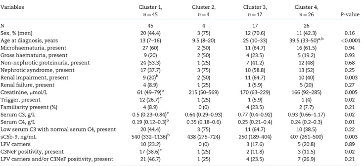
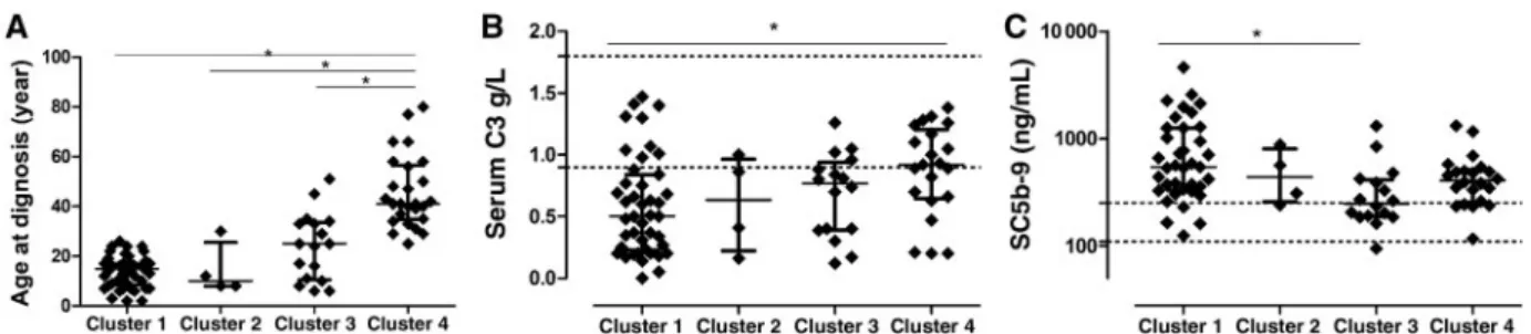
Generation of clinical patterns by cluster analysis Clinical characteristics (Tables 1and2) of the patients and com- parison of the two cohorts (Supplementary data,Figure S2) are summarized inSupplementary results. Similar to the study of Iatropoulos et al. [15], four distinct clusters were generated (Supplementary data, Figure S3). Forty-five patients were regrouped into Cluster 1, 4 patients into Cluster 2, 17 subjects into Cluster 3 and 26 patients into Cluster 4 (Tables 3and4). In our analysis Cluster 3, including mainly young adults with lower sC5b-9 levels and higher prevalence of sclerotic glomeruli, was completely separated from the remaining clusters.
Thereafter, Cluster 4 was separated from Clusters 1 and 2, which includes adult patients with later disease onset and higher C3 levels (Supplementary data,Figure S3).
Cluster 1 was characterized by low C3 levels (median 0.5 g/L), very high sC5b-9 (median 540 ng/mL) levels and high prevalence of LPVs and C3NeF. The median age of onset was 13 years. Renal impairment and creatinine levels were low in that cluster at the time of onset. On light microscopy, no sclerotic glomeruli or crescents were seen, the strong C3 staining was not isolated and Ig staining was also seen in Cluster 1 on immunofluores- cence microscopy, in concordance with the original study of Iatropouloset al. [15].
The four patients in Cluster 2 had the strongest glomerular staining for IgA, IgG and C1q, with substantial crescent forma- tion, three of them had nephrotic-range proteinuria; hence, the descriptive characteristics are very similar to those published by Iatropouloset al. However, due to the low case number, we excluded these patients from further analysis.
In Cluster 3, the lowest sC5b-9 levels (median 250 ng/mL) were observed, compared with other clusters, along with mod- erately decreased C3 concentration (median 0.77 g/L) and with high prevalence of LPVs (Figure 1). On light microscopy, the highest degree of interstitial fibrosis, interstitial inflammation
and the highest prevalence of sclerotic glomeruli were seen with low prevalence of crescents, compared with the other clusters.
In Cluster 4, the median age of onset (39.5 years) was the highest compared with the other clusters, and this cluster was also characterized by near-normal C3 levels (median 0.93 g/L;
Table 3andFigure 1) and with lower prevalence of intramem- branous deposits (P¼0.046). The prevalence of thrombotic microangiopathy (TMA) was the highest in this cluster in Italian cluster [15]. We have only two patients with data for TMA, but they were excluded because of missing data, which is why we could not examine this aspect of Cluster 4.
The key pathogenic features of the different clusters in the two cohorts are shown in comparative manner in Supplementary data,Figure S4.
Decision tree analysis was done to select the minimum set of features to predict membership of patients in the clusters (Figure 2), and predictor variables were ranked according to their classification importance (Supplementary data,Table S3). The top two classifiers were the presence and extent of sclerotic glomeruli and age at diagnosis. Using three cut-points of these variables, all but one patient were correctly classified into Clusters 1, 3 and 4.
Variable ranking indicated that besides age at diagnosis and extent of glomerular sclerosis, the most important predictor variables for cluster membership were the presence of interstitial fibrosis, inter- stitial inflammation, arteriolar sclerosis and sC5b-9 level.
No difference was observed with regard to the occurrence or distribution of LPVs between the different clusters, but LPVs af- fecting C3 and CFH were identified only in Cluster 1. The gener- ated clusters did not show significant connection with the original histology-based diagnosis (Supplementary data,Table S4) although patients with C3GN and IC-MPGN fall mainly into Clusters 1 and 4.
Prognostic significance of the clusters
Clinical relevance of the clusters was evaluated by analysis of their association with renal outcome. During the follow-up
period, we managed to collect data on the development of end- stage renal disease (ESRD), defined as estimated glomerular fil- tration rate decrease to<15 mL/min/1.73 m2 with or without renal replacement therapy (such as haemodialysis, peritoneal dialysis or kidney transplantation). Ultimately, 10 out of the 79 successfully followed patients progressed to ESRD including 5 patients with acute kidney injury at the time of diagnosis but later temporary improvement of kidney function. Renal out- come was found to be worse in Clusters 3 and 4 compared with Cluster 1 with Kaplan–Meier analysis (P<0.05 for Clusters 3 ver- sus 1 and Clusters 4 versus 1), indicating that patients with higher age at diagnosis, more prevalent glomerular sclerosis at diagnosis and lower prevalence of C3NeF and/or LPV have infe- rior renal outcome. There was no difference in renal survival according to the histology groups (Figure 3). We have data of patients’ treatment strategy at the time of diagnosis, which may affect renal survival. Going through the data, most of the patients received antihypertensive drugs [angiotensin-convert- ing enzyme-I and angiotensin-receptor blockers (ARB) and immunosuppressants (steroid, cyclophosphamide and myco- phenolate mofetil). In some cases, plasmatherapy and renal re- placement therapy were indicated at the time of diagnosis. No relevant differences were seen between patients’ initial therapy in different clusters (Supplementary data,Table S5). The various treatment modifications during follow-up period were not collected.
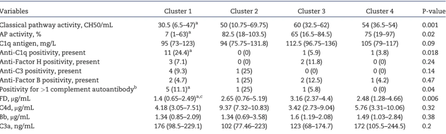
Complement activation and regulation in relation to clinical patterns
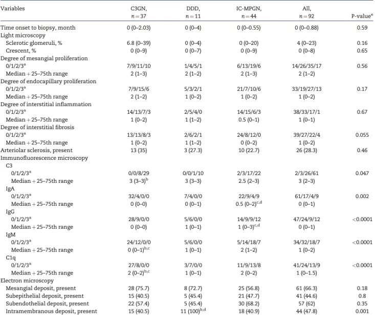
The findings of the cluster analysis motivated us to examine an extended set of complement parameters. Due to the association of high sC5b-9 levels and C3 consumption with clusters (Table 3), we measured additional complement proteins, path- way activities and activation markers (Table 5). Significantly lower classical pathway (CP) (P¼0.001) and alternative pathway (AP) (P¼0.02) activity, along with decreased FD concentrations (P¼0.006) were observed in Cluster 1, where C1q levels were also the lowest. These results together with decreased C3 and Table 1. Clinical characteristics of patients with histologically proven diagnosis of C3G or IC-MPGN
Variables C3GN,
n¼37
DDD, n¼11
IC-MPGN, n¼44
All,
n¼92 P-valueb
Sex, % men 17 (46) 3 (27.3) 26 (59.1) 46 (50) 0.14
Age at diagnosis, years 23.5 (13–39) 17 (13.5–27.5) 15 (9–33) 16 (10–33) 0.1
Microhaematuria, present 24 (64.8) 8 (72.7) 24 (54.5) 56 (60.9) 0.44
Gross haematuria, present 7 (19) 2 (18.2) 11 (25) 20 (21.7) 0.76
Non-nephrotic proteinuria, present 18 (48.6) 4 (36.4) 22 (50) 44 (48.5) 0.71
Nephrotic syndrome, present 15 (40.5) 7 (63.6) 21 (47.7) 43 (47.3) 0.39
Renal impairment, present 13 (35.1) 4 (36.4) 15 (34.1) 32 (35.2) 0.98
Renal failure, present 4 (10.8) 1 (9) 6 (13.6) 11 (12.1) 0.88
Creatinine,lmol/L 86 (53–240) 79 (52–121) 82 (55–168) 83 (54–196) 0.89
Trigger, present 5 (13.5) 3 (27.3) 7 (16) 15 (16.5) 0.55
Familiarity, present 5 (13.2) 0 (0) 5 (11.4) 10 (10.9) 0.44
Serum C3, g/L 0.68 (0.34–1.05) 0.5 (0.25–0.86) 0.69 (0.31–0.96) 0.67 (0.33–0.99) 0.93
Serum C4, g/L 0.28 (0.19–0.41) 0.21 (0.16–0.36) 0.2 (0.13–0.25)a 0.23 (0.17–0.32) 0.02
Low serum C3 with normal serum C4 19 (51.3) 7 (63.6) 18 (41) 44 (47.8) 0.34
sC5b-9, ng/mL 421 (271–661) 509 (327–1069) 375 (243–731) 406 (261–690) 0.7
LPV carriers 7 (20) 2 (18.2) 9 (21.4) 18 (20.5) 0.96
C3NeF positivity, present 7 (19.4) 5 (45.4) 11 (25) 23 (25.3) 0.2
LPV carriers and/or C3NeF positivity, present 11 (29.7) 5 (45.4) 17 (38.6) 33 (35.8) 0.18
The data are given as median and interquartile range or number and percentages. Reference range: C3, 0.9–1.8 g/L; C4, 0.15–0.55 g/L; sC5b-9, 110–252 ng/mL.
aSignificantly different from C3GN.
bP-values are given as the results of Chi-square or Kruskal–Wallis tests of the patients with IC-MPGN, C3GN and DDD.
C4 levels in Cluster 1 indicating overactivation and consump- tion of CP/AP compared with the Clusters 3 and 4.
Concentrations of the complement activation split products C4d, Bb and C3a were low and did not show any significant dif- ference among the different clusters.
C3NeF positivity was more prevalent in Cluster 1; therefore, additional complement autoantibodies including anti-C1q, anti- Factor H, anti-Factor B and anti-C3 were also determined.
Cluster 1 captured 11 out of 13 anti-C1q-positive patients (P¼0.018), whereas no difference in the prevalence of anti- Factor H, anti-C3 and anti-Factor B among the four clusters was observed. Multiple positivity for the examined autoantibodies was observed more frequently in Cluster 1. We did not find any association between the studied parameters and the histology- based groups (Supplementary data,Table S6).
DISCUSSION
Current classification of MPGN is based on immunofluorescence microscopy and classifies patients as C3G/IC-MPGN [3].
Recently, an unsupervised, data-driven method was applied by Iatropoulos and associates for a cohort of MPGN patients to see whether clinically meaningful groups can be identified based on clinical, biochemical, histological or genetic data [15]. This new statistical approach generated clusters with small within- cluster, as opposed to large between-cluster, differences.
The clusters showed clear associations with AP abnormalities, additional pathogenic features and risk of progression to ESRD.
As was pointed out in an editorial [18] commenting the results of Iatropoulos et al., repetition of these observations in an independent cohort is essential to validate if such clusters exist or not.
Table 2. Histological characteristics of patients with histologically proven diagnosis of C3G or IC-MPGN
Variables C3GN,
n¼37
DDD, n¼11
IC-MPGN, n¼44
All,
n¼92 P-valuee
Time onset to biopsy, month 0 (0–2.03) 0 (0–4) 0 (0–0.55) 0 (0–0.88) 0.59
Light microscopy
Sclerotic glomeruli, % 6.8 (0–39) 0 (0–4) 0 (0–20) 4 (0–23) 0.16
Crescent, % 0 (0–9) 0 (0–7) 0 (0–9) 0 (0–8) 0.65
Degree of mesangial proliferation
0/1/2/3a 7/9/11/10 1/4/5/1 6/13/19/6 14/26/35/17 0.56
Medianþ25–75th range 2 (1–3) 2 (1–2) 2 (1–3) 2 (1–2)
Degree of endocapillary proliferation
0/1/2/3a 7/9/15/6 5/3/2/1 21/7/10/6 33/19/27/13 0.17
Medianþ25–75th range 2 (1–2) 1 (0–2) 1 (0–2) 1 (0–2)
Degree of interstitial inflammation
0/1/2/3a 14/13/7/3 2/5/4/0 14/15/6/3 38/33/17/1 0.67
Medianþ25–75th range 1 (0–2) 1 (1–2) 0.5 (0–1) 1 (0–1)
Degree of interstitial fibrosis
0/1/2/3a 13/13/8/3 2/6/2/1 24/8/12/0 39/27/22/4 0.055
Medianþ25–75th range 1 (0–2) 1 (1–2) 0 (0–2) 1 (0–2)
Arteriolar sclerosis, present 13 (35) 3 (27.3) 10 (22.7) 26 (28.3) 0.46
Immunofluorescence microscopy C3
0/1/2/3a 0/0/8/29 0/0/1/10 2/3/17/22 2/3/26/61 0.047
Medianþ25–75th range 3 (3–3)b 3 (3–3) 2.5 (2–3) 3 (2–3)
IgA
0/1/2/3a 32/4/0/0 7/4/0/0 22/9/4/9 61/17/4/9 0.002
Medianþ25–75th range 0 (0–0) 0 (0–1) 0.5 (0–2)c,d 0 (0–1)
IgG
0/1/2/3a 28/9/0/0 5/6/0/0 14/9/9/12 47/24/9/12 <0.0001
Medianþ25–75th range 0 (0–0) 1 (0–1) 1 (0–3)c,d 0 (0–1)
IgM
0/1/2/3a 24/12/0/0 5/6/0/0 5/14/18/7 34/32/18/7 <0.0001
Medianþ25–75th range 0 (0–1)b,c 1 (0–1) 2 (1–2) 1 (0–2)
C1q
0/1/2/3a 27/8/0/0 3/7/0/0 11/9/13/8 41/24/13/9 <0.0001
Medianþ25–75th range 2 (0–2)b,c 1 (0–1) 2 (0–2) 1 (0–1.5)
Electron microscopy
Mesangial deposit, present 28 (75.7) 8 (72.7) 25 (56.8) 61 (66.3) 0.18
Subepithelial deposit, present 15 (40.5) 5 (45.4) 21 (47.7) 41 (44.6) 0.8
Subendothelial deposit, present 22 (57.4) 5 (45.4) 30 (68.2) 57 (62) 0.35
Intramembranous deposit, present 15 (40.5) 11 (100)b,d 18 (40.9) 44 (47.8) 0.001
The data are given as median and interquartile range or number and percentages if not otherwise stated. Some patients have missing values in the following data: IgA, IgM, C1q immunofluorescence staining.
aDegree of light microscopy, immunofluorescence findings were defined using scales 0–3.
bSignificantly different from IC-MPGN.
cSignificantly different from DDD.
dSignificantly different from C3GN.
eP-values are given as the results of Chi-square or Kruskal–Wallis tests of the patients with IC-MPGN, C3GN and DDD.
The aim of our study was to replicate the unsupervised hier- archical cluster analysis made by Iatropouloset al. [15] in an in- dependent C3G/IC-MPGN cohort and to validate the existence of clinically meaningful clusters. We recruited 92 patients and de- termined the same clinical, biochemical, histological and ge- netic parameters as in the original study [15]. Our independent cohort seemed to be appropriate for validation study since the most important characteristics were similar to the original co- hort (Supplementary data,Figure S2). With unsupervised, data- driven clustering method, four clusters were generated yielding Clusters 1, 3 and 4 with sufficiently high patient numbers, whereas Cluster 2 with only four patients was excluded from further analysis. Clusters 1, 3 and 4 showed clear associations with clinical, biochemical or histological variables with a very similar pattern to that of Iatropouloset al. (Supplementary data, Figure S4). Clinical relevance of the clusters was demonstrated by the significant association of Clusters 3 and 4 with develop- ment of ESRD during follow-up (Figure 3).
In the study of Iatropouloset al. [15], a three-step algorithm was created to assign patients into different clusters.
Unfortunately, we have no data to differentiate between highly electron-dense ribbon-like and granular deposits in basement membrane. That is why the minimum data set necessary to predict membership in the clusters was determined by decision tree analysis. Based on the presence and extent of sclerotic glo- meruli, and age at onset, we could divide the patients into four clusters (Figure 2). Patients with higher percentages of sclerotic glomeruli and older age fall into Clusters 3 and 4, and have infe- rior renal survival. These findings are consistent with the well- known fact that renal survival is connected with the chronicity of the glomerular lesions, but the pathomechanism of the resulting pathological image is not known. It is noteworthy and would be important to explore why the pattern of some
important clinical and complement parameters show associa- tion with these clusters and with outcome, too.
The patterns of clinical and pathogenetic factors of the dif- ferent clusters were very similar in our study to those in Iatropouloset al. Cluster 1 identified patients with very high sC5b-9 and decreased C3 levels, along with the high prevalence of LPVs and/or C3NeF in younger patients. Patients in Cluster 3 were typically young adults characterized also by complement consumption (decreased C3) but lower sC5b-9 levels, whereas the histological picture was more severe as indicated by higher degree of interstitial inflammation, fibrosis and presence of sclerotic glomeruli. Based on these results, we could not un- equivocally match the current Cluster 3 to the Italian Cluster 3.
The importance of the difference is also supported by the differ- ent renal survival of Cluster 3. Cluster 4 included adults with normal levels of C3, lack of severe complement activation and inferior renal survival.
Beyond the similarities, some differences were also noted between our study and that of Iatropouloset al. [15], the most important being some discrepancies in histology. We enrolled patients from several centres, without the option of centralized second review of biopsy. Rather, aggregate evaluation of biopsy descriptions was done by a structured questionnaire.
Furthermore, the mean ages in Clusters 3 and 4 are higher by 10 years in our study, and this may explain why the prevalence of sclerotic glomeruli is also higher in Cluster 3. Cluster 3 differs in our study from the original study in additional aspects too, including the lack of crescents and lower C3NeF prevalence.
The difference of C3NeF prevalence between the two studies could be explained by the different method of the detection of C3NeF activity. In contrast to the Italian study where purified IgG was used for the detection, we used patients’ sera following the protocol of Rother [17]. This fact can make this slight differ- ence between the prevalence of C3NeF.
Table 3. Clinical characteristics of patients according to the classification by cluster analysis
Variables Cluster 1,
n¼45
Cluster 2, n¼4
Cluster 3, n¼17
Cluster 4,
n¼26 P-value
N 45 4 17 26
Sex, % (men) 20 (44.4) 3 (75) 12 (70.6) 11 (42.3) 0.16
Age at diagnosis, years 13 (7–16) 9.5 (8–20) 25 (10–33) 39.5 (33–50)a,b <0.0001
Microhaematuria, present 27 (60) 2 (50) 11 (64.7) 16 (61.5) 0.94
Gross haematuria, present 9 (20) 2 (50) 4 (23.5) 5 (19.2) 0.93
Non-nephrotic proteinuria, present 24 (53.3) 1 (25) 7 (41.2) 12 (48) 0.68
Nephrotic syndrome, present 17 (37.7) 3 (75) 10 (58.8) 13 (52) 0.25
Renal impairment, present 9 (20)b 2 (50) 11 (64.7) 10 (40) 0.003
Renal failure, present 4 (8.9) 1 (25) 1 (5.9) 5 (20) 0.27
Creatinine,lmol/L 61 (49–79)b 215 (50–569) 170 (63–229) 166 (92–285) 0.005
Trigger, present 12 (26.7)c 1 (25) 1 (5.9) 1 (4) 0.02
Familiarity present (%) 4 (8.9) 0 (0) 4 (23.5) 2 (7.7) 0.21
Serum C3, g/L 0.5 (0.23–0.84)c 0.64 (0.29–0.93) 0.77 (0.4–0.92) 0.93 (0.66–1.17) 0.02
Serum C4, g/L 0.19 (0.12–0.3)b 0.35 (0.18–0.6) 0.25 (0.21–0.4) 0.24 (0.2–0.3) 0.01
Low serum C3 with normal serum C4, present 20 (44.4) 3 (75) 11 (64.7) 10 (38.5) 0.22
sC5b-9, ng/mL 540 (332–1136)b 438 (275–724) 250 (189–404) 407 (261–500) 0.003
LPV carriers 10 (23.2) 0 (0) 3 (17.6) 5 (20.8) 0.89
C3NeF positivity, present 17 (38.6)c 1 (25) 2 (11.8) 3 (11.5) 0.02
LPV carriers and/or C3NeF positivity, present 21 (46.7) 1 (25) 4 (23.5) 7 (26.9) 0.21
The data are given as median and interquartile range or number and percentages. P-values are given as the results of Chi-square or Kruskal–Wallis tests of patients in Clusters 1, 3 and 4. Reference range: C3 0.9–1.8 g/L; C4 0.15–0.55 g/L; sC5b-9 1110–252 ng/mL. Some patients have missing values in the following data: proteinuria, renal impairment/failure, sC5b-9, LPV and C3NeF.
aSignificantly different from Cluster 1.
bSignificantly different from Cluster 3.
cSignificantly different from Cluster 4.
Higher age, and lack of crescents but presence of sclerosis, indicating advanced disease stage may explain the inferior re- nal survival of patients in Cluster 3. Cluster 4 is best character- ized by lack of systemic signs of complement activation and consumption, but with intense glomerular C3 stain and inferior survival. The answer to why patients in Clusters 3 and 4 have advanced glomerular lesions and higher age at presentation with inferior renal survival, and whether these features are re- lated to the molecular pathophysiology, remains elusive today.
The presence of complement consumption and a high sC5b-9 level in Cluster 1 prompted us to explore additional comple- ment biomarkers and autoantibodies to understand the under- lying pathophysiology. We found that Cluster 1 encompassed most of the patients with anti-C1q autoantibodies. Remarkably, 2/11 and 5/11 anti-C1q-positive patients in Cluster 1 were
double or triple positive for additional complement autoanti- bodies listed inTable 5. The observation that different comple- ment autoantibodies occur together in Cluster 1 (P¼0.043 when compared with Clusters 3 and 4) also raises the possibility of strong polyclonal response and epitope spreading towards com- plement proteins. In addition to the autoantibodies presented in this study (Table 5), presence of C4NeF also seems to be more prevalent in Cluster 1 (N. Garam et al., unpublished work).
We acknowledge the potential limitations of our study.
Some of our observations and conclusions stem from under- powered analysis and may suffer from Type I error. However, since this is an independent replication study, and most of the key observations and differences were similar in the two studies, the appearance of false-positive decisions twice is highly unlikely. In addition, our study lacked centralized second Table 4. Histological characteristics of patients according to the classification by cluster analysis
Variables Cluster 1,
n¼45
Cluster 2, n¼4
Cluster 3, n¼17
Cluster 4,
n¼26 P-valuee
Time onset to biopsy (months) 0 (0–0.69) 1.85 (0.15–4.91) 0 (0–0.6) 0 (0.46–2.1) 0.22
Light microscopy
Sclerotic glomeruli, % 0 (0–5) 0 (0–0) 60 (47–70)a,b 6 (0–19) <0.0001
Crescent, % 0 (0–4) 55 (46–62)a,b,c 0 (0–6) 0 (0–9) 0.06
Degree of mesangial proliferation
0/1/2/3d 5/11/18/11 1/1/2/0 2/3/6/4 4/11/9/2 0.34
Medianþ25–75th range 2 (1–2) 1.5 (0.5–2) 2 (1–2) 1 (1–2)
Degree of endocapillary proliferation
0/1/2/3d 13/8/18/6 2/0/1/0 7/4/3/3 11/7/5/3 0.48
Medianþ25–75th range 2 (0–2) 1 (0–2.5) 1 (0–2) 1 (0–2)
Degree of interstitial inflammation
0/1/2/3d 24/18/3/0 2/1/1/0 2/6/6/3 10/8/7/1 0.001
Medianþ25–75th range 0 (0–1)b,c 0.5 (0–1.5) 2 (1–2)b 1 (0–2)
Degree of interstitial fibrosis
0/1/2/3d 27/15/3/0 2/1/1/0 1/3/10/3 9/8/8/1 <0.0001
Medianþ25–75th range 0 (0–1)b,c 0.5 (0–1.5) 2 (2–2)b 1 (0–2)
Arteriolar sclerosis, present 3 (6.7)b,c 1 (25) 10 (58.8) 12 (46.2) <0.0001
Immunofluorescence microscopy C3
0/1/2/3d 1/1/12/31 0/1/0/3 0/1/4/12 1/0/10/15 0.72
Medianþ25–75th range 3 (2–3) 3 (2–3) 3 (2–3) 3 (2–3)
IgA
0/1/2/3d 28/9/3/5 2/1/0/1 12/3/0/1 19/4/1/2 0.89
Medianþ25–75th range 0 (0–1) 0.5 (0–2) 0 (0–0.5) 0 (0–1)
IgG
0/1/2/3d 22/8/7/8 0/2/0/2 9/7/0/1 16/7/2/1 0.13
Medianþ25–75th range 1 (0–2) 2 (1–3) 0 (0–1) 0 (0–1)
IgM
0/1/2/3d 14/16/10/5 1/3/0/0 7/5/2/2 12/8/6/0 0.5
Medianþ25–75th range 1 (0–2) 1 (0.5–1) 1 (0–1) 1 (0–1.5)
C1q
0/1/2/3d 18/11/9/7 1/1/1/1 10/5/0/1 12/7/3/0 0.16
Medianþ25–75th range 1 (0–2) 1.5 (0.5–2.5) 0 (0–1) 0 (0–1)
Electron microscopy
Mesangial deposit, present 30 (66.7) 2 (50) 12 (70.6) 17 (65.4) 0.93
Subepithelial deposit, present 24 (53.3) 3 (75) 7 (41.2) 7 (26.9) 0.09
Subendothelial deposit, present 31 (68.9) 3 (75) 10 (58.8) 13 (50) 0.28
Intramembranous deposit, present 24 (53.3) 2 (50) 11 (64.7) 7 (26.9)a 0.07
The data are given as median and interquartile range or number and percentages if not otherwise stated. Some patients have missing values in the following data: IgA, IgM, C1q immunofluorescence staining.
aSignificantly different from Cluster 1.
bSignificantly different from Cluster 4.
cSignificantly different from Cluster 3.
dDegree of light microscopy, immunofluorescence findings were defined using scales 0–3.
eP-values are given as the results of Chi-square or Kruskal–Wallis tests of patients in Clusters 1, 3 and 4.
look-up of biopsy results, potentially leading to higher variance in histology-based scores. However, the most important cluster- defining histological marker was presence and extent of sclerotic glomeruli, a feature not prone to suffer from large inter-observer differences. We could not distinguish between granular and ribbon-like highly electron-dense intramembra- nous deposits in our study because only intramembranous highly electron-dense deposits were mentioned in most of the pathology reports.
Considering the extremely low prevalence of C3G (2–3/1 000 000 people), large multicentre studies are essentials to confirm the original studies of Iatropouloset al. [15]. Although we could not collect some data in exactly the same way as it was de- scribed in the Italian study, this study is the first that managed to collect enough patients to run this analysis again in a very similar manner and this international collaboration allowed us to validate the main findings of the original analysis in this rare disorder. With the ultimate goal to support treatment decisions for patients with these rare diseases, our results confirmed the existence of Clusters 1 and 4. Patients in Cluster 1 are best char- acterized by younger age, lack of glomerular sclerosis, high prevalence of LPVs and autoantibodies, and signs of systemic complement activation and consumption, hence, intense C5-
convertase activity. These signs support the presence of fluid- phase dysregulation; hence, patients falling into Cluster 1 are potential candidates for C5-targeted therapies. In contrast, patients in Cluster 4 had no signs of systemic complement acti- vation or consumption despite intense C3 deposition in the kid- ney, indicating the presence of surface-limited complement dysregulation. The fact that four out of five LPV carriers in Cluster 4 carried LPVs in the genes of surface expressed cofac- tors CD46 and THBD seems to support this idea. Therefore, the proposition of Iatropouloset al. to consider complement inhibi- tors, targeting C3 activation products on cell surfaces (like TT30) [19], seems to be rational and is also supported by our observa- tions. Finally, despite independent validation of the original observations [15], it is still a question whether this new, data- driven, pathogenesis-connected classification can change the histology-based classification in the long term, with the final goal of better classification to support appropriate treatment decisions.
SUPPLEMENTARY DATA
Supplementary dataare available at ckj online.
FIGURE 1:Scatterplots of age at diagnosis (A), serum C3 levels (B) and plasma sC5b-9 levels (C) across the different clusters. Horizontal lines show medians, whiskers interquartile ranges of the groups. Horizontal dotted lines indicate reference range limits. For the comparison of Clusters 1, 3 and 4, Kruskal–Wallis analysis of variance was used (panel A, P¼0.018; panel B, P¼0.003; panel C, P<0.0001) with Dunn’spost hoctest. *P<0.05.
FIGURE 2:Decision tree to assign patients into the different clusters. Analysis based on classification and regression model built to predict independent clusters as cat- egorical variable, and on the same predictor variable set that was used for the cluster analysis (for details see Materials and methods section). Four patients in Cluster 2 were excluded from this analysis.
FIGURE 3:Kaplan–Meier renal survival curves for groups of MPGN patients based on the histological diagnosis (A) or cluster (B). P-values of log-rank tests are indicated.
Cluster 2 was excluded from Kaplan–Meier analysis. To better visualize the renal survival of the patients, we stopped the curves at 6 years because of a shorter follow- up in the majority of the cases. ICGN, immune complex-mediated MPGN.
Table 5. Additional complement factors, activation markers and autoantibodies in the different clusters
Variables Cluster 1 Cluster 2 Cluster 3 Cluster 4 P-value
Classical pathway activity, CH50/mL 30.5 (6.5–47)a 50 (10.75–69.75) 60 (32.5–62) 54 (36.5–54) 0.001
AP activity, % 7 (1–63)a 82.5 (18–103.5) 65 (16.5–84.5) 75 (19–97) 0.02
C1q antigen, mg/L 95 (73–123) 94 (75.75–131.8) 112.5 (96.75–136) 105 (79–117) 0.09
Anti-C1q positivity, present 11 (24.4)a 0 (0) 1 (5.9) 1 (3.8) 0.018
Anti-Factor H positivity, present 3 (7.1) 0 (0) 2 (11.8) 0 (0) 0.24
Anti-C3 positivity, present 4 (9.3) 1 (25) 0 (0) 0 (0) 0.14
Anti-Factor B positivity, present 2 (4.7) 1 (25) 2 (12.5) 1 (4.2) 0.47
Positivity for>1 complement autoantibodyb 5 (11.1)a 1 (25) 1 (5.8) 0 (0) 0.04
FD,mg/mL 1.4 (0.65–2.49)a,c 2.65 (0.76–5.19) 3.16 (2.37–4.4) 2.48 (1.28–4.66) 0.006 C4d,mg/mL 4.18 (3.05–7.51) 9.37 (7.32–10.83) 3.42 (2.73–9.04) 5.76 (3.31–10.06) 0.32 Bb,mg/mL 1.34 (0.85–2.09) 1.34 (0.69–3.58) 1.6 (1.19–2.08) 1.49 (1.03–2.84) 0.38
C3a, ng/mL 176 (98.5–229.1) 102 (77.46–223) 123 (68–174.7) 172 (105.5–244.5) 0.2
The data are given as median and interquartile range or number and percentages. P-values are given as the results of Chi-square or Kruskal–Wallis tests of patients in Clusters 1, 3 and 4. Reference range: sC5b-9 110–252 ng/mL; C3a 70–270 ng/mL; C4d 0.7–6.3mg/mL; Bb 0.49–1.42mg/mL; FD 0.51–1.59mg/mL. Some patients have missing values in the following data: C1q antigen, anti-C1q, anti-FH, anti-C3 and anti-FB, FD, C4d, Bb, C3.
aSignificantly different from Cluster 4.
bOut of C3NeF, anti-C1q, anti-FH, anti-C3 and anti-FB .
cSignificantly different from Cluster 3.
ACKNOWLEDGEMENTS
The authors would like to thank Edina Szabo´, Bea´ta Taka´cs, Zsuzsanna Szendrei, Ma´rta Ko´kai and Erika Kerte´szne´ for expert technical assistance. They also thank No´ra Veszeli, Blanka Mez}o and Luca Laszip for their supporting work in this study.
Special thanks to all of the pathologists for their work to evaluate the biopsy: Marijana C´oric, Merica Glavina Durdov, Prof. Dusan Ferluga, Cosmin Florescu, Danica Galesic, Jaromir Hacek, Prof. Eva Honsova, Be´la Iva´nyi, Magdolna Kardos, Ilona Kasza´s, Arvydas Laurinavicius, Prof. Violina Minkova, Dr Oleksiy Tsybrovskyy, Nicoleta Petre, Kristyna Pivovarcı´kova´, Thomas Tichy and Prof. Alenka Vizjak.
FUNDING
This work was supported by the grant of the National Research Fund (National Research, Development and Innovation Office) of Hungary, PD116119 and by the Bolyai Ja´nos Research Fellowship (2015–18) to D.Csuka. The research was financed by the Higher Education Institutional Excellence Program of the Ministry of Human Capacities in Hungary, within the framework of the molecular biology thematic pro- gramme of Semmelweis University. N.G. received financial support from the EFOP-3.6.3-VEKOP-16-2017-00009 grant.
AUTHORS’ CONTRIBUTIONS
Study designation was performed by Z.P., D.Csuka, A´ .Szila´gyi and N.G. Experiments are conducted by D.Csuka and N.G. Data analysis was done by Z.P., D.Csuka, A´ .Szila´gyi and N.G. Data collection was performed by C.A., A.Schmidt, M.G., G.S.-P., D.B., J.B., A.D., D.Cejka, S.F., H.F., A´ .Haris, A´ .Hartmann, A.Heilos, T.M., K.R., K.A., J.H., D.J., M.Sinko, E.Szigeti, C.B., V.J., K.K., G.S.R., A.J.S., N.Klenk, K.K, N.Kojc, M.K., M.L., A.C.L., A.M., R.R., T.K.-L., E.M., M.M., A.P., T.Stompo´r, L.P., M.R., G.M., R.R., J.R., M.Saraga, T.Seeman, J.Z., E.Sla´dkova´, T.Szabo´, A.C., S.S., M.T., K.G., A.T., I.V., M.W., T.Z., G.Z. Z.P., D.Csuka, A´ .Szila´gyi and N.G. All authors revised the article and approved the final version of the manuscript.
CONFLICT OF INTEREST STATEMENT
None declared. The results presented in this article have not been published previously in whole or part, except in ab- stract format.
REFERENCES
1. Iatropoulos P, Noris M, Mele Cet al. Complement gene var- iants determine the risk of immunoglobulin-associated MPGN and C3 glomerulopathy and predict long-term renal outcome.Mol Immunol2016; 71: 131–142
2. Bu F, Borsa NG, Jones MBet al. High-throughput genetic test- ing for thrombotic microangiopathies and C3 glomerulopa- thies.J Am Soc Nephrol2016; 27: 1245–1253
3. Pickering MC, D’Agati VD, Nester CMet al. C3 glomerulop- athy: consensus report.Kidney Int2013; 84: 1079–1089 4. Licht C, Heinen S, Jo´zsi Met al. Deletion of Lys224 in regula-
tory domain 4 of Factor H reveals a novel pathomechanism for dense deposit disease (MPGN II).Kidney Int2006; 70: 42–50 5. Abrera-Abeleda MA, Nishimura C, Frees Ket al. Allelic var- iants of complement genes associated with dense deposit disease.J Am Soc Nephrol2011; 22: 1551–1559
6. Wong EKS, Anderson HE, Herbert APet al. Characterization of a factor H mutation that perturbs the alternative pathway of complement in a family with membranoproliferative GN.
J Am Soc Nephrol2014; 25: 2425–2433
7. Marinozzi MC, Roumenina LT, Chauvet Set al. Anti-factor B and Anti-C3b Autoantibodies in C3 glomerulopathy and Ig- associated membranoproliferative GN.J Am Soc Nephrol2017;
28: 1603–1613
8. Zhang Y, Meyer NC, Fervenza FCet al. C4 nephritic factors in C3 glomerulopathy: a case series.Am J Kidney Dis2017; 70:
834–843
9. Blom AM, Corvillo F, Magda Met al. Testing the activity of complement convertases in serum/plasma for diagnosis of C4NeF-mediated C3 glomerulonephritis.J Clin Immunol2016;
36: 517–527
10. Sethi S, Fervenza FC, Zhang Yet al. C3 glomerulonephritis:
clinicopathological findings, complement abnormalities, glomerular proteomic profile, treatment, and follow-up.
Kidney Int2012; 82: 465–473
11. Zhang Y, Meyer NC, Wang Ket al. Causes of alternative path- way dysregulation in dense deposit disease.Clin J Am Soc Nephrol2012; 7: 265–274
12. Hou J, Markowitz GS, Bomback ASet al. Toward a working definition of C3 glomerulopathy by immunofluorescence.
Kidney Int2014; 85: 450–456
13. Figue`res M-L, Fre´meaux-Bacchi V, Rabant M et al.
Heterogeneous histologic and clinical evolution in 3 cases of dense deposit disease with long-term follow-up.Hum Pathol 2014; 45: 2326–2333
14. Servais A, Noe¨l L-H, Roumenina LTet al. Acquired and ge- netic complement abnormalities play a critical role in dense deposit disease and other C3 glomerulopathies.Kidney Int 2012; 82: 454–464
15. Iatropoulos P, Daina E, Curreri Met al. Cluster analysis iden- tifies distinct pathogenetic patterns in C3 glomerulopathies/
immune complex-mediated membranoproliferative GN.
J Am Soc Nephrol2018; 29: 283–294
16. Fetterhoff TJ, McCarthy RC. A micromodification of the CH50 test for the classical pathway of complement. J Clin Lab Immunol1984; 14: 205–208
17. Rother U. A new screening test for C3 nephritis factor based on a stable cell bound convertase on sheep erythrocytes.
J Immunol Methods1982; 51: 101–107
18. Cook HT, Pickering MC. Clusters not classifications: making sense of complement-mediated kidney injury. J Am Soc Nephrol2018; 29: 9–12
19. Fridkis-Hareli M, Storek M, Mazsaroff Iet al. Design and de- velopment of TT30, a novel C3d-targeted C3/C5 convertase inhibitor for treatment of human complement alternative pathway-mediated diseases.Blood2011; 118: 4705–4713