Contents lists available atScienceDirect
Molecular Aspects of Medicine
journal homepage:www.elsevier.com/locate/mam
Quantification of mitochondrial DNA from peripheral tissues: Limitations in predicting the severity of neurometabolic disorders and proposal of a novel diagnostic test
Christos Chinopoulos
Department of Medical Biochemistry, Semmelweis University, Tuzolto St. 37-47, Budapest, 1094, Hungary
A R T I C L E I N F O Keywords:
Fibroblast Blood Lymphocyte
Oxidative phosphorylation Neuropathology Ragged-blue fibers
A B S T R A C T
Neurometabolic disorders stem from errors in metabolic processes yielding a neurological phenotype. A subset of those disorders encompasses mitochondrial abnormalities partially due to mitochondrial DNA (mtDNA) deple- tion. mtDNA depletion can be attributed to inheritance, spontaneous mutations or acquired from drug-related toxicities. In the armamentarium of diagnostic procedures, mtDNA quantification is a standard for disease classification. However, alterations in mtDNA obtained from peripheral tissues such as skin fibroblasts and blood cells do not often reflect the severity of the affected organ, in this case, the brain. The purpose of this review is to highlight the pitfalls of quantitating mtDNA from peripheral –and not limited to-tissues for diagnosing patients suffering from a variety of mtDNA depletion syndromes exhibiting neurologic abnormalities. In lieu, a quali- tative test of mitochondrial substrate-level phosphorylation –even from peripheral tissues-reflecting the ability of mitochondria to rely on glutaminolysis in the presence of respiratory chain defects is proposed as a novel di- agnostic assessment of mitochondrial functionality.
1. Neurometabolic disorders
Stated simply, neurometabolic disorders are those due to enzyme or cofactor deficiencies leading to significant alterations in metabolism to the extent of affecting the developing brain. Enzyme deficiencies may be caused by genetic mutations inherited from the parents or acquired during the fetal stage (Willemsen et al., 2016), (El-Hattab and Scaglia, 2013), (Suomalainen and Battersby, 2018). Even though mtDNA is maternally inherited (see section 2) the diseases mentioned below follow Mendelian inheritance since all mutations affect nuclear-en- coded proteins. These disorders encompass a wide variety of conditions including organic acidurias, amino-acidopathies (including defects of the urea cycle), lipofuscinoses, leukodystrophies, and disorders of the peroxisomes, lysosomes and mitochondria. In the vast majority of cases there is no cure and treatment remains palliative, highly dependent on the diagnostic workup of patients. To this end, “five new things” have emerged in the literature reviewed in (Willemsen et al., 2016), speci- fically: i) next-generation sequencing combined with next-generation metabolic screening; ii) recognition of disease-specific MRI patterns; iii) realization that some neurometabolic disorders do not follow a classic autosomal recessive mode of inheritance in all families; iv) gene therapy may soon be an option for treatment; v) neurometabolic dis- orders also emerge in the adult life of some patients, thus future
professionals should be aware and prepared. The above “five new things” not only shorten the time for definite diagnosis but also protect the patients from unnecessary, often invasive procedures as well as decreasing costs.
There are a number of mitochondrial disorders that may yield a neurometabolic phenotype; among them, depletion of mitochondrial DNA (mtDNA) -due to a variety of reasons, see section4is a potential underlying cause which has rightfully earned own entries in the Men- delian inheritance in Man, termed mtDNA depletion syndromes (MDS).
Before exploring this further, a background on mtDNA is warranted.
2. Mitochondrial DNA
In humans, mtDNA is a 16,589 base-pair long, double-stranded circular DNA sequence coding for 13 polypeptides of electron transport chain components, the 12S and 16S ribosomal RNAs and 22 transfer RNAs critical for mitochondrial protein synthesis. Its two strands ex- hibit different sedimentation rates due to enrichment of guanine in one of them, thus they are referred to as “heavy” and “light” strand.
Nucleated cells harbor hundreds or thousands of mtDNA copies (Ylikallio and Suomalainen, 2012), (Miller et al., 2003); furthermore, mtDNA content may vary over time within the same cell (Moraes, 2001). It is a recurrent question how many mtDNA copies are present
https://doi.org/10.1016/j.mam.2019.11.004
Received 12 September 2019; Received in revised form 7 November 2019; Accepted 12 November 2019 E-mail address:chinopoulos.christos@eok.sote.hu.
Available online 16 November 2019
0098-2997/ © 2019 The Author. Published by Elsevier Ltd. This is an open access article under the CC BY license (http://creativecommons.org/licenses/BY/4.0/).
T
per mitochondrion, but it is best to consider mitochondria as a highly dynamic intracellular network (Chan, 2006); thus, it is perhaps more correct to report mtDNA copies per cell per some other mitochondrial marker rather than mitochondrial count number. These copies are bound to specific proteins forming so-called nucleoids in the shape of foci or puncta attached to the inner mitochondrial membrane (Hensen et al., 2014), (Spelbrink, 2010). The mtDNA/nucleoid molecule ratio in cultured human cells is 1.4 (Kukat et al., 2011), thus, it is probably safe to assume that one nucleoid binds to one mtDNA molecule (Johnston et al., 2015).
In one end of the mtDNA content spectrum, the human oocyte contains hundreds of thousands of mtDNA copies. However, following fertilization, mtDNA replicates only after the blastocyst stage; until this occurs, zygote mtDNA copies are being distributed to all blastocyst cells. Consequently, each female primordial germ cell harbors a rela- tively small number of mtDNA copies. The reduction of mtDNA copies in the primordial germ cells is known as “bottleneck”, a phenomenon that we now know to exist in other cell types as well, reviewed in (Zhang et al., 2018). The number of mtDNAs transmitted through this bottleneck in humans has been estimated to vary from ~9 (Li et al., 2016) to ~30–35 (range 9–141) (Rebolledo-Jaramillo et al., 2014). The time elapsed for one round of mtDNA replication is about 1 h (Clayton, 1982) and it takes place independently from cell division, obviously also occurring in post-mitotic cells. During fetal life, mtDNA amount increases but not appreciably (Suomalainen and Isohanni, 2010); en- ergy provision by glycolysis is more prevalent thus, even the most se- vere mitochondrial abnormalities do not yet manifest (Suomalainen and Isohanni, 2010). mtDNA content as well as respiratory chain sub- units increase dramatically in the first year of life, a period coinciding with the manifestation of several mtDNA depletion syndromes (Morten et al., 2007).
Up until recently it was believed that mtDNA is exclusively mater- nally inherited; however, it was recently shown that mtDNA can also be inherited from the father (Luo et al., 2018), although this seems to be a very rare event, and currently contested (Lutz-Bonengel and Parson, 2019). In any case, paternal contribution of mtDNA to the fertilized egg is expected to be negligible, because the number of mtDNA molecules in the oocyte outnumber those in the sperm by a factor of 10,000 (Pyle et al., 2015). Furthermore, it was postulated that the results of (Luo et al., 2018) could be explained by the presence of multicopies of mtDNA in the nuclear genome termed “Mega-NUMT” that segregate in an autosomal dominant manner regardless of parental origin (Balciuniene and Balciunas, 2019). Thus, in the vast majority –if not all- of cases the notion that male and female mtDNAs don't recombine imply that the mtDNA sequence can only be altered by accumulating mutations along the lineage.
3. mtDNA maintenance
mtDNA is maintained by securing a sufficiently high number of healthy copies (a phenomenon termed “homoplasmy”, see below in this section), rather than proofreading a single copy; this is because mi- tochondria harbor only one DNA polymerase as opposed in the nucleus.
This, and in addition to the fact that DNA in mitochondria are devoid of protective histones while being in proximity to reactive oxygen species emanating from the electron transport chain, substantiate the reasons why mtDNA is ~10 times more prone to uncorrected mutagenesis. It is not the subject of the present work to review mtDNA maintenance mechanisms, though some background is hereby outlined so as to assist the reader, focusing on processes that are associated with neurometa- bolic diseases. mtDNA maintenance mechanisms are exhaustively re- viewed elsewhere (Gustafsson et al., 2016), (Suomalainen and Battersby, 2018), (El-Hattab et al., 2017), (Viscomi and Zeviani, 2017).
Pathways known to be directly involved in mtDNA maintenance are shown in Fig. 1 (reproduced by permission from (El-Hattab et al., 2017)). From this figure it is apparent that mtDNA maintenance
Fig. 1.Obtained from (El-Hattab et al., 2017), by permission. A diagram showing the proteins that are involved in mtDNA maintenance and known to be associated with MDMDs: 1) Enzymes of mitochondrial nucleotide salvage pathway that convert the deoxyribonucleosides (thymidine, deoxycytidine, deoxyguanosine, and deoxyadenosine) to deoxyribonucleotide monopho- sphates (dNMPs: thymidine monophosphate (TMP), deoxycytidine monopho- sphate (dCMP), deoxyguanosine monophosphate (dGMP), and deoxyadenosine monophosphate (dAMP)), then to deoxyribonucleotide diphosphates (dNDPs:
thymidine diphosphate (TDP), deoxycytidine diphosphate (dCDP), deox- yguanosine diphosphate (dGDP), and deoxyadenosine diphosphate (dADP)), then to deoxyribonucleotide triphosphates (dNTPs: thymidine triphosphate (TTP), deoxycytidine triphosphate (dCTP), deoxyguanosine triphosphate (dGTP), and deoxyadenosine triphosphate (dATP)). Thymidine kinase 2 (TK2;
encoded by TK2) and deoxyguanosine kinase (DGK; encoded by DGUOK) convert the deoxyribonucleosides to dNMPs. Nucleotide monophosphate kinase (NMPK) converts dNMPs to dDMPs. Nucleotide diphosphate kinase (NDPK) converts dNMPs to dNTPs. NDPK forms complex with both succinyl-CoA ligase (SUCL composed of an alpha subunit encoded by SUCLG1 and a beta subunit encoded by either SUCLA2 or SUCLG2) and gamma-aminobutyrate transami- nase (GABAT; encoded by ABAT). 2) Enzymes of cytosolic nucleotide metabo- lism: ribonucleotide reductase (RNR; composed of 2 catalytic subunits and two small subunits either R2 or p53-inducible small RNR subunit (p53R2; encoded by RRM2B)) converts ribonucleotide diphosphates (NDPs) to dNDPs. Thymi- dine phosphorylase (TP; encoded by TYMP) converts thymidine to thymine. 3) Proteins involved in mitochondrial nucleotide transport: adenine nucleotide translocator 1 (ANT1; encoded by SLC25A4), acylglycerol kinase (AGK; en- coded by AGK), and MPV17 protein (encoded by MPV17). 4) Enzymes involved in mtDNA synthesis: Twinkle (encoded by TWNK) which is a DNA helicase that separates the DNA stands (presented as blue lines), DNA polymerase γ (pol γ;
consisting of catalytic subunit encoded by POLG and two accessory subunits encoded by POLG2) which needs RNA primer (presented as red lines) to initiate DNA synthesis (presented as blue arrows), and mitochondrial transcription factor A (TFAM; encoded by TFAM) that is required for the generation RNA primer (presented as red lines). 5) Nucleases removing RNA primers and flap intermediate (presented as red and blue lines): RNase H1 (encoded by RNASEH1), DNA helicase/nuclease 2 (DNA2; encoded by DNA2), and mi- tochondrial genome maintenance exonuclease 1 (MGME1; encoded by MGME1). 6) Proteins involved in mitochondrial fusion: mitofusin 1 (MFN1;
encoded by MFN1), mitofusin 2 (MFN2; encoded by MFN1), dynamin-related GTPase OPA1 (encoded by OPA1), and F-box and leucine-rich repeat 4 (FBXL4;
encoded by FBXL4). (For interpretation of the references to colour in this figure legend, the reader is referred to the Web version of this article.)
depends on many nuclear gene-encoded proteins. Furthermore, unin- terrupted provision of nucleotides -achieved by intramitochondrial re- cycling as well as import from the cytosol-is required. In addition, mtDNA maintenance also depends on regulated exchange of in- tramitochondrial contents through mitochondrial fission/fusion and bioenergetic parameters that affect protein or other metabolite import, critical for mtDNA. Perturbations in one or more of the above me- chanisms may impact mtDNA by conferring depletion or large fragment deletions. In rarer circumstances an excess of mtDNA can be observed to lead to disease, see section4.
4. Mitochondrial DNA maintenance defects (MDMDs)
Defects in mtDNA maintenance are due to mutations in nuclear genes coding for proteins involved in mtDNA synthesis, nucleotide pool maintenance, mitochondrial fission/fusion or regulating basic bioe- nergetic processes, rendering them among the most common of the inherited disorders of metabolism, reviewed in (Schaefer et al., 2004).
Originally reported in 1988 (Holt et al., 1988) and (Wallace et al., 1988) and considered rare, we now know that the prevalence of mi- tochondrial diseases can be as high as 1 in 2,000 individuals (Suomalainen and Battersby, 2018). Secondary causes of mtDNA de- pletion are reviewed in (Nogueira et al., 2014), (Niyazov et al., 2016), (Rotig and Poulton, 2009).
The mechanisms by which mtDNA gets depleted are incompletely understood: until recently, two theories have been formulated, i) the
“slipped-strand model” (Shoffner et al., 1989) a consequence of the guanine-cytosine bias of the heavy and light strand, and ii) a second model proposing that deletions are caused by double strand breaks followed by DNA repair (Krishnan et al., 2008). Most recently though, evidence was put forward suggesting that mtDNA deletions are formed by copy-choice recombination during active DNA synthesis of the light strand (Persson et al., 2019), arguing against both previously proposed models.
mtDNA depletion can also be drug-induced (Arnaudo et al., 1991), as it occurs during anti-retroviral treatment (Montaner et al., 2004), (Casula et al., 2007), (Morse et al., 2012), (Lewis et al., 2003), (Young, 2017). One-fifth of patients undergoing anti-retroviral treatment de- velop a phenotype stemming from mtDNA depletion, although in most cases this is reversible upon cessation of treatment (Lewis et al., 2003), (Young, 2017), (Dagan et al., 2002), (Lewis et al., 2001). Whatever the cause of damaging mtDNA maintenance, the result may be quantitative (mtDNA depletion) or qualitative (multiple mtDNA deletions). mtDNA deletion syndromes are caused by large-scale deletions of mtDNA on the order of several thousand base pairs and are usually sporadic (Holt et al., 1988). This author supports the classification by El-Hattab, Craigen and Scaglia, including the concept that defects in mtDNA maintenance genes result in mtDNA depletion andmultiple mtDNA deletions, both representing the spectrum of a single disease group (El- Hattab et al., 2017), thus adopting the term of mitochondrial DNA maintenance defects (MDMDs). Rarely, mtDNA maintenance defects may be because of an excess: by supplementing HeLa cells with surplus thymidine for just 4 h, mitochondrial dTTP and dGTP pools increased to such an extent that multiple mtDNA deletions were apparent after 8 months of culture (Song et al., 2003); furthermore, a very high mtDNA copy number was associated with several pathologies associated with mtDNA maintenance defects in transgenic mice (Ylikallio et al., 2010);
in humans, this possibility remains unexplored.
When inherited, MDMDs do so in an autosomal recessive or domi- nant manner and can basically manifest in any organ at any time (Suomalainen and Battersby, 2018). Pathogenic variants for all nuclear genes depicted inFig. 1have been reported to yield an MDMD. mtDNA depletion syndromes are reported and periodically updated in the OMIM database; currently, the search string ["mtDNA depletion” OR
“mitochondrial DNA depletion"] in OMIM database yields 65 entries.
What is important to realize is that the knowledge of a specific defect in
mtDNA due to one reason or another, does not afford the investigator with the capacity of predicting the severity of disease, simply because the latter depends on at least three parameters: i) the severity of mu- tation, partially dictated by its position in the affected gene, ii) per- centage of variable mtDNA content, and iii) likelihood for disease manifestation (phenotype). The latter parameter also depends on the affected organ; furthermore, with lesser amount of mtDNA, chances for pathogenic mtDNA depletion increases. Thus, these three parameters are strongly interrelated.
Despite that MDMDs may appear in any organ at any time, on the basis of an apparent preponderance for tissue-specific manifestation these disorders are usually classified as myopathic, encephalomyo- pathic, hepatocerebral or neurogastrointestinal (El-Hattab and Scaglia, 2013). In most cases it is thought that because adequate amount of mtDNA is required for the production of subunits of mitochondrial re- spiratory chain complexes and therefore energy-harnessing through oxidative phosphorylation (OXPHOS), mtDNA depletion results in en- ergy-shortage (Moraes et al., 1991), (Sarzi et al., 2007), (Spinazzola et al., 2009). Notably though, MDMDs leading to respiratory chain dysfunction let subunits of complex II intact, simply because they are all nuclear-encoded. However, mindful that mitochondria participate in calcium homeostasis, biosynthesis of heme and steroid hormones, apoptosis, cell cycle regulation –to name a few-it must be pointed out that exactly because they do so many things, it is oversimplifying to consider that mtDNA depletion –or any mitochondrial problem for that matter-yields phenotypes exclusively from issues regarding OXPHOS.
Indeed, mitochondria require ~1,500 proteins for proper operation (Pagliarini et al., 2008), (Calvo and Mootha, 2010) out of only ~100 of are directly involved in oxidative phosphorylation and the production of ATP (Gorman et al., 2016). This concept is critical for understanding the novel diagnostic test outlined below in which mitochondria are challenged to benefit from glutaminolysis in the absence of an active respiratory chain, see section6.
5. Insufficient correlation between mtDNA quantification and neurometabolic disease severity
When a neurometabolic disease is suspected of having a mi- tochondrial origin a host of clinical assessments and multitude of in- vestigations are at hand which differ for pediatric vsadult patients (Bernier et al., 2002), (Walker et al., 1996). Historically, serum creatine kinase, alanine and lactate levels have been used as indices of mi- tochondrial diseases, but these measures are known to be nonspecific and somewhat insensitive (Debray et al., 2007). Muscle biopsy followed by histologic and functional assessment are the “gold standard”
(Mitochondrial Medicine Society's Committee on et al., 2008), (Suomalainen, 2011), (Viscomi and Zeviani, 2017), (Nishigaki et al., 2003), (Nishino et al., 1999), (Nolden et al., 2005), (Ostergaard et al., 2007a), (Chretien et al., 1998), however, carrying a number of un- certainties (Tucker et al., 2011). Functional assays encompass re- spiratory chain component activities including that of the Fo-F1ATP synthase, blue-native gel electrophoresis, semi-quantitation of the var- ious protein components within complexes and supercomplexes via western blots and gel electrophoresis, oxygen consumption rate esti- mations and their alterations in the presence of various specific in- hibitors. However, a muscle biopsy is invasive, exhibits high false-ne- gative and false-positive rates and a disappointingly high irreproducibility among laboratories evaluating respiratory chain en- zyme activities (Rodenburg et al., 2013), (Medja et al., 2009). In ad- dition, in muscle homogenate the activity of respiratory chain compo- nents may vary from normal to about 50% of the controls' mean (Servidei et al., 1991). Despite that MDMDs were discovered by mea- suring enzymatic activities of the respiratory chain complexes in af- fected tissues based on measurements prone to inaccuracies, exactly because of the complexity and high degree of irreproducibility, Viscomi and Zeviani have tactfully suggested that “these assays should be
carried out in specialized centers, as they require ad hoc expertise and standardized, verified and complex technologies and diagnostic proto- cols” (Viscomi and Zeviani, 2017). Finally, the costs of surgical proce- dures especially for children are considerable, since they require an- esthesia (Suomalainen, 2011). On the other hand, new biomarkers show promise in diagnosing mitochondrial diseases, such as measuring the serum levels of fibroblast growth factor 21 (FGF21) and growth/
differentiation factor 15 (GDF15) (Suomalainen et al., 2011), (Davis et al., 2013), (Yatsuga et al., 2015).
Mindful that normal respiratory chain activities in muscle do not exclude respiratory chain deficiency in another tissue, an under- appreciated factor for diagnosis of mitochondrial diseases is tissue se- lection. As stated above, the amount of depleted mtDNA may vary among cells and therefore within and between tissues. In general, tis- sues harboring the highest levels of mtDNA depletion tend to be those that are difficult or almost impossible to obtain such as the brain, heart, retina and cochlea whereas more accessible tissues such as skin fibro- blasts and blood cells exhibit low mutation levels (Sue et al., 1998). It is considered prudent to culture fibroblasts obtained from a skin biopsy from a patient who donated muscle for diagnostic procedures under the impression that fibroblasts may harbor the deficiency in oxidative phosphorylation as shown in skeletal muscle and serving as a much easier tissue for diagnostic workup regarding assigning pathogenicity to new mutations (Gorman et al., 2016). This may assist in the following:
once an index case has been identified, relatives can also be tested in a much less invasive manner by providing buccal, urine and blood DNA samples (Gorman et al., 2016). However, reports of classical MDMDs are outlined below in which mtDNA was not found to be depleted in several tissues. In almost all cases, mtDNA was quantified by Southern- blot analysis or real-time PCR (Gourlain et al., 2003). In both ap- proaches, mtDNA amount is compared to a specific nuclear reference gene, and always examined using age- and tissue-matched controls as well (Clay Montier et al., 2009), (Hakonen et al., 2007), (Gotz et al., 2008). An extent of mtDNA decrease of more than 35–40% compared to controls is deemed pathologic (Poulton and Holt, 2009).
In (Tzoulis et al., 2014) it was shown that autosomal recessive mutations in POLG (see alsoFig. 1) mtDNA depletion and secondary mtDNA mutations were cell- and tissue-specific. In (Nishino et al., 1999) and (Nishino et al., 2000), only half of the patients exhibiting mutations in TP causing mitochondrial neurogastrointestinal en- cephalomyopathy (MNGIE) syndrome harbored mtDNA depletions. In patients with deoxyguanosine kinase (DGUOK) mutations, mtDNA de- pletion was observed in liver but not in muscle (Mandel et al., 2001), even though this disease affects the muscle as well (Freisinger et al., 2006). The effect of DGUOK mutations on mtDNA content obtained from fibroblasts is variable (Morten et al., 2007). In (Gauthier-Villars et al., 2001), it was shown that patients suffering from Alpers-Hutten- locher syndrome usually exhibit respiratory chain defects and mtDNA depletion in liver, but not always in skeletal muscle; fibroblasts were shown to be mosaic in nature in respect of their mtDNA content. Fi- nally, mtDNA depletion was found to be brain-specific in patients with infantile-onset spinocerebellar ataxia (IOSCA) syndrome while no al- terations were found in muscle (Hakonen et al., 2008). In (Ostergaard, 2008), it was shown that in patients with mutations in SUCLG1 and SUCLA2 a combined respiratory chain defect was observed in liver or muscle, but not in skin fibroblasts. Likewise, in muscle of SUCLA2-de- ficient individuals mtDNA content was reduced to 20–60% compared to age-matched controls (Elpeleg et al., 2005), (Carrozzo et al., 2007), (Ostergaard et al., 2007b), (Carrozzo et al., 2016). In patients suffering from TK2 deficiency, only 66% of those exhibited a greater than 30%
mtDNA depletion (Garone et al., 2018).
In the same line of thought,i.e.that mtDNA depletion may not be necessarily leading to mitochondrial pathology in humans, the MPV17−/−mouse with severe mtDNA depletion in liver failed to de- monstrate an appreciable decrease in respiratory chain activities, not even overt liver failure with age, only grey hair early in adulthood and
focal segmental glomerulosclerosis with massive proteinuria and co- chlear lesions (Viscomi et al., 2009).
Overall, mtDNA depletion in a tissue does not imply depletion in another; by the same token, mtDNA depletion is not necessarily asso- ciated with respiratory chain defects.
6. Mitochondrial substrate-level phosphorylation (mSLP) in the absence of electron transport and its use as a mitochondrial diagnostic marker
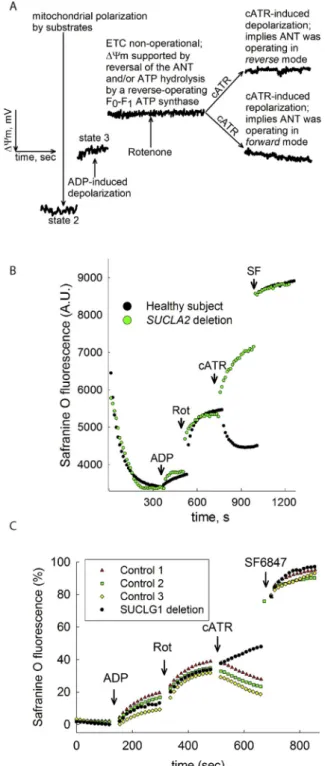
It has been firmly established that when the respiratory chain is pharmacologically inhibited or in the presence of anoxia, mitochondria are still able to utilize downstream metabolites of glutamine (gluta- mate, α-ketoglutarate) using NAD+obtained from sources other than complex I (Chinopoulos, 2019), (Ravasz et al., 2018), (Kiss et al., 2013), (Chinopoulos et al., 2010), a concept which is likely to be exploited in cancer cells thriving in adverse tumor microenvironments (Chinopoulos and Seyfried, 2018). This serves the purpose of generating high-energy phosphate nucleotides within the mitochondrial matrix through the process of mitochondrial substrate-level phosphorylation (mSLP) for i) fueling a reverse-operating F0–F1ATP synthase (Chinopoulos, 2011b) and ii) maintaining the ANT in forward mode, thus un-straining gly- colysis (Chinopoulos, 2011a). An mSLP test is qualitative (presencevs absence of mSLP, there is no parameter that needs to be quantified) and can be performed in skin fibroblasts requiring no specialized machinery (spectrofluorimetry in plate or cuvette format is standard laboratory equipment). The test that we have devised is based on an “interroga- tion” of the directionality of succinate-CoA ligase (SUCL), a citric acid cycle enzyme that interconverts succinyl-CoA and ADP (or GDP) to CoASH, succinate and ATP (or GTP) (Johnson et al., 1998), essentially performing mSLP. This enzyme is a heterodimer composed of an in- variant α subunit encoded by SUCLG1 and a substrate-specific β sub- unit, encoded by either SUCLA2 or SUCLG2. This dimer combination results in either an ATP-forming (EC 6.2.1.5) or a GTP-forming SUCL (EC 6.2.1.4). The possibility of co-presence of an ATP- and GTP-forming SUCL in the same mitochondrion has only been recently investigated in human fibroblasts, where it was found that in these cells the majority of in situ mitochondria co-express both isoforms (Chinopoulos et al., 2019), seeFig. 2(reproduced from (Chinopoulos et al., 2019) by per- mission). This seems redundant, mindful that an intramitochondrial diphosphate kinase (NME4) can transfer the high-energy bond between adenine and guanine nucleotides (Lacombe et al., 2018). In any case, mSLP supports the production of sufficient amounts of [ATP] in the mitochondrial matrix for both a reverse-operating F0–F1ATP synthase (Chinopoulos, 2011b) and maintain the ANT in forward mode (Chinopoulos, 2011a). Presence of mSLP maintains ANT in forward mode for reasons outlined in (Chinopoulos, 2019), (Ravasz et al., 2018), (Kiss et al., 2013), (Chinopoulos et al., 2010), and this can be easily assessed by checking the effect of a highly-specific ANT inhibitor –carboxyatractyloside- in permeabilized cells with blocked complex I (using rotenone). Under these conditions, mSLP can only occur in the presence of suitable substrates specifically those stemming from glu- taminolysis, but not succinate (Kiss et al., 2013). Such a test is depicted inFig. 3(obtained from (Tretter et al., 2016), by permission). As shown in figure panel 3A, the test relies on the principle that the ATP-ADP exchange through the ANT is electrogenic, since one molecule of ATP4
−is exchanged for one molecule of ADP3 −(Klingenberg, 2008). If the ANT operates in forward mode, abolition of its operation by carbox- yatractyloside (cATR) will lead to a gain of ΔΨm. If the ANT operates in reverse, abolition of its operation by the inhibitor will lead to a loss of ΔΨm. Note that addition of the ANT inhibitor must take placeafter complex I is inhibited (i.e. by rotenone), thus, it cannot be over- emphasized that the mSLP test addresses the extent of compensation (i.e. remaining mitochondrial functionality) when the electron trans- port chain is non-functional. It does not yield any information regarding the respiratory chain, it only provides information if the in situ
mitochondria of the examined cells are able to harness energy from glutamine-derived metabolites when the respiratory chain is inhibited.
In essence, this test will hint on the ability of mitochondria to cope with a defect in their respiratory chain. By employing this test, we have reported that defects in SUCLA2 and SUCLG1, respectively, yield an mSLP negative test, i.e. no mSLP present. As shown in figure panels 3B and 3C,in situfibroblast mitochondria of a SUCLA2-deficient patient (B) or SUCLG1-deficient patient (C) exhibited no mSLP compared to age-matched controls. Note that patients with defects in SUCLA2 and SUCLG1 exhibit mtDNA depletion in some but not all tissues, while the phenotypes are debilitating leading to life termination at a young age (Ostergaard, 2008). Obviously, this test has been applied in fibroblasts from patients suffering from MDMDs due to mutations in the enzyme directly related to mSLP; efforts are ongoing in our laboratory to verify the validity of this test using fibroblasts from patients suffering from various mtDNA depletion syndromes.
Finally, it may be relevant to ponder on the fact that mSLP relies on a sufficient clearance of succinate, which can occur by increasing suc- cinate dehydrogenase (SDH) activity; it is thus not surprising that in several MDMDs, ragged-blue fibers are observed in histological ex- amination of muscle tissue, which is none other than an increase in SDH activity (DiMauro et al., 1999).
6.1. mSLP protocol
The mSLP protocol can be performed in cultured fibroblasts exactly as in (Kacso et al., 2016) and (Chinopoulos et al., 2019). It is best to compare cultured fibroblasts from the patient and at least three con- trols. It is not important to age- and gender-match the controls to the patient; the controls serve the purpose of ensuring that the mSLP pro- tocol is being performed correctly; it is not a quantitative test, and for this reason it is also not necessary to normalize results to protein con- tent or a housekeeping protein. Basically, fibroblasts [two 75 cm^2 (midi) or one 175 cm^2 (maxi) flask)] are cultured to approximately 80% confluence. On the day of the experiment, two buffers need to be prepared: 500 ml of ice-cold buffer “A”, containing 110 mM K-gluco- nate, 10 mM HEPES (free acid), 10 mM KH2PO4, 10 mM mannitol, 10 mM NaCl, 8 mM KCl, 1.5 mM MgCl2, 0.01 mM EGTA, 0.5 mg/ml BSA (essentially fatty acid-free), with the pH adjusted to 7.25 with KOH, and 5 ml of buffer “B” which in addition to the substances mentioned for buffer “A” it contains the following: 5 μM safranine O, 5 mM glutamate, 5 mM malate, 5 mM α-ketoglutarate and 175 μl of 2.5 mM digitonin (purity > 90%, dissolved in DMSO). For exact catalogue numbers of chemicals, the reader is referred to http://antactivity.com/Buffers_
chemicals.html. Buffer “B” must be kept at 37 °C. Feeding media are aspirated and cells are washed once with ice-cold buffer “A”, then harvested by scraping in buffer “A” (no need for trypsinization, cells will be eventually permeabilized for the mSLP assay within a few Fig. 2.Confocal images of immunolabeling of human fibroblasts from healthy subjects. Blue: anti-SUCLA2; Red: anti-SUCLG2; Green: Mitotracker orange (MTO).
Reproduced from (Chinopoulos et al., 2019) by permission. (For interpretation of the references to colour in this figure legend, the reader is referred to the Web version of this article.)
minutes, it is redundant to maintain high viability of the cells). Pellet cells by centrifugation at 3,500 rpm for 2.5 min using an angular rotor (or at 1,100 rpm for 10 min if a swing-bucket rotor is used) in at 50 ml tubes containing at least 40 ml of buffer (to dilute calcium of the feeding media as much as possible). Wash cells once with buffer “A”, and repeat the centrifugation step. Resuspend final pellet in 0.7 ml of buffer “B”. Use a plate reader for recording safranine O fluorescence (495ex/585em). Split the 0.7 ml pellet in three 0.2 ml (record in tri- plicates) and add to individual wells of a 96 white plate suitable for fluorescence, and insert into the plate reader. The interior of the plate reader must be kept at 37 °C; possibility for automatic shaking of the plate is imperative (i.e. every 3 s). Acquisitions should be made every 10–15 s. Allow 5 min for adequate polarization ofin situmitochondria and cell membrane permeabilization by digitonin. Record baseline for 3 min, add 2 mM ADP (from a 6.9 pH-ed 200 mM stock, see http://
antactivity.com/Buffers_chemicals.html), seeFig. 3. Record for 3 more minutes, then add 5 μM rotenone. Record for an additional 3 min, then add 1 μM carboxyatractyloside. Record for an additional 3 min, then add 250 nM of the uncoupler SF6847. The volume of each addition to each well should be not less than 1 μl and no more than 2 μl. Convert fluorescence traces to percentages and compare the 3 controls with that from the patient cells. If carboxyatractyloside leads to hyperpolariza- tion in the controls but to a depolarization in the patient, mSLP in the patient is impaired. This means that the fibroblasts of the patient are unable to harness ATP from mitochondrial substrate-level phosphor- ylation from glutaminolysis, thus his/her mitochondria cannot com- pensate for a potential electron transport chain dysfunction to an ap- preciable degree.
7. Conclusions
The present review does not make an attempt to indicate how much mtDNA depletion is required for disease manifestation; it merely dis- cusses that quantitating mtDNA on peripheral –but not limited to-tis- sues is not pathognomonic. Furthermore, the concept of mSLP as a qualitative and not quantitative test for evaluating the extent of mi- tochondrial functionality in the absence of an electron transport chain is put forward. Having said that, it cannot be overemphasized that the mSLP test cannot be used to differentiate among the numerous defects described in mtDNA translation, or involving a single complex, or secondary to other diseases involving mitochondrial dysfunction; it can only provide an indication if the affected mitochondria are able to energetically compensate and maintain –albeit- minor- ATP production and not act as sinks of cytosolically made ATP.
Funding
This work was supported by grants from NKFIH [FIKP-61822- 64888-EATV], [VEKOP 2.3.3-15-2016-00012], [2017-2.3.4-TET-RU- 2017-00003] and [KH129567] to C.C.
References
Arnaudo, E., Dalakas, M., Shanske, S., Moraes, C.T., DiMauro, S., Schon, E.A., 1991.
Depletion of muscle mitochondrial DNA in AIDS patients with zidovudine-induced myopathy. Lancet 337 (8740), 508–510.
Balciuniene, J., Balciunas, D., 2019. A nuclear mtDNA concatemer (Mega-NUMT) could mimic paternal inheritance of mitochondrial genome. Front. Genet. 10, 518.
Bernier, F.P., Boneh, A., Dennett, X., Chow, C.W., Cleary, M.A., Thorburn, D.R., 2002.
Diagnostic criteria for respiratory chain disorders in adults and children. Neurology 59 (9), 1406–1411.
Calvo, S.E., Mootha, V.K., 2010. The mitochondrial proteome and human disease. Annu.
Rev. Genom. Hum. Genet. 11, 25–44.
Carrozzo, R., Dionisi-Vici, C., Steuerwald, U., Lucioli, S., Deodato, F., Di Giandomenico, S., Bertini, E., Franke, B., Kluijtmans, L.A., Meschini, M.C., Rizzo, C., Piemonte, F., Rodenburg, R., Santer, R., Santorelli, F.M., van Rooij, A., Vermunt-de Koning, D., Morava, E., Wevers, R.A., 2007. SUCLA2 mutations are associated with mild me- thylmalonic aciduria, Leigh-like encephalomyopathy, dystonia and deafness. Brain: J.
Neurol. 130 (Pt 3), 862–874.
Fig. 3.Reproduced from (Tretter et al., 2016), (Kacso et al., 2016), (Chinopoulos et al., 2019), by permission. A: Explanatory cartoon graph of ΔΨm measurements of mitochondria regarding the ‘biosensor test’ addressing the directionality of the ANT, and as an extension of this, the directionality of SUCL. Black trace represents a safranine O signal calibrated to ΔΨm, in mV. B:
Reconstructed time course of safranin O signal from permeabilized fibroblasts of a control subject (black dots) and a patient suffering from complete SUCLA2 deletion (green dots). Rot: rotenone, 1 μM. C: Reconstructed time courses of safranine O signal (expressed as percentage) indicating ΔΨm from permeabi- lized fibroblasts of three age-matched control subjects (red triangles, green quadrangles and yellow diamonds) and the patient (IP) exhibiting SUCLG1 defect (black circles). Rot: rotenone, 5 μM; cATR: carboxyatractyloside, 1 μM;
SF6847 500 nM. (For interpretation of the references to colour in this figure legend, the reader is referred to the Web version of this article.)
Carrozzo, R., Verrigni, D., Rasmussen, M., de Coo, R., Amartino, H., Bianchi, M., Buhas, D., Mesli, S., Naess, K., Born, A.P., Woldseth, B., Prontera, P., Batbayli, M., Ravn, K., Joensen, F., Cordelli, D.M., Santorelli, F.M., Tulinius, M., Darin, N., Duno, M., Jouvencel, P., Burlina, A., Stangoni, G., Bertini, E., Redonnet-Vernhet, I., Wibrand, F., Dionisi-Vici, C., Uusimaa, J., Vieira, P., Osorio, A.N., McFarland, R., Taylor, R.W., Holme, E., Ostergaard, E., 2016. Succinate-CoA ligase deficiency due to mutations in SUCLA2 and SUCLG1: phenotype and genotype correlations in 71 patients. J. Inherit.
Metab. Dis. 39 (2), 243–252.
Casula, M., Vrisekoop, N., Wit, F.W., de Baar, M.P., de Ronde, A., Miedema, F., Reiss, P., 2007. Mitochondrial DNA decline in T cells of HIV-1 seroconverters may be depen- dent on immune activation. J. Infect. Dis. 196 (3), 371–376.
Chan, D.C., 2006. Mitochondria: dynamic organelles in disease, aging, and development.
Cell 125 (7), 1241–1252.
Chinopoulos, C., 2011a. The "B space" of mitochondrial phosphorylation. J. Neurosci. Res.
89 (12), 1897–1904.
Chinopoulos, C., 2011b. Mitochondrial consumption of cytosolic ATP: not so fast. FEBS Lett. 585 (9), 1255–1259.
Chinopoulos, C., 2019. Succinate in ischemia: where does it come from? Int. J. Biochem.
Cell Biol. 115, 105580.
Chinopoulos, C., Seyfried, T.N., 2018. Mitochondrial substrate-level phosphorylation as energy source for glioblastoma: review and hypothesis. ASN neuro 10
1759091418818261.
Chinopoulos, C., Gerencser, A.A., Mandi, M., Mathe, K., Torocsik, B., Doczi, J., Turiak, L., Kiss, G., Konrad, C., Vajda, S., Vereczki, V., Oh, R.J., Adam-Vizi, V., 2010. Forward operation of adenine nucleotide translocase during F0F1-ATPase reversal: critical role of matrix substrate-level phosphorylation. FASEB J. : Off. Publ. Fed. Am. Soc.
Exp. Biol. 24 (7), 2405–2416.
Chinopoulos, C., Batzios, S., van den Heuvel, L.P., Rodenburg, R., Smeets, R., Waterham, H.R., Turkenburg, M., Ruiter, J.P., Wanders, R.J.A., Doczi, J., Horvath, G., Dobolyi, A., Vargiami, E., Wevers, R.A., Zafeiriou, D., 2019. Mutated SUCLG1 causes mis- localization of SUCLG2 protein, morphological alterations of mitochondria and an early-onset severe neurometabolic disorder. Mol. Genet. Metab. 126 (1), 43–52.
Chretien, D., Gallego, J., Barrientos, A., Casademont, J., Cardellach, F., Munnich, A., Rotig, A., Rustin, P., 1998. Biochemical parameters for the diagnosis of mitochondrial respiratory chain deficiency in humans, and their lack of age-related changes.
Biochem. J. 329 (Pt 2), 249–254.
Clayton, D.A., 1982. Replication of animal mitochondrial DNA. Cell 28 (4), 693–705.
Dagan, T., Sable, C., Bray, J., Gerschenson, M., 2002. Mitochondrial dysfunction and antiretroviral nucleoside analog toxicities: what is the evidence? Mitochondrion 1 (5), 397–412.
Davis, R.L., Liang, C., Edema-Hildebrand, F., Riley, C., Needham, M., Sue, C.M., 2013.
Fibroblast growth factor 21 is a sensitive biomarker of mitochondrial disease.
Neurology 81 (21), 1819–1826.
Debray, F.G., Mitchell, G.A., Allard, P., Robinson, B.H., Hanley, J.A., Lambert, M., 2007.
Diagnostic accuracy of blood lactate-to-pyruvate molar ratio in the differential di- agnosis of congenital lactic acidosis. Clin. Chem. 53 (5), 916–921.
DiMauro, S., Bonilla, E., De Vivo, D.C., 1999. Does the patient have a mitochondrial encephalomyopathy? J. Child Neurol. 1 (14 Suppl. l), S23–35.
El-Hattab, A.W., Scaglia, F., 2013. Mitochondrial DNA depletion syndromes: review and updates of genetic basis, manifestations, and therapeutic options. Neurotherapeutics.
J. Am. Soc. Exp. NeuroTherap. 10 (2), 186–198.
El-Hattab, A.W., Craigen, W.J., Scaglia, F., 2017. Mitochondrial DNA maintenance de- fects. Biochimica et biophysica acta. Mol. Basis Dis. 1863 (6), 1539–1555.
Elpeleg, O., Miller, C., Hershkovitz, E., Bitner-Glindzicz, M., Bondi-Rubinstein, G., Rahman, S., Pagnamenta, A., Eshhar, S., Saada, A., 2005. Deficiency of the ADP- forming succinyl-CoA synthase activity is associated with encephalomyopathy and mitochondrial DNA depletion. Am. J. Hum. Genet. 76 (6), 1081–1086.
Freisinger, P., Futterer, N., Lankes, E., Gempel, K., Berger, T.M., Spalinger, J., Hoerbe, A., Schwantes, C., Lindner, M., Santer, R., Burdelski, M., Schaefer, H., Setzer, B., Walker, U.A., Horvath, R., 2006. Hepatocerebral mitochondrial DNA depletion syndrome caused by deoxyguanosine kinase (DGUOK) mutations. Arch. Neurol. 63 (8), 1129–1134.
Garone, C., Taylor, R.W., Nascimento, A., Poulton, J., Fratter, C., Dominguez-Gonzalez, C., Evans, J.C., Loos, M., Isohanni, P., Suomalainen, A., Ram, D., Hughes, M.I., McFarland, R., Barca, E., Lopez Gomez, C., Jayawant, S., Thomas, N.D., Manzur, A.Y., Kleinsteuber, K., Martin, M.A., Kerr, T., Gorman, G.S., Sommerville, E.W., Chinnery, P.F., Hofer, M., Karch, C., Ralph, J., Camara, Y., Madruga-Garrido, M., Dominguez- Carral, J., Ortez, C., Emperador, S., Montoya, J., Chakrapani, A., Kriger, J.F., Schoenaker, R., Levin, B., Thompson, J.L.P., Long, Y., Rahman, S., Donati, M.A., DiMauro, S., Hirano, M., 2018. Retrospective natural history of thymidine kinase 2 deficiency. J. Med. Genet. 55 (8), 515–521.
Gauthier-Villars, M., Landrieu, P., Cormier-Daire, V., Jacquemin, E., Chretien, D., Rotig, A., Rustin, P., Munnich, A., de Lonlay, P., 2001. Respiratory chain deficiency in Alpers syndrome. Neuropediatrics 32 (3), 150–152.
Gorman, G.S., Chinnery, P.F., DiMauro, S., Hirano, M., Koga, Y., McFarland, R., Suomalainen, A., Thorburn, D.R., Zeviani, M., Turnbull, D.M., 2016. Mitochondrial diseases. Nature reviews. Dis. Primers 2, 16080.
Gotz, A., Isohanni, P., Pihko, H., Paetau, A., Herva, R., Saarenpaa-Heikkila, O., Valanne, L., Marjavaara, S., Suomalainen, A., 2008. Thymidine kinase 2 defects can cause multi-tissue mtDNA depletion syndrome. Brain: J. Neurol. 131 (Pt 11), 2841–2850.
Gourlain, K., Amellal, B., Ait Arkoub, Z., Dupin, N., Katlama, C., Calvez, V., 2003.
Quantitative analysis of human mitochondrial DNA using a real-time PCR assay. HIV Med. 4 (3), 287–292.
Gustafsson, C.M., Falkenberg, M., Larsson, N.G., 2016. Maintenance and expression of mammalian mitochondrial DNA. Annu. Rev. Biochem. 85, 133–160.
Hakonen, A.H., Isohanni, P., Paetau, A., Herva, R., Suomalainen, A., Lonnqvist, T., 2007.
Recessive Twinkle mutations in early onset encephalopathy with mtDNA depletion.
Brain: J. Neurol. 130 (Pt 11), 3032–3040.
Hakonen, A.H., Goffart, S., Marjavaara, S., Paetau, A., Cooper, H., Mattila, K., Lampinen, M., Sajantila, A., Lonnqvist, T., Spelbrink, J.N., Suomalainen, A., 2008. Infantile- onset spinocerebellar ataxia and mitochondrial recessive ataxia syndrome are asso- ciated with neuronal complex I defect and mtDNA depletion. Hum. Mol. Genet. 17 (23), 3822–3835.
Hensen, F., Cansiz, S., Gerhold, J.M., Spelbrink, J.N., 2014. To be or not to be a nucleoid protein: a comparison of mass-spectrometry based approaches in the identification of potential mtDNA-nucleoid associated proteins. Biochimie 100, 219–226.
Holt, I.J., Harding, A.E., Morgan-Hughes, J.A., 1988. Deletions of muscle mitochondrial DNA in patients with mitochondrial myopathies. Nature 331 (6158), 717–719.
Johnson, J.D., Mehus, J.G., Tews, K., Milavetz, B.I., Lambeth, D.O., 1998. Genetic evi- dence for the expression of ATP- and GTP-specific succinyl-CoA synthetases in mul- ticellular eucaryotes. J. Biol. Chem. 273 (42), 27580–27586.
Johnston, I.G., Burgstaller, J.P., Havlicek, V., Kolbe, T., Rulicke, T., Brem, G., Poulton, J., Jones, N.S., 2015. Stochastic modelling, Bayesian inference, and new in vivo mea- surements elucidate the debated mtDNA bottleneck mechanism. eLife 4, e07464.
Kacso, G., Ravasz, D., Doczi, J., Nemeth, B., Madgar, O., Saada, A., Ilin, P., Miller, C., Ostergaard, E., Iordanov, I., Adams, D., Vargedo, Z., Araki, M., Araki, K., Nakahara, M., Ito, H., Gal, A., Molnar, M.J., Nagy, Z., Patocs, A., Adam-Vizi, V., Chinopoulos, C., 2016. Two transgenic mouse models for beta-subunit components of succinate-CoA ligase yielding pleiotropic metabolic alterations. Biochem. J. 473 (20), 3463–3485.
Kiss, G., Konrad, C., Doczi, J., Starkov, A.A., Kawamata, H., Manfredi, G., Zhang, S.F., Gibson, G.E., Beal, M.F., Adam-Vizi, V., Chinopoulos, C., 2013. The negative impact of alpha-ketoglutarate dehydrogenase complex deficiency on matrix substrate-level phosphorylation. FASEB J.: Off. Publ. Fed. Am. Soc. Exp. Biol. 27 (6), 2392–2406.
Klingenberg, M., 2008. The ADP and ATP transport in mitochondria and its carrier.
Biochim. Biophys. Acta 1778 (10), 1978–2021.
Krishnan, K.J., Reeve, A.K., Samuels, D.C., Chinnery, P.F., Blackwood, J.K., Taylor, R.W., Wanrooij, S., Spelbrink, J.N., Lightowlers, R.N., Turnbull, D.M., 2008. What causes mitochondrial DNA deletions in human cells? Nat. Genet. 40 (3), 275–279.
Kukat, C., Wurm, C.A., Spahr, H., Falkenberg, M., Larsson, N.G., Jakobs, S., 2011. Super- resolution microscopy reveals that mammalian mitochondrial nucleoids have a uni- form size and frequently contain a single copy of mtDNA. Proc. Natl. Acad. Sci. U. S.
A 108 (33), 13534–13539.
Lacombe, M.L., Tokarska-Schlattner, M., Boissan, M., Schlattner, U., 2018. The mi- tochondrial nucleoside diphosphate kinase (NDPK-D/NME4), a moonlighting protein for cell homeostasis. Laboratory investigation. J. Tech. Methods Pathol. 98 (5), 582–588.
Lewis, W., Copeland, W.C., Day, B.J., 2001. Mitochondrial DNA depletion, oxidative stress, and mutation: mechanisms of dysfunction from nucleoside reverse tran- scriptase inhibitors. Laboratory investigation. J. Tech. Methods and Pathol. 81 (6), 777–790.
Lewis, W., Day, B.J., Copeland, W.C., 2003. Mitochondrial toxicity of NRTI antiviral drugs: an integrated cellular perspective. Nature reviews. Drug discovery 2 (10), 812–822.
Li, M., Rothwell, R., Vermaat, M., Wachsmuth, M., Schroder, R., Laros, J.F., van Oven, M., de Bakker, P.I., Bovenberg, J.A., van Duijn, C.M., van Ommen, G.J., Slagboom, P.E., Swertz, M.A., Wijmenga, C., Genome of Netherlands, Kayser, C., Boomsma, M., Zollner, D.I., de Knijff, S., toneking M, P., 2016. Transmission of human mtDNA heteroplasmy in the Genome of The Netherlands families: support for a variable-size bottleneck. Genome Res. 26 (4), 417–426.
Luo, S., Valencia, C.A., Zhang, J., Lee, N.C., Slone, J., Gui, B., Wang, X., Li, Z., Dell, S., Brown, J., Chen, S.M., Chien, Y.H., Hwu, W.L., Fan, P.C., Wong, L.J., Atwal, P.S., Huang, T., 2018. Biparental inheritance of mitochondrial DNA in humans. Proc. Natl.
Acad. Sci. U. S. A 115 (51), 13039–13044.
Lutz-Bonengel, S., Parson, W., 2019. No further evidence for paternal leakage of mi- tochondrial DNA in humans yet. Proc. Natl. Acad. Sci. U. S. A 116 (6), 1821–1822.
Mandel, H., Szargel, R., Labay, V., Elpeleg, O., Saada, A., Shalata, A., Anbinder, Y., Berkowitz, D., Hartman, C., Barak, M., Eriksson, S., Cohen, N., 2001. The deox- yguanosine kinase gene is mutated in individuals with depleted hepatocerebral mi- tochondrial DNA. Nat. Genet. 29 (3), 337–341.
Medja, F., Allouche, S., Frachon, P., Jardel, C., Malgat, M., Mousson de Camaret, B., Slama, A., Lunardi, J., Mazat, J.P., Lombes, A., 2009. Development and im- plementation of standardized respiratory chain spectrophotometric assays for clinical diagnosis. Mitochondrion 9 (5), 331–339.
Miller, F.J., Rosenfeldt, F.L., Zhang, C., Linnane, A.W., Nagley, P., 2003. Precise de- termination of mitochondrial DNA copy number in human skeletal and cardiac muscle by a PCR-based assay: lack of change of copy number with age. Nucleic Acids Res. 31 (11), e61.
Mitochondrial Medicine Society's Committee on, Haas, D., Parikh, R.H., Falk, S., Saneto, M.J., Wolf, R.P., Darin, N.I., Wong, N., Cohen, L.J., Naviaux, B.H., RK, 2008. The in- depth evaluation of suspected mitochondrial disease. Mol. Genet. Metab. 94 (1), 16–37.
Montaner, J.S., Cote, H.C., Harris, M., Hogg, R.S., Yip, B., Harrigan, P.R., O'Shaughnessy, M.V., 2004. Nucleoside-related mitochondrial toxicity among HIV-infected patients receiving antiretroviral therapy: insights from the evaluation of venous lactic acid and peripheral blood mitochondrial DNA. Clin. Infect. Dis. : Off. Publ. Infect. Dis. Soc.
America 2 (38 Suppl. l), S73–79.
Montier, Clay, Deng, L.L., Bai Y, J.J., 2009. Number matters: control of mammalian mitochondrial DNA copy number. J. Genet. Genom. = Yi chuan xue bao 36 (3), 125–131.
Moraes, C.T., 2001. What regulates mitochondrial DNA copy number in animal cells? TIG (Trends Genet.) 17 (4), 199–205.
Moraes, C.T., Shanske, S., Tritschler, H.J., Aprille, J.R., Andreetta, F., Bonilla, E., Schon,
E.A., DiMauro, S., 1991. mtDNA depletion with variable tissue expression: a novel genetic abnormality in mitochondrial diseases. Am. J. Hum. Genet. 48 (3), 492–501.
Morse, C.G., Voss, J.G., Rakocevic, G., McLaughlin, M., Vinton, C.L., Huber, C., Hu, X., Yang, J., Huang da, W., Logun, C., Danner, R.L., Rangel, Z.G., Munson, P.J., Orenstein, J.M., Rushing, E.J., Lempicki, R.A., Dalakas, M.C., Kovacs, J.A., 2012. HIV infection and antiretroviral therapy have divergent effects on mitochondria in adi- pose tissue. J. Infect. Dis. 205 (12), 1778–1787.
Morten, K.J., Ashley, N., Wijburg, F., Hadzic, N., Parr, J., Jayawant, S., Adams, S., Bindoff, L., Bakker, H.D., Mieli-Vergani, G., Zeviani, M., Poulton, J., 2007. Liver mtDNA content increases during development: a comparison of methods and the importance of age- and tissue-specific controls for the diagnosis of mtDNA depletion.
Mitochondrion 7 (6), 386–395.
Nishigaki, Y., Marti, R., Copeland, W.C., Hirano, M., 2003. Site-specific somatic mi- tochondrial DNA point mutations in patients with thymidine phosphorylase defi- ciency. J. Clin. Investig. 111 (12), 1913–1921.
Nishino, I., Spinazzola, A., Hirano, M., 1999. Thymidine phosphorylase gene mutations in MNGIE, a human mitochondrial disorder. Science 283 (5402), 689–692.
Nishino, I., Spinazzola, A., Papadimitriou, A., Hammans, S., Steiner, I., Hahn, C.D., Connolly, A.M., Verloes, A., Guimaraes, J., Maillard, I., Hamano, H., Donati, M.A., Semrad, C.E., Russell, J.A., Andreu, A.L., Hadjigeorgiou, G.M., Vu, T.H., Tadesse, S., Nygaard, T.G., Nonaka, I., Hirano, I., Bonilla, E., Rowland, L.P., DiMauro, S., Hirano, M., 2000. Mitochondrial neurogastrointestinal encephalomyopathy: an autosomal recessive disorder due to thymidine phosphorylase mutations. Ann. Neurol. 47 (6), 792–800.
Niyazov, D.M., Kahler, S.G., Frye, R.E., 2016. Primary mitochondrial disease and sec- ondary mitochondrial dysfunction: importance of distinction for diagnosis and treatment. Mol. Syndromol. 7 (3), 122–137.
Nogueira, C., Almeida, L.S., Nesti, C., Pezzini, I., Videira, A., Vilarinho, L., Santorelli, F.M., 2014. Syndromes associated with mitochondrial DNA depletion. Ital. J. Pediatr.
40, 34.
Nolden, M., Ehses, S., Koppen, M., Bernacchia, A., Rugarli, E.I., Langer, T., 2005. The m- AAA protease defective in hereditary spastic paraplegia controls ribosome assembly in mitochondria. Cell 123 (2), 277–289.
Ostergaard, E., 2008. Disorders caused by deficiency of succinate-CoA ligase. J. Inherit.
Metab. Dis. 31 (2), 226–229.
Ostergaard, E., Christensen, E., Kristensen, E., Mogensen, B., Duno, M., Shoubridge, E.A., Wibrand, F., 2007a. Deficiency of the alpha subunit of succinate-coenzyme A ligase causes fatal infantile lactic acidosis with mitochondrial DNA depletion. Am. J. Hum.
Genet. 81 (2), 383–387.
Ostergaard, E., Hansen, F.J., Sorensen, N., Duno, M., Vissing, J., Larsen, P.L., Faeroe, O., Thorgrimsson, S., Wibrand, F., Christensen, E., Schwartz, M., 2007b. Mitochondrial encephalomyopathy with elevated methylmalonic acid is caused by SUCLA2 muta- tions. Brain. J. Neurol. 130 (Pt 3), 853–861.
Pagliarini, D.J., Calvo, S.E., Chang, B., Sheth, S.A., Vafai, S.B., Ong, S.E., Walford, G.A., Sugiana, C., Boneh, A., Chen, W.K., Hill, D.E., Vidal, M., Evans, J.G., Thorburn, D.R., Carr, S.A., Mootha, V.K., 2008. A mitochondrial protein compendium elucidates complex I disease biology. Cell 134 (1), 112–123.
Persson, O., Muthukumar, Y., Basu, S., Jenninger, L., Uhler, J.P., Berglund, A.K., McFarland, R., Taylor, R.W., Gustafsson, C.M., Larsson, E., Falkenberg, M., 2019.
Copy-choice recombination during mitochondrial L-strand synthesis causes DNA deletions. Nat. Commun. 10 (1), 759.
Poulton, J., Holt, I.J., 2009. In: 163rd ENMC International Workshop: Nucleoid and Nucleotide Biology in Syndromes of Mitochondrial DNA Depletion Myopathy 12-14 December 2008, vol 19. NMD, Naarden, The Netherlands. Neuromuscular disorders, pp. 439–443 6.
Pyle, A., Hudson, G., Wilson, I.J., Coxhead, J., Smertenko, T., Herbert, M., Santibanez- Koref, M., Chinnery, P.F., 2015. Extreme-Depth Re-sequencing of mitochondrial DNA finds No evidence of paternal transmission in humans. PLoS Genet. 11 (5), e1005040.
Ravasz, D., Kacso, G., Fodor, V., Horvath, K., Adam-Vizi, V., Chinopoulos, C., 2018.
Reduction of 2-methoxy-1,4-naphtoquinone by mitochondrially-localized Nqo1 yielding NAD(+) supports substrate-level phosphorylation during respiratory in- hibition. Biochimica et biophysica acta. Bioenergetics 1859 (9), 909–924.
Rebolledo-Jaramillo, B., Su, M.S., Stoler, N., McElhoe, J.A., Dickins, B., Blankenberg, D., Korneliussen, T.S., Chiaromonte, F., Nielsen, R., Holland, M.M., Paul, I.M., Nekrutenko, A., Makova, K.D., 2014. Maternal age effect and severe germ-line bot- tleneck in the inheritance of human mitochondrial DNA. Proc. Natl. Acad. Sci. U. S. A 111 (43), 15474–15479.
Rodenburg, R.J., Schoonderwoerd, G.C., Tiranti, V., Taylor, R.W., Rotig, A., Valente, L., Invernizzi, F., Chretien, D., He, L., Backx, G.P., Janssen, K.J., Chinnery, P.F., Smeets, H.J., de Coo, I.F., van den Heuvel, L.P., 2013. A multi-center comparison of diag- nostic methods for the biochemical evaluation of suspected mitochondrial disorders.
Mitochondrion 13 (1), 36–43.
Rotig, A., Poulton, J., 2009. Genetic causes of mitochondrial DNA depletion in humans.
Biochim. Biophys. Acta 1792 (12), 1103–1108.
Sarzi, E., Bourdon, A., Chretien, D., Zarhrate, M., Corcos, J., Slama, A., Cormier-Daire, V., de Lonlay, P., Munnich, A., Rotig, A., 2007. Mitochondrial DNA depletion is a pre- valent cause of multiple respiratory chain deficiency in childhood. J. Pediatr. 150 (5), e531–536 531-534, 534.
Schaefer, A.M., Taylor, R.W., Turnbull, D.M., Chinnery, P.F., 2004. The epidemiology of mitochondrial disorders–past, present and future. Biochim. Biophys. Acta 1659 (2–3), 115–120.
Servidei, S., Zeviani, M., Manfredi, G., Ricci, E., Silvestri, G., Bertini, E., Gellera, C., Di Mauro, S., Di Donato, S., Tonali, P., 1991. Dominantly inherited mitochondrial myopathy with multiple deletions of mitochondrial DNA: clinical, morphologic, and biochemical studies. Neurology 41 (7), 1053–1059.
Shoffner, J.M., Lott, M.T., Voljavec, A.S., Soueidan, S.A., Costigan, D.A., Wallace, D.C., 1989. Spontaneous Kearns-Sayre/chronic external ophthalmoplegia plus syndrome associated with a mitochondrial DNA deletion: a slip-replication model and metabolic therapy. Proc. Natl. Acad. Sci. U. S. A 86 (20), 7952–7956.
Song, S., Wheeler, L.J., Mathews, C.K., 2003. Deoxyribonucleotide pool imbalance sti- mulates deletions in HeLa cell mitochondrial DNA. J. Biol. Chem. 278 (45), 43893–43896.
Spelbrink, J.N., 2010. Functional organization of mammalian mitochondrial DNA in nucleoids: history, recent developments, and future challenges. IUBMB Life 62 (1), 19–32.
Spinazzola, A., Invernizzi, F., Carrara, F., Lamantea, E., Donati, A., Dirocco, M., Giordano, I., Meznaric-Petrusa, M., Baruffini, E., Ferrero, I., Zeviani, M., 2009. Clinical and molecular features of mitochondrial DNA depletion syndromes. J. Inherit. Metab. Dis.
32 (2), 143–158.
Sue, C.M., Quigley, A., Katsabanis, S., Kapsa, R., Crimmins, D.S., Byrne, E., Morris, J.G., 1998. Detection of MELAS A3243G point mutation in muscle, blood and hair follicles.
J. Neurol. Sci. 161 (1), 36–39.
Suomalainen, A., 2011. Biomarkers for mitochondrial respiratory chain disorders. J.
Inherit. Metab. Dis. 34 (2), 277–282.
Suomalainen, A., Battersby, B.J., 2018. Mitochondrial diseases: the contribution of or- ganelle stress responses to pathology. Nature reviews. Mol. Cell Biol. 19 (2), 77–92.
Suomalainen, A., Isohanni, P., 2010. Mitochondrial DNA depletion syndromes–many genes, common mechanisms. Neuromuscul. Disord.: NMD 20 (7), 429–437.
Suomalainen, A., Elo, J.M., Pietilainen, K.H., Hakonen, A.H., Sevastianova, K., Korpela, M., Isohanni, P., Marjavaara, S.K., Tyni, T., Kiuru-Enari, S., Pihko, H., Darin, N., Ounap, K., Kluijtmans, L.A., Paetau, A., Buzkova, J., Bindoff, L.A., Annunen-Rasila, J., Uusimaa, J., Rissanen, A., Yki-Jarvinen, H., Hirano, M., Tulinius, M., Smeitink, J., Tyynismaa, H., 2011. FGF-21 as a biomarker for muscle-manifesting mitochondrial respiratory chain deficiencies: a diagnostic study. The Lancet. Neurology 10 (9), 806–818.
Tretter, L., Patocs, A., Chinopoulos, C., 2016. Succinate, an intermediate in metabolism, signal transduction, ROS, hypoxia, and tumorigenesis. Biochim. Biophys. Acta 1857 (8), 1086–1101.
Tucker, E.J., Compton, A.G., Calvo, S.E., Thorburn, D.R., 2011. The molecular basis of human complex I deficiency. IUBMB Life 63 (9), 669–677.
Tzoulis, C., Tran, G.T., Coxhead, J., Bertelsen, B., Lilleng, P.K., Balafkan, N., Payne, B., Miletic, H., Chinnery, P.F., Bindoff, L.A., 2014. Molecular pathogenesis of polymerase gamma-related neurodegeneration. Ann. Neurol. 76 (1), 66–81.
Viscomi, C., Zeviani, M., 2017. MtDNA-maintenance defects: syndromes and genes. J.
Inherit. Metab. Dis. 40 (4), 587–599.
Viscomi, C., Spinazzola, A., Maggioni, M., Fernandez-Vizarra, E., Massa, V., Pagano, C., Vettor, R., Mora, M., Zeviani, M., 2009. Early-onset liver mtDNA depletion and late- onset proteinuric nephropathy in Mpv17 knockout mice. Hum. Mol. Genet. 18 (1), 12–26.
Walker, U.A., Collins, S., Byrne, E., 1996. Respiratory chain encephalomyopathies: a di- agnostic classification. Eur. Neurol. 36 (5), 260–267.
Wallace, D.C., Singh, G., Lott, M.T., Hodge, J.A., Schurr, T.G., Lezza, A.M., Elsas 2nd, L.J., Nikoskelainen, E.K., 1988. Mitochondrial DNA mutation associated with Leber's hereditary optic neuropathy. Science 242 (4884), 1427–1430.
Willemsen, M.A., Harting, I., Wevers, R.A., 2016. Neurometabolic disorders: five new things. Neurol. Clin. Pract. 6 (4), 348–357.
Yatsuga, S., Fujita, Y., Ishii, A., Fukumoto, Y., Arahata, H., Kakuma, T., Kojima, T., Ito, M., Tanaka, M., Saiki, R., Koga, Y., 2015. Growth differentiation factor 15 as a useful biomarker for mitochondrial disorders. Ann. Neurol. 78 (5), 814–823.
Ylikallio, E., Suomalainen, A., 2012. Mechanisms of mitochondrial diseases. Ann. Med. 44 (1), 41–59.
Ylikallio, E., Tyynismaa, H., Tsutsui, H., Ide, T., Suomalainen, A., 2010. High mi- tochondrial DNA copy number has detrimental effects in mice. Hum. Mol. Genet. 19 (13), 2695–2705.
Young, M.J., 2017. Off-target effects of drugs that disrupt human mitochondrial DNA maintenance. Front. Mol. Biosci. 4, 74.
Zhang, H., Burr, S.P., Chinnery, P.F., 2018. The mitochondrial DNA genetic bottleneck:
inheritance and beyond. Essays Biochem. 62 (3), 225–234.