III. 1. THE ROLE OF SULFUR IN SOME METAL-PROTEINS
Irving M. Klotz and Themis A. Klotz
Department of Chemistry, Northwestern University, Evanston, Illinois
I. Introduction
Page 127
II. Linkage of Metal to Protein 127
1. Thioether Bridge . 2. Mercaptide Formation
127 128
III. Interaction Effects of Sulfur 133
IV. Conclusions 136
I. Introduction
The proteins which shall be considered in this discussion will be pri- marily certain naturally occurring iron and copper proteins. These will include the oxygen-carrying pigments hemoglobin, hemerythrin, and hemocyanin, as well as nonoxygen-combining proteins such as cytochrome c, ferritin, and ceruloplasmin. In the context of the title of this symposium we shall want to know what role sulfur may play in determining the be- havior of these macromolecules.
For purposes of this discussion it will be convenient to separate our analysis into two parts. First we shall consider those situations in which sulfur is involved in the holding of the metal to the protein. In this con- nection we shall describe some procedures which have been used to estab- lish direct sulfur-to-metal linkages where they exist. Thereafter, we shall examine at least one example in which it seems certain that sulfur does not participate in retaining the metal in the protein, but yet in which the state of sulfur has a marked effect on the properties of the macromolecule.
It is this second topic which has wide implications as to the molecular basis of the role of sulfur in the biological activities of nonmetal proteins as well as metal-proteins.
1. THIOETHER BRIDGE
Two different types of involvement of sulfur have been established so far. Historically the first one recognized was the thioether type of linkage
127
II. Linkage of Metal to Protein
suggested by Theoreil (1) for heme-protein bonding in cytochrome c. In this protein the sulfur linkage is not directly to the metal but to the porphyrin which in turn holds the metal ion. This picture has been re- cently clearly established by the isolation and characterization of a porphyrin-undecapeptide (I) from
Val-Glu(NH2)-Lys-Cys-Ala-Glu(NH2)-Cys-His-Thr-Val-Glu
1 I
S Porphyrin S (I) III!
Fe
controlled proteolytic digests of cytochrome c (2). This work will be de- scribed in detail by Dr. Tuppy.
2. MERCAPTIDE FORMATION
Let us turn then to the direct thiol-metal linkage. Such a bond has been suggested for iron in hemerythrin (3, 4) and ferritin (δ) and for copper in hemocyanin (6-8). We shall describe the approach used with hemerythrin in some detail, in part because it has been a protein with which we have had direct experience and also because it is probably the case in which a direct sulfur-metal linkage has been most clearly dem- onstrated.
To review briefly, hemerythrin is the oxygen-carrying pigment found primarily among the sipunculid worms. It contains iron at its active sites but is devoid of heme groups (9). This pigment is, therefore, a biologically active metalloprotein in which the metal seems to be attached directly to the protein. It has been established recently (3, 4,10) that two iron atoms hold a molecule of O2 at each active site (II).
Fe · · · Oo · · · Fe
S S (H) PROTEIN
The problem then remains, how are the iron atoms attached to the pro- tein.
A number of protein functional groups (III) might provide linkages for ionic iron.
—COO- — c = C H /} λ
I I —NH, — f y - o - — s-
H \ / W (HD
c Η
One approach to distinguish between these possibilities would be to add reagents which have strong preferences for specific groups and to see how
their presence affects the iron. It was on this basis that Mazur et al. (δ) added p-chloromercuribenzoate (PCMB) to ferritin; when the ferrous iron content dropped by 65% in the presence of P C M B , they concluded that ferrous metal was normally attached to a mercaptan group. A simi- lar reagent was used at first (11) with hemerythrin. Afterwards, several metals with strong preferences for mercaptan sulfur—Hg++, Ag+, C u+ +, P b + + , Z n+ +— w e r e also added to hemerythrin and each one decolorized the protein. Clearly sulfur is critically involved in the oxygenation process.
Such an observation in itself does not prove, however, that each of these
Ι.5Γ
300 350 400
300 350 400 450
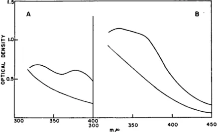
FIG. 1. Displacement of Fe from hemerythrin by equimolar Ag ion. A, spectra of hemerythrin in aqueous solution containing 8 Ai urea and 0.15 M NaCl, top curve without AgNOa, bottom with AgNOa; B, spectra of hemerythrin in tris buffer, pH 7.4, containing 8 M urea and 0.05 M KNOa, top curve without AgNOa, bottom with AgNOa. Blank cell in spectrophotometer contained same solutes, except for protein, to compensate for their absorption, particularly that of the nitrate ion.
metals displaced Fe from the protein. To do so we followed changes in spectra of the metal-protein as Ag+ was added in amounts equimolar to the iron. As is illustrated in Fig. 1, formation of the silver mercaptide is accompanied by the disappearance of the two-prong absorption peak characteristic of this iron-protein. Simultaneously, colorimetric tests re- veal iron in solution, free from protein. Silver is known to combine more avidly with thiols than any of the other groups listed in (III). Since one mole of Ag per mole of Fe displaces the latter from the protein, it seems evident that Fe must be attached to hemerythrin through a mercaptide linkage.
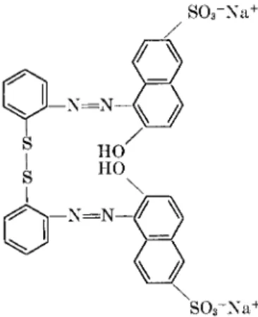
One might argue, nevertheless, that a particularly favorable juxtapo- sition of histidine and lysine side chains could produce a site with unusu- ally high affinity for Ag or Fe. Doubts on this ground were removed, however, by some experiments with a disulfide compound (IV), which certainly should
- N = N -
(IV)
not react with any of the groups in (III) except the thiol (12). The num- ber of mercaptan groups found in titrations with the disulfide dye agreed
P - S - C u
2000h
Cu + Alb.
Cu+ Hg(n) +Alb.
300
mμ 400
FIG. 2 . Near-ultraviolet absorption band of copper-serum albumin complex, and chemical tests showing that metal remains in cupric state. State of Cu: Cu (total), 0.00300 M ; C u d ) , 0.00002 M. State of Protein-S: bovine serum albumin, 0.63 SH per mole; bovine serum albumin-Cu, 0.61 SH per mole.
with that obtained in amperometric silver titrations (4). There can hardly be any doubt, therefore, that silver reacts with the same group as does the specific disulfide dye, i.e. with the thiol group.
The presence of a direct thiol-metal bond in hemerythrin thus seems clearly established. The status of the metal-protein bond in hemocyanin, however, is much more uncertain. Our primary reason for suggesting a copper-mercaptide is the presence in the spectrum of hemocyanin of a near ultraviolet absorption peak (340-350 m^) reminiscent of that in cop- per-serum albumin (Fig. 2 ) . In the latter, copper was added in the form of cupric ion. We have now established that this copper remains in the cupric state. As the data appended to Fig. 2 illustrate, the copper dis- placed from the protein by acid is essentially entirely in the cupric state.
Furthermore, even after addition of cupric ion, the —SH titer of the pro- tein remains essentially unchanged ; in other words, the one group capable of reducing Cu(II) to Cu(I) does not become oxidized. Since, as has been shown previously, both H g+ + and Ag+ abolish the near-ultraviolet peak of copper-albumin (Fig. 2 ) , it seems clear that this absorption is due to a Cu(II)—S linkage. One might reasonably attribute the 340 τημ peak of hemocyanin to the same type of bond.
It has seemed appropriate, to others as well as to us, to try to confirm the conclusion obtained from spectroscopy by using amperometric Ag+
or Hg+ + titrations (13, 14) to test for mercaptan groups in hemocyanin.
T A B L E I
AMPEROMETRIC TITRATIONS FOR MERCAPTAN IN HEMOCYANIN
Titrant SH/Cu
Hemocyanin Metal Ion Solution mole/mole
Busy con Ag+ Tris* 0.2
Busy con A g+ Tris, 8 M urea 0.26
Busy con A g+ Tris, 8 M urea, 0.003 M citrate 1.1
Busy con Ag+ Tris, 0.06 M dodecyl sulfate 0.2
Busy con, dialyzed
vs. NaCN Ag+ Tris 0.15
Busy con, dialyzed
vs. NaCN and CaClo Ag+ Tris, 8 M urea 0.6
Busycon H g+ + Citrate pH 4, 8 M urea, 0.5 M KCl 0.05 Busy con H g+ + Acetate pH 4, 8 M urea, 0.003 M
citrate 0.15
Busycon H g+ + Acetate pH 5, 5 M guanidine,
0.5 M KCl 0.03
Busycon H g+ + Phosphate, pH 7, 8 M urea 0.1
Limulus A g+ Tris, 8 M urea 0.5-1.5
» Tris refers to tris(hydroxymethyl)aminomethane buffer, pH approximately 7.3.
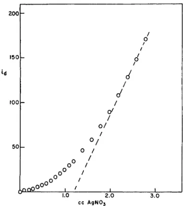
Results reported by others vary from zero to one thiol per atom of copper (15-17). Our results are in agreement with all of these; depending on the conditions, we also obtain values from zero to one (Table I ) . However, none of the amperometric titrations gives well-defined results. A typical curve is shown in Fig. 3. The upward curvature, as more H g+ + titrant is added, is typical of the reaction of a masked sulfhydryl group. Likewise, the current shows a slow drop with time if followed for a fixed amount of
2 0 0 h
150
l O O r -
2.0 3.0
cc AgN03
FIG. 3. Amperometric Ag+ titration of Busycon hemocyanin in tris buffer, 8 M urea and citrate. Citric acid was added to protein and the mixture added to tris buffer with urea.
H g+ +. As we shall show in a moment, it is not unreasonable that Ag+, or even H g+ +, should have difficulty in displacing copper if it is attached to sulfur in hemocyanin, and hence that the amperometric titration should be slow. Unfortunately, over long periods of time, the titrant metal ions may also catalyze the hydrolysis of disulfide bonds (18). In view of these uncertainties, the results of silver or mercury titrations must be consid- ered unreliable.
We have also carried out some amperometric titrations with a sample
of ceruloplasmin kindly supplied to us by Dr. L H. Scheinberg, but the results were just as equivocal as those with hemocyanin.
Some reasons for the difficulty in displacing copper from hemocyanin, assuming the metal is bound through a sulfur linkage, become evident on consideration of the properties of this protein. In the deoxygenated pro- tein, the metal is in the cuprous state. Even if linked to the protein only through sulfur (an unlikely situation since other co-ordination sites of the metal are probably additionally bonded to side chains of residues such as tyrosine, lysine, or histidine), one can estimate a minimum strength of the bond by assuming it is at least that of Cu(I) -S in cuprous cysteinate.
From Stricks and Kotthoffs (19) studies one can compute an association constant of 1.5 X 1 01 9 for
Cu(I) + - S — = Cu-S— ( 1 ) Competition experiments of Felsenfeld (8) between cyanide and hemo-
cyanin for Cu(I) likewise point to an association constant of 1 01 9- 1 02 0
for reaction (1) if ~ S — is in the protein. Of the two metal ions, H g+ +
and Ag+, used in amperometric titrations, the former should form the stronger bond with sulfur. Estimates of about 1 02 0 for the association constant in
Hg(II) + - - S — = H g - S - ( 2 ) can be made from Stricks et ai. (20). Clearly, then, Hg (II) does not have
the substantially greater affinity for sulfur than does C u ( I ) , in order that the latter may be displaced by the former from a mercaptide linkage.
Ag(I) would undoubtedly be in an even less favorable position to dis- place the copper.
In addition to the fact that the copper bonding to sulfur is likely to be very strong, another hindrance to the displacement of the metal by H g+ + or Ag+ lies in the protein itself. It is a difficult one to denature.
Hemocyanin in 8 M urea still shows its full blue color. Masking as con- tributed by the protein configuration or its hydration sheath can thus be quite strong even in urea and would provide a barrier for the penetration of H g + + or Ag+.
For the copper proteins it is clearly necessary, therefore, to develop further techniques to establish the nature of the metal-to-protein linkage.
III. Interaction Effects of Sulfur
The state of sulfur may have a marked influence on the behavior of a metalloprotein, even though the sulfur atom is definitely not attached to the metal. Hemoglobin serves as a prime example of such a phenome- non. Since it is sulfur in the mercaptan form which is involved in these
interactions, let us review briefly the status of quantitative measurements of this group in, for example, human hemoglobin. This is a subject upon which Dr. Huisman will expand.
Widely separated workers have obtained 8 —SH groups per mole of hemoglobin A by amperometric silver titrations, and values consistent with 8 by titration with mercury (13, 21-23). Total sulfur analyses indi- cate that aside from methionine only 8 sulfur atoms are available. Thus no disulfide sulfur seems to be present in hemoglobin. More recently, somewhat lower values (approximately 5 —SH groups) have been ob- tained on lyophilized samples, both by amperometric titration and by analysis for cysteic acid (24). Whether or not lyophilization is the reason for these differences, the conclusion that no disulfide sulfur is present is valid from these analyses also.
In the course of amperometric titrations some very interesting observa- tions have been made by Murayama (23) and by Ingram (25) which are pertinent to some of our subsequent discussion. Murayama in ampero- metric titrations found 8 —SH groups for crystallized dialyzed (deoxy- genated) hemoglobin at 38° ; at 0°, however, only 4 were detected. Similar results have been reported by Ingram. It is apparent that hemoglobin un- dergoes some important changes as the temperature is lowered.
With these observations in mind, let us now turn to the experiments described by Riggs (26, 27) on the effect of mercurials on hemoglobin- oxygen equilibria. We may note first that the addition of PCMB, even more than 8 moles per mole hemoglobin, did not change the oxygen capac- ity of hemoglobin ; clearly thiol is not involved in a direct bond to the iron
(26). On the other hand, PCMB as well as other mercury compounds de- creases markedly the interaction between sites. Oxygenation curves be- come less S-shaped and even approach the hyperbolic form characteristic of completely independent sites. Clearly, blocking of thiol groups in some way interferes with the means of communication between heme groups.
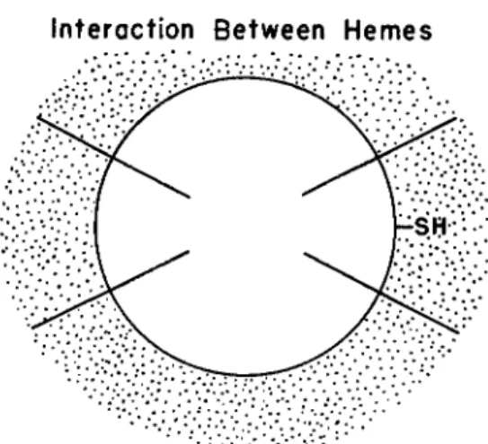
Riggs and Wolbach (27), following St. George and Pauling (28) and Wyman and Allen (29), have suggested that interactions between hemes depend upon a rearrangement of the polypeptide architecture and that this in turn can be controlled by the state of the mercaptan group. An alternative explanation of interactions between sites in terms of the lat- tice structure of the hydration water of the protein (Fig. 4) has been previously proposed (30). The binding site of the deoxygenated protein may be visualized as having an aqueous covering sheath whose structure is determined by the state of the metal in the heme as well as by the porphyrin group and amino acid side chains in the vicinity of the site.
Under appropriate conditions these crystalline water islands may be suf- ficiently extensive that they merge into an interwoven lattice. The at-
135 tachment of an 02 molecule to the binding site can then be visualized as producing an incongruity in this lattice because of the larger size of 02 as compared to H20 and because of differences in Η-bonding properties. As a result the hydration lattice might be disturbed for an appreciable dis- tance around the active site. This disturbance could affect the hydration
Interaction Between Hemes
-SH-:'..
FIG. 4. Schematic diagram to illustrate interaction between sites through hydra- tion sheath of protein.
structure of neighboring sites and hence change their affinity toward oxy- gen molecules. In this way the typical S-shaped oxygenation curve may arise.
On this basis one can also interpret readily the effect of mercurials. As shown in Fig. 4, one can readily visualize the water lattice between hemes as having adapted itself to the presence of an — S H side chain. When, however, the character of this side chain is altered, as in combination with a mercurial, the lattice of communication between hemes is also disturbed. Consequently, the subsequent attachment of an 02 molecule to the heme group will not have as much of an effect on the water bridge to a neighboring heme as it would have had if the lattice had not been previously disturbed.
On the basis of this iceberg model one can also understand the results of Murayama (28) and Ingram (25) showing fewer —SH groups at lower temperatures. The masking of these groups we attribute to the "frozen"
water of the hydration layer (31 ). As the temperature of the solution is lowered, it is only reasonable that the hydration layer become more ex- tensive. Since ions do not diffuse readily in ice, the metal ion of the amper- ometric titration would have difficulty penetrating the covering on the thiol group.
Likewise, on the basis of the hydration picture, one can understand why sulfhydryl-blocking agents and oxygen have comparable effects in the prevention of sickling of sickle-cell hemoglobin (23, 32), despite the fact that each acts at a very different site on the protein. What these two reagents have in common is an ability to modify the nature of the hydra- tion lattice on the protein. The very phenomenon of sickling implies, at the molecular level, an alteration in the interaction of the protein molecule with solvent molecules.
IV. Conclusions
The metal-proteins and particularly the oxygen-carrying ones are par- ticularly suitable for studying the effects of molecular changes on bio- chemical function. The reaction involved with these pigments is well- defined and easy to observe. One can follow readily not only perturbations which produce gross inactivation but also changes in the kinetics and in the equilibria.
In discussing the role of sulfur in the metal-proteins, we have been limited to descriptions of the effects of the thiol group, either in covalent thioether linkage or in its mercaptan or mercaptide state. N o mention has been made of sulfur in methionine or cystine residues. These, of course, must make an important contribution in determining the configuration of the polypeptide as well as in interactions with the solvent, but at pres- ent nothing definitive can be said in these respects. It is conceivable, fur- thermore, that S—S and C H3— S — C - form linkages to the metal, but no way of studying these has been possible yet, primarily because simple model complexes in aqueous solution cannot be examined.
As regards the mercaptan group, we see that it may influence the be- havior of the metal-proteins in either of two ways. There can be a direct linkage to the active site. Secondly, there can be an important indirect influence due to a change in configuration of the protein or a modification in the lattice structure of its hydration sheath. In hemoglobin, the indirect influence may be expressed by an activation as well as an inactivation when the mercaptan group is blocked. The direct type of effect has been postulated, although not so clearly demonstrated, to explain the effects of thiol-blocking agents in many other proteins besides the oxygen-car- riers. The second type of effect has not been so clearly recognized in other protein systems except in connection with the possibility of configura- tional changes due to oxidation of —SH to S—S. In fact in most protein systems it would be difficult to distinguish between these different types of effect because one can study usually only the kinetics and it may be
difficult to tell whether the number of active sites or their reactivity has been changed. Changes in the behavior of the oxygen-carrying proteins, on the other hand, can readily be assigned to stoichiometric or affinity factors. For this reason as well as others cited above, these proteins should continue to provide attractive systems for the interpretation of biological specificity in molecular terms.
ACKNOWLEDGMENT
These investigations have been carried out with the aid of a grant (H-2910) from the National Institutes of Health, United States Public Health Service. We are also indebted to Dr. H. A. Fiess for his assistance in some of the amperometric titrations.
REFERENCES 1. H. Theorell, Biochem. Z. 298, 242 (1939).
2. H. Tuppy and S. Paléus, Acta Chem. Scand. 9, 353 (1955).
3. I. M. Klotz and T. A. Klotz, Science 121, 477 (1955).
4. I. M. Klotz, T. Α. Klotz, and Η. Α. Fiess, Arch. Biochem. Biophys. 68, 284 (1957).
δ. A. Mazur, S. Balz, and Ε. Shorr, J. Biol. Chem. 213, 147 (1955).
6. J. B. Conant, F. Dersch, and W. E. Mydans, J. Biol. Chem. 107, 755 (1935).
7. I. M. Klotz, J. M. Urquhart, and H. A. Fiess, J. Am. Chem. Soc. 74, 5537 (1952).
8. G. Felsenfeld, / . Celluhr Comp. Physiol. 43, 23 (1954).
9. Α. Β. Griffiths, Compt. rend. 115, 669 (1892).
10. Ε. Boeri and A. Ghiretti-Magaldi, Biochem. Biophys. Acta 23, 489 (1957).
11. I. M. Klotz, S. Rapaport, and Ε. V. H. Rosenberg, Biol. Bull. 105, 377 (1953).
12. I. M. Klotz, J. Ayers, J. Y. C. Ho, M. G. Horowitz, and R. E. Heiney, / . Am.
Chem. Soc. 80, 2132 (1958).
18. R. E. Benesch, H. A. Lardy, and R. Benesch, J. Biol. Chem. 216, 663 (1955).
14. I. M. Kolthoff, W. Stricks, and L. Morren, Anal. Chem. 26, 366 (1954).
Ιδ. R. Lontie, Behringwerk-Mitteilungen 32, 64 (1957).
16. K Yasunobu, L. C. G. Thomson, and H. S. Mason, Federation Proc. 16, 275 (1957).
17. E. S. G. Barron, private communication, 1956.
18. R. Cecil, Biochem. J. 47, 572 (1950)
19. W. Stricks and I. M. Kolthoff, J. Am. Chem. Soc. 73, 1723 (1951).
20. W. Stricks, I. M. Kolthoff, and A. Heyndrickx, / . Am. Chem. Soc. 76, 1515 (1954).
21. V. M. Ingram, Biochem. J. 59, 653 (1955).
22. F. A. Hommes, J. S. Drinkwaard, and T. H. J. Huisman, Biochem. Biophys. Acta 20,564 (1956).
23. M. Murayama, J. Biol. Chem. 228, 231 (1957).
24. W. H. Stein, H. G. Kunkel, R. D . Cole, D . H. Spackman, and S. Moore, Biochem.
Biophys. Acta 24, 640 (1957).
2δ. V .M. Ingram, Biochem. J. 65, 760 (1957).
26. A. F. Riggs, Λ Gen. Physiol. 36, 1 (1952).
27. A. F. Riggs and R. A. Wolbach, J. Gen. Physiol. 39, 585 (1956).
28. R. C. C. St. George and L. Pauling, Science 114, 629 (1951),
29. J. Wyman, Jr. and D. W. Allen, J. Polymer Sei. 7, 499 ( 1 9 5 1 ) .
30. I. M. Klotz and R . Ε. Heiney, Proc. Natl. Acad. Sei. U. S. A3, 7 1 7 ( 1 9 5 7 ) . 31. I. M. Klotz and J. Ayers, J. Am. Chem. Soc. 79, 4078 ( 1 9 5 7 ) .
32. A. C . Allison, Biochem. J. 65, 2 1 2 ( 1 9 5 7 ) .
Discussion
KAUZMANN : A number of years ago F. H. Johnson and F. M. Schlegel (/. Cellular Comp. Physiol. 31, 421, 1948) measured the effect of hydrostatic pressure on the oxy- genation curve of hemoglobin and found there was no appreciable effect up to 10,000 lb. per sq. in., which proves that there is no volume change when oxygen in solution combines with hemoglobin. If ice were melting in any considerable quantities when the oxygen combined with the hemoglobin, they would certainly have found an in- crease in oxygen affinity at high pressures. I merely toss that in for what it is worth.
KLOTZ: Maybe so. I would like to see the original data. For example does pressure affect also the pH of the buffer solution used?
RIGGS: I believe that the heme-heme interaction is largely independent of tem- perature, and I would expect that it would be highly dependent upon temperature because of the changes in the crystallinity of this ice.
KLOTZ: Whether you would observe any effect would depend on how far ice
"melting" had progressed at the highest temperature which has been measured. Or turning around in the direction of temperature change, if you have already really at room temperature filled all of the channels of communication with frozen water, then it will not matter a great deal if you lower the temperature further. To decrease in- teraction you would have to raise the temperature. There is surely some temperature at which the heme-heme interaction disappears.
RIGGS : Yes, when the whole protein is destroyed.
KLOTZ : In terms of my viewpoint, I would say that at any temperature which has been measured so far and with the heme groups sufficiently close to the surface, ice chains between them have been pretty thoroughly formed.
RIGKSS: This is describing the boat in terms of the water it is in. It seems to me the specificity of these effects and the specificity of mercurials on different hemoglo- bins means that the protein structure as such must be intimately involved here. It is certainly true that this protein structure may determine to some extent the lattice arrangement of the ice, but I have a little bit the feeling that this is a case of the cart before the horse. You have the ice determining the properties of the protein and I prefer to think the protein determines the properties of the ice.
KLOTZ: NO, I think both determine each other.
RIGGS: This is certainly true.
KLOTZ : As I indicated here, I think the ice structure, and particularly its pathway in the protein must be determined by which side chains are present and where they are present. In turn their particular orientation might be determined by how the solvent stabilizes a particular structure. I think the interaction would work both ways. I admit I have emphasized the water aspects, not only here, but in writing. I do not think the protein aspect has to be emphasized. Others are going to emphasize that. I am not going to argue that protein configuration plays no role or deny that some of these effects are due to a change in configuration. I would merely like to emphasize that solvent also may be important in determining structure and behavior.
MORALES: D O you consider this crystallizable water to be accessible for ions to dissolve in the solution?
KLOTZ: It would depend on the substance. I would reason as follows. Diffusion of ions like lithium in ice and in water has been measured and compared. The diffu- sion of lithium ion is very slow in ice. Consequently I would feel that an ion like silver ion would have trouble getting into this lattice. That is the basis, from this view point, of the so-called masked nature of some S H groups. I would not say that a long chain molecule would necessarily have a hard time getting in because such organic molecules do form good crystal ice hydrates, which indicates that they can fit into this ice structure.
EDSALL: Wouldn't there be a time factor there? The thing has to make its way in. It might well form a stable structure when it got in, but I should think the time required might be appreciable.
KLOTZ: If the substance can fit into an established ice-like lattice, it would not be as difficult for it to get in as in the case of a lithium or silver ion where it will have to disorder the established ice-like lattice in order to impose a new lattice on the system. On that basis, I would make a distinction in rate as well as in equilibrium.
MORALES: The sequel to that question was, would you then expect changes in a predictable direction by addition of substances that interfere with the lattice? Does this happen? Are there any instances you already know?
KLOTZ: I think you can say this. There are lots of substances that change the interaction in hemoglobin. The S H group is usually emphasized because a great deal of work has been done with it.
MORALES: I meant things that you might add to the solution—lithium ion, for instance. Does it affect the binding of oxygen in a predictable way from your theory?
KLOTZ : No, not in a predictable way except to say they do affect it. Once a lithium or sodium ion is in an aqueous environment, and there is enough water, would it really want to go into the ice? I think perhaps not, except maybe in a very concentrated solution. The same may not be true of anions, however, judging from differences in binding to proteins. There are salt effects on the oxygenation curves of which every- one is aware. I think they are a little easier to rationalize from this hydration ice viewpoint than from that of a configurational change in the protein. What you would consider more reasonable may be a matter of taste. I have become accustomed to this viewpoint now and feel a lot of things fit in a lot better from this viewpoint than from the view of changes in protein configuration.
HOCH: I wonder if the interpretation of your data on hemerythrin is necessarily as unique as you indicate : namely, that the iron is bound by sulfhydryl groups to the protein. The possibility exists that added silver ions, for example, actually bind to sulfhydryl groups involved in the maintenance of the tertiary or secondary protein structure, and that changes in the structure result in the liberation of the iron atom from sites other than sulfhydryl groups. The net result would be the same as if the iron were bound to sulfhydryls and directly displaced by Ag+, and the loss of iron from the protein is, therefore not diagnostic of a specific chemical binding. Is it possible to reverse the reaction leading to the liberation of the iron, and to return the metal to its original locus in a functioning hemerythrin? This might be of value in deciding whether its binding is as simple as you have indicated.
KLOTZ : With respect to your second question, we have never actually tried it. In any event from your viewpoint even if iron could be put back in, the group involved would not be known. As to your first question, I showed the curves of the changes in absorption spectra as silver is added. At the same time as silver is attached to the protein you find iron in solution detached from the protein. It is difficult to see why there should be such good stoichiometry if a configurational change caused the loss of
this spectrum. You can carry your argument to the limit and say that certain amino groups have to be in the proper configurational position to hold iron. This is pos- sible, but I think far-fetched, for it is not readily apparent how —SH as such would maintain the configuration. There is also one thing I left out at the last moment here in order to cut down my time. We have also done another experiment not with a metal but a much more specific compound, the dye which has been alluded to several times which has S S groups; and this surely would be specific only for SH.
This, too, will abolish the spectrum. Nevertheless, if you are absolutely insistent that the S H merely maintains the protein structure in some way and that silver or dye merely loosen it up, I do not know any way of getting around that until we can ob- tain and interpret X-ray diffraction data of proteins in solution.
HOCH: I agree that theories can be carried to extreme limits. However, experi- mental evidence may be of aid in avoiding deductional impasses. A possible way to resolve the question of whether structural changes are involved in, or are the cause of, the loss of iron in these experiments might be the experimental demonstra- tion that there is or is not a structural change.
KLOTZ: I do not know that such an experiment would prove anything because there might be some structural changes accompanying the loss of iron simply because you have lost the iron. There must be other co-ordination positions involved in the protein besides the one to sulfur, since iron is hexa-coordinated. I say that the sulfur forms the critical bond because if you break that linkage the iron comes off. After that the protein structure might fall apart because several ligand groups are no longer restrained.
BENESCH: With regard to denaturation by high pressures, there was a lot of Russian work which I read in detail in Chemical Abstracts on renaturation of pro- teins with high pressures. These people claim that they can renature denatured pro- teins by exposing them to very high pressures.
KLOTZ: Several people who tried to repeat the Russian work say they cannot do it—Sanger in particular, and some of the French in Macheboeuf's laboratory.
BOYER: One brief comment. Is it correct to assign a dominant role to sulfur when you need a precise positional arrangement of several groups to interact with iron?
If you had a specific way of blocking any of these groups you would probably block interaction with the iron. So is it fair at this time to say that it is dominantly sulfur because this is the group you happened to get hold of?
KLOTZ : If you look at a skyscraper, you may be sure it is being held up in part by the windows. On the other hand, if you take out the steel and find the building col- lapses you could claim that it is really held up primarily by the steel. There is a relative order. The iron-sulfur bond is certainly the strongest of the bonds. If you blocked any other ligand, but not the sulfur, I doubt that iron would be released from the protein.
BOYER: This is another case where reason by analogy might lead us into difficulty.
KLOTZ: The real answer is that the iron-sulfur linkage is the strongest bond. In this sense I think the analogy is an appropriate one.