CHAPTER 21
Mercaptide-Forming Agents
1Ν. B. Madsen
I. Introduction 119 II. Metal Cations 120
A. Chemistry of Mercaptide Formation 120 B. Application to Estimation of Thiols 123
C. Inhibition of Enzymes 125 III. Organic Mercurials 127
A. Chemistry of Mercaptide Formation 127 B. Methods Employing Organic Mercurials 131
IV. T w o Individual Enzymes 136 A. Glyceraldehyde-3-phosphate Dehydrogenase 136
B. Phosphorylase 138 V. Summary 140
References 141
I. INTRODUCTION
Mercaptide formation is one of the chief reactions used in research on sulfhydryl groups. The whole question of the role of — S H 2 groups in enzymic catalysis appears just now to be going through a period of doubt and uncertainty, whereas in the early part of the last decade it appeared to be firmly fixed on a course whose ultimate destination was thought to be known. A t that time the generally accepted criteria for the recogni
tion of a "sulfhydryl enzyme" were its inhibition by P C M B and its reactivation by cysteine or other thiols. The satisfaction of these criteria usually led to speculation about the role of the — S H groups in the
1 Contribution No. 511, from the Microbiology Research Institute, Research Branch, Canada Department of Agriculture, Central Experimental Farm, Ottawa, Canada.
2 The following abbreviations are used: — S H , sulfhydryl; PCMB, p-chloro- mercuribenzoate; N E M , ΛΓ-ethylmaleimide; E D T A , ethylenediaminetetraace- tate; PCMPS, p-chloromercuriphenylsulfonate; D P N , diphosphopyridine nucleotide; D P N H , reduced form of D P N ; Tris, tris(hydroxymethyl)amino- methane.
119
mechanism of catalysis. Recent research has indicated, however, that the problem is more complex than was formerly thought; for example, evi
dence of the direct transfer of hydrogen in reactions involving pyridine nucleotides has rendered untenable certain postulated reaction sequences.
Sulfhydryl groups have also been demonstrated to have important roles in the maintenance of protein structure. Many of the results which were interpreted as indicating a participation of —SH groups in the mechanism of enzyme action can also be explained by postulating changes in protein configuration. Keeping this rather unsettled stage of development in mind, the present chapter will attempt to evaluate the advantages and limita
tions of mercaptide-forming agents in promoting our knowledge of this important subject.
The subject of the — S H groups of enzymes has been the theme of many excellent reviews, which have, naturally, discussed mercaptide for
mation. Among these, Barron (1) may be consulted for an insight into the literature and prevailing opinions prior to 1950. "A Symposium on Sulfur in Proteins," edited by R. E. Benesch et al. (#), presents a good picture of the remarkable scope of biological processes in which —SH groups play an important role, as well as a critical appraisal of modern methods and results. Boyer (3) has given an account of the chemistry of mercaptide formation and has dealt capably with the application of this and other phenomena to the problem of enzyme action. Cecil and McPhee (4) have delved extensively into the sulfur chemistry of proteins, and their review is recommended for its thoughtful discussions of analytical methods.
Because of the extensive recent reviews available, and in keeping with the emphasis which this book places on the inhibitor rather than on the biological system under investigation, this chapter will confine itself mostly to discussions of the principles and applications of the method
ology now available. The limitations of space and purpose have dictated a highly selective rather than inclusive approach. For this reason, it has been possible to choose only a few enzymes for detailed examination to illustrate various points. Similarly, work with crude biological prepara
tions has been excluded because of space and because it is difficult to interpret the results of such work.
II. METAL CATIONS
A. Chemistry of M e r c a p t i d e Formation
Gurd and Wilcox (5) have presented a comprehensive review on the binding of metallic cations by proteins, peptides, and amino acids, in
21. MERCAPTIDE-FORMING AGENTS 121 which they discuss the chemistry of mercaptide formation in detail.
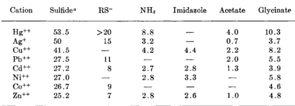
Table I is presented to give a rough idea of the relative affinities of metal
T A B L E I
FIRST ASSOCIATION CONSTANTS (LOG ki) FOR THE COMBINATION OF SOME CATIONS AND SMALL MOLECULES
Cation Sulfide" RS~ N H3 Imidazole Acetate Glycinate
H g+ + 53.5 > 2 0 8.8 4 . 0 10.3
Ag+ 50 15 3.2 — 0.7 3.7
C u+ + 41.5 — 4.2 4 . 4 2.2 8.2
P b+ + 27.5 11 — — 2.0 5.5
Cd++ 27.2 8 2.7 2.8 1.3 3.9
N i+ + 27.0 — 2 . 8 3.3 — 5.8
C o+ + 26.7 9 — — — 4.6
Zn++ 25.2 7 2.8 2.6 1.0 4.8
° pK of the solubility product, taken from Kolthoff (6). The remainder of the data are abstracted from the data collected by Gurd and Wilcox (5). Many of the constants, particularly those for the mercaptides, should be regarded as approximations.
ions for various groups typical of those found in proteins. As noted before by Klotz ( 7 ) , there is a rough correlation between these affinities and the order of the solubility product constants of the metal sulfides. It may be seen also that the specificity of mercury and silver for — S H groups is only relative. Thus, one might expect that when these metals are added to a protein solution they will saturate the — S H binding sites first but that any excess will immediately begin associating with other groups. The first association constants only are listed in Table I, with the exception of the association constants for the formation of sulfides, because metals bound to proteins may not be able to form higher complexes, as they would with smaller molecular weight analogues. A small anion from the medium would usually be bound to the metal.
The constants given in Table I are for the reaction given in Eq. (1),
Metal + ligand ^=± metal — ligand (1) whereas, under experimental conditions, hydrogen ion competes with the
metal for the ligand, while anions from the medium compete with the ligand for the metal. If we use the formation of a mercaptide from mer- curic chloride and a protein-bound — S H group as an example [Eq. ( 2 ) ] , the over-all equation is:
R—SH + HgCl2 ^ R—S—HgCl + H+ + Cl" (2)
but this contains two dissociations, namely:
R—SH ^± R—S~ + H+ (3)
HgCl2 ^± HgCl+ + Cl- (4) plus the actual formation of the mercaptide, for which the equation analogous to Eq. (1) is Eq. (5).
The equilibrium constant for Eq. (2) does not appear to have been determined, but a rough estimate may be calculated if we assume that the association of HgCl+ to a protein-bound —SH group is similar to that of mercury to small molecular weight thiols where the "log associa
tion constant"3 is a minimum of 20 (8, 9). The logarithms of the associa
tion constants for R—SH and HgCl2 [reverse of Eqs. (3) and (4) ] are taken as 8.5 (10) and 6.5 (11), respectively. This leads to a log equilib
rium constant for Eq. (2) of 5, which indicates that the equilibrium is still far to the right.
It should be noted that one of the important factors in determining the equilibrium of Eq. (2) is the dissociation of an anion from the mercury [Eq. ( 4 ) ] . The relative affinities of various anions for mercury are C N - > I - > Br~ > C I - > N 03- . It may be calculated from the data of Sillen (11) that the logs of the association constants for the reaction shown in Eq. (6),
where Ζ is a halogen, are 11.0, 8.3, and 6.5 for I - , B r - , and CI", respectively. Thus, the equilibrium of Eq. (2), as well as the specificity of the reaction of mercury with — S H as opposed to other available groups, can be influenced by the type of small anions present. This theme will be taken up again in relation to one of the organic mercurials.
It is also seen readily that the pH will have a great influence on the equilibrium of Eq. (2). What may not be so apparent is that pH will affect the specificity of the reactions of metals with various protein groups because of the different pi£'s of the latter. For example, at pH 7.0, all of the carboxyl groups and approximately half of the imidazole groups of a protein are free of protons and may therefore react readily with metal R—S~ + HgCl+ ^± R—S—HgCl (5)
HgX+ + X " ^ HgX; (6)
3 The term "log association constant" is used here to mean the same as "log k" as defined by Gurd and Wilcox (5) on p. 316 of their review.
21. MERCAPTIDE-FORMING AGENTS 123 cations. On the other hand, only a small fraction of the —SH and c-amino groups (perhaps of the order of 1% and 0.1%, respectively) are available.
Although the association constants listed in Table I indicate that sev- eral of the protein-bound groups would not be expected to be very effec- tive in binding metals, the binding by glycinate ion shows that chelation would increase affinities to a significant extent. Tanford and Epstein (12) showed that the tight binding of zinc to insulin would be consistent with the chelation of each Zn++ to two imidazole groups. Ingram (13) has found that native horse hemoglobin will bind four Ag+ per molecule, as measured amperometrically, but X-ray analysis indicates only two silver spots. Furthermore, only two H g + + or P C M B molecules were bound. The results have been interpreted as indicating that there are two clusters of two —SH groups each, the two —SH groups in each cluster lying so close together that they can chelate the H g + + . The P C M B bound to one of the
—SH groups in a cluster would block a second mercurial by steric hindrance.
Vallee and associates (14) have recently made some very interesting observations on bovine pancreatic carboxypeptidase, a zinc metallo- enzyme containing 1 gm atom of zinc per mole of protein. The enzymic activity is abolished by removing the zinc with chelating agents and is restored upon adding zinc. N o — S H groups are found in the native en- zyme, but one group per mole of protein can be titrated in the zinc-free enzyme. Furthermore, the addition of silver or P C M B to the zinc-free enzyme prevents the restoration of activity by adding zinc. The authors suggest that zinc is bound as a mercaptide but that it is also bound by a second linkage; the chelation thus produced would explain the relatively high affinity of zinc for this enzyme. These results open up several new possibilities in connection with sulfhydryl enzymes. First, a chelation of a metal involving an — S H group may occur in nature. Second, this chelation may prevent any normal reactivity of the — S H group. A new type of sulfhydryl enzyme is introduced which can be detected only with special techniques. Third, one may speculate on the possibility that such a chelated metal may act as a structural link serving to maintain an enzyme in a conformation necessary for activity.
B. Application to Estimation of Thiols
Mercaptide formation with silver and mercury ions has been the basis of a number of amperometric procedures, which have been developed to a large extent by Benesch and Benesch and by Kolthoff and Stricks. Silver nitrate has been used extensively, being added to a solution of the thiol containing ammonium ions which serve to complex the silver (15-17).
The conditions required were not suitable for most proteins, but Benesch et al. (18) introduced the use of Tris buffer as the complexing ion, thus making it possible to carry out the titrations at neutral pH. Their method could be used to estimate the speed of reaction of various classes of —SH groups. Urea was used as a denaturing agent to determine the total sul
fhydryl content of a protein. Some of their results, however, are con
siderably higher than those obtained by other methods. Cecil and McPhee (4) review the whole question of the use of silver to titrate — S H groups.
They note that silver ion forms complexes with the mercaptides of a stability comparable to the mercaptides themselves, and high results are obtained. Ammonia does not complex the silver strongly enough to pre
vent this, and Tris appears to be still less effective. It is on this basis that Cecil and McPhee explain the high titration value for —SH groups in hemoglobin compared to the values obtained by titration with mercury or organic mercurials. Ingram (13) presents an ingenious alternative explanation, discussed in Section II, A.
One may note, however, that the amount of P C M B bound to hemo
globin, as measured by equilibrium dialysis, agreed with the amperometric silver titration (18). Here again, as in so many instances, at least some of the discrepancies in results from different laboratories may be due to the different rates of reaction of various types of — S H groups on the same protein. This reviewer believes that the method of Benesch et al.
(18) has, in general, yielded results in excellent agreement with those obtained by other methods and that those discrepancies so far found are not yet numerous enough or sufficiently well documented to justify dis
carding this procedure.
Mercuric ion is, in some respects, more suitable for the titration of thiols because the mercaptides formed are considerably more stable than the complexes between the mercaptides and mercury (but see Section II, C, where it is pointed out that silver may be more effective than mercury in reacting with certain "masked" — S H groups). The latter complexes can be suppressed by a high concentration of suitable anions, such as chloride (8, 9). Mercuric chloride has been used as the basis of various amperometric titrations (19), but the stoichiometry of the mercaptide formation tends to vary under different conditions because of the divalency of mercury. Cecil and McPhee (4) advise that it may be difficult to tell if the end point of a titration corresponds to RSHgX, (RS)2Hg, or ( R S )2H g2. They feel that certain organic mercury deriva
tives fulfill the need for a univalent reagent with some of the advantages of mercuric ion. The use of organic mercurials will be discussed in a later section.
21. MERCAPTIDE-FORMING AGENTS 125 C. Inhibition of Enzymes
Heavy metals were at one time used widely for inhibitory studies, but they fell into disfavor after the development of the more specific organic mercurials. The use of metals for inhibitions is subject to the limitations of specificity and reaction conditions which were discussed earlier.
Those enzymes which possess — S H groups should form complexes with mercuric ion, and such complexes would be expected to be less soluble (5). Several enzymes have been isolated as crystalline mercury complexes, including enolase {20), rat lactic dehydrogenase (21), papain (22), and lysozyme from papaya latex (23). The papain crystallizes as a dimer with one molecule of mercury, in a manner reminiscent of the crystalline mer
cury complex of mercaptalbumin (24). The lysozyme has no — S H groups, and it is believed that the mercury is bound to an imidazole group.
Urease has been the subject of considerable experimentation with regard to inhibition by metals. Jacoby (25) showed that it could be inhibited by mercuric ion, and Bersin (26) found that H2S would reverse this inhibi
tion. The data of Hellerman et al. (27), when recalculated for the molecu
lar weight of 483,000 given by Sumner et al. (28), indicate that the first 22 moles of P C M B added per mole of urease produce no inhibition, while there is a linear titration of the activity with the next 22 equivalents. It is therefore interesting that Ambrose et al. (29) found that as little as 6 equivalents of silver ion were sufficient to cause complete inhibition. Shaw (30) investigated the relative efficiency of inhibition by a number of metal ions and found that it was directly related to the logarithm of the solubility constant of the metal sulfides. Silver, however, was considerably more efficient as an inhibitor than mercury, which may be related to the greater ability of silver to react with masked —SH groups, as discussed below. There is also the possibility, as suggested by Hellerman (31), that PCMB causes structural changes, such as a dissociation of the urease molecule. Urease shows many interesting facets and it might be interest
ing to reinvestigate the inhibition by mercaptide-forming agents, using the more recent methods discussed above and below.
Hellerman et al. (32) employed silver in an interesting series of experi
ments on crystalline glutamic acid dehydrogenase. If their results with the enzyme from calf liver are expressed on the basis of a molecular weight of 1 Χ 106, then 40 gm atoms of silver may be added without any inactivation. Further addition of silver produces a proportional inhibition which is complete at a total of 80 gm atoms per mole of protein. An additional 40 — S H groups can be titrated with silver only after denatura- tion. Organic mercurials can react with the first 40 — S H but only with
difficulty with the second 40, as they produce little inhibition. Ampero- metric titration with silver agrees with the enzymic titration in yielding a total of 80 —SH groups per mole of native protein. The authors suggest that the organic mercurials fail to inhibit because of steric hindrance. The
—SH groups are believed to play a direct role in the catalytic function of the enzyme.
Cecil and McPhee (4) have criticized the above interpretation on the grounds that if steric hindrance prevents the organic mercurials from approaching the —SH groups there would also be difficulty in the ap
proach of the glutamic acid molecule. They suggest that the —SH groups are present in some form of combination which is broken more easily by silver ion than by the mercurials or mercury, as is apparently the case with disulfides. This suggestion would imply that the — S H groups are more concerned with maintenance of protein structure than directly with the catalytic function.4
This reviewer agrees with Cecil and McPhee, but would also like to point out that P C M B was allowed to react with the glutamic dehydro
genase for only 2 minutes before enzymic activity was measured. At the low concentration of protein-bound — S H groups employed (5.6 X 1 0 -7 M), the extent of inhibition reported with varying concentrations of P C M B is consistent with the occurrence of a second-order reaction having a rate constant similar to those reported for other enzymes and proteins which react slowly with P C M B (see Section III, A and Table I I I ) . That P C M B is capable of reacting with all of the groups available to silver is shown in the same paper by amperometric measurement of the —SH groups after reaction with the mercurial. Here the reaction went to completion because of the much higher concentrations of both protein and mercurial which were used in this experiment. This paper presents some fascinating contrasts in the effectiveness of silver versus organic mercurials as inhibitors of certain types of sulfhydryl enzymes and again illustrates the fact that slowly reacting or masked —SH groups react very much more quickly with silver than with organic mercurials or even with mercuric ion.
Heavy metal inhibition of sulfhydryl enzymes may occur when it is undesirable and, possibly, undetected. Hoch et al. (33) have recently presented evidence that the inhibition of yeast alcohol dehydrogenase by iV'-methylnicotinamide is caused by silver present in the chemical as
4 Note added in proof: A more recent communication indicates that silver ion does cause glutamic dehydrogenase to dissociate. Eighty equivalents of A g+ per 10e gm of protein changed the S2o,«> from 24.4 to 10.5. [K. Rogers, T.
E. Thompson, and L. Hellerman, Biochim. Biophys. Acta 64, 202 (1962).]
21. MERCAPTIDE-FORMING AGENTS 1 2 7 supplied. The inhibition by hydroxylamine is also due to contaminating metals. Another facet of the dangers of contamination of chemicals by heavy metals may possibly be illustrated by the same enzyme. The results of Barron and Levine (84), when recalculated for a molecular weight of 150,000 indicate that the addition of D P N causes the disappearance of 8 —SH groups per mole of enzyme, an average of two per D P N bound (85). This has led to speculation on the role of the — S H groups in this enzyme; but if the D P N used has been contaminated with metals, the results may be in error.
III. ORGANIC MERCURIALS
A. Chemistry of Mercaptide Formation
P C M B is the most commonly used compound of this type and was introduced by Hellerman and his associates (27). p-Chloromercuriphenyl- sulfonate was recommended by Velick (86) because of its greater solu- bility. Cecil and McPhee (4) prefer phenylmercuric hydroxide, while compounds of methylmercury have been used extensively by Hughes (87) and co-workers. Other organic mercurials which have been used include mersalyl and 4- (p-dimethylaminobenzeneazo) phenylmercuric acetate.
The latter compound must be used in a two-phase system with heptanol and is chiefly useful for quantitative measurements (88).
The reaction of an organic mercurial with an —SH group may be depicted as in Eq. (7).
R—HgX + R'—SH ;=± R—Hg—S—R' + H+ + X~ (7) The nature of the anion attached to the mercurial, here denoted by X—,
may have a significant effect on the equilibrium and specificity of the reaction. Boyer (8, 89) has pointed out that, when P C M B is solubilized in base, the chloride is probably replaced by OH~\ Pyrophosphate or glycylglycine increase the solubility of P C M B , presumably by displacing the chloride. Harris and Hellerman (40) have found that E D T A may overcome the inhibition of some enzymes by P C M B , and certainly the interaction of the two reagents is shown by the shift in the ultraviolet absorption spectrum of the P C M B (89). Chinard and Hellerman (41) caution about the use of chelating agents in conjunction with P C M B , but in at least one case the presence of E D T A does not affect the inhibition by PCMB (42). Hughes (48) has found that methylmercury hydroxide will
combine in nonspecific fashion in great excess of the number of protein
—SH groups, but methylmercury iodide is much more specific because only the —SH groups can overcome the strong affinity between the mer
curial and iodide. Boyer (39) has contributed a detailed and systematic study of the effect of various anions on the reaction between P C M B and certain model proteins. This should be of considerable benefit in guiding future studies, but caution will still be needed when using new systems.
Hoch and Vallee (44), for example, found a significant difference in the number of —SH groups titratable in alcohol dehydrogenase by P C M B in different buffers (16 vs. 31 —SH groups per protein molecule in Tris versus phosphate buffer).
The equilibrium of Eq. (7) is generally considered to be far to the right, but the actual value has seldom been determined. An indication that P C M B is bound tightly to protein —SH groups is given by the adher
ence to stoichiometry when the latter are titrated. Ingram (13) has shown that the order of tightness of binding is HgCl2 > P C M B > A g N 03. This would place the association constant for P C M B with —SH groups between 1 01 5 and 1 02 0 M, since these are the approximate association constants for silver and mercury (Table I ) . Hughes (37) determined the equilibrium constant for the reaction of methylmercury iodide with mercaptalbumin [Eq. (8) ] and found that it corresponded to a ipK of 4.45.
CH3HgI + albumin-SH ^± albumin-S-HgCH3 + H+ + I" (8) If we assume that the association constant of C H3H g + and I"" is the
same as for H g l + and I ~ , namely 1 01 1 [from Sillen (11)], and that the association constant of albumin-S~ and H + is equivalent to that of - O O C C H ( N H3+ ) C H2- S - and H + , taken as 3.4 Χ 108, then it may be calculated that
i ^ a e s o c = Τ Τ Γ Ι α - \ / η τ τ τ τ +\ ~ 1·2Χ101 δ (9)
(albumin-S ) (CH3Hg+)
This result is in fair agreement with that estimated for P C M B above. It may be noted that pH will have a considerable effect on the equilibrium of Eq. (8), and Hughes found that a pH of 7.3 or more was necessary to obtain a quantitative reaction. Table II gives a few of the association constants which have been estimated.
A few estimations of the binding of P C M B to protein have been made.
Benesch et al. (18) found association constants of 4.6 Χ 104 and 2.4 χ 104 with sheep and human hemoglobins by the method of equilibrium dialysis.
Madsen and Gurd (48), using the ultracentrifugal separation method,
21. MERCAPTIDE-FORMING AGENTS 129
FIRST ASSOCIATION CONSTANTS (LOG hi) FOR THE FORMATION OF SOME MERCAPTIDES
Thiogly- Gluta Mercaptal-
Ions Cysteine colate thione bumin
(1) Hg++« 20 22 20
(2) R S — H g+ · 20 22 20 12.6,6 13.2C
(3) RS—Hg—R'—Hg+ d — — — 17.2«
(4) C H3H g + ' 15 — — 15
α The values of log kx for the small molecular weight thiols are estimated by taking half of the p2£i for the dissociation of the mercaptide Hg(RS)2 given by Stricks, et al., (9) i.e., the sum of lines (1) and (2) add up to the pKj. R S — H g+ in line (2) is the respec
tive mercaptide formed in the first step of the complex formation.
6 Calculated by Gurd and Wilcox (δ) from the data of Edelhoch et al. (4b~) for human mercaptalbumin.
c Calculated from the data of Kay and Edsall (46) for bovine mercaptalbumin.
d R is mercaptalbumin; R' is 3,6-bis(acetatomethyl)dioxane.
e Calculated by Gurd and Wilcox (δ) from the data of Edsall et al. (47) for human mercaptalbumin
f see text for the calculation based on the data of Hughes (37) for human mercaptal
bumin.
found an association constant of 1 Χ 106 for P C M B and phosphorylase, but this result is complicated by the accompanying dissociation of the protein. The data so far accumulated, sketchy and preliminary though they may be, already show some intriguing differences and suggest that a systematic study might provide an additional parameter of considerable value in assessing the effects of these inhibitors.
Slightly more information is available about the rates of reaction be
tween organic mercurials and — S H groups. Cysteine and other simple thiols apparently react "instantaneously" in the sense that the reaction is complete as soon as it can be measured by present methods. Protein-bound
— S H groups vary considerably in their rate of reaction with P C M B and other inhibitors. P C M B reacts with the first — S H groups of papain, causing complete inhibition in less than 2 minutes, but the reaction with the remaining five groups is very slow (49). Swenson and Boyer (50) have found that adolase exhibits at least three classes of — S H groups with respect to rate of reaction with P C M B . The first 5-7 groups react in less than 15 seconds and another 3-4 in about 90 minutes, full activity still being maintained. On more prolonged standing at 37° an additional 4-5 groups react, accompanied by a progressive loss of activity. Finally, the remaining — S H groups, to a total of 28 per molecule, can be titrated only
TABLE II
in 5 Μ urea. Benesch et al. (18) found 29 groups per molecule with their amperometric silver titration. It is now usually considered that those protein-bound —SH groups which are "masked," "sluggish," or not "freely reacting" are protected by either steric hindrance or some type of weak bonding, and the latter explanation is gaining favor. Swenson and Boyer (50) have pointed out the possible significance of their results in relation to the role of —SH groups in maintaining the structural integrity of aldolase.
Various factors have been found to influence the rate of reaction of PCMB with a given class of protein-bound — S H group. Boyer (39) showed that increasing the sulfate concentration increased the rate of reaction with egg albumin. He suggested that the sulfate would displace an undissociated hydroxyl group from the mercury. Another contributing factor could, however, be an effect on protein configuration of inter- molecular association, since these are influenced by the salt concentration.
Boyer (89) also showed that pH had a considerable effect on both the rate and extent of reaction. For example, at pH 4.6 in 0.33 Μ acetate, 4.0 moles of PCMB reacted rapidly with 1 mole of egg albumin; at pH 7.0 in 0.05 Μ phosphate, only 3.2 moles reacted in 24 hours. Further increases in the pH had little effect.
Boyer (39) was the first to establish that the reaction between P C M B and protein-bound —SH groups follows second-order kinetics. This is true of the reaction with egg albumin at pH 7.0 and leads to the conclusion that the three —SH groups are essentially homogeneous with respect to their reaction with P C M B . Under certain conditions, however, an in
crease in reaction rate can be observed after one-third of the — S H groups have been titrated. It is possible that the formation of the first mercaptide initiates a slow change in protein configuration which, when complete, facilitates the reaction of the other — S H groups. A somewhat similar hypothesis has been advanced for the "all-or-none" reaction of P C M B with phosphorylase, which nevertheless also follows second-order kinetics
(48). This will be discussed in more detail later.
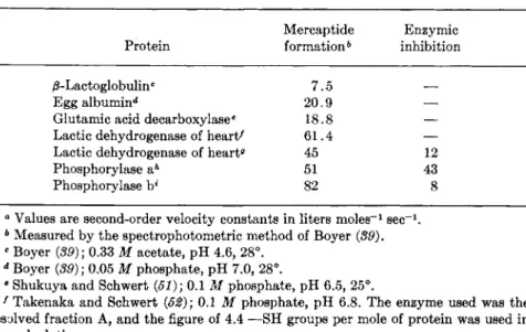
Shukuya and Schwert (51) showed that the reaction between P C M B and glutamic acid decarboxylase follows second-order reaction kinetics, as does that of PCMB and lactic dehydrogenase [Takenaka and Schwert (52)]. The data of Neilands (53) for the latter reaction may also be analyzed to show adherence to second-order kinetics for both the mer
captide formation and the inhibition. Some data on the rates of reaction between P C M B and proteins are tabulated in Table III. Perhaps not too much significance can yet be attached to the fact that the enzymic inhibition, in the few cases where data are available, is slower than the
2 1 . M E R C A P T I D E - F O R M I N G A G E N T S 1 3 1
TABLE III
RATES OF REACTION BETWEEN P C M B AND SOME SULFHYDRYL PROTEINS"
Mercaptide Enzymic Protein formation6 inhibition /3-Lactoglobulinc 7.5
Egg albumin** 20.9 —
Glutamic acid decarboxylase6 18.8 —
Lactic dehydrogenase of heart' 61.4 —
Lactic dehydrogenase of heartff 45 12
Phosphorylase aA 51 43
Phosphorylase b* 82 8
° Values are second-order velocity constants in liters m o l e s- 1 s e c- 1.
6 Measured by the spectrophotometric method of Boyer (89).
e Boyer (89); 0.33 Μ acetate, pH 4.6, 28°.
d Boyer (39); 0.05 Μ phosphate, pH 7.0, 28°.
β Shukuya and Schwert (51); 0.1 Μ phosphate, pH 6.5, 25°.
f Takenaka and Schwert (52); 0.1 Μ phosphate, pH 6.8. The enzyme used was the resolved fraction A, and the figure of 4.4 —SH groups per mole of protein was used in the calculation.
0 Estimates calculated by the reviewer from the data published in graphical form by Neilands (53); 0.1 Μ phosphate, pH 7.0, 25°. The unresolved enzyme was used, which has been shown to contain 3 —SH groups per mole of protein reacting uder these con
ditions (54-56).
A Madsen and Cori (42); 0.02 Μ sodium glycerophosphate, pH 6.7, 21°.
* Madsen and Cori (42). Conditions as in h.
mercaptide formation. Nevertheless, one is tempted to speculate that the mercaptide formation is not directly responsible for the inhibition but that an intermediary step is required. I t is hoped that more such data will become available in the future so that this idea may be either documented or refuted.
B. Methods Employing O r g a n i c Mercurials
1. I N H I B I T O R Y S T U D I E S
The use of P C M B for the inhibition of enzymes was introduced by Hellerman and his co-workers. Methods for synthesizing and handling the compound have been given by Chinard and Hellerman (41). Further useful information is found in the article by Boyer (39). In determining the effect of P C M B on an enzyme or enzyme system, the mercurial should be preincubated with the enzyme for various lengths of time and at
various concentrations before the substrate is added for the determination of enzymic activity. The necessity for varying the concentration and time is dictated by the second-order nature of the reaction with many —SH groups. In the past, several enzymes which have now been shown to be dependent on intact —SH groups for activity were classed as non- sulfhydryl enzymes because the investigator did not allow sufficient time for the reaction. A study of the effect with time has the further advantage that the data may, in some cases, be used to calculate rate constants, which may then be compared with the rate of mercaptide formation. Of course, if the reaction is of the "instantaneous" type, this will soon be apparent and an elaborate study of the effect with time may not be neces
sary or possible.
The use of a single concentration of inhibitor can be criticized not only on the basis of the effect on reaction rate but also because, in order to make sure of demonstrating any possible inhibition, the concentration chosen may be so high that nonspecific interaction may occur. Nonspecific binding of P C M B to phosphorylase began to occur at 2.4 Χ 1 0 ~4 Μ PCMB (48). Benesch et al. (18) had previously demonstrated nonspecific binding.
It is advisable to add the inhibitor before the substrate even when the mercaptide formation is extremely rapid because the presence of substrate may protect the enzyme against inhibition. For example, acetyl phosphate prevents P C M B from reacting with two to three —SH groups of glycer- aldehyde-3-phosphate dehydrogenase in spite of the fact that the mercu
rial should be able to cleave a thio ester bond (57) ,5 This will be discussed further in Section IV, A.
As indicated in Section III, A, the composition of the solution with regard to pH concentration and type of anions can have a considerable effect on the reaction between P C M B and proteins. After suitable condi
tions for time and concentrations have been established for the inhibition of an enzyme by an organic mercurial, it is advisable to try to reactivate the enzyme by adding a thiol such as cysteine or glutathione. Since mer
captide formation is a reversible reaction, the addition of an excess of thiol should regenerate the — S H groups of the enzyme and restore ac
tivity. Failure to achieve this indicates that more than simple mercaptide formation must have occurred. While it has been said that protein denaturation could account for such results, this only raises the question as to why such denaturation should have occurred. If, for example, the
5 Note added in proof: Data published recently indicates that PCMB does not split thio esters at neutral pH. [T. Sanner and A. Pihl, Biochim. Biophys.
Acta 62, 171 (1962).]
21. MERCAPTIDE-FORMING AGENTS 133
—SH groups of the enzyme were concerned directly with substrate activa- tion or binding, it is difficult to see why tying them up as mercaptides should lead to irreversible denaturation. On the other hand, the reversal of inhibition with a thiol, while it can no longer be considered conclusive evidence of specificity or function, does eliminate certain troublesome complications.
The prevention of inhibition by substrate or cofactors has been in- vestigated fairly extensively, but only a few positive results have been obtained (3). Since a test of this relationship requires incubation of inhibitor, substrate, and enzyme together, it is obvious that certain techni- cal difficulties are presented when a single substrate is involved, particu- larly if the reaction with the mercurial is slow. In the latter case it might be possible to determine the effect of substrate on the rate of mercaptide formation, as measured by the spectrophotometric method of Boyer (39).
The effect of substrates can more easily be determined where two are involved, since one can be preincubated with the inhibitor and the en- zyme, and the reaction can be started with the second substrate. Some investigators have determined the effect of the presence of substrate on the titration of enzyme-bound — S H groups by amperometric or spectro- photometric means. Caution must be observed here that the results are not caused by contaminating metals, as discussed in Section II, C.
The failure of a substrate to protect an enzyme against inhibition by a mercurial does not necessarily mean that the — S H groups are not directly concerned with substrate binding because the mercurial will usually have a greater affinity. Where negative results are obtained it might be profita- ble to devise a more sensitive test, such as the effect on the rate of either inhibition or mercaptide formation. Where the binding of substrate or cofactor to enzyme can be detected by spectrophotometric of fluorometric means, the effects of mercurials on these phenomena should be investi- gated. The results of using the latter techniques have been reviewed by Shifrin and Kaplan (58).
This chapter is concerned only with mercaptide-forming agents, but consideration of the effects of quinones, arsenicals, and alkylating agents, treated elsewhere in this volume, in conjunction with the results from mercaptide formation, can be very valuable in differentiating the func- tions of — S H groups. Thus, Strittmatter (59) used both P C M B and N E M to study the — S H groups of microsomal cytochrome reductase. He found that P C M B reacts with three — S H groups per protein molecule, as measured spectrophotometrically, and with a fourth group after denatura- tion with Duponol. Inactivation began during addition of the first equiva- lent of P C M B but was not completed at one equivalent, although the
curve could be extrapolated to 100% inactivation at one equivalent. The presence of D P N H afforded partial protection at low P C M B concentra
tions. N E M reacted quickly with two — S H groups, causing 100% inacti
vation, and more slowly with a third — S H group. In the presence of D P N H , however, N E M reacted quickly with only one —SH group and more slowly with a second, producing a derivative which was fully active.
This derivative was now completely inactivated by one equivalent of PCMB, and D P N H afforded no protection. B y these experiments the four—SH groups [lettered (a)-(d)] have been completely differentiated:
Group (a) reacts quickly with P C M B and N E M and appears to be intimately related to enzymic activity and D P N H binding in particular.
D P N H protects it against reaction with N E M .
Group (b) reacts almost as quickly with N E M as does (a) but some
what more slowly with PCMB, and does not appear to be related to activity.
Group (c) reacts slowly with N E M and is not essential for activity.
Group (d) can only be titrated with P C M B when the enzyme is de
natured.
Sometimes, different types of organic mercurials will yield different results. Halsey (60) found that the first two equivalents of methylmer- curic nitrate caused only a small inhibition of yeast glyceraldehyde- 3-phosphate dehydrogenase and that a total of four equivalents were required for complete inhibition. Two equivalents of P C M B produced complete inhibition.
2. E S T I M A T I O N O P T H I O L S
Mercaptide-forming agents are widely used to measure sulfhydryl groups quantitatively, and organic mercurials are achieving more popu
larity for this purpose. Chinard and Hellerman (41) discussed the meth
ods used up to 1953 for the titration of — S H groups by PCMB. The end point of the titration has been determined with nitroprusside as an outside indicator. Cecil and McPhee (4) suggest that amperometric titration is probably the best method for determining the end point.
Hughes (87) investigated the use of methylmercury iodide with mer- captalbumin as a model thiol. The mercurial in toluene was equilibrated with the protein in an equal volume of buffer, and excess mercurial was titrated with dithizon. Horowitz and Klotz (38) introduced an azomer- curial which was used in much the same manner but which would be determined directly by colorimetry.
Boyer (89) observed that when P C M B reacts to form a mercaptide there is an increase in its absorption spectrum, this increase being maxi-
21. M E R C A P T I D E - F O R M I N G A G E N T S 135 mal at 250 m/x at pH 7.0 and at 255 τημ at pH 4.6. Based on this phenomenon, he has worked out quantitative methods for the titration of — S H groups in proteins. Since the total spectral shift accompanying mercaptide formation varies slightly with different thiols, it is necessary to titrate a known quantity of P C M B with increasing amounts of the thiol until there is no further increase in absorbancy (after suitable corrections have been made for the reactants). Care must be exercised to ensure that none of the reagents used cause spectral changes, as E D T A and iodide ion were found to interfere. The spectral shift was shown, however, to be quite specific for — S H groups, and this fact enables the investigator to demonstrate that an inhibition by P C M B is really caused by mercaptide formation. Careful use of high concentrations of urea have been employed to determine the total — S H content of a protein, with results that agree well with amperometric methods. The Boyer technique has by now been used successfully in many different laboratories. For the enzymologist it offers the considerable advantages of not requiring spe
cialized equipment and of being simple and rapid.
Boyer also showed that the method was applicable to kinetic studies of mercaptide formation, and the results so obtained have been reviewed in Section III, A.
Equilibrium dialysis (18) and the ultracentrifugal separation method (48) have also been used to determine the binding of mercurials to proteins. These methods are more cumbersome and time consuming than most, but they appear to be accurate and apparent dissociation constants may be calculated from the data.
3. E S T I M A T I O N O F S T R U C T U R A L C H A N G E S
Edelhoch et al. (45) may have been the first to introduce physical methods to measure the effect of mercaptide formation on protein struc
ture when they used light scattering to study the dimerization of mer
captalbumin with mercury salts. Light scattering also made it possible to determine the equilibria and kinetics of the reactions involved. Madsen et al. (42, 48, 61) used the analytical ultracentrifuge and light scattering to demonstrate the effects of P C M B on the structure of phosphorylase.
The ultracentrifuge, which has also been used elsewhere (62), makes it possible to demonstrate gross changes only, such as occur on dimerization or dissociation of protein units. Light scattering may make it possible to observe more subtle changes, in addition to kinetic studies. The introduc
tion of the charged P C M B molecule into a protein should result in a change of electrophoretic mobility, but the application of electrophoretic techniques to the phosphorylase problem did not meet with much success.
It might be more useful where a protein has a higher ratio of — S H groups to weight.
Elodi (63), in studying the effect of P C M B on the structure of swine muscle glyceraldehyde-3-phosphate dehydrogenase, was able to show that configurational changes, of a finer detail than had hitherto been found, could be demonstrated by measuring the specific optical rotation [ a ]D 2 0 and the intrinsic viscosity. This is discussed in Section IV, A.
The two methods used by Elodi, as well as optical rotatory dispersion, should prove to be of great value in determining the effects of organic mercurials on the configuration of proteins. N o doubt, as newer techniques of greater sensitivity to slight changes in protein configuration are devel
oped, they will be applied with profit. Those methods which make possible a kinetic analysis of structural changes will probably yield the most significant information.
IV. T W O INDIVIDUAL ENZYMES
Results obtained with several enzymes have been reviewed above in the course of discussing the effects of inhibitors. Because of space, only two more of the many possible choices can be reviewed here. The first is chosen because of the large amount of data which has accumulated about it, and because it appears to fulfill most closely the original and classic concept of a sulfhydryl enzyme. The second was chosen because of the reviewer's personal familiarity with it and because it is representative of a second class of sulfhydryl enzymes in which the — S H groups are involved in the maintenance of protein structure.
A. G l y c e r a l d e h y d e- 3- p h o s p h a t e D e h y d r o g e n a s e
This enzyme has been studied intensively, and the experiments with sulfhydryl reagents are particularly instructive for the variety of both experimental techniques and the results obtained, as well as the manner in which the latter have been fitted into a general reaction mechanism.
The enzyme has been crystallized from yeast with a molecular weight of 122.000. The enzyme crystallized from rabbit muscle contains two moles of D P N which can be removed by charcoal. Its molecular weight was taken as 120,000 in earlier studies, but is now considered to be 137,000 (64, 65). Both enzymes catalyze reaction (10) in which the normal alde
hyde substrate is glyceraldehyde-3-phosphate. Rapkine and his coworkers
21. M E R C A P T I D E - F O R M I N G A G E N T S 137
RCHO + DPN+ + H P 04 2- ^± RCOOPCV" + D P N H + H+ (10) showed that the yeast enzyme is inhibited by iodoacetate (66) and that D P N protected the enzyme against inhibition by certain oxidizing agents (67). Velick (86) found that the addition of P C M P S to the muscle enzyme caused a proportional loss of activity until inactivation was com
plete at 3 moles of P C M P S per mole of enzyme. Ultracentrifugal separa
tion analysis indicated that the combination of inhibitor with enzyme was accompanied by a release of the 2 moles of bound D P N and was complete when three moles of inhibitor had been bound. Both effects were reversed instantaneously upon addition of cysteine. In the case of the yeast enzyme, two equivalents of P C M B were sufficient to produce com
plete inactivation, and the activity was restorable with cysteine. The inhibitor-enzyme complex could be crystallized, and it would not bind D P N . Thus, the evidence would appear to point to participation of —SH groups in the binding of the nucleotide, but Velick pointed out that steric hindrance or configurational changes provide alternative explanations.
More recently, he has presented evidence that conformational changes in the protein do occur upon inhibition (68).
Considerable evidence, both kinetic and chemical in nature, has accu
mulated that an acyl enzyme is an intermediate in Eq. (10) and that an
—SH group on the enzyme provides the protein-bound part of this com
pound. Equation (10) may thus be formulated in the steps (adapted from the schemes of Racker and of Boyer) shown in Scheme I.
Η I
HOCR O—C—R
Ο DPN SH DPN S DPNH S I I
R - C (
+ J L ^ ± J L — J L
+ H+Η Enzyme Enzyme Enzyme
0 = C — R
DPN S „ „ DPN SH / /
ι ο
D P N ^ Η3Ρ Ρ4 ν > , , + R _ C
^DPNH J L ^ 1— L \
Enzyme Enzyme W J :
S C H E M E I
Observations on the relationships between substrate and sulfhydryl rea
gents are among the evidence favoring the participation of — S H groups
as shown in Scheme I. Segal and Boyer (69) showed that glyceraldehyde- 3-phosphate gives almost complete protection against inactivation by iodoacetate. Amperometric iodosobenzoate titration gave 15 — S H groups per mole of enzyme, 5 of which disappeared after treatment with iodo
acetate, but only 3 disappeared when the aldehyde was present along with the iodoacetate. That two — S H groups react with the substrate was corroborated by the later finding (70) that acetyl phosphate blocks the reaction of two —SH groups with PCMB. Spectrophotometric titration with P C M B indicates that 11 groups react rapidly and three more slowly at pH 4.6. Acetyl phosphate decreases the number of titratable groups by two. The acyl-enzyme compounds formed from acetyl phosphate or 1,3-diphosphoglycerate have the properties of thiol esters [Krimsky and Racker ( 7 ί ) ] .
The apparent involvement of two —SH groups in the binding of both nucleotide and substrate is further substantiated by the results of Racker and Krimsky (72), who showed that the absorption at 360 τημ resulting from the binding of D P N to the muscle enzyme can be abolished by 1,3-diphosphoglycerate, acetyl phosphate or acetyl glutathione, as well as by PCMB or iodoacetate. They favor an aldehydolysis of a D P N - sulfhydryl bond with the concomitant formation of a thiol ester during the first stage of the enzymic catalysis.
Elodi (63) has recently presented evidence that the role of —SH groups in the swine muscle enzyme may be more extensive than is suggested above. He showed that the addition of successive increments of P C M B to the enzyme cause proportionate changes in both the specific optical rotation and the intrinsic viscosity. This suggests that pronounced struc
tural changes had occurred, probably involving a considerable unfolding of the α-helical structure. Both properties ceased changing at a total of 14 equivalents of inhibitor. There was no lag in commencing changes with the first increment, suggesting that even the first 2 or 3 —SH groups reacting, which are generally considered to be involved in the enzymic reaction, may also be involved in maintaining structural integrity. It might be interesting to attempt to find out if structural changes occur during the enzymic process. Boyer and Schultz (73) did find that there was a slight, reversible change in optical rotation on removing D P N from the rabbit muscle enzyme.
B. Phosphorylase
The studies on the interaction of P C M B with crystalline muscle phos
phorylase (42, 48, 61) are another example of the application of several
21. MERCAPTIDE-FORMING AGENTS 139 techniques differing widely in principle to ascertain the nature of the reaction and the effect on the protein. Phosphorylase a has a molecular weight of 495,000 (74). Approximately 18 equivalents of P C M B were required for complete inactivation of the enzyme, a figure which agrees well with the amino acid analysis of 9 cystine equivalents (75), as well as with titrations of the — S H groups by the spectrophotometry method of Boyer, and the binding of P C M B as measured by the ultracentrifugal separation technique. Ultracentrifrugal analysis indicated that the in- hibited enzyme had a molecular weight one-quarter that of the native enzyme and that during the course of reactivation by cysteine the re- associating protein passed through a dimeric stage. Enzymic reactivation proceeded more slowly than the return to the original molecular weight.
The kinetics of the reaction were studied by spectrophotometry, enzymic activity, and light scattering. The spectrophotometrie method indicated that the reaction of P C M B with the — S H groups followed second-order kinetics. The enzymic inactivation also followed second-order kinetics but was slightly slower than the mercaptide formation. The light scattering showed that the dissociation of the protein molecule followed first-order kinetics and was very much slower than the other two processes. Thus, the inactivation is not caused by the dissociation.
The reaction of P C M B with phosphorylase was shown to be an "all-or- none" process because partially inhibited enzyme showed two molecular species in the ultracentrifuge, the ratio of monomer to total protein being the same as the ratio of P C M B added to total — S H content. This was substantiated by chemical analysis of the two molecular species sepa- rated by ultracentrifugation; the monomer fraction contained sufficient mercurial to saturate all its — S H groups, whereas the tetramer con- tained only a little mercurial. Thus, whereas the 18 — S H groups of phos- phorylase a have not yet been differentiated by kinetic means, the preceding experiment suggests that the reaction of the first few — S H groups on a protein molecule increases the reactivity of the remainder.
Finally, the binding of P C M B to phosphorylase was measured by the ultracentrifugal separation method. This showed that the first approxi- mately 19 equivalents of mercurial were bound tightly to the protein
(apparent association constant of 106), while a further larger number of equivalents were bound more loosely and, presumably, to groups other than sulfhydryl.
The results obtained with phosphorylase prompt a certain amount of speculation about the sequence of events during inhibition. The first —SH groups of a protein molecule to form mercaptides facilitate the reaction of the remainder, so that all the — S H groups of a molecule form mer-
captides very quickly. The inhibition appears to be slightly slower than the mercaptide formation, although the difference in rate is small enough that it may not be significant. If it is assumed to be significant, then some factor other than mercaptide formation must account for the inhibi
tion. Local structural changes may be the cause. Finally, after inhibition, the protein molecule dissociates. One of the remaining problems, the events occuring immediately upon mercaptide formation which lead to inhibition, might prove amenable to study by a method which is rapid and sensitive to small changes in protein configuration, such as optical rotation.
The experiments with phosphorylase provided decisive evidence that the blocking of — S H groups may lead to pronounced changes in protein structure and, by implication, that —SH groups may be important in the maintenance of a given protein configuration. Similar results have since been found for other proteins, but the nature of the linkages in which the — S H groups might be involved remains unknown and the subject of considerable speculation (8, 4, 76, 77). Mercurials can be of considerable use in detecting those cases where —SH groups may be involved in protein structure, since a slow reaction of a mercurial with
—SH groups appears to be associated with such involvement.
V. SUMMARY
The chemistry of mercaptide formation has been discussed from the standpoint of specificity in comparison to other possible reactions.
Reaction conditions which may affect the application of mercaptide for
mation have been considered. After appraisal of the analytical use of mercaptide-forming agents in estimating the — S H groups of proteins, it was concluded that an amperometric silver titration method and a spec- trophotometric method employing P C M B are the most suitable among those currently available. The use of silver and mercuric ions for inhibi
tory studies offer certain advantages, but care must be exercised that the results obtained are specifically due to mercaptide formation. Organic mercurials, and P C M B in particular, are the most generally useful and specific agents available for inhibitory studies designed to delineate the relationships between — S H groups and the mechanism of enzyme action.
As research continues, an increasing number of phenomena are ob
served to result from mercaptide formation in enzymes. Some of these phenomena and their significance with respect to enzyme action have been discussed briefly. Considerable further data must be assembled, however,
21. MERCAPTIDE-FORMING AGENTS 141 before truly sound deductions can be made about the role of — S H groups in enzymic catalysis and protein structure. Fortunately, the present techniques appear adequate for achieving much of this objective.
REFERENCES
1. E . S. G. Barron, Advances in Enzymol. 11, 201 (1951).
2. R. E . Benesch et al., eds., "Sulfur in Proteins." Academic Press, N e w York, 1959.
3. P. D. Boyer, in "The Enzymes" ( P . D. Boyer, H. Lardy and K. Myrback, eds.), 2nd ed., Vol. I, p. 511. Academic Press, N e w York, 1959.
4. R . Cecil and J. R . McPhee, Advances in Protein Chem. 14, 255 (1959).
5. F . R . N . Gurd and P. E . Wilcox, Advances in Protein Chem. 11, 311 (1956).
6. I. M. Kolthoff, J. Phys. Chem. 35, 2711 (1931).
7. I. M. Klotz, in "The Mechanism of Enzyme Action" (W. D. McElroy and B. Glass, eds.), p. 257. Johns Hopkins Press, Baltimore, Maryland, 1954.
8. W. Stricks and I. M. Kolthoff, J. Am. Chem. Soc. 75, 5673 (1953).
9. W. Stricks, I. M. Kolthoff, and A. Heyndrickx, J. Am. Chem. Soc. 76, 1515 (1954).
10. R . E . Benesch and R . Benesch, J. A. Chem. Soc. 77, 5877 (1955).
11. L. G. Sillen, Acta Chem. Scand. 3, 539 (1949).
12. C. Tanford and J. Epstein, J. Am. Chem. Soc. 76, 2170 (1954).
13. V. M. Ingram, Biochem. J. 59, 653 (1955).
14. B. L. Vallee, T. L. Coombs, and F . L. Hoch, J. Biol. Chem. 235, PC 45 (1960).
15. I. M. Kolthoff and W. E. Harris, Ind. Eng. Chem. Anal. Ed. 18, 161 (1946).
16. R . Benesch and R . E. Benesch, Arch. Biochem. Biophys. 19, 35 (1948).
17. I. M. Kolthoff and W. Stricks, J. Am. Chem. Soc. 72,1952 (1952).
18. R . E. Benesch, H. A. Lardy, and R . Benesch, J. Biol. Chem. 216, 663 (1955).
19. I. M. Kolthoff, W. Stricks, and L. Morren, Anal. Chem. 26, 366 (1954).
20. 0 . Warburg and W. Christian, Biochem. Z. 310, 384 (1942).
21. F. Kubowitz and P. Ott, Biochem. Z. 314, 94 (1943).
22. E . L. Smith, J. R . Kimmel, and D. M. Brown, J. Biol. Chem. 207, 533 (1954).
23. E . L. Smith, J. R . Kimmel, D. M. Brown, and E . O. P. Thompson, J. Biol.
Chem. 215, 67 (1955).
24. W. L. Hughes, Jr., J. Am. Chem. Soc. 69,1836 (1947).
25. M. Jacoby, Biochem. Z. 181, 194 (1927).
26. T. Bersin, Ergeb. Enzymforsch. 4, 68 (1935).
27. L. Hellerman, F. P. Chinard, and V. R . Deitz, J. Biol. Chem. 147, 443 (1943).
28. J. B. Sumner, N . Gralen, and I.-B. Eriksson-Quensel, J. Biol. Chem. 125, 37 (1938).
29. J. F. Ambrose, G. B. Kistiakowski, and A. G. Kridl, J. Am. Chem. Soc.
73, 1232 (1951).
30. W. H. R . Shaw, J. Am. Chem. Soc. 76, 2160 (1954).
31. L. Hellerman, Personal communication; also see Boyer (8).
32. L. Hellerman, Κ. Α. Schellenberg, and Ο. Κ. Reiss, J. Biol. Chem. 233, 1468 (1958).
33. F. L. Hoch, R. G. Martin, W. E. C. Wacker, and B. L. Vallee, Arch. Bio- chem. Biophys. 91, 166 (1960).
34. E. S. G. Barron and S. Levine, Arch. Biochem. Biophys. 41, 175 (1952).
35. J. V a n E y s and N. 0 . Kaplan, Biochim. et Biophys. Acta 23, 574 (1957).
36. S. F. Velick, J. Biol. Chem. 203, 563 (1953).
37. W. L. Hughes, Jr., Cold Spring Harbor Symposia on Quant. Biol. 14, 79 (1950).
38. M. G. Horowitz and I. M. Klotz, Arch. Biochem. Biophys. 63, 77 (1956).
39. P. D. Boyer, J. Am. Chem. Soc. 76, 4331 (1954).
40. J. Harris and L. Hellerman, Federation Proc. 12, 215 (1953).
41. F. P. Chinard and L. Hellerman, Methods Biochem. Anal. 1, 1 (1954).
42. Ν . B. Madsen and C. F . Cori, J. Biol. Chem. 223, 1055 (1956).
43. W. L. Hughes, Jr., in "The Mechanism of Enzyme Action" (W. D. Mc- Elroy and B. Glass, eds.), p. 286. Johns Hopkins Press, Baltimore, Maryland, 1954.
44. F. L. Hoch and B. L. Vallee, Arch. Biochem. Biophys. 91, 7 (1960).
45. H. Edelhoch, E . Katchalski, R. H. Maybury, W. L. Hughes, Jr., and J.
T. Edsall, J. Am. Chem. Soc. 75, 5058 (1953).
46. C. M. Kay and J. T. Edsall, Arch. Biochem. Biophys. 65, 354 (1956).
47. J. T. Edsall, R. H. Maybury, R. B. Simpson, and R. Straessle, J. Am.
Chem. Soc. 76, 3131 (1954).
48. Ν. B. Madsen and F. R. N . Gurd, J. Biol. Chem. 223, 1075 (1956).
49. B. J. Finkle and E . L. Smith, J. Biol. Chem. 230, 669 (1958).
50. A. D. Swenson and P. D. Boyer, J. Am. Chem. Soc. 79, 2174 (1957).
51. R. Shukuya and G. W. Schwert, J. Biol. Chem. 235,1658 (1960).
52. Y. Takenaka and G. W. Schwert, J. Biol. Chem. 223, 157 (1956).
53. J. B. Neilands, J. Biol. Chem. 208, 225 (1954).
54. A. P. Nygaard, Acta Chem. Scand. 9, 1048 (1955).
55. A. P. Nygaard, Acta Chem. Scand. 10, 397 (1956).
56. G. Pfleiderer, D. Jeckel, and Th. Wieland, Arch. Biochem. Biophys. 83, 275 (1959).
57. F. Lynen, E.Reichert, and L. Rueff, Ann. 574, 1 (1951).
58. S. Shifrin and N. O. Kaplan, Advances in Enzymol. 22, 337 (1960).
59. P. Strittmatter, J. Biol. Chem. 234, 2661 (1959).
60. Y. D. Halsey, J. Biol. Chem. 214, 589 (1955).
67. Ν. B. Madsen, J. Biol. Chem. 223, 1067 (1956).
62. P. J. Snodgrass, B. L. Vallee, and F. L. Hoch, J. Biol. Chem. 235, 504 (1960).
63. P. Elodi, Biochim. et Biophys. Acta 40, 272 (1960).
64. W. B. Dandliker and J. B. Fox, J. Biol Chem. 214, 275 (1955).
65. J. B. F o x and W. B. Dandliker, / . Biol. Chem. 218, 53 (1956).
66. L. Rapkine, Biochem. J. 32,1329 (1938).
67. L. Rapkine, S. M. Rapkine, and P. Trpinac, Compt. rend. acad. sci. 209, 253 (1939).
68. S. F. Velick, J. Biol. Chem. 233, 1455 (1958).
69. H. L. Segal and P. D. Boyer, J. Biol. Chem. 204, 265 (1953).