Accepted manuscript
Title: Nitro-oxidative stress correlates with Se tolerance of Astragalus species
Authors:Kolbert Zs, Molnár Á+, Szőllősi R, Feigl G, Erdei L, Ördög A
DOI: 10.1093/pcp/pcy099
Cite as: Kolbert, Z., Molnár, Á., Szőllősi, R., Feigl, G., Erdei, L., & Ördög, A. (2018). Nitro- oxidative stress correlates with Se tolerance of Astragalus species. Plant and Cell Physiology.
This is a PDF file of an unedited manuscript that has been accepted for publication.
1 REGULAR PAPER
2 (2) Environmental and stress responses 3
4 Title: Nitro-oxidative stress correlates with Se tolerance of Astragalus species 5
6 Kolbert Zs1+*, Molnár Á1+, Szőllősi R1, Feigl G1, Erdei L1, Ördög A1 7 1 Department of Plant Biology, University of Szeged, Hungary
8 +these Authors contributed equally to this work 9
10 Running title: Selenate-induced nitro-oxidative stress in Astragalus species 11 * corresponding Author e-mail address: kolzsu@bio.u-szeged.hu
12
13 Number of black and white figures: 2 14 Number of color figures: 7
15 Number of tables: 1
16 Number of Supplementary files: 8
1 ABSTRACT
2 At high concentrations selenium (Se) exerts phytotoxic effects in non-tolerant plant
3 species partly due to the induction of nitro-oxidative stress; however, these processes
4 are not fully understood. In order to get a more accurate view about the involvement of nitro- 5 oxidative processes in plant Se sensitivity, this study aims to characterize and compare Se- 6 triggered changes in reactive oxygen (ROS) and nitrogen species (RNS) metabolism and the 7 consequent protein tyrosine nitration as a marker of nitrosative stress in non-accumulator 8 Astragalus membranaceus and in Se hyperaccumulator Astragalus bisulcatus.
9 The observed parameters (Se accumulation, microelement homeostasis, tissue-level 10 changes in the roots, germination, biomass production, root growth, cell viability) supported 11 that A. membranaceus is Se sensitive while the hyperaccumulator A. bisulcatus tolerates high 12 Se doses. We first revealed that in A. membranaceus, Se sensitivity coincides with the Se- 13 induced disturbance of superoxide metabolism leading to its accumulation. Furthermore, Se 14 increased the production or disturbed the metabolism of RNS (nitric oxide, peroxynitrite, S- 15 nitrosoglutathione) consequently resulting in intensified protein tyrosine nitration in sensitive 16 A. membranaceus. In the (hyper)tolerant and hyperaccumulator A. bisulcatus, Se-induced
17 ROS/RNS accumulation and tyrosine nitration proved to be negligible suggesting that this 18 species is able to prevent Se-induced nitro-oxidative stress.
19 Keywords: Astragalus ssp; nitro-oxidative stress; reactive oxygen species; reactive 20 nitrogen species; selenate
22
23 Abbreviations: CAT, catalase; DAF-FM DA, 4-amino-5-methylamino- 2′,7′- 24 difluorofluorescein diacetate; DHE, Dihydroethidium; DHR, dihydrorhodamine 123; DPI, 25 diphenylene iodonium; EDTA, ethylene-diamine-tetraacetic acid; FDA, fluorescein diacetate;
1 GSNO, S-nitrosoglutathione; GSNOR, S-nitrosoglutathione reductase; H2O2, hydrogen 2 peroxide; MES, morpholine-ethansulphonic acid; NBT, nitroblue tetrazolium; NBT/BCIP,
3 nitroblue tetrazolium/ 5-Bromo-4-chloro-3-indolyl phosphate; NO, nitric oxide; .NO2, nitrogen 4 dioxide radical; N2O3, dinitrogen-trioxide; N2O4, dinitrogen tetroxide; NOX, NADPH oxidase;
5 NR, nitrate reductase; O2.-
, superoxide; OH., hydroxyl radical; ONOO-, peroxynitrite; PODs, 6 peroxidases; RNS, reactive nitrogen species; ROS, reactive oxygen species; SDS-PAGE, 7 sodium dodecyl sulphate-polyacrylamide gel electrophoresis; Se, selenium; SIN-1, 3- 8 morpholino-sydnonimine; SMT, selenocysteine methyltransferase; SNO, S-nitrosothiol; SOD, 9 superoxide dismutase.
1 INTRODUCTION 2
3 Selenium (Se) is a non-metal element which seems to be non-essential for higher plants.
4 Still, its chemical similarity with sulphur (S) results in its uptake and metabolism via S 5 transporters and pathways (Pilon-Smits and Quinn, 2010). Moreover, a few plant species not 6 only take up but accumulate or hyperaccumulate high Se levels in their tissues.
7 The ability of Se hyperaccumulation has been described in 45 plant taxa in six families
8 (White, 2016). The Astragalus (Fabaceae) genus is the most representative since large number 9 of species (25) in the genus have the ability to take up and tolerate high concentrations of 10 selenium (Shrift, 1969). Species like Astragalus bisulcatus grow on seleniferous soils and can
11 accumulate over 1000 μg g−1 DW Se (up to 1% of its dry weight). Hyperaccumulators possess 12 10-100-fold higher endogenous Se content as well as higher Se:S ratio compared to non- 13 accumulators (White et al., 2007). Another distinctive feature of hyperaccumulators is the
14 active sulphate/selenate assimilation which is suggested by the dominance of organic Se forms 15 (gamma-glutamyl-methyl-selenocysteine) in their tissues. Hyperaccumulators can be 16 characterized by notable root-to-shoot Se translocation (Mehdawi and Pilon-Smits, 2012).
17 Species like A. bisulcatus are able to sequester Se in their epidermis and trichomes, which may 18 have a role both in defence and in Se stress mitigation (Freeman et al., 2006). The mechanism 19 responsible for Se hyperaccumulation is the constitutive expression of several SULTR 20 transporters, which contributes to the preferential uptake of selenate over sulphate (Cabannes
21 et al., 2011). Also, the expression of certain enzymes involved in selenate/sulphate assimilation
22 is enhanced in hyperaccumulators resulting in greater inorganic-organic conversion (Freeman 23 et al., 2010). Moreover, hyperaccumulators express selenocysteine methyltransferase (SMT) 24 which is responsible for the conversion of toxic selenocysteine to methyl-selenocysteine (Sors
1 et al., 2009). Selenium tolerance is also typical for hyperaccumulators; however, the molecular
2 mechanism of this ability is only partly understood.
3 High tissue concentrations of inorganic selenium forms can induce the production of 4 reactive oxygen species (ROS) such as superoxide (O2.-
), hydrogen peroxide (H2O2), hydroxyl 5 radical (OH.) leading to oxidative stress (Van Hoewyk, 2013). The amount of the generated 6 ROS and consequently the redox homeostasis is precisely controlled by antioxidant 7 mechanisms. Beyond the enzymatic components like superoxide dismutase (SOD), catalase
8 (CAT) and peroxidases (PODs), non-enzymatic antioxidants such as ascorbate and glutathione 9 (GSH) play crucial role in the defence against oxidative damage (Das and Roychoudhury,
10 2014). For Se-induced ROS accumulation, GSH and its depletion seems to be responsible (Van 11 Hoewyk, 2013). According to previous data, hyperaccumulators prefer to produce organic Se
12 forms presumably in order to avoid oxidative stress (Freeman et al., 2006, Van Hoewyk, 2013).
13 Besides ROS, reactive nitrogen species (RNS) are also formed as the effect of
14 environmental stresses like Se exposure (reviewed by Kolbert et al., 2016). This group of nitric 15 oxide (NO)-related molecules consists of peroxynitrite (ONOO-), S-nitrosoglutathione 16 (GSNO), dinitrogen trioxide (N2O3), dinitrogen tetroxide (N2O4), nitrogen dioxide radical
17 (.NO2) (Corpas et al., 2007). The overproduction of RNS leads to nitrosative stress during which 18 one of the principle mechanism is the nitration of tyrosine residues in certain proteins yielding 19 3-nitrotyrosine (Corpas et al., 2013a). This modification causes structural and functional 20 changes in the affected proteins. In most published cases, tyrosine nitration results in activity 21 loss of the target plant proteins (Kolbert et al., 2017) or it can negatively affect signal 22 transduction through the prevention of tyrosine phosphorylation (Galetskiy et al., 2011).
23 Selenium-induced increase in protein tyrosine nitration and in oxidative parameters (ROS
24 levels, lipid peroxidation, antioxidants) has been revealed in the leaves of non-accumulator pea 25 (Lehotai et al., 2016). Also, the relationship between the toxicity of selenium forms and protein
1 tyrosine nitration has been evaluated in non-accumulator Arabidopsis thaliana and secondary 2 accumulator Brassica juncea (Molnár et al., 2018ab) but there is no knowledge about RNS 3 metabolism and protein nitration in Se hyperaccumulator plants such as A. bisulcatus. Another 4 species in the Astragalus genus is Astragalus membranaceus, which is considered to be
5 pharmacologically relevant. The root of this Astragalus species has been used in Chinese 6 medicine for thousands of years because of its general strengthening effect. Based on the 7 literature, in modern medicine it can provide perspective for the prevention and therapy of 8 cerebrovascular, cardiovascular, neurodegenerative and liver diseases (Yang et al., 2013).
9 Despite the significance of A. membranaceus, we know little about its Se accumulation and 10 tolerance as well as about reactive species metabolism and nitrosative stress.
11 Therefore, this comparative study aims to explore the possible differences in selenium- 12 modified ROS and RNS metabolism and the consequent protein tyrosine nitration using the 13 hyperaccumulator Astragalus bisulcatus and Astragalus membranaceus as another species in 14 the same genus. The better understanding of tolerance mechanisms of Se hyperaccumulator 15 plant species is of particular significance in phytoremediation (Gupta and Gupta, 2017) and in 16 biofortification (Wu et al., 2015) as well as in ecological (Schiavon and Pilon-Smits, 2017) 17 point of view. Furthermore, examination of Se accumulation and tolerance of the medicinal 18 herb A. membranaceus can have importance in human health aspect.
1 RESULTS
2 Selenium uptake, accumulation and microelement imbalance 3
4 Selenate-induced selenium accumulation showed differences in the organs of Astragalus
5 species (Fig 1). In the root tissues of A. membranaceus, Se concentration significantly enhanced 6 as the effect of increasing exogenous selenate supplementation (Fig 1A). In case of A.
7 membranaceus cotyledons, Se accumulation was not concentration-dependent and proved to be
8 lower compared to the root (Fig 1B). The Se content measured in cotyledons of 50 or 100 µM 9 selenate-treated A. membranaceus, did not reach the endogenous Se content of the control A.
10 bisulcatus. The root of the hyperaccumulator A. bisulcatus showed moderate Se accumulation
11 (Fig 1A), while in the cotyledons a remarkable, concentration-dependent increase of Se content 12 was observed (Fig 1B). In case of 100 µM selenate supplementation, the accumulated Se 13 exceeded 1700 µg g-1 DW concentration in the cotyledons of A. bisulcatus. It has to be 14 mentioned that significant difference was observed in the endogenous Se contents of untreated
15 Astragalus plants. Cotyledons of A. bisulcatus contained 200-fold more selenium than the same
16 organs of A. membranaceus (Fig 1B). Regarding the root, similar but much smaller (16-fold) 17 difference was revealed (Fig 1A).
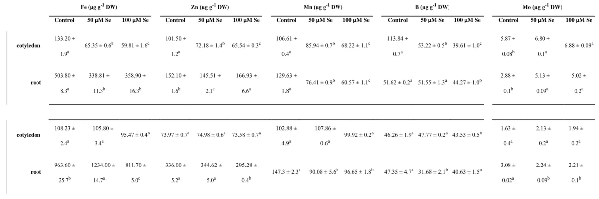
18 Selenate exposure led to the modification of microelement concentrations in the organs 19 of Astragalus species (Table 1). Of the examined microelements, the contents of the essential 20 Fe, Zn, Mn and B showed notable reduction especially in the cotyledons of A. membranaceus.
21 However, the concentration of the above mentioned elements were not affected at all or just 22 slightly changed by selenate in A. bisulcatus cotyledons. Regarding the root system, more 23 serious effects were observed in case of A. membranaceus compared to A. bisulcatus. E.g. Fe 24 concentration decreased by 30% in A. membranaceus but only by 15% in A. bisulcatus.
25 Contrary to the other microelements, Mo concentrations in A. membranaceus organs
1 significantly increased as the effect of Se treatments. In A. bisulcatus, the concentrations of Mo 2 were decreased or were not modified by selenium (Table 1).
3
4 Growth and Se tolerance of Astragalus species
5 Selenium tolerance of Astragalus species was evaluated by germination capacity, 6 biomass production, root meristem viability and root elongation on selenate-supplemented 7 medium.
8 Both species showed ~85% germination under control conditions and this good
9 germination capability was retained by A. bisulcatus on 50- and 100 µM selenate-treated plates 10 (Fig 2A). In contrast, the presence of selenate significantly and concentration-dependently
11 reduced the germination percentage of A. membranaceus. In case of 100 µM Se treatment, 55%
12 of A. membranaceus seeds placed on the medium were germinated, while A. bisulcatus showed 13 better (~70%) germination performance.
14 With regard to biomass production, 14-days-old, untreated individuals of the species 15 possessed similar shoot weight (Fig 2B). Although, the root fresh weight of control A.
16 membranaceus was significantly smaller (Fig 2C) and the phenotype of the root system notably
17 differed from that of A. bisulcatus (Fig 2D). Both concentrations of exogenous Se (50 and 100 18 µM) negatively affected shoot (40 and 46% reduction, respectively) and root growth (57 and 19 75% reduction, respectively) of A. membranaceus (Fig 2BC) and a brown discoloration was 20 visible on the root surface of Se-treated plants (Fig 2D). In contrast, A. bisulcatus showed
21 significantly slighter growth inhibition, since the root biomass was affected only by the highest 22 Se dose (30% reduction) and none of the treatments inhibited shoot growth (Fig 2 BCD).
23 Selenium tolerance correlates with the capability of maintaining primary root (PR)
24 elongation, therefore Se tolerance index can be calculated from PR length data (Tamaoki et al., 25 2008). Compared to the 100% tolerance of the untreated plants (indicated by dashed line in Fig
1 3A), 50 or 100 µM selenate resulted in 35 or 25% tolerance index of A. membranaceus,
2 respectively (Fig 3A). However, A. bisulcatus was able to maintain its root growth and Se even 3 slightly increased elongation resulting in tolerance indexes around or above 100%.
4 Furthermore, we examined the Se tolerance of the species by evaluating viability of the root 5 meristem cells using fluorescein diacetate staining (Fig 3BC). As expected from the previous 6 data, the meristem cells of A. membranaceus showed 50 or 85% viability loss as the effect of
7 50 or 100 µM selenate exposure, respectively. Even though root elongation of A. bisulcatus was 8 negatively affected by none of the applied Se doses (Fig 3A), root meristem cells suffered 50%
9 viability loss as the effect of the highest Se concentration (Fig 3BC). We acknowledge that the 10 application of plant tissues with highly reduced viability might limit the reliability of the data.
11 At the same time, the choice of the 14 days-long treatment period proved to be necessary for 12 the appearance of the effect, as well as for the emergence of tolerance in this comparative 13 Astragalus system (Suppl Fig 1).
14
15 Se-induced tissue-level changes in the roots
16 To evaluate the Se-induced tissue-level changes in the root structure of both Astragalus 17 species, we measured the diameter of the root, the thickness of the cortex and the diameter of 18 the vascular cylinder (stele). Both untreated and Se-treated A. membranaceus plants had thick
19 roots and Se application did not significantly affect root diameter (F= 1.25, p= 0.29). In case of 20 control and 50 µM Se-treated plants, A. membranaceus had nearly twice as thick roots as A.
21 bisulcatus (Fig 4A). When 100 µM Se was added to the media, the roots of A. bisulcatus
22 exhibited remarkable thickening which value was similar to that of A. membranaceus. This 23 tendency was also confirmed by analysis of correlation (r= 0.82, p< 0.001). Similarly, the 24 sensitive A. membranaceus had significantly thicker root cortex than the Se-hyperaccumulator 25 A. bisulcatus in both control and 50 µM Se-treated plants, but it was almost the same in the
1 roots of 100 µM Se-treated plants of both species (Fig 4B). Increasing Se levels significantly
2 enhanced the thickness of the cortex in the case of A. bisulcatus (F= 403.88, p< 0.001; r= 0.88, 3 p< 0.001), while remarkable increase was found only in the root cortex of 50 µM Se-treated A.
4 membranaceus (F= 33.88; p< 0.001; r= 0.34, p<0.001). There was a remarkable increase of
5 stele diameter in A. membranaceus roots exposed to 50 µM Se, while it significantly decreased 6 compared to control after 100 µM Se application. The size of the stele in the roots of A.
7 bisulcatus was only affected by the highest Se stress (Fig 4C). The stele of control and 50 µM
8 Se-treated A. membranaceus roots was at least twice thicker than that of A. bisulcatus. Selenium 9 stress-induced deposition of callose was investigated in aniline blue (AB)-stained root sections 10 taken from the mature zone. Significantly higher fluorescence was found in Se-treated roots of
11 A. membranaceus compared to control, while it diminished after Se application in A. bisulcatus
12 (Fig 4D). Lignin and suberin deposition was visualized using Auramine O staining in the root 13 sections. An intense fluorescence was found in the stele in control roots of both species due to 14 the xylem vessels (Fig 4E). In the Se-treated roots of A. membranaceus a slight fluorescence 15 appeared on the surface (exodermis) of the roots. This staining exhibited both the endodermis 16 and the exodermis in the roots of Se-treated A. bisulcatus.
17
18 Se-induced changes in ROS and RNS metabolism in root and shoot tissues
19 The ROS and RNS-inducing effects of Se were compared in the organs of
20 Astragalus species (Fig 5) in order to reveal the possible link between Se tolerance or sensitivity 21 and Se-induced oxidative and nitrosative (together nitro-oxidative) stress.
10
1 In root tips of A. membranaceus, both Se concentrations increased superoxide levels, 2 although the highest and significant superoxide production was observed in case of 50 µM Se
3 resulting in 170% increase (Fig 5AB). In the root tips of tolerant A. bisulcatus, selenate had no 4 effect on superoxide levels (Fig 5 AB). In intact cotyledons, superoxide levels were examined 5 qualitatively by NBT staining (Fig 5C). In case of 50 or 100 µM Se-treated A. membranaceus 6 plants, the intense presence of blue colorization indicated superoxide production. In A.
7 bisulcatus, slightly intensified blue staining was detected only as the effect of 100 µM Se
8 treatment (Fig 5C). In order to reveal the mechanism of the different superoxide-response of 9 the species, we examined the metabolism of this reactive intermediate. The superoxide- 10 generating NADPH oxidase (NOX) isoenzymes were separated by native-PAGE and a protein 11 band being strongly present in all samples was determined (Fig 5D “main band”). In the 12 cotyledons of A. bisulcatus one, while in A. membranaceus four additional putative NOX
13 isoenzymes were detected. As the effect of Se, only slight changes occurred in NOX isoenzyme 14 activities especially in A. bisulcatus, while more protein bands showed increased activity in A.
15 membranaceus cotyledons (Fig 5D, Suppl Fig 3). In the roots of both species, the activity of
16 the main NOX protein band was less pronounced; although Se induced its activity in A.
17 bisulcatus roots. In addition to the main protein band, four other isoenzymes were detected in
18 A. bisulcatus roots, three of which showed induction as the effect of selenate exposure (Fig 5D,
19 Suppl Fig 3). In case of A. membranaceus roots, Se reduced the activity of the main NOX band, 20 which seemed to be substituted by the appearance and strong activation of additional, putative 21 NOX isoenzymes (indicated by asterisks in Fig 5D, Suppl Fig 3).
22 Both concentrations of selenate caused notable (~30% and 38%) induction of superoxide- 23 eliminating SOD enzymes in A. membranaceus roots, while the effect of selenate in the root 24 system of A. bisulcatus proved to be much slighter (~10%, Fig 5E). Regarding the cotyledons,
1 selenate exposure resulted in SOD activation only in A. membranaceus and the effect proved 2 to be slighter compared to the root (~15%, Fig 5F).
3 We separated SOD isoforms by native-PAGE, and differences were observed between the 4 species and also between the organs (Fig 5E). In both organs of A. bisulcatus, four activity
5 bands (MnSODI, FeSOD I, FeSOD II, Cu/Zn SOD) were identified, while in A. membranaceus 6 cotyledons six bands were detected (MnSODII, FeSOD I, FeSOD II, Cu/Zn SOD I, Cu/Zn SOD 7 II, Cu/Zn SOD III). Moreover, in the root system of A. membranaceus only three SOD activity 8 bands (MnSODII, FeSODI, FeSODII) were observed. Quantification showed, that selenate at 9 the largest applied concentration exerted slight effect on SOD isoenzymes in A. bisulcatus 10 cotyledons (Suppl Fig 4). In contrast, five SOD isoforms of six showed intensified activity as 11 the effect of 50 µM Se in cotyledons of A. membranaceus. Regarding to the root system, both 12 applied Se treatments induced the activity of Mn, Fe, Cu/Zn SODs in A. bisulcatus, but these 13 inductions were much intense in A. membranaceus (Suppl Fig 4).
14 Similar to superoxide, nitric oxide formation significantly enhanced as the effect of 50 µM 15 Se in the root of sensitive A. membranaceus (Fig 6 AB). Regarding peroxynitrite, 50 µM Se 16 resulted in its accumulation, but the highest Se dose decreased its level in the root tips of the 17 sensitive species (Fig 6 EF). Interestingly, none of the applied selenium treatments had any 18 observable effect on the examined RNS levels in A. bisulcatus root tips (Fig 6 AB and EF).
19 Unlike the roots, both species showed NO accumulation in their cotyledons as the effect of 20 50 µM Se (Fig 6 CD). Both Se concentrations triggered significant peroxynitrite generation in 21 the cotyledons of A. membranaceus, while in the tolerant species only slight, non-significant 22 changes were observed (Fig 6 GH). Selenium-induced alterations in GSNO levels were also 23 determined in the root and shoot tissues of the species (Fig 6 I-L). Under control conditions, 24 significantly higher GSNO content was determined in both organs of A. bisulcatus compared 25 to A. membranaceus. Selenate treatments caused significant reduction in GSNO levels of both
1 A. bisulcatus organs. Similar Se-induced diminution of GSNO content was found in A.
2 membranaceus roots (Fig 6 IJ); however, in the cotyledons Se exposure led to the significant
3 and concentration-dependent increase of GSNO levels (Fig 6 KL). Significantly increased 4 fluorescence was detected in GSNO pre-treated sections, which served as positive controls, 5 while light-inactivated GSNO did not result in fluorescence increase (Suppl Fig 7). In their 6 cotyledons, both species showed relatively high S-nitrosoglutathione reductase (GSNOR)
7 activity compared to the root system during control conditions (Fig 6 M, Suppl Fig 6). Selenate 8 exerted inhibitory effect on GSNOR activity in A. bisulcatus cotyledons, while notably induced 9 it in the cotyledons of 50 µM selenate-treated A. membranaceus. As for the control root system, 10 A. bisulcatus showed higher GSNOR activity than A. membranaceus where the activity was
11 barely detectable (Fig 6M, Suppl Fig 6). In case of A. bisulcatus, selenate exerted concentration- 12 dependent reducing effect on GSNOR activity. As opposed to this, selenate did not modify the 13 enzyme activity in the root of A. membranaceus (Fig 6M, Suppl Fig 6).
14
15 Selenium-induced protein tyrosine nitration
16 Protein tyrosine nitration as a consequence of RNS accumulation was investigated by both 17 immunofluorescence (Fig 7) and western blot analysis (Fig 8). In cross sections of A.
18 membranaceus primary roots, immunofluorescent signal related to 3-nitrotyrosine was
19 observable mainly in endodermal cell layer and within the central cylinder (Fig 7B). Selenium 20 exposure led to the significant increase in 3-nitrotyrosine-dependent fluorescent signal in all 21 tissues of the root (Fig 7A), but this elevation was the most pronounced in the central cylinder
22 (Fig 7B). Under control conditions, 3-nitrotyrosine located mainly in the endodermal cell layer 23 of A. bisulcatus roots (Fig 7B). Milder Se treatment caused a slight increase of the fluorescence 24 in the endodermis and the most serious Se exposure induced 3-nitrotyrosine accumulation in 25 all tissues of the primary root, although this increase was smaller than in A. membranaceus
1 roots (Fig 7A). In cotyledons, Astragalus species showed differences in physiological 3-
2 nitrotyrosine levels, since A. bisulcatus showed higher 3-nitrotyrosine-related fluorescence (Fig 3 7C). Moreover, high levels of 3-nitrotyrosine were found to be located in cotyledon veins (Fig
4 7D). Both selenate treatments significantly decreased the 3-nitrotyrosine content of A.
5 bisulcatus cotyledons but in case of A. membranaceus, 100 µM selenate induced 3- 6 nitrotyrosine formation (Fig 7C). As positive and negative controls, sections were treated with 7 SIN-1 and enhanced fluorescence intensity was detected while urate pre-treatment remarkably 8 mitigated 3-nitrotyrosine-dependent fluorescence (Suppl Fig 7).
9 In whole protein extract, tyrosine nitration was determined by western blot analysis (Fig 10 8). In A. membranaceus cotyledons, selenate intensified tyrosine nitration of five protein bands 11 (~27, 22, 17, 12, 10 KDa, indicated by grey arrows) but newly nitrated protein band could not 12 be observed. In cotyledons of A. bisulcatus, both Se treatments resulted in the appearance of a
13 highly nitrated protein band (with high molecular weight, indicated by black arrows) but Se did 14 not cause any other nitration-related change in the proteome. In A. bisulcatus roots, Se did not 15 intensify protein tyrosine nitration, even caused decrease in three protein bands (~75, 12, 10 16 KDa). In contrast, the Se-sensitive Astragalus species showed several protein bands which 17 immunopositivity towards anti-3-nitrotyrosine showed Se-dependent appearance.
1 DISCUSSION
2 Both species were able to take up selenate from the external media (Fig 1). Even though 3 A. membranaceus accumulated large amount of Se in its root, the root-to-shoot Se translocation
4 proved to be slight. In contrast, in A. bisulcatus cotyledons, more than 7-fold Se concentrations 5 were measured compared to A. membranaceus indicating a high rate of Se translocation. Indeed, 6 the root-to-shoot Se ratio was 3.8 in A. bisulcatus plants grown on 100 µM selenate suggesting 7 that it is a hyperacumulator species (Freeman et al., 2010). Furthermore, the relative high 8 endogenous Se content in the organs of control A. bisulcatus indicates its hyperaccumulator
9 nature. Also the amount of the accumulated Se (~1800 µg g-1 DW in the cotyledons of 100 µM 10 selenate-exposed plants) supports hyperaccumulation capability of A. bisulcatus (Mehdawi and 11 Pilon-Smits, 2012). In addition to Se, exogenous selenate affected the concentrations of 12 essential microelements like Fe, Zn, Mn and B (Table 1) especially in A. membranaceus
13 inhibiting their absorption and consequently causing disturbances in their homeostasis. Similar 14 antagonism between Se and macro- or microelements has earlier been described by others
15 (Pazurkiewicz-Kocot et al., 2003; Zembala et al., 2010; Filek et al., 2010). Reduced availability 16 of essential microelements may worsen growth and physical condition of the plant. Boron is 17 needed to maintain cell wall integrity, while Zn protects membrane lipids and proteins and 18 together with Mn, Cu and Fe is the metal component of SOD antioxidant enzymes (Cakmak, 19 2000). In case of the Se hyperaccumulator A. bisulcatus the microelement homeostasis seems 20 to be more stable, since Se did not cause disturbance in it which may contribute to the better 21 tolerance of this species.
22 Selenium negatively affected the germination capability and the biomass production of 23 young A. membranaceus, but the germination and growth of A. bisulcatus proved to be
24 insensitive to selenium (Fig 2). Although, root elongation concentration-dependently decreased 25 as the effect of elevating Se concentrations suggesting the higher sensitivity of the root system
1 to Se compared to the aerial plant parts (Lehotai et al., 2016). Because of the Se concentration- 2 dependent response of elongation, root growth can be used as an indicator of selenium tolerance 3 (Tamaoki et al., 2008; Molnár et al., 2018a). The hyperaccumulator A. bisulcatus was able to 4 maintain its root growth on Se-containing medium (Fig 3A) even though meristem cells 5 suffered certain degree viability loss (Fig 3BC). The reduced root elongation (Fig 3A) and 6 meristem viability (Fig 3BC) of A. membranaceus indicates its sensitivity to Se. Beyond the
7 viability of the root apical meristem, in the background of Se-inhibited organ development, the 8 disturbances of hormone homeostasis or unfavourable alterations in primary metabolism can 9 also be determined (reviewed Kolbert et al., 2016). Based on the observed parameters 10 (germination, biomass production, root elongation, cell viability), young A. membranaceus 11 proved to be Se sensitive, while the hyperaccumulator A. bisulcatus showed remarkable Se 12 tolerance which supports the previously described connection between Se hyperaccumulation
13 and (hyper)tolerance (Mehdawi and Pilon-Smits, 2012). The main reason for Se tolerance of A.
14 bisulcatus is that this species expresses SMT enzyme which prevent toxic seleno-amino acid
15 formation (Neuhierl and Bock, 1996). Considering the high shoot Se accumulation (Fig 1B), it 16 can be assumed that the notable Se tolerance of A. bisulcatus is due to detoxification and not 17 exclusion.
18 We observed selenium-induced alterations in root structure of both Astragalus species.
19 Thicker roots of control and 50 µM Se-treated sensitive A. membranaceus compared to A.
20 bisulcatus were probably due to the thicker cortex (Fig 4AB). The increment of the root
21 diameter, including the thickening of the cortex is common in heavy metal stressed plant roots 22 (Arduini et al., 1995; Maksimović et al., 2007; Potters et al., 2007). The hyperaccumulator 23 species, A. bisulcatus showed more intense Se-induced root thickening than A. membranaceus 24 (Fig 4 ABC), which is in agreement with the results of Li et al. (2009) where in the 25 hyperaccumulating ecotype of Sedum alfredii, lead/zinc-triggered increment in root diameter
1 and other root morphological parameters was observed. The deposition of callose seems to be 2 a good marker of stress induced cell wall alterations. It was formerly found that copper can 3 induce callose formation in onion epidermal cells and in the root tips of Brassica species 4 (Kartusch 2003; Feigl et al., 2013). In our study, the sensitive Astragalus species showed both
5 Se-triggered callose accumulation (Fig 4D) and exodermal suberin lamellae deposition (Fig 4E, 6 Dalla Vecchia et al., 1999; Rahoui et al., 2017) which together may serve as an extracellular
7 barrier limiting water and mineral uptake. This may result in Se exclusion and at the same time 8 the inhibition of growth. In case of A. bisulcatus, not only the exodermis but also the endodermis 9 exhibited the presence of suberin (Fig 4E). Since exodermal suberin deposition occurs earlier
10 in time followed by the appearance of endodermal suberin as the effect of metal stress (Vaculík 11 et al., 2012), we can conclude that in case of A. membranaceus, the delayed formation of Se-
12 induced endodermal suberin lamellae formation is associated with Se sensitivity. Moreover, the 13 development of apoplastic barriers (exodermal and endodermal) can be considered as an 14 adaptive trait (Vaculík et al., 2012).
15 For the toxic effect of Se the accumulation of ROS and the consequent oxidative stress
16 is partly responsible (Van Hoewyk, 2013). The accumulation of the rapidly generating, harmful 17 ROS, superoxide anion (Fig 5 ABC) as well as the induction of SOD activity (Fig 5 EF) suggest 18 Se-triggered oxidative stress in A. membranaceus organs while no sign of serious oxidative 19 damage was observed in A. bisulcatus. The expression of superoxide generating NOX 20 isoenzymes showed species-specificity in A. membranaceus roots, and newly expressed NOX 21 isoenzymes were observed as the effect of selenate (Fig 5D). Regarding SOD isoenzymes, A.
22 membranaceus cotyledons express more Cu/Zn SODs than A. bisulcatus and selenate
23 remarkably increased the activity of most of the isoenzymes (Fig 5G). Selenium-triggered 24 superoxide accumulation has been observed in the non-accumulator Stanleya albescens and 25 Arabidopsis thaliana and in secondary accumulators like Brassica napus, Brassica rapa and
1 Brassica juncea (Freeman et al., 2010, Tamaoki et al., 2008, Dimkovikj and Van Hoewyk,
2 2014; Chen et al., 2014; Molnár et al., 2018ab). In the hyperaccumulator species Stanleya 3 pinnata, elevated levels of ROS scavenging compounds (ascorbate and glutathione) were
4 observed which are involved in the prevention of selenium-induced oxidative stress (Freeman 5 et al., 2010). In our study, A. bisulcatus showed moderately higher SOD activities (especially
6 Cu/Zn SODs) in the roots compared to A. membranaceus (Fig 5F) which may contribute to 7 endurance against Se-induced oxidative stress. At the same time, Se hyperaccumulators are 8 known to accumulate organic selenium forms (mainly methyl-seleno-cysteine) instead of the 9 oxidative stress-inducing inorganic Se compounds which may be a relevant protection 10 mechanism against oxidative stress (Schiavon and Pilon-Smits, 2017).
11 Additionally, Se exposure has been earlier shown to disturb the metabolism of RNS.
12 Milder selenate dose triggered NO production mainly in the non-accumulator species (Fig 6 A- 13 D) similarly to selenite-exposed Pisum sativum (Lehotai et al., 2016) or selenate-treated 14 secondary accumulator Brassica rapa (Chen et al., 2014). Based on the results of Rios et al.
15 (2010) it is conceivable that selenate induces nitrate reductase (NR) which is the main 16 enzymatic NO source in the root system and is also involved in NO production in the aerial 17 plant parts (Zhang et al., 2011). The effect of selenium on NR activity can be direct or indirect 18 since Se-induced S-deficiency may increase Mo content thus inducing NR (Shinmachi et al., 19 2010, Yu et al., 2010). In our experiments, significantly higher Mo concentrations were 20 measured in both organs of selenate-treated A. membranaceus (Table 1) which can be
21 connected to the elevated NO production. Peroxynitrite can be formed in vivo in the fast reaction 22 between superoxide radical and NO (Kissner et al., 1997) thus their accumulation may predict 23 and explain Se-induced ONOO- generation. The concentration of this strong oxidative and 24 nitrosative agent could reflect overall stress severity (Arasimowicz-Jelonek and Floryszak- 25 Wieczorek, 2011) therefore we can suspect that A. membranaceus suffers more severe Se-
1 triggered nitro-oxidative stress compared to A. bisulcatus. However, as the effect of the highest 2 Se dose in A. membranaceus root, peroxynitrite level decreases (Fig 6 E) due to the possible
3 activation of scavenging mechanisms. GSNO is a mobile NO storage in plants being responsible 4 for protein S-nitrosylation. The spontaneous decomposition of GSNO leads to NO production
5 while it is enzymatically reduced by GSNOR or it can catalyse the transnitrosylation of protein 6 thiols leading to its decomposition (Lindermayr, 2018; Begara-Morales et al., 2018). Both 7 species responded to the presence of selenate by decreasing the endogenous GSNO reservoir 8 of their roots (Fig 6 I), however this resulted in NO accumulation only in A. membranaceus 9 (Fig 6 A). Presumably, in A. bisulcatus the originally high GSNO content participated in 10 transnitrosylation reactions with cysteine thiols in proteins leading to S-nitrosothiol (SNO) 11 formation and GSNOR-catalysed reduction is not involved in GSNO metabolism under Se 12 stress. In the cotyledon of A. bisulcatus, the level of GSNO decreased (Fig 6 KL) possibly due 13 to spontaneous decomposition yielding NO but not to GSNOR activity.
14 Similar to other species (reviewed by Corpas et al., 2013a), both A. membranaceus and 15 A. bisulcatus can be characterized by a certain physiological nitropoteome which means that a
16 part of their protein pool is nitrated even at control state. Both the Se-induced increase in 17 fluorescence intensity (Fig 7) and the presence of several newly nitrated protein bands (Fig 8) 18 indicated more intense protein tyrosine nitration in the organs of A. membranaceus compared 19 to the hyperaccumulator A. bisulcatus. Moreover, both immunofluorescence and western blot 20 results showed that the tolerant species possesses large physiological nitroproteome as well as 21 large mobile NO storage (GSNO) with which it is able to buffer NO radical content. The 22 selenium-induced GSNO and 3-nitro-tyrosine decompositions without the accumulation of the
23 reactive .NO may contribute to tolerance against nitro-oxidative stress in A. bisulcatus. The Se- 24 triggered decrease in the amount of 3-nitrotyrosine may be conceivable via proteasomal 25 degradation (Castillo et al., 2015).
1 Our experiments examined the sensitivity of young non-accumulator A. membranaceus 2 and hyperaccumulator A. bisulcatus to selenium in connection to secondary oxidative and 3 nitrosative processes and the obtained results are summarized in Fig 9. As expected, the 4 observed parameters (Se accumulation, microelement homeostasis, tissue-level changes in the 5 roots, germination, biomass production, root growth, cell viability) indicated that A.
6 membranaceus is Se sensitive while A. bisulcatus tolerates the presence of high selenium doses.
7 We first revealed that in A. membranaceus, Se sensitivity coincides with the Se-induced 8 disturbance of superoxide metabolism involving NOXs and SODs leading to superoxide 9 accumulation. Furthermore, this study points out for the first time that Se induced the 10 production or disturbed the metabolism of RNS (NO, ONOO-, GSNO) consequently resulting
11 in intensified protein tyrosine nitration in the sensitive A. membranaceus. In the (hyper)tolerant 12 and hyperaccumulator A. bisulcatus, Se decreased large GSNO content and tyrosine
13 nitroproteome without the accumulation of NO radical resulting in the lack of tyrosine nitration.
14 These suggest that this species is able to prevent Se-induced nitro-oxidative stress to which 15 enhanced ROS/RNS scavenging capability may also contribute. Given that the elevated levels 16 of other elements (e.g. zinc, arsenic, cadmium) have been reported to induce protein nitration 17 and cause similar disturbances in ROS and RNS metabolism like selenium (Feigl et al., 2015;
18 Feigl et al., 2016; Letterier et al., 2012; Liu et al., 2018), excess selenium-induced nitro- 19 oxidative stress can be considered rather a general than a Se-specific phenomenon. Future
20 research should focus on the evaluation of the antioxidative system in order to get more accurate 21 view about nitro-oxidative processes in relation to Se tolerance.
1 MATERIALS & METHODS
2 Plant material and growing conditions
3 Astragalus bisulcatus (Hook.) A. Gray seeds were obtained from B&T World Seeds
4 (Aigues-Vives, France) and Astragalus membranaceus (Fisch.) Bunge seeds were provided by
5 Professor Aaron Chang (Kaohsiung Medical University, Graduate Institute of Natural Products, 6 Kaohsiung, Taiwan).
7 Seeds were surface sterilized with 20 % (v/v) sodium hypochlorite for 20 minutes, and 8 were washed with sterile distilled water for four times in 20 minutes. Seeds were dried on a 9 sterile metal filter and we polished them one by one using P-400 sanding paper in order to
10 scratch the external seed coat. Seeds were placed on agar medium (the scratched surface of the 11 seeds contacted the medium). Plastic, square Petri dishes contained half-strength Murashige- 12 Skoog medium (0.8% v/v agar, 1% sucrose) supplemented with 0 (control), 50 or 100 μM 13 sodium selenate (Na2SeO4). Both plant species were grown during controlled conditions (150 14 µmol m-2 s-1 photon flux density, 12h/12h light/dark cycle, relative humidity 55–60% and 15 temperature 25±2 ºC) for 14 days. All chemicals were purchased from Sigma-Aldrich (St.
16 Louis, USA) unless stated otherwise.
17
18 Se and microelement content analysis
19 Cotyledon and root materials of both Astragalus species were harvested separately and 20 rinsed with distilled water then dried at 70 ºC for 72 hours. Nitric acid (65% w/v, Reanal,
21 Budapest, Hungary) and hydrogen peroxide (30%, w/v, VWR Chemicals, Poole, England) were 22 added to dried plant material. The samples were destructed in microwave destructor (MarsX-
23 press CEM, Matthews, USA) at 200 ºC and 1600 W for 15 min. After appropriate dilutions with 24 distilled water, the samples were transferred to 20 mL Packard glasses. Element concentrations 25 were determined by inductively coupled plasma mass spectrometry (ICP-MS) (Agilent 7700
1 Series, Santa Clara, USA). Concentrations of Se and essential microelements (Fe, Zn, Mn, Mo, 2 B) are given in µg g-1 dry weight (DW). These analyses were carried out two times with three 3 samples each (n=3).
4
5 Evaluation of germination, growth parameters, root cell viability and Se tolerance
6 index
7 Germinated seeds were counted in each Petri dish and germination percentages (%) were 8 calculated. Fresh weights of root and shoot materials were measured using a balance and the 9 values are given in mg. Length of primary roots were measured manually. From the data 10 selenium tolerance index (%) was calculated according to the following formula: tolerance 11 index (%)= (treated root length/mean control root length) * 100
12 Cell viability in root apical meristem was determined by using fluorescein diacetate 13 (FDA) fluorophore. Root tips were incubated in 10 µM FDA solution (prepared in 10/50 mM 14 MES/KCl buffer, pH 6.15) for 30 min in darkness and were washed four times in buffer.
15 These data were acquired from three separate generations and in each generation 15 16 plants/seeds were examined (n=15).
17
18 Evaluation of tissue-level changes in the roots induced by selenium
19 Small pieces of root samples derived from the mature zone were fixed in 4 % (w/v) 20 paraformaldehyde according to Barroso et al. (2006). After the fixation root samples were 21 washed in distilled water and embedded in 5% agar (bacterial; Zelko et al., 2012 with
22 modifications). Then 100 µm thick cross sections were prepared using a vibratome (VT 1000S, 23 Leica, Wetzlar, Germany). The sections were placed on a slide with a drop of water and were 24 stained with aniline blue (AB; 0.5 % w/v) to detect the deposition of callose. The root sections 25 were observed by light microscope and inverted fluorescent microscope (Zeiss Axiovert 200
1 M, Carl Zeiss, Jena, Germany) equipped with a digital camera (AxiocamHR, HQ CCD, Carl 2 Zeiss, Jena, Germany). Images obtained by light microscopy were applied to measure several
3 parameters of the root such as root diameter, the thickness of the cortex and the diameter of the 4 stele according to Arduini et al. (1995). All data are given in µm.
5 Fluorescent microscopy was applied to observe the fluorescence of secondary cell wall
6 compounds like lignin and suberin (Auramine O staining) as well as the formation of callose as 7 a result of Se stress, using filter set 9 (exc.:450–490 nm, em.:515–∞ nm) and filter set 49 (exc.:
8 365 nm, em.: 445/50 nm) (Feigl et al., 2013; Rahoui et al., 2017). In both cases, fluorescence 9 intensity (pixel intensity) was measured on digital images applying Axiovision Rel. 4.8 10 software (Carl Zeiss, Jena, Germany) within circles of 100 µm radii which were set to cover 11 the largest area of the vascular cylinder. The data of the Se-treated plants were calculated in 12 control %.
13 These experiments were carried out on two separate plant generations with 6 plants 14 examined each (n=6).
15
16 In situ detection of ROS and RNS in the root tips and in cotyledons
17 Dihydroethidium (DHE) at 10 µM concentration was applied for the detection of 18 superoxide anion levels in the roots. Root segments were incubated for 30 min in darkness at 19 37 ºC, and washed two times with Tris-HCl buffer (10 mM, pH 7.4) (Kolbert et al., 2012). In 20 cotyledons, instead of DHE, nitroblue-tetrazolium (NBT) was used for visualizing superoxide
21 production. Excised cotyledons were incubated in Falcon tubes containing 5 mL NBT solution 22 (1 mg mL-1 in 10 mM phosphate buffer, pH 7.4) for 30 min under illumination. Pigments were 23 removed by incubating the cotyledons in 80% (v/v) ethanol at 70 °C for 30 minutes.
24 Nitric oxide level of the root tips and in handmade cross-sections from cotyledons was 25 monitored with the help of 4-amino-5-methylamino- 2′,7′-difluorofluorescein diacetate (DAF-
1 FM DA) according to Kolbert et al. (2012). Root and cotyledon segments were incubated in
2 10 µM dye solution for 30 min (darkness, 25±2 °C), and washed twice with Tris-HCl (10 mM, 3 pH 7.4).
4 Peroxynitrite was visualised also in root tips and in handmade cross sections of
5 cotyledons. Samples were incubated in 10 µM dihydrorhodamine 123 (DHR) prepared in Tris- 6 HCl buffer. After 30 min of incubation at room temperature, root tips and cotyledon segments 7 were washed two times with the buffer solution (Sarkar et al., 2014).
8 These analyses were carried out two times with 10 samples each (n=10).
9
10 Determining SOD, NADPH oxidase izoenzymes and GSNOR activity by native
11 PAGE
12 Fresh cotyledon and root tissues of A. bisulcatus and A. membranaceus were grounded 13 with double volume of extraction buffer (50 mM Tris–HCl buffer pH 7.6–7.8) containing 0.1 14 mM EDTA, 0.1% Triton X-100 and 10% glycerol and centrifuged at 12,000 rpm for 20 min at 15 4 °C. The protein extract was treated with 1% protease inhibitor cocktail and stored at -20 °C.
16 Protein concentration was determined using the Bradford (1976) assay with bovine serum 17 albumin as a standard. In order to avoid the effect of the changes in protein concentration and 18 composition induced by the treatments, our data are standardized to fresh weight by loading
19 equal volumes of protein extracts in each well. Silver staining was performed according to Blum 20 et al. (1987) with slight modifications. The gel was fixated with methanol and acetic acid, then 21 treated with a sensitizing solution and staining solution containing AgNO3. The gel was
22 developed in a solution containing sodium carbonate and formaldehyde (Suppl. Fig 2 and Suppl 23 Fig 5).
24 NADPH oxidase (NOX) activity was examined on 10% native polyacrylamide gels by 25 the NBT reduction method of López-Huertas et al. (1999) with slight modifications. In case of
1 cotyledons 15 µl and in case of roots 25 µl protein extracts were loaded in each well. Following 2 electrophoresis, the gel was incubated in reaction buffer (50 mM Tris-HCl pH 7.4, 0.1 mM
3 MgCl2, 1mM CaCl2) containing 0.2 mM NBT and 0.2 mM NADPH for 20 minutes in darkness.
4 As positive control, NADPH oxidase specific inhibitor diphenylene iodonium (DPI) was used
5 at a final concentration of 50 µM. In addition, NADPH-independent superoxide production was 6 examined on a gel without NAPDH supplementation.
7 SOD activity was measured based on the ability of the enzyme to inhibit photochemical 8 reduction of nitro blue tetrazolium (NBT) catalysed by riboflavin, as described by Dhindsa et 9 al. (1981). 250 mg of plant biomass was grounded with 10 mg polyvinyl polypyrrolidone 10 (PVPP) in 1 ml 50 mM pH 7.0 phosphate buffer containing 1 mM of EDTA. The enzyme 11 activity is expressed in specific activity (U/g fresh weight), where on unit of SOD activity 12 means 50% inhibition of NBT reduction in light.
13 For the examination of SOD activity and isoenzymes, protein extracts (15 µl and 25 µl 14 in case of cotyledons and roots, respectively) were subjected to native gel electrophoresis on 10 15 % polyacrylamide gel (Beauchamp and Fridovich, 1971). The gel was rinsed in 50 mM
16 potassium phosphate buffer (pH 7.8) two times, then incubated for 20 minutes in 2.45 mM NBT 17 in darkness then for 15 minutes in freshly prepared 28 mM TEMED solution containing 2.92 18 µM riboflavin. After the incubation, the gels were washed two times and developed by light 19 exposure. SOD isoforms were identified by incubating gels in 50 mM potassium phosphate 20 containing 2 mM potassium cyanide to inhibit Cu/Zn SOD activity or 5 mM H2O2 which 21 inhibits Cu/Zn and Fe SOD activity for 30 min before staining with NBT. Mn SODs are 22 resistant to both inhibitors.
23 GSNOR activity was visualised using a slightly modified method described by Seymour 24 and Lazarus (1989). Native polyacrylamide gel electrophoresis was performed using 6%
25 acrylamide gels with Tris-boric-EDTA buffer (8.9 mM Tris base, 8.9 mM boric acid and 0.2
1 mM Na2EDTA, pH 8). In case of cotyledons 30 µl and in case of roots 50 µl protein extracts 2 were loaded in each well. Gels were incubated for 15 minutes at 4 °C in the presence of 2 mM 3 NADH solution prepared in 100 mM sodium phosphate buffer (pH 7.4). Excess buffer was 4 removed and a filter paper containing freshly prepared 3 mM GSNO solution (prepared in 100 5 mM sodium phosphate buffer, pH 7.4) was added (15 min, darkness, 4 °C). NADH UV 6 fluorescence was visualised at 312 nm wavelength using a gel documentation system (Image 7 System Felix 1000/2000, Biostep, Burkhardtsdorf, Germany). GSNOR enzyme activity 8 consumed NADH resulting in dark bands in the gel.
9 Relevant bands showing NOX, SOD or GSNOR signals were quantified by Gelquant 10 software (provided by biochemlabsolutions.com) and the data are presented as Suppl Fig 3,4 11 and 6, respectively.
12 These experiments were carried out on two separate plant generations with 3 samples 13 examined each (n=3).
14
15 Immunofluorescent detection of GSNO and 3-nitro-tyrosine in root and cotyledon 16 cross sections
17
18 Cross sections were prepared using a vibratome as described earlier and 19 immunodetection was performed according to Corpas et al. (2008) with slight modifications.
20 Free-floating sections were incubated at room temperature overnight with rat antibody against 21 GSNO (VWR Chemicals, Poole, England) diluted 1:2500 in TBSA-BSAT solution containing 22 5 mM Tris buffer (pH 7.2), 0.9% (w/v) NaCl, 0.05% (w/v) sodium azide, 0.1% (w/v) bovine 23 serum albumin (BSA) and 0.1% (v/v) Triton X-100. Samples were washed three times with 24 TBSA-BSAT solution within 15 min. After the washings, cross sections were incubated with 25 FITC-conjugated rabbit anti-rat IgG secondary antibody (1:1000 in TBSA-BSAT, Agrisera,
1 Vännäs, Sweden) for one hour at room temperature. Samples were placed on microscopic slides 2 in PBS:glycerine (1:1). As a positive control, cross-sections were treated with 250 μM GSNO
3 (prepared in TBSA-BSAT) for one hour prior to the labelling process. Light-inactivated GSNO 4 was prepared as described by Wodala and Horváth (2008) and was applied for one hour prior 5 to labelling.
6 Immunodetection of 3-nitro-tyrosine was carried out according to Valderrama et al.
7 (2007). Samples were incubated for 3 days at 4 °C with polyclonal rabbit antibody against 3- 8 nitrotyrosine (Sigma-Aldrich, St. Louis, USA) diluted in TBSA-BSAT (1:300). After three 9 washings with TBSA-BSAT, sections were incubated for 1h at room temperature in FITC-
10 conjugated goat anti-rabbit IgG (1:1000 in TBSA-BSAT, Agrisera, Vännäs, Sweden). Samples 11 were placed on microscopic slides in PBS:glycerine (1:1). As a positive control, samples were 12 incubated with 3-morpholino-sydnonimine (SIN-1, 1 mM in TBSA-BSAT) for one hour prior 13 to the labelling process. Urate at 2 mM concentration (prepared in distilled water) was applied 14 for one hour prior to the labelling process in order to quench endogenous peroxynitrite.
15 All microscopic analysis was accomplished under Zeiss Axiovert 200 M inverted 16 microscope (Carl Zeiss, Jena, Germany) equipped with a digital camera (AxiocamHR, HQ
17 CCD, Carl Zeiss, Jena, Germany). Filter set 10 (exc.: 450–490, em.: 515–565 nm) was used for 18 FDA, DAF-FM, DHR and FITC, filter set 9 (exc.:450–490 nm, em.:515–∞ nm) for DHE and 19 filter set 49 (exc.: 365 nm, em.: 445/50 nm) was applied for UV autofluorescence. Pixel 20 intensity was measured in area of circles using Axiovision Rel. 4.8 software (Carl Zeiss, Jena, 21 Germany). The radii of circles were set to cover the largest sample area.
22 Immunofluorescent detections were carried out on two separate plant generations with 23 5-6 plants examined each (n=5-6).
24 25
1 Detection of nitrated proteins using SDS-PAGE and western blot
2 Protein extracts were prepared as described earlier. To evaluate the electrophoresis and
3 transfer we used Coomassie Brilliant Blue R-350 according to Welinder and Ekblad (2011). As 4 a protein standard, actin from bovine liver (Sigma-Aldrich, cat. no. A3653) was used (Suppl 5 Fig 8). Silver staining was carried out as previously described.
6 25 µg of denaturated root and shoot protein were subjected to sodium dodecyl sulphate- 7 polyacrylamide gel electrophoresis (SDS-PAGE) on 12 % acrylamide gels. The proteins were 8 transferred to PVDF membranes using the wet blotting procedure (25 mA, 16h) for 9 immunoblotting. After transfer, membranes were used for cross-reactivity assays with rabbit 10 polyclonal antibody against 3-nitrotyrosine diluted 1:2000. Immunodetection was performed 11 by using affinity isolated goat anti-rabbit IgG-alkaline phosphatase secondary antibody in 12 dilution of 1:10000, and bands were visualized by using NBT/BCIP reaction. Nitrated bovine 13 serum albumin served as positive control. Western blot was applied to 2 separate protein
14 extracts from different plant generations, multiple times per extract, meaning a total of 6 blotted 15 membranes (n=2).
16
17 Statistical analysis
18 Root morphological data (Fig 4) were analysed using STATISTICA 10.0 software. To 19 ascertain the effect of Se treatment on the anatomical parameters examined one-way analysis
20 of variance (ANOVA) was applied. Since most of the data showed non-normal distribution, we 21 took a non-parametric test (Kruskal-Wallis ANOVA) to test the differences of means. In order 22 to determine the relationship between Se concentration and the measured parameters, a non- 23 parametric analysis of correlation (Spearman’s Rank Order Correlation) was used. Data are 24 given as mean values ± standard deviation (SD), the level of significance was * p<0.05, **
25 p<0.01 and *** p<0.001. In case of any additional data the results are shown as mean±SE. Data
1 were statistically evaluated by Duncan’s multiple range test (One-way ANOVA, P≤0.05) using 2 SigmaPlot 12 or by Student’s T-test applying Microsoft Excel 2010.
3
4 FUNDING
5 This work was supported by the János Bolyai Research Scholarship of the Hungarian 6 Academy of Sciences (Grant no. BO/00751/16/8) by the National Research, Development and
7 Innovation Fund (Grant no. NKFI-6, K120383) and by the EU-funded Hungarian grant EFOP- 8 3.6.1-16-2016-00008. Zs. K. was supported by UNKP-17-4 New National Excellence Program 9 of the Ministry of Human Capacities.
10
11 ACKNOWLEDGEMENTS
12 The Authors thank Professor Aaron Chang (Kaohsiung Medical University, Graduate 13 Institute of Natural Products, Kaohsiung, Taiwan) the Astragalus membranaceus (Fisch.) 14 Bunge seeds. We are grateful also to Dr. Attila Pécsváradi (Department of Plant Biology, 15 University of Szeged) for his valuable advices and help.
1 SUPPLEMENTARY INFORMATION
2 Suppl Fig 1 Cell viability (control%) in root tips of Astragalus species treated with 0, 50, 75
3 or 100 µM sodium selenate for 3, 7, 11 and 14 days (n=10). On the 3rd and 7th days, the viability 4 of both species remarkably decreased and the difference between the viability of the species
5 proved to be small as well as the difference caused by Se treatment concentrations. For the 11th 6 day, the viability of the tolerant species increased and a good correlation between 7 concentrations and viability could be seen. Meanwhile the sensitive plant showed further 8 viability loss for the 14th day, and the tolerant and the sensitive species were well separated in 9 terms of their viability as well as the clear Se concentration-dependence of root cell viability 10 could be observed.
11 Suppl Fig 2 Silver-stained native gel (10%) as a control for NADPH oxidase activity gel. The 12 gel shows a good run-off, and major protein bands do not show any greater decomposition in 13 the protein extract. It has to be noted; however, that in 100 µM Se-treated A. membranaceus 14 root, the formation of selenoproteins may trigger protein turnover.
15 Suppl Fig 3 Quantification of NADPH oxidase in gel activities using Gelquant software.
16 Because of the several isoforms the position of “the main band” was determined as “0”. The
17 izoforms which are slower compared to the main band are labelled with positive numbers, while 18 the faster isoforms are indicated with negative numbers in order to indicate their position within 19 the gel. The obtained intensities are depicted in graphs.
20 Suppl Fig 4 Quantification of SOD in gel activities using Gelquant software. The values of the
21 individual isoforms are depicted on separate graphs except Cu/Zn SOD isoforms in A.
22 membranaceus cotyledon.
23 Suppl Fig 5 Silver-stained native gel (6%) as a control for GSNOR activity gel. The gel shows 24 a good run-off, and major protein bands do not show any greater decomposition in the protein
1 extract. It has to be noted; however, that in 100 µM Se-treated A. membranaceus root, the 2 formation of selenoproteins may trigger protein turnover.
3 Suppl Fig 6 Quantification of GSNOR in gel activities using Gelquant software. The data are 4 depicted in graphs.
5 Suppl Fig 7 Representative images showing root cross sections (A,B,C,G,H,I) and cotyledon 6 cross sections (D,E,F,J,K,L) labelled for GSNO (A-F) or 3-nitrotyrosine (G-L) 7 immunodetection. Cross sections were prepared from the organs of 14-days-old A. bisulcatus
8 grown on half-strength MS medium under control conditions. Immunolocatization of GSNO in 9 control root cross-section (A), in control cotyledon cross section (D) and in root and cotyledon 10 cross sections pre-treated with 250 µM GSNO (B and E) or 250 µM decomposed GSNO (C 11 and F) for one hour prior labelling. 3-nitro-tyrosine immunodetection in control root cross- 12 section (G), in control cotyledon cross section (J) and in root and cotyledon cross sections pre-
13 treated with 1 mM SIN-1 (H and K) or 2 mM urate (I and L) for one hour prior labelling. Mean 14 values of pixel intensities and standard errors are indicated. Bars=200 or 500 µm.
15 Suppl Fig 8 Astragalus proteins separated by SDS gel electrophoresis and transferred to PVDF 16 membrane. Actin from bovine was used as standard. In both organs of both species, actin bands 17 are observable, which proves the intactness of the samples.