PATHOPHYSIOLOGY AND PHARMACOLOGY OF NITRIC OXIDE – CYCLIC GUANOSINE
MONOPHOSPHATE SIGNALING IN DIABETIC CARDIOMYOPATHY
PhD thesis
Csaba Mátyás, M.D.
Doctoral School of Basic Medicine Semmelweis University
Supervisor: Tamás Radovits, M.D., Ph.D.
Official reviewers: Zsuzsanna Miklós, M.D., Ph.D.
Attila Borbély, M.D., Ph.D.
Head of the Final Examination Committee: Péter Ferdinandy, M.D., D.Sc.
Members of the Final Examination Committee: Beatrix Sármán, M.D., Ph.D.
Béla Juhász, Pharm.D., Ph.D.
Budapest
2017
Table of contents
1. THE LIST OF ABBREVIATIONS ... 6
2. INTRODUCTION ... 10
2.1. TYPE ONE DIABETES MELLITUS ... 11
2.2. TYPE TWO DIABETES MELLITUS ... 11
2.3. DIABETIC CARDIOMYOPATHY ... 12
2.3.1. Diastolic dysfunction in diabetes mellitus ... 13
2.3.2. Systolic dysfunction in diabetes mellitus ... 14
2.3.3. Metabolic alterations in diabetes mellitus contribute to the development of diabetic cardiomyopathy... 15
2.3.4. Mitochondrial dysfunction in diabetic cardiomyopathy ... 16
2.3.5. The role of oxidative stress in the diabetic myocardium ... 16
2.3.6. Apoptosis and cell death in diabetic cardiomyopathy ... 19
2.3.7. Myocardial hypertrophy in diabetic cardiomyopathy ... 19
2.3.8. Fibrotic remodeling of the diabetic heart ... 20
2.4. THE ROLE OF CYCLIC GUANOSINE MONOPHOSPHATE (CGMP) SIGNALING IN DIABETIC CARDIOMYOPATHY ... 21
2.4.1. Generation, degradation and effects of cGMP ... 21
2.4.2. Deterioration of cGMP-signaling in oxidative stress and in diabetic cardiomyopathy ... 23
2.4.3. Pharmacological modulation of the NO-sGC-cGMP-PKG pathway ... 24
2.4.4. Pharmacology and cardioprotective effects of the sGC activator cinaciguat . ... 26
2.4.5. Pharmacology and cardioprotective effects of the PDE5A inhibitor vardenafil ... 27
3. OBJECTIVES ... 29
4. METHODS ... 30
4.1. ANIMAL MODELS ... 30
4.1.1. Rat model of type 1 diabetes mellitus ... 30
4.1.2. Rat model of type 2 diabetes mellitus ... 30
4.2. STUDY PROTOCOL ... 31
4.2.1. Comparative investigation of T1DM or T2DM related diabetic
cardiomyopathy ... 31
4.2.2. Experiments on the effect of sGC activation in T1DM related diabetic cardiomyopathy ... 31
4.2.3. Experiments on the effect of PDE5A inhibition in T2DM related diabetic cardiomyopathy ... 32
4.3. BIOCHEMICAL MEASUREMENTS ... 33
4.4. ECHOCARDIOGRAPHY ... 34
4.5. HEMODYNAMIC INVESTIGATION ... 34
4.6. FORCE MEASUREMENT IN PERMEABILIZED LV CARDIOMYOCYTES ... 35
4.7. QUANTITATIVE REVERSE TRANSCRIPTION POLYMERASE CHAIN REACTION (QRT- PCR) MEASUREMENTS ... 36
4.8. WESTERN BLOT ANALYSIS ... 39
4.9. HISTOPATHOLOGY ... 42
4.10. IMMUNOHISTOCHEMISTRY ... 43
4.11. DETECTION OF APOPTOSIS BY TERMINAL DEOXYNUCLEOTIDYL TRANSFERASE DUTP NICK END LABELING (TUNEL) ASSAY ... 44
4.12. STATISTICAL ANALYSIS ... 44
4.12.1. Comparative investigation of T1DM and T2DM-related diabetic cardiomyopathy ... 45
4.12.2. Experiments on the effect of sGC activation in T1DM-related diabetic cardiomyopathy ... 45
4.12.3. Experiments on the effect of PDE5A inhibition in T2DM-related diabetic cardiomyopathy ... 45
4.13. DRUGS ... 46
5. RESULTS ... 47
5.1. COMPARATIVE INVESTIGATION OF T1DM- AND T2DM-RELATED DIABETIC CARDIOMYOPATHY ... 47
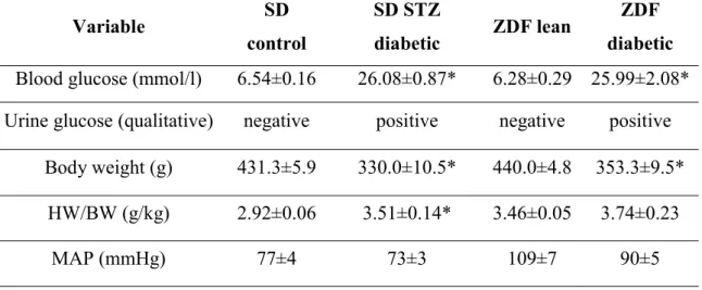
5.1.1. Heart weight (HW), body weight and glucose levels ... 47
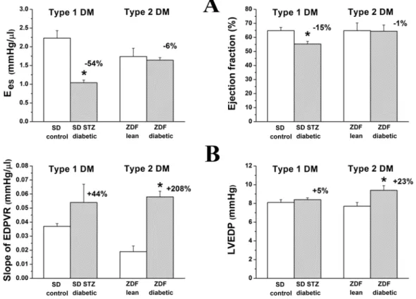
5.1.2. Cardiac contractility and ventricular stiffness ... 48
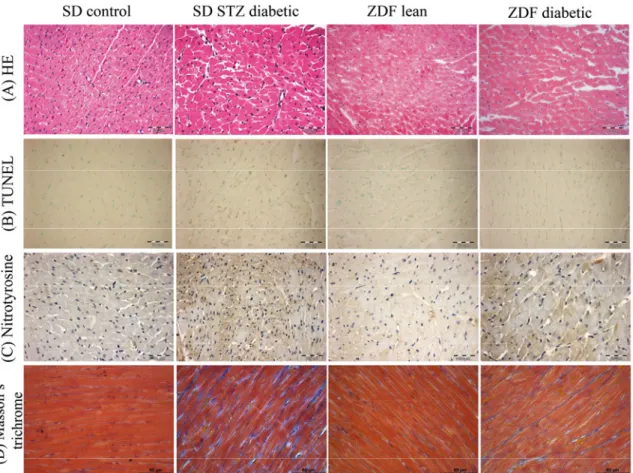
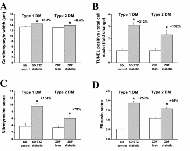
5.1.3. Histopathology ... 49
5.1.4. DNA strand breaks and nitro-oxidative stress ... 50
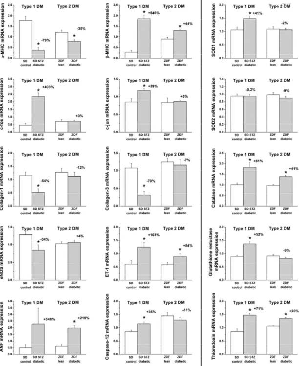
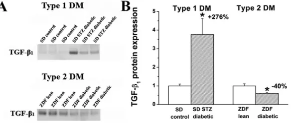
5.1.5. Myocardial fibrosis ... 51 5.1.6. Myocardial expression of genes involved in the development of diabetic cardiomyopathy ... 52 5.1.7. Western blot analysis of TGF-β1 ... 54 5.2. EXPERIMENTS ON THE EFFECT OF SGC ACTIVATION IN T1DM-RELATED
DIABETIC CARDIOMYOPATHY ... 54 5.2.1. Body weight, heart weight and glucose levels ... 54 5.2.2. Effects of cinaciguat on plasma and myocardial cGMP levels in DM ... 56 5.2.3. Effects of diabetes mellitus and cinaciguat treatment on myocardial
oxidative stress ... 58 5.2.4. Cinaciguat protects against DM related alteration of the NO-sGC-cGMP- PKG signaling ... 60 5.2.5. Cinaciguat treatment protects against DM related fibrotic remodeling of the myocardium ... 61 5.2.6. DM-related myocardium hypertrophy and apoptosis is alleviated by
cinaciguat ... 64 5.2.7. In vivo cardiac function is improved by cinaciguat in DM ... 66 5.3. EXPERIMENTS ON THE EFFECT OF PDE5A INHIBITION IN T2DM-RELATED
DIABETIC CARDIOMYOPATHY ... 71 5.3.1. The effect of vardenafil on basic characteristics in T2DM ... 71 5.3.2. Vardenafil prevented T2DM-associated LV dysfunction in vivo ... 73 5.3.3. Vardenafil prevented T2DM-associated stiffening of LV cardiomyocytes 76 5.3.4. Vardenafil decreased myocardial hypertrophy in T2DM rats ... 76 5.3.5. Vardenafil reduced alterations associated with nitro-oxidative stress in T2DM ... 80 5.3.6. Vardenafil suppressed myocardial fibrotic remodeling... 82 5.3.7. Phosphodiesterase-5A inhibiton prevented cardiomyocyte apoptosis in ZDF rats 84
5.3.8. Vardenafil protected against the disturbances of myocardial cGMP-PKG signaling in ZDF rats ... 85 6. DISCUSSION ... 90
6.1. STRUCTURAL AND MOLECULAR HALLMARKS OF TYPE 1 AND TYPE 2DM-
ASSOCIATED DIABETIC CARDIOMYOPATHY ... 90
6.1.1. Nitro-oxidative stress ... 90
6.1.2. DNA damage and apoptosis ... 91
6.1.3. Myocardial hypertrophy ... 92
6.1.4. Fibrotic remodeling ... 93
6.1.5. Deterioration of the NO-sGC-cGMP-PKG signaling pathway ... 94
6.1.6. Characteristics of cardiac dysfunction T1DM and T2DM ... 95
6.1.7. Effects of sGC activation by cinaciguat on diabetic cardiomyopathy in T1DM ... 96
6.1.8. Effects of PDE5A inhibition by vardenafil on T2DM-associated heart failure ... 98
7. CONCLUSIONS ... 100
8. SUMMARY ... 101
9. ÖSSZEFOGLALÁS ... 102
10. BIBLIOGRAPHY ... 103
11. BIBLIOGRAPHY OF THE CANDIDATE’S PUBLICATIONS ... 126
11.1. PUBLICATIONS RELATED TO THE DISSERTATION ... 126
11.2. PUBLICATIONS NOT RELATED TO THE DISSERTATION ... 127
12. ACKNOWLEDGEMENTS ... 131
1. The list of Abbreviations
3-NT 3-nitrotyrosine A wave atrial filling
ADP adenosine diphosphate
AGE advanced glycation end-product ANF atrial natriuretic factor
ANOVA analysis of variance ANP atrial natriuretic peptide ATP adenosine triphosphate
AW left ventricular anterior wall thickness BAX Bcl2-associated X protein
Bcl-2 B-cell CLL/lymphoma 2
BG blood glucose
BNP B-type natriuretic peptide
BW body weight
cGMP cyclic guanosine monophosphate
CO cardiac output
Co vehicle-treated control CoCin cinaciguat-treated control Col1a1 Collagen 1a1
Col3a1 Collagen 3a1
CTGF connective tissue growth factor
d end-diastole
DiabCin cinaciguat-treated diabetic DiabCo vehicle-treated diabetic DM diabetes mellitus
dP/dtmax maximal slope of systolic pressure increment dP/dtmin maximal slope of diastolic pressure decrement DT deceleration time
E wave early diastolic filling
EDPVR left ventricular end-diastolic P-V relationship
EF ejection fraction
EIA enzyme immunoassay
eNOS endothelial nitric oxide synthase ET-1 endothelin-1
F forward
FFA free fatty acids
GAPDH glyceraldehyde-3-phosphate dehydrogenase GSR glutathione reductase
H2O2 hydrogen peroxide HE hematoxylin and eosin
HF heart failure
HFmrEF heart failure with mid-range ejection fraction HFpEF heart failure with preserved ejection fraction HFrEF heart failure with reduced ejection fraction HgbA1c haemoglobin A1C
HR heart rate
HRP horseradish peroxidase HSP70a1 heat shock 70kDa protein 1A
HW heart weight
ID left ventricular internal diameter ISO isolating solution
IVRT isovolumic relaxation time LV left ventricular
LVEDP left ventricular end-diastolic pressure LVSP maximal left ventricular systolic pressure MAP mean arterial blood pressure
MMP-2 matrix metallopeptidase 2 MMP-9 matrix metallopeptidase 9
MT Masson’s trichrome
NOX nicotinamide adenine dinucleotide phosphate-oxidases PARP poly adenosine diphosphate ribose polymerase enzyme PDE phosphodiesterase
PKC protein kinase C PKG protein kinase G
PLB phospholamban
PPAR-α peroxisome proliferator-activated receptor α p-PLB phospho-phospholamban
PRSW preload recruitable stroke work
P-V pressure-volume
p-VASP phosphorylated vasodilator-stimulated phosphoprotein PW left ventricular posterior wall thickness
qRT-PCR quantitative reverse transcription polymerase chain reaction
R reverse
RGB red green blue
RIPA radioimmunoprecipitation assay lysis buffer RNS reactive nitrogen species
ROS reactive oxygen species
RT room temperature
rTdT recombinant Terminal Deoxynucleotidyl Transferase
s end-systole
SD Sprague-Dawley rat
SEM standard errors of the mean sGC soluble guanylate cyclase sGC β1 soluble guanylate cyclase β1 SOD1 superoxide dismutase 1 SOD2 superoxide dismutase 2 STZ streptozotocin
SW stroke work
T1DM type 1 diabetes mellitus T2DM type 2 diabetes mellitus
TauW time constant of LV pressure decay TGF-β transforming growth factor β
TIMP tissue inhibitor of matrix metallopeptidase TIMP-1 tissue inhibitor of matrix metallopeptidase 1
TIMP-2 tissue inhibitor of matrix metallopeptidase 2
TL tibia length
TUNEL terminal deoxynucleotidyl transferase dUTP nick end labeling UPL Universal Probe Library
VASP vasodilator-stimulated phosphoprotein WHO World Health Organization
ZDF vehicle-treated diabetic ZDF Zucker Diabetic Fatty ZDF+Vard vardenafil-treated diabetic ZDFLean vehicle-treated controls ZDFLean+Vard vardenafil-treated controls α-MHC myosin heavy chain alpha β-MHC myosin heavy chain beta
2. Introduction
Diabetes mellitus (DM) is a chronic metabolic disease, characterized by elevated blood glucose level which leads to a serious damage of different organ systems including the heart, liver, kidneys, vessels and eyes. According to the World Health Organization (WHO) diabetes mellitus is one of the most common chronic diseases globally and the number of people living with diabetes has nearly quadrupled since 1980. According to the Global Diabetes Report in 2016, 442 million adults have diabetes mellitus and 1.5 million deaths are directly attributed to diabetes mellitus each year (World Health Organization 2016b). Moreover, higher-than-optimal blood glucose level caused an additional 2.2 million deaths by increasing the risk of cardiovascular and other diseases.
One in three adults aged over 18 years is overweight and one of 10 adults is obese globally. In Hungary, the prevalence of diabetes is 10 %, while 63.3 % is overweight and 26 % is obese of the adult population (World Health Organization 2016a).
The diabetes epidemic is becoming the biggest one in the twenty-first century due to the constant growth in the number of diabetic patients (Tabish 2007). There are several contributing factors that play a key role in the development of diabetes mellitus including low physical activity, unhealthy western-type diet and the ageing population.
The constantly growing incidence of diabetes is not only a great epidemiological, but also an economical burden for the health care system (Tabish 2007). For example, the total cost of diabetes was 245 billion USD including the direct medical costs and the reduced productivity in the United States in 2012 (American Diabetes Association 2013). Therefore, novel therapies that aim at decreasing the incidence and prevalence of diabetes and its complications are urgently needed.
Diabetes mellitus, due to the elevated blood glucose level and the disturbance in insulin signaling leads to the development of different complications and co-morbidities including stroke, heart attack, hypertension, dyslipidaemia, blindness and eye problems, kidney failure and heart failure. Diabetic cardiomyopathy is a special form of heart failure that develops in diabetes independently of coronary artery disease or hypertension (Huynh et al. 2014).
According to the American Diabetes Association, diabetes can be classified into four major types that include type 1, type 2, gestational and specific types of diabetes (due to
other causes) (American Diabetes Association 2016). In the diagnosis of diabetes mellitus the value of plasma/blood glucose is a reliable measure. A patient is considered diabetic if fasting blood glucose is over 7.0 mmol/l or blood glucose is above 11.0 mmol/l at 2 hours in oral glucose tolerance test. In addition to the measurement of blood glucose, measurement of the glycated haemoglobin A1C (HgbA1c) is a reliable test in patients follow-up. A patient is considered to be prediabetic if the HgbA1c is between 5.7 – 6.4 % and should be counseled about strategies to lower their risks. Several forms of diabetes are known, in the current work I will focus on the two major types of the disease.
2.1. Type one diabetes mellitus
Type 1 diabetes, which accounts approximately for 5-10 % of all diabetes cases, is a cellular-mediated autoimmune disease leading to the destruction of the pancreatic β–
cells and the lack of insulin (American Diabetes Association 2016). Type 1 diabetes usually occurs in childhood or adolescence but patients can develop it at any age.
Patients are often presented with acute symptoms of diabetes including high blood glucose level and ketoacidosis (American Diabetes Association 2016). In the development of immune-mediated diabetes several factors play a key role. The disease is strongly associated with HLA DQA and DQB genes (American Diabetes Association 2016). On the other hand, the autoimmune process in type 1 diabetes is also related to different environmental factors (such as viral infections) (American Diabetes Association 2016). Patients with type 1 diabetes mellitus are not obese but are prone to develop other autoimmune diseases such as Addison disease, vitiligo, Hashimoto thyroiditis or autoimmune hepatitis (American Diabetes Association 2016).
2.2. Type two diabetes mellitus
Type 2 diabetes mellitus is responsible for 90 % of all diabetes cases (American Diabetes Association 2016). In type 2 diabetes patients have insulin resistance and usually relative insulin deficiency (American Diabetes Association 2016). In spite of type 1 diabetes mellitus, autoimmune destruction of the pancreatic β–cells does not
occur. However, there are different causes that lead to the development of type 2 diabetes such as overweight or obesity, lack of physical activity and frequent consumption of westernized-diet (Zimmet et al. 2001). It is often associated with strong genetic predisposition, more often than type 1 diabetes (American Diabetes Association 2016). Family history and ethnic background are also important in the development of the disease (Huynh et al. 2014). The risk for developing the disease is increasing with age and obesity. As opposite to the insulinopenia in type 1 diabetes, patients usually have hyperinsulinemia with hyperglycemia in type 2 diabetes. Despite the higher level of insulin the blood glucose level remains elevated due to developed insulin resistance of different organs (American Diabetes Association 2016).
2.3. Diabetic cardiomyopathy
Diabetes mellitus (independently of the type of diabetes) is associated with cardiovascular complications, such as myocardial infarction, chronic heart failure (HF), micro- and macrovascular diseases. Literature data indicate that there is a casual relationship between hyperinsulinemia and the development of hypertension or coronary artery disease (Fontbonne et al. 1991, Lender et al. 1997). Diabetes acts as an independent risk factor for coronary atherosclerosis and ischemic heart disease, however, the altered metabolic state – mainly due to elevated glucose levels – has a direct effect on cardiac structure and function independently of coronary artery disease.
The special cardiac disease entity which develops due to the direct effects of diabetes on the myocardium called diabetic cardiomyopathy (Huynh et al. 2014). Several key morphological (described below in detail) and functional (diastolic and systolic dysfunction) (Joffe et al. 1999, Poirier et al. 2001, Radovits et al. 2009a, Radovits et al.
2009b) features have been described in the development and progression of diabetic cardiomyopathy, however, the exact pathomechanism is still under debate (Cai 2006, Sharma et al. 2006, Mourmoura et al. 2013, Pearson et al. 2013, Huynh et al. 2014).
Nevertheless, depending on the severity of diabetic cardiomyopathy it leads to the development of HF. HF is a complex clinical syndrome characterized by specific clinical signs and symptoms. It has a great epidemiological burden, as its prevalence is approximately 1-2 % in the adult population of the Western countries and it is one of
the most common causes leading to hospitalization (Mosterd et al. 2007). The latest
„ESC Guidelines for the diagnosis and treatment of acute and chronic HF” determines 3 main forms of HF by the value of left ventricular (LV) ejection fraction (EF): 1) HF with reduced EF (HFrEF; LVEF < 40 %), 2) HF with mid-range EF (HFmrEF; LVEF 40-49 %) and 3) HF with preserved EF (HFpEF; LVEF ≥ 50 %) (Ponikowski et al.
2016).
2.3.1. Diastolic dysfunction in diabetes mellitus
Diastolic dysfunction is an early sign of diabetic cardiomyopathy that develops before systolic function deteriorates (Huynh et al. 2014). The prevalence of diastolic heart failure is constantly growing and it is one of the most important features of HFpEF (Ponikowski et al. 2016). HFpEF is associated with diastolic dysfunction characterized by prolonged LV isovolumic relaxation, increased LV stiffness, increased LV end- diastolic pressure and slow LV filling (Zouein et al. 2013). Despite the lack of systolic dysfunction, diastolic heart disease has a similar prognosis as systolic HF (Huynh et al.
2014). Several measures are used in the everyday clinical setting in order to evaluate the severity of diastolic dysfunction including Doppler and tissue Doppler echocardiography and cardiac magnetic resonance imaging. However, on the experimental level diastolic dysfunction can be characterized by invasive hemodynamic measurements and pressure-volume (P-V) loop analysis. P-V loop analysis provides a reliable assessment of cardiac diastolic dysfunction whereas cardiac stiffness and active relaxation can be investigated in detail (Pacher et al. 2008). Doppler echocardiography is a useful application to detect early deterioration and to perform follow-up studies on diabetic cardiomyopathy. Doppler echocardiography measures the peak blood flow across the mitral valve during the cardiac cycles, whereas a characteristic pattern of early diastolic filling (E wave) and the atrial filling (A wave) is seen and isovolumic relaxation time (IVRT) and deceleration time (DT) can be measured (Huynh et al.
2014). Diabetes is associated with a reduced E/A ratio and with prolonged IVRT (Schannwell et al. 2002). Tissue Doppler echocardiography is another useful tool in the assessment of diastolic function as it measures the movement (velocity) of a particular myocardial segment and provides similar indices as above (including E’, A’).
Additionally, by cardiac magnetic resonance imaging similar alterations in diastolic function are detectable (increased left ventricular filling pressures, reduced E/A ratio, prolonged IVRT) (Rijzewijk et al. 2009). Similarly to the human conditions diastolic cardiac dysfunction is a common phenomenon among experimental animal models of type 1 and type 2 diabetes mellitus. For example, in streptozotocin (STZ)-induced diabetes mellitus and in type 2 diabetic db/db mice an early deterioration of diastolic function is seen (as evidenced by E/A ratio, deceleration time, prolonged IVRT) (Joffe et al. 1999, Semeniuk et al. 2002). Additionally, invasive hemodynamic investigation and P-V loop analysis revealed the severe deterioration of diastolic dysfunction in STZ- induced type 1 and in the Zucker Diabetic Fatty (ZDF) rat model of type 2 diabetes mellitus (as represented by increased relaxation time, increased filling pressures and increased cardiac stiffness) (Radovits et al. 2009b). Not only the in vivo measurement of cardiac function, but isolated cardiomyocytes from diabetic animals and humans showed impaired relaxation and increased stiffness in vitro (Fulop et al. 2007, van Heerebeek et al. 2008).
2.3.2. Systolic dysfunction in diabetes mellitus
In spite of the early manifestation of diastolic dysfunction, systolic abnormalities become more evident at later stages in the disease (Boudina et al. 2010). In addition to that, subclinical alterations of the contractile function are difficult to detect with conventional 2-dimensonal echocardiography due to its load- and frequency dependence (Pacher et al. 2008). However, tissue Doppler or speckle tracking strain analysis is able to reveal early signs of contractile dysfunction in diabetes (Fang et al. 2005, Shepherd et al. 2016). In vivo animal studies using magnetic resonance imaging or an invasive hemodynamic approach have shown the impairment of contractile function in different types of animal models (Van den Bergh et al. 2006, Yue et al. 2007). However, it is believed that the development of systolic dysfunction in an experimental setting is dependent on the experimental model used given that contractility is preserved when multiple low-dose STZ is applied in an animal model (Huynh et al. 2013). In spite of that, STZ-induced diabetic C57Bl/6 mice develop severe contractile dysfunction (Westermann et al. 2007). Recently, a study by our research group showed that there is
significant difference regarding the cardiac dysfunction in STZ-induced type 1 diabetic and the ZDF type 2 diabetic animal models whereas type 1 diabetes was associated with decreased cardiac contractility, impaired systolic function coupled with prolonged relaxation and diastolic dysfunction, while type 2 diabetes showed preserved systolic performance and markedly increased cardiac stiffness (Radovits et al. 2009b).
2.3.3. Metabolic alterations in diabetes mellitus contribute to the development of diabetic cardiomyopathy
Impaired glucose homeostasis due to the lack of insulin or insulin resistance with hyperinsulinemia are the hallmarks of diabetes associated metabolic alterations in the heart. In addition to these effects, disturbances in insulin signaling and the downregulation and diminished activity of the glucose transporter type 4 directly contribute to the substrate switching that characterize the diabetic heart (Bugger et al.
2014). Thus, diabetes mellitus is associated with reduced glucose and lactate metabolism (Stanley et al. 1997) which makes it more vulnerable to different harmful stimuli including ischemia/reperfusion in myocardial infarction (Huynh et al. 2014).
The levels of free fatty acids (FFA) in the plasma and their rate of metabolism in the heart are increased in diabetes mellitus (Boudina et al. 2007). Furthermore, diabetes can cause a significant increase in myocardial triacylglycerol content (Murthy et al. 1977).
Taken together, as FFA uptake exceeds the rate of its oxidation in the heart, lipid accumulation occurs in the diabetic myocardium which may promote lipotoxicity and greatly contributes to apoptosis and tissue injury (McGavock et al. 2006). On the other hand, Oakes et al. pointed out that diabetic hearts are less able to adapt their metabolic substrate use when the concentration of the metabolic substrates were altered (Oakes et al. 2006). In conclusion, the diabetic heart has lost its metabolic flexibility in vivo which phenomenon is an important contributor to the inability of the diabetic myocardium to adapt itself to different stress situations. FFAs can directly inhibit glucose oxidation by activating the peroxisome proliferator-activated receptor (PPAR)- α. In response, PPAR-α increases the expression of pyruvate dehydrogenase kinase-4 thereby mediates the increased mitochondrial FFA uptake and β-oxidation (Hopkins et al. 2003). Additionally, in the diabetic myocardium the acetyl-CoA carboxylase is
inhibited, the level of malonyl-CoA is increased which processes contribute to the enhanced FFA oxidation in the mitochondria resulting in increased level of acetyl-CoA that enters the Szent-Györgyi – Krebs - cycle (Stanley et al. 1997). The above findings further deteriorate the flexibility of metabolism in diabetes mellitus.
2.3.4. Mitochondrial dysfunction in diabetic cardiomyopathy
Proteomic analysis by Turko et al. revealed that myocardial mitochondria in diabetic cardiomyopathy have decreased expression of proteins of the electron transport chain, creatine kinase, the voltage-dependent anion channel 1, however, the level of β- oxidation proteins was increased (Turko et al. 2003b). As described above, the rate of β- oxidation is greatly elevated in the heart of diabetics that delivers more reducing equivalents to the electron transport chain (Boudina et al. 2007). However, due to the decreased expression of electron transport chain proteins the oxidative phosphorylation is significantly limited thus leading to increased production of superoxide anions and reactive oxygen species (ROS) from the mitochondria and to the decreased production of high energy phosphates (Boudina et al. 2007). As a result of the above process the mitochondrial electron transport chain becomes uncoupled and begins to produce large amounts of ROS and promotes oxidative stress (Boudina et al. 2005). Furthermore, ROS in oxidative stress and reactive nitrogen species (RNS) in nitrosative stress directly deteriorates the mitochondrial proteins thus a vicious cycle of oxidative mitochondrial injury develops (Turko et al. 2003a). However, blunted antioxidant protection in the failing myocardium (e.g. in diabetes mellitus) further enhances mitochondrial injury and leads to mitochondrial dysfunction in diabetes (Shen et al. 2006).
2.3.5. The role of oxidative stress in the diabetic myocardium
The presence of increased oxidative stress has been widely described in diabetic cardiomyopathy (Pacher et al. 2007, Bugger et al. 2014, Huynh et al. 2014, Varga et al.
2015), however, the exact mechanism in its development is still unknown (Boudina et al. 2010). Most probably an interplay of several processes leads to the enhanced production of ROS and RNS in diabetes (Varga et al. 2015): 1) lipid overload with
excessive FFA generation (described above in detail) (Boudina et al. 2010); 2) hyperglycemia-induced glucotoxicity and inflammatory response; 3) endothelial nitric oxide (NO) synthase (eNOS) uncoupling; 4) formation of the highly reactive molecule peroxynitrite ; 5) overactivation of ROS producing enzymes.
In spite of the available treatment therapies for high blood glucose, to achieve the optimal anti-hyperglycemic treatment is still challenging and high blood sugar level is poorly controlled in patients (Ross et al. 2011). High or fluctuating glucose levels lead to the enhancement of oxidative stress (Giacco et al. 2010). Glucotoxicity, high glucose levels and the non-specific glycosylation of different proteins (as forming advanced glycation end-products [AGEs]) contribute to the dysfunction of enzymes, contractile proteins and they provoke a whole body inflammatory response (Varga et al. 2015).
Inflammatory cell response, including neutrophils, is a major drive in the development of diabetes mellitus associated cardiovascular abnormalities. Neutrophils are sources of nicotinamide adenine dinucleotide phosphate-oxidases (NOX) that produce large amounts of superoxide anions as part of their inflammatory response (Varga et al.
2015). Superoxide directly deteriorates proteins or forms highly reactive molecules including peroxynitrite (together with NO) (Pacher et al. 2007) or hydrogen peroxide (Pacher et al. 2007). Overexpression or overactivation of different NOX enzymes have been described in diabetic cardiomyopathy (Rajesh et al. 2010, Roe et al. 2011, Maalouf et al. 2012) and in hypercholesterolemia (Varga et al. 2013) as well. Furthermore, overactivation of the renin-angiotensin-aldosterone system plays decisive role in diabetes-associated cardiovascular oxidative stress, as vascular NOX is a downstream target of angiotensin II (Kajstura et al. 2001, Westermann et al. 2007) thereby it promotes the production of vascular ROS (Varga et al. 2015). The above theory is supported by experimental data whereas angiotensin II type 1 receptor blockade inhibited glucose-derived cardiac dysfunction by the attenuation of angiotensin 1 receptor – NOX signaling (Privratsky et al. 2003). Beside the role of NOX enzymes in diabetes-related oxidative stress, other enzyme systems can also importantly contribute to ROS production. One of these enzymes is xanthine oxidase that has been shown to be overactivated in diabetes in animal models (Desco et al. 2002, Rajesh et al. 2009). This theory has been evidenced by the protective effects of allopurinol on the hearts of diabetic mice and rats (Rajesh et al. 2009, Gao et al. 2012).
Beside the above mentioned enzyme systems, there is another important player in oxidative stress in diabetes mellitus. Namely, the eNOS enzyme which is located in the endothelial system becomes uncoupled in diabetes as it loses its cofactor tetrahydrobiopterin (Zou et al. 2002). As eNOS becomes uncoupled it produces superoxide anions instead of NO thus NO bioavailability markedly decreases and the level of ROS increases in the diabetic heart (Zou et al. 2002). On the other hand, iNOS and eNOS expression has been shown to be induced in diabetic cardiomyopathy that might further contribute to increased generation of NO. However, NO rapidly reacts with the excessive amount of superoxide anions in oxidative stress and peroxynitrite, a highly reactive molecule, is formed (Pacher et al. 2007). Peroxynitrite is able to directly damage (via protein nitration) different cellular elements including DNA, contractile proteins, proteins of calcium handling, elements of the antioxidant defence system and induces apoptosis and necrosis (Pacher et al. 2007). Furthermore, in cooperation with other ROS peroxynitrite activates matrix metallopeptidases (MMPs) in the diabetic myocardium leading to the remodeling of the myocardium and may impair the NO – soluble guanylate cyclase (sGC) axis (Evgenov et al. 2006). The importance of peroxynitrite in cardiovascular complications of diabetes has been shown in patients as well, as the level of 3-nitrotyrosine (a marker of nitrosative stress) correlated with the presence of high blood pressure (Hoeldtke et al. 2003). The presence of constant myocardial oxidative and nitrosative stress may further deteriorate the antioxidant defence system by the inactivation (probably by protein nitration and oxidation) of its key elements like superoxide dismutases and catalase (Pacher et al. 2007). In fact, oxidative and nitrosative stress aggravate the severity of diabetes mellitus as they promote the oxidation and/or nitration of insulin receptors in peripheral tissues that further compromise insulin resistance of the organs (Stadler 2011). Nonetheless, hyperglycemia coupled with oxidative/nitrosative stress causes nonspecific and nonenzymatic glycation (AGEs), oxidation and nitration of the proteins and lipids that significantly contribute to diabetes mellitus-associated cardiac dysfunction (e.g.
contractile proteins – systolic dysfunction, calcium transporters – diastolic dysfunction).
2.3.6. Apoptosis and cell death in diabetic cardiomyopathy
Increased rate of cell death and apoptosis is often observed in the heart of diabetic individuals and in the myocardium of experimental animal models (Frustaci et al. 2000, Chowdhry et al. 2007, Rajesh et al. 2010, Rajesh et al. 2012). Apoptosis is a well- known form of programmed cell death and it is essential in the regulation and for the maintenance of tissue homeostasis (Huynh et al. 2014). In the pathogenesis of diabetes- induced cell death many processes have been identified such as the activation of caspases, increased amount of ROS, increased rate of inflammation, increased activation of transforming growth factor β (TGF-β) signaling and activation of the poly adenosine diphosphate (ADP) ribose polymerase (PARP) enzymes (Bugger et al. 2014).
The overactivation of PARP-1 is a major drive of apoptosis and necrosis in diabetic cardiomyopathy (Varga et al. 2015). PARP-1, a major regulator of DNA repair and cell death becomes activated when ROS and RNS directly bind to the DNA causing its oxidative injury (Bai et al. 2012). During its repair operation, PARP-1 binds to the damaged DNA, forms homodimers and cleaves its substrate NAD+ into nicotinamide and ADP-ribose (Bai et al. 2012, Varga et al. 2015). Then long branches of ADP-ribose polymers are formed on target proteins (e.g. histones and PARP-1) and energy (adenosine triphosphate [ATP]) depletion, mitochondrial dysfunction and subsequent necrosis/apoptosis will occur (Varga et al. 2015). This hypothesis is underlain by literature data where PARP inhibition successfully improved diabetes-associated endothelial and cardiac dysfunction (Garcia Soriano et al. 2001, Soriano et al. 2001, Pacher et al. 2002). Moreover, PARP inhibition also prevented the pathological activation of protein kinase C (PKC) pathways, the hexosamine pathways and the AGE formation induced by hyperglycemia in vitro (Pacher et al. 2005).
2.3.7. Myocardial hypertrophy in diabetic cardiomyopathy
Myocardial hypertrophy is a structural hallmark of diabetic cardiomyopathy characterized by the hypertrophy of the individual cardiomyocytes (Huynh et al. 2014).
The Strong Heart Study, conducted in Native Americans, found that myocardial hypertrophy (as represented by increased LVmass and increased wall thicknesses)
develops both in men and women with type 2 diabetes mellitus (Devereux et al. 2000).
Moreover, a study conducted in a multi-ethnic population described that the likelihood of developing cardiac hypertrophy (increased LVmass) is greater in patients with type 2 diabetes mellitus (Eguchi et al. 2008). Pathological myocardial hypertrophy is a risk factor for the development of myocardial infarction, stroke and death from HF (Bernardo et al. 2010). Myocardial hypertrophy occurs in response to hemodynamic stress and due to oxidative stress-induced apoptosis and as compensatory mechanisms of the remaining functional cardiomyocytes and due to the neurohormonal (renin- angiotensin-aldosterone system) activation (Huynh et al. 2014). However, concentric hypertrophy of the heart is a long-term consequence of diabetes and obesity regardless of the presence of hypertension (Woodiwiss et al. 2008, Boudina et al. 2010). In the development of LV hypertrophy, adipose tissue derived cytokines (e.g. leptin) play a key role. Leptin has been shown to induce cardiomyocyte hypertrophy in vitro probably via an endothelin-1 mediated oxidative stress response (Xu et al. 2004). Furthermore, hyperglycemia directly propagates the overexpression of endothelin-1 thus it maintains and contributes to the already elevated level of ROS (Huynh et al. 2014). Endothelin-1 is a powerful vasoconstrictor (Kiowski et al. 1995) and induces an inflammatory response (Davidson et al. 2010) thereby it might contribute to the hemodynamic stress and ROS production in diabetes mellitus. Furthermore, LV hypertrophy was associated with elevated levels of atrial and B-type natriuretic peptides (NP; ANP/ANF, BNP) and was shown to be associated with hypertrophic gene signaling and with the reactivation of the fetal gene program in diabetes mellitus (Huynh et al. 2014).
2.3.8. Fibrotic remodeling of the diabetic heart
Interstitial and perivascular fibrotic remodeling of the myocardium is frequently observed in diabetes mellitus in humans (Shimizu et al. 1993) and in experimental animal models as well (Mizushige et al. 2000, Korkmaz-Icoz et al. 2016). In the background of increased fibrosis an over-activation of the TGF-β signaling and its downstream effector connective tissue growth factor (CTGF) might play a role (Radovits et al. 2009a, Bugger et al. 2014). Furthermore, due to the enhanced apoptosis the cardiomyocytes are replaced by fibroblasts and extracellular matrix. However, the
dysregulation of MMPs (e.g. MMP-2) and their inhibitors tissue inhibitor of matrix metallopeptidases (TIMPs) could significantly contribute to the accumulation of extracellular matrix in the myocardium (Westermann et al. 2007, Van Linthout et al.
2008). Increase of collagen is often observed in the heart of diabetic patients (Shimizu et al. 1993). Additionally, the elevated levels of extracellular matrix proteins and the elevation of their gene expression have been observed in the heart of diabetic animals (Mizushige et al. 2000, Westermann et al. 2007, Rajesh et al. 2009, Rajesh et al. 2012).
Nevertheless, hyperglycemia only is sufficient to induce cardiac fibroblast and vascular smooth muscle cell proliferation and to trigger myocardial pro-growth signaling pathways (Begum et al. 2000, Tokudome et al. 2004, Yeshao et al. 2005).
2.4. The role of cyclic guanosine monophosphate (cGMP) signaling in diabetic cardiomyopathy
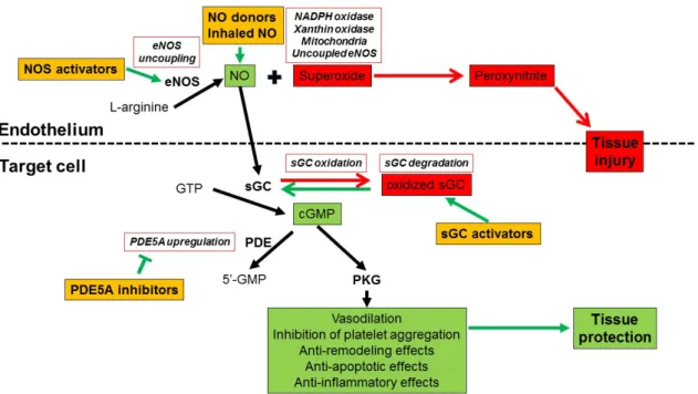
The main steps of the signaling pathway and its pharmacology are summarized on Figure 1.
2.4.1. Generation, degradation and effects of cGMP
The NO-sGC-cGMP pathway. NO is a key transmitter molecule synthetized from L- arginine by NO synthases (endothelial (eNOS), neuronal (nNOS) and inducible (iNOS)) and is involved in many regulatory and biological processes in mammals (e.g.
inflammatory processes, vascular and cardiac homeostasis and neuronal transmission).
As a gaseous transmitter it diffuses into the target cells such as vascular smooth muscle cells or cardiomyocytes. Thereafter, NO binds to the sGC and activates the enzyme to produce massive amounts of intracellular cGMP from GTP (Takimoto 2012).
The NP - particulate GC (pGC) - cGMP pathway. NPs are polypeptide hormones. ANP and BNP are derived from atria and ventricles while C-type NP is originated from endothelial cells (Kuhn 2003). cGMP is generated by the pGC when NPs are bound to the enzyme. The pGC enzyme is widely expressed in different tissues including the myocardium.
Degradation of cGMP by phosphodiesterases (PDEs). The biological effects of the second messenger cGMP are regulated by PDEs. PDEs are responsible for the degradation of cGMP as they convert it into 5’-GMP (Zhao et al. 2016). Altogether 11 types of PDE (1-11) are known to date and the 1, 2, 3, 4, 5, 8 and 9 are expressed in the heart (Takimoto 2012). Among all PDEs, the PDE5A subtype is a cGMP-specific esterase expressed in vascular smooth muscle cells, however, recent evidence suggest that PDE5A is presented in cardiac myocytes (Tsai et al. 2009). Moreover, PDE5A has been shown to be overexpressed in different cardiac diseases associated with oxidative stress (Nagendran et al. 2007, Lu et al. 2010). PDE5A is localized near to the Z-band of cardiomyocytes (Takimoto 2012). According to a recent publication, PDE9A is also upregulated in cardiac disorders and pathological myocardium hypertrophy thus it could significantly contribute to cGMP-depletion in cardiomyocytes. However, PDE9A regulates mainly the NO-independent pool of cGMP (Lee et al. 2015).
cGMP-dependent effects and protein kinase G (PKG). PKG is a serine/threonine kinase that mediates the main effects of cGMP (Moncada et al. 1991, Evgenov et al. 2006).
The two subtypes, PKGI and PKGII are encoded by two different genes and they contain three functional domains that determines cGMP-sensitivity of the enzyme (Takimoto 2012). cGMP, through the activation of PKG has several downstream effects including the negative regulation of cardiac contractility by the reduction of myofilament Ca2+ responsiveness. NO-derived cGMP has also anti-adrenergic effects that might be attributable to cardiac troponin I phosphorylation by PKG (Lee et al.
2010). Cardiac diastolic function is regulated by both NO and NP-derived cGMP pools as exogenous BNP increases the speed and extent of ventricular relaxation (Lainchbury et al. 2000). Moreover, PKG contributes to diastolic relaxation by PKG-mediated phosphorylation of the giant protein titin (Takimoto 2012) and by exerting inhibitory effects on L-type Ca2+ channels and activating phospholamban-mediated calcium reuptake to the sarcoplasmic reticulum (Kovacs et al. 2016). As discussed above, cardiac hypertrophy is an important contributor to myocardial remodeling in different cardiac diseases. However, the cGMP-signaling mediated effects of PKG activate anti- hypertrophic pathways in the heart. These processes include Gq-coupled and calcineurin related pathways. Recent studies showed that calcineurin pathway is deactivated via L- type Ca2+ channel inhibition (Fiedler et al. 2002) and by the phosphorylation of TRCP6
(transient receptor potential canonical 6) receptor (Koitabashi et al. 2010). Other studies also implicated the role of regulator of G protein signaling 2 and 4 (RGS2 and 4) in the anti-hypertrophic effects as PDE5A inhibition failed to modulate cardiac hypertrophy in mice lack RGS2 (Takimoto et al. 2009). On the other hand, cGMP signaling mediates anti-apoptotic effects via the regulation of the mitochondrial KATP channel and the stress responsive survival signaling cascades (Burley et al. 2007, Kukreja et al. 2011). As a result of PKG activation opening of the mitochondrial KATP channel and subsequent inhibition of the mitochondrial transition pore occur thus the membrane potential of the mitochondria become stabilized. Other features of cGMP-mediated anti-apoptotic effects are phosphorylation of extracellular signal-regulated kinases and the glycogen synthase kinase 3β and upregulation of anti-apoptotic Bcl2 (Takimoto 2012). PKG also inhibits the pro-apoptotic p38 mitogen-activated protein kinase (Fiedler et al. 2006) and exerts anti-oxidative effects through the upregulation of the mitochondrial superoxide dismutase (Kukreja et al. 2011).
2.4.2. Deterioration of cGMP-signaling in oxidative stress and in diabetic cardiomyopathy
Diseases that are associated with excessive production of RNS and ROS (including diabetes mellitus) have been shown to lead to the impairment of NO-sGC-cGMP-PKG signaling (Huynh et al. 2014). Another important contributor to the impaired NO- signaling is microvascular inflammation coupled with the dysfunction of the vascular endothelial layer (Paulus et al. 2013). In pathological cases associated with oxidative stress (including diabetes mellitus), eNOS can become uncoupled as it loses its co- factor tetrahydrobiopterin. Thus, it produces massive amounts of superoxide anions which further propagates oxidative stress (Silva et al. 2012). Moreover, phosphorylation of eNOS might alter the enzyme’s activity, e.g. phosphorylation at Thr495 decreases its activation, thus reducing NO bioavailability (Bouloumie et al. 1997). Furthermore, superoxide anions rapidly react with NO in oxidative stress and peroxynitrite is formed which further decreases the bioavailability of NO (Pacher et al. 2007). Not only the decreased bioavailability of NO, but also superoxide and peroxynitrite-mediated damage of sGC (and many other enzymes and structural proteins) coupled with the
upregulation of different PDEs are contributors of the deteriorated cGMP-signaling (Pacher et al. 2007, Pokreisz et al. 2009, Das et al. 2015) in diabetes mellitus, in particular. Oxidative stress directly leads to the oxidative damage of the sGC enzyme which in turn loses its heme prosthetic group and becomes inactivated (Evgenov et al.
2006). As a result, sGC will not be able to produce intracellular cGMP even if NO is available. Moreover, upregulation of PDEs (especially PDE5A) further decreases the amount of intracellular cGMP and this contributes to the insufficiency of PKG-mediated cardioprotective signaling pathways and causes consequent tissue injury.
2.4.3. Pharmacological modulation of the NO-sGC-cGMP-PKG pathway
NO donors and NOS activators. The first class of drugs that has been tested to treat chest pain are the so called nitrovasodilators including glycerine trinitrate. These drugs have been used since 1879. These compounds release NO by spontaneous decomposition or by bioconversion to activate sGC. Although they are widely used drugs to treat different cardiovascular disorders, everyday use of such drugs is limited due to the development of nitrate tolerance, nonspecific interactions of NO with other molecules (e.g. peroxynitrate) and by the lack of response due to insufficient metabolism (Evgenov et al. 2006). Recently, inhaled NO has been approved for the treatment of persistent pulmonary hypertension of the newborn (Stasch et al. 2011).
Besides, tetrahydrobiopterin and eNOS activators have been shown to reduce diastolic dysfunction in animal experiments (Kovacs et al. 2016).
sGC activation and stimulation. Recently discovered classes of drugs are the sGC activators and stimulators that modulate sGC activity. sGC activators and stimulators are ideal substitutes of NO in case of endothelial dysfunction (e.g. in oxidative stress) as they directly bind to sGC and increase its ability to produce cGMP (Kovacs et al. 2016).
The difference between the two types of these compounds is that sGC activators are able to reactivate even the oxidized, inactive form of the enzyme, while the stimulators bind to the reduced, heme-containing form of sGC by replacing NO. However, sGC stimulators do not reactivate the inactive, heme-free enzyme (Evgenov et al. 2006). The sGC stimulator riociguat has been approved to treat patients with pulmonary
hypertension. Currently, a trial investigating another sGC stimulator, the vericiguat is conducted in heart failure patients.
PDE5A inhibition. PDE5A inhibitors (sildenafil, vardenafil, tadalafil, avanafil) are on- demand treatment for erectile dysfunction (Korkmaz-Icoz et al. 2017). The role of PDE5A in myocardial remodeling has been shown by Takimoto and colleagues on PDE5A knockout mice (Takimoto et al. 2005). The mechanism of action of PDE5A inhibitors include increased tissue and plasma levels of cGMP which results in vasorelaxation and vasodilation (Kukreja et al. 2011). Among the different PDE5A inhibitors vardenafil is a highly selective one and it displays the highest potency compared to its comparators. Sildenafil and tadalafil have also been approved by the US Food and Drug Administration for the treatment of pulmonary hypertension.
Figure 1. Schematic diagram of physiology, pathophysiology and pharmacology of nitric-oxide (NO) – cyclic guanosine monophosphate (cGMP) pathway
Detailed description of the signaling pathway is available in the text. NO: nitric oxide, NOS: NO synthase, eNOS: endothelial NOS, sGC: soluble guanylate cyclase, GTP:
guanosine triphosphate, GMP: guanosine monophosphate, PDE5A: phosphodiesterase- 5A, PKG: protein kinase G, NADPH: nicotinamide adenine dinucleotide phosphate, Target cell: cardiomyocytes and/or smooth muscle cells. Physiological effects of the signaling pathway are depicted as green, pathophysiological processes are shown in red and the possible pharmacological interventions are orange-colored.
2.4.4. Pharmacology and cardioprotective effects of the sGC activator cinaciguat
The sGC activator cinaciguat (BAY 58-2667; Figure 2.) has been reported to bind to the heme pocket of NO-insensitive sGC (Stasch et al. 2006) thus stabilizing the enzyme to increase cGMP generation and to augment cGMP-PKG signaling. Cinaciguat produces an additive effect when combined with NO donors (Evgenov et al. 2006). Cinaciguat replaces the oxidized heme prostethic group of the sGC enzyme resulting in the activation of sGC. One of the biggest advantage of the compound is that it is able to discriminate between both redox states of the enzyme (Evgenov et al. 2006).
Additionally, cinaciguat increases the maximal catalytic rate of sGC and stabilizes the nitrosyl-heme complex (Evgenov et al. 2006). Interestingly, cinaciguat inhibits the PDE5A enzyme with an IC50 of 10 µmol/l (Evgenov et al. 2006). Recent studies described the beneficial cardioprotective effects of cinaciguat in experimental myocardial infarction (Korkmaz et al. 2009), myocardial ischemia/reperfusion injury (Radovits et al. 2011, Salloum et al. 2012), endothelial dysfunction induced by nitro- oxidative stress (Korkmaz et al. 2013), vascular neointima formation (Hirschberg et al.
2013) or in prevention of cardiomyocyte hypertrophy (Irvine et al. 2012, Nemeth et al.
2016). Moreover, the safety and tolerability of the sGC activator cinaciguat have been assessed by phase-I human clinical trials (Frey et al. 2008). Although cinaciguat has demonstrated efficacy in a proof-of-concept study in patients with acute decompensated heart failure (Lapp et al. 2009), the phase-IIb clinical studies for the same indication has been prematurely terminated due to the high number of adverse events (mainly a significant drop of blood pressure) during acute intravenous application (Erdmann et al.
2013, Levy et al. 2014). However, the investigators concluded that rather a chronic, long-term oral administration of the compound could improve endothelial and preserve myocardial function in heart failure patients (Gheorghiade et al. 2012).
Figure 2. Molecular structure of cinaciguat
2.4.5. Pharmacology and cardioprotective effects of the PDE5A inhibitor vardenafil
As discussed above, inhibitors of PDEs (PDE5A in particular) enhance cGMP-driven effects and increase cardiomyocyte relaxation via PKG-mediated signaling pathways (Kovacs et al. 2016). Vardenafil (Figure 3.) is a highly selective PDE5 inhibitor with a potency about 10-fold higher than that of sildenafil (Saenz de Tejada et al. 2001). The protective effects of enhanced cGMP-signaling via pharmacological inhibition of PDE5A has been demonstrated in several cardiovascular diseases (Kukreja et al. 2012) including ischemia/reperfusion injury related to heart transplantation (Loganathan et al.
2008), cardiopulmonary bypass (Szabo et al. 2009) and myocardial infarction (Kukreja et al. 2004), neointima hyperplasia formation (Hirschberg et al. 2009) and in type 1 diabetes mellitus-associated vascular and cardiac dysfunction in particular (Schafer et al. 2008, Radovits et al. 2009a). Interestingly, vardenafil has been shown to improve blood flow in 70 % of patients with Raynaud’s disease and improved clinical signs in 68
% of patients (Kukreja et al. 2011). Moreover, the beneficial cardiac effects of sildenafil on HFpEF patients have been investigated in the RELAX trial. The outcome of the RELAX trial, however, failed to show improvement in exercise capacity and on the clinical outcomes in advanced HFpEF patients treated with sildenafil (Redfield et al.
might be of therapeutic importance in subgroup of patients who have diabetes mellitus (Das et al. 2015). Moreover, sildenafil and tadalafil have been approved for the treatment of pulmonary hypertension (Kukreja et al. 2012).
Figure 3. Molecular structure of vardenafil.
3. Objectives
Recent advances of the research on heart failure pointed out that the clinical outcome and the success of drug therapy of heart failure patients depend a lot on the co- morbidities of the patients. Among these co-morbidities, diabetes mellitus is a significant one which leads to the development of a special disease entity, diabetic cardiomyopathy. Diabetic cardiomyopathy is a complex disease characterized by special features including increased oxidative and nitrosative stress, fibrotic and hypertrophic remodeling and the deterioration of cGMP signaling. Therefore, enhancement of cGMP signaling in the myocardium might provide a novel and promising therapeutic option to treat diabetic heart failure.
Based upon that, the aims of the present studies were:
1. Comparison of the functional (systolic and diastolic) and the structural hallmarks (oxidative stress, fibrotic and hypertrophic remodeling and apoptosis) of diabetic cardiomyopathy in animal models of T1DM and T2DM
2. Revelation of the alterations of NO-cGMP signaling in diabetic cardiomyopathy in the two types of DM
3. Explore the possible therapeutic effects of sGC activation by the novel sGC activator cinaciguat in T1DM-associated diabetic cardiomyopathy
4. Study the potential cardioprotective effects of the PDE5A inhibitor vardenafil on the development of T2DM-related diabetic cardiomyopathy
4. Methods
The investigation conformed to the EU Directive 2010/63/EU guidelines and the Guide for the Care and Use of Laboratory Animals used by the US National Institutes of Health (NIH Publication No. 85–23, revised 1996). The experimental protocol was reviewed and approved by the institutional ethical committee (permission number:
22.1/1162/3/2010). Animals received humane care.
4.1. Animal models
4.1.1. Rat model of type 1 diabetes mellitus
Depending on the experimental setting, eight- or twelve-week-old male Sprague- Dawley rats (SD; Charles River, Sulzfeld, Germany) were housed individually in a cage in a room (constant temperature (22±2 ºC), 12/12h light/dark cycles) and received standard laboratory rat diet and water ad libitum. T1DM was induced with a single intraperitoneal injection of STZ (60 mg/kg) (Sigma Aldrich, Saint Louis, MO, USA) in our rats. STZ was freshly dissolved in citrate buffer (pH = 4.5; 0.1 mol/l). Control animals received only the buffer. 72 h after the injection a drop of blood was collected from the tail vein and blood glucose concentration was determined by using a digital blood glucose meter (Accu-Chek® Sensor, Roche, Mannheim, Germany). Animals with a random blood glucose level >15 mmol/l were considered to be diabetic and were included into the study.
4.1.2. Rat model of type 2 diabetes mellitus
The inbred ZDF rat was used as an animal model of T2DM. Rats develop T2DM due to a genetic mutation of the leptin receptor and a special diet (Purina #5008) recommended by the supplier. Homozygous recessive males (fa/fa) develop obesity, fasting hyperglycemia and T2DM. Homozygous dominant (+/+) and heterozygous (fa/+) lean genotypes remain normoglycemic. Six-week old male ZDF diabetic (fa/fa) and ZDF lean (+/?) rats (Charles River) were acclimatized for one week before starting the
experimental protocol. Rats were housed individually in a cage in a room at a constant temperature (22±2 ºC) and humidity, with a 12/12h light/dark cycle and fed Purina
#5008 diet and water ad libitum. Experiments on the rats were performed at the age of 30-32 weeks.
4.2. Study protocol
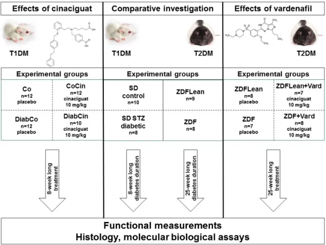
Summary of our study protocol is shown on Figure 4.
4.2.1. Comparative investigation of T1DM or T2DM related diabetic cardiomyopathy
Rat models of T1DM (STZ-induced DM) and T2DM (ZDF) were used to compare the pathophysiology of T1DM and T2DM-related diabetic cardiomyopathy. For T1DM 12- week old SD rats were used and DM was induced by a single i.p. injection of STZ as described above (groups: SD control; n=10 and SD diabetic; n=8). Experiments were performed after eight week of diabetes duration. For T2DM ZDF rats were used (for detailed description of the model see above). Experiments were performed on 32-week old male ZDF Lean (n=9) and ZDF (n=8) rats. The time of sacrifice was chosen based on our pilot and previous studies where cardiac dysfunction is detectable in these models.
4.2.2. Experiments on the effect of sGC activation in T1DM related diabetic cardiomyopathy
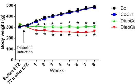
Eight-week old SD rats were used. After confirmation of DM induced by STZ injection, rats were randomized into four groups: vehicle-treated control (Co; n=12), cinaciguat- treated control (CoCin; n=12), vehicle-treated diabetic (DiabCo; n=12) and cinaciguat- treated diabetic (DiabCin; n=10) groups. Animals were treated for 8 weeks with 0.5 % methylcellulose (Co, DiabCo) vehicle or with the sGC activator cinaciguat in suspension per os (CoCin, DiabCin; 10 mg/kg/day), starting immediately after DM
confirmation. Water intake was measured every day. Body weight (BW) of the animals were recorded every other day and the dose of cinaciguat was adjusted accordingly.
4.2.3. Experiments on the effect of PDE5A inhibition in T2DM related diabetic cardiomyopathy
6-week old male ZDF diabetic (fa/fa) and ZDF lean (+/?) rats (Charles River) were acclimatized for one week before starting the experimental protocol. 7-week old animals were randomized into four groups: vehicle-treated controls (ZDFLean; n=8), vardenafil- treated controls (ZDFLean+Vard; n=7), vehicle-treated diabetic (ZDF; n=7) and vardenafil-treated diabetic (ZDF+Vard; n=8). Rats were fed Purina #5008 diet and water ad libitum. Per os drug treatment (10 mg/kg BW vardenafil continuously, dissolved in 0.01 mol/l citrate buffer) or vehicle (0.01 mol/l citrate buffer) administration (in water bottle) was initiated at the 7th week of age and continued until the end of the experimental period. Animal cages were handled with care and were not moved after water bottle replacement to prevent spilling of water from the bottles.
Functional measurements were performed at the age of 32 weeks. BW of the animals were measured every two days and the dose of vardenafil was adjusted accordingly.
Figure 4. Study protocol
Abbreaviations: Type 1 diabetes mellitus (T1DM), type 2 diabetes mellitus (T2DM).
Comparative investigation: Sprague-Dawley (SD), Zucker Diabetic Fatty (ZDF).
Cinaciguat effects: vehicle-treated control (Co), cinaciguat-treated controls (CoCin), vehicle-treated diabetic (DiabCo), cinaciguat-treated diabetic (DiabCin). Vardenafil effects: vehicle-treated controls (ZDFLean), vardenafil-treated controls (ZDFLean+Vard), vehicle-treated diabetic (ZDF), vardenafil-treated diabetic (ZDF+Vard).
4.3. Biochemical measurements
Urine samples were collected at the end of the invasive hemodynamic measurements and urine glucose level was determined by a digital blood glucose meter (Accu-Chek®
Sensor, Roche).
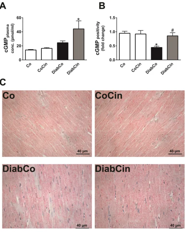
Blood was collected at the end of the invasive hemodynamic investigation from the inferior caval vein. Blood glucose (BG) level was determined by a digital blood glucose meter (Accu-Chek® Sensor, Roche). Thereafter, plasma was prepared and stored at -80 ºC for long term. Plasma cGMP was measured by a cGMP enzyme immunoassay (EIA)
kit (Amersham cGMP EIA Biotrak System, GE Healthcare, Buckinghamshire, UK) according to the recommendations of the manufacturer. Plasma nitrite/nitrate levels were determined by by Nitric Oxide Colorimetric Assay Kit (#K262-200, Biovision, Milpitas, CA, USA) in order to assess nitric oxide bioavailability.
4.4. Echocardiography
In case of ZDF rats, animals underwent echocardiographic examination at the age of 32 weeks. Animals were anesthetized with 1-2 % isoflurane in 100 % oxygen, placed on a heating pad (body temperature was maintained at 37 °C). Echocardiography was conducted by using a 13-MHz linear transducer (GE 12L-RS, GE Healthcare) coupled with an echocardiography machine (Vivid i, GE Healthcare). Images were analyzed by an independent investigator with an image analysis software (EchoPac, GE Healthcare).
B-mode images, acquired in the long-axis and in the short-axis at the mid-papillary level, were used to measure LV anterior, posterior wall thicknesses (LVAW, LVPW) and LV internal diameter (ID) in end-diastole (d) and in end-systole (s). M-mode images were taken on short-axis plane at the mid-papillary level. End-systole was defined at minimal, while end-diastole was defined at maximal LVID. Values of three consecutive cycles were averaged and used for statistical analysis. LV mass was calculated by the following equation: LVmass=1.04x[(LVAWd+LVIDd+LVPWd)3- LVIDd3]. Relative wall thickness (RWT) was calculated as RWT=(LVAWd+LVPWd)/LVIDd. LVmass was normalized to the BW (in kg) (LVmass index) and to the TL (LVmass/TL) to compare cardiac hypertrophy in the animals.
4.5. Hemodynamic investigation
Invasive hemodynamic measurements were carried out. Briefly, at the end of the experimental period, the animals were anesthetized, tracheotomized and intubated to facilitate breathing. Rats were placed on controlled heating pads, core temperature was maintained at 37 ºC. The left external jugular vein was cannulated with a polyethylene catheter for fluid administration. A 2F microtip pressure-conductance microcatheter
(SPR-838, Millar Instruments, Houston, TX, USA) was inserted into the right carotid artery and advanced into the ascending aorta. After 5 min stabilization, mean arterial blood pressure (MAP) and heart rate (HR) were recorded. Afterwards, the catheter was advanced into the LV under pressure control. After 5 min stabilization, signals were continuously recorded at a sampling rate of 1000 samples/s using a P-V conductance system (MPVS-Ultra, Millar Instruments) connected to the PowerLab 16/30 data acquisition system (AD Instruments, Colorado Springs, CO, USA), stored, and displayed on a personal computer by the LabChart7 Software System (AD Instruments).
A special P-V analysis programme (PVAN, Millar Instruments) was used to compute and calculate MAP, maximal LV systolic pressure (LVSP), LV end-diastolic pressure (LVEDP), maximal slope of systolic pressure increment (dP/dtmax) and diastolic pressure decrement (dP/dtmin), time constant of LV pressure decay (TauW; according to the Weiss method), ejection fraction (EF), stroke work (SW) and cardiac output (CO).
LV P-V relations were determined at different preloads during transient occlusion of the inferior caval vein. The slope of the LV end-systolic P-V relationships (Ees; according to the parabolic curvilinear model) and preload recruitable stroke work (PRSW) were calculated as load-independent indexes of LV contractility and the slope of the LV end- diastolic P-V relationship (EDPVR) was calculated as reliable index of LV diastolic stiffness.
After completing the hemodynamic measurements in vivo and in vitro volume calibrations were performed. In vivo volume calibration was performed with hypertonic saline (100 µl) intravenous bolus injection. From the shift of P-V loops parallel conductance (Vp) was calculated and cardiac mass volume was corrected appropriately.
Animals were euthanized by exsanguination and in vitro cuvette calibration was carried out as follows: nine cylindrical holes in a block 1 cm deep and with known diameters (ranging from 2 to 11 mm) were filled with fresh heparinized whole blood of the animal. In this calibration, the linear volume-conductance regression of the absolute volume in each cylinder versus the raw signal acquired by the conductance catheter was used as the volume calibration formula.
4.6. Force measurement in permeabilized LV cardiomyocytes
Force measurements were performed as described by Papp and colleagues (Papp et al.
2002). Briefly, frozen LV tissue samples were mechanically disrupted in isolating solution (ISO) (1 mmol/l MgCl2, 100 mmol/l KCl, 2 mmol/l EGTA, 4 mmol/l ATP, 10 mmol/l imidazole, pH 7.0) containing 0.5 mmol/l phenylmethylsulfonyl fluoride, 40 μmol/l leupeptin and 10 μmol/l E-64 protease inhibitors (Sigma-Aldrich). The mechanically isolated cardiomyocytes were incubated in ISO supplemented with 0.5 % (v/v) Triton X-100 (Sigma–Aldrich) for 5 min at 4 °C. Single permeabilized cells were attached with silicone adhesive (DAP 100 % all-purpose silicone sealant; Baltimore, MD, USA) between stainless insect needles, connected to a sensitive force transducer (SensoNor, Horten, Norway) and an electromagnetic high-speed length controller (Aurora Scientific Inc., Aurora, Canada) in ISO at 15 °C. Isometric force measurements were performed by transferring the cardiomyocytes from relaxing to activating solution.
The compositions of activating and relaxing solutions were calculated as described previously (Fabiato et al. 1979). All solutions supplemented with protease inhibitors (0.5 mmol/l phenylmethylsulfonyl fluoride, 40 μmol/l leupeptin and 10 μmol/l E-64 (Sigma-Aldrich). Myocyte cross sectional-area was calculated from the widths and heights of cardiomyocytes, and force values were expressed in kN/m2.
4.7. Quantitative reverse transcription polymerase chain reaction (qRT-PCR) measurements
We performed qRT-PCR experiments from LV samples that were harvested immediately after euthanasia, snap frozen in liquid nitrogen and stored at -80 ºC. After homogenization total RNA was isolated from LV tissue by using RNeasy Fibrous Tissue Kit (Qiagen, Hilden, Germany) according to the manufacturer’s instructions.
RNA concentration was measured photometrically at 260 nm. RNA purity was ensured by obtaining a 260/280 nm optical density ratio of ∼2.0. Reverse transcription was performed with QuantiTect Reverse Transcription Kit (Qiagen) by using 1 µg RNA of each sample and random primers. PCR reactions were performed either on the StepOnePlus™ Real-Time PCR System (Applied Biosystems, Foster City, CA, USA) with TaqMan® Universal PCR MasterMix (Applied Biosystems) and TaqMan® Gene Expression Assay (Applied Biosystems) or on the LightCycler480 system (Roche)
coupled with LightCycler480 Probes Master (Roche) and Universal Probe Library (UPL) probes (Roche). Every sample was quantified in triplicates for the targets specified in Table 1. Gene expression data were normalized to glyceraldehyde-3- phosphate dehydrogenase (GAPDH) and expression levels were calculated using the CT comparative method (2−ΔΔCT). All results are expressed as values normalized to a positive calibrator (a pool of cDNAs from all samples of the respective control group).
Table 1. Identification numbers of TaqMan® Gene Expression Assays and sequence for the forward (F) and reverse (R) primers (from 5’ to 3’) and Universal Probe Library (UPL) probes used in qRT-PCR
Gene target (abbreviation)
Target full
name TaqMan® Gene Expression Assay ID
Catalase Rn00560930_m1
GAPDH
glyceraldehyde- 3-phosphate dehydrogenase
Rn01775763_g1
GSR glutathione
reductase Rn01482159_m1
SOD2 superoxide
dismutase 2 Rn00690587_g1
Thioredoxin-1 Rn00587437_m1
ANF atrial natriuretic
factor Rn00561661_m1
α-MHC myosin heavy
chain alpha Rn00568304_m1
β-MHC myosin heavy
chain beta Rn00568328_m1
Bcl-2
B-cell CLL/lymphoma
2
Rn99999125_m1
BAX Bcl2-associated Rn02532082_g1