Addition of Vardena fi l into Storage Solution Protects the Endothelium in a Hypoxia-Reoxygenation Model
G. Veresa,b,*,d, P. Heged}usa,c,d, E. Barnucza,c, R. Zöllera, T. Radovitsa,c, S. Korkmaza, F. Kolonicsa,c, A. Weymanna, M. Karcka, G. Szabóa
aDepartment of Cardiac Surgery, University of Heidelberg, Germany
bDepartment of Cardiac Surgery, Semmelweis University, Hungary
cHeart Center, Semmelweis University, Hungary
WHAT THIS PAPER ADDS
During bypass surgery, preservation of vascular grafts in preservation solutions (saline or Custodiol) causes endothelial dysfunction, leading to an enhanced rate of complications and unfavourable early and late outcome (e.g., early graft thrombosis, vasospasm, restenosis, and occlusion). Additionally, there are unavoidable situa- tions in vascular or transplantation surgery in which a long ischaemic period is necessary and therefore current storage protocols need further improvement. We proved in our model that vardenafil-enriched solution effi- ciently preserved the endothelium after long-term cold ischaemic storage and warm reperfusion, and could be a new therapeutic option for an optimal preservation of vascular grafts.
Objective:Based upon the well known protective effect of intracellular cyclic guanosine monophosphate (cGMP) accumulation, we tested the hypothesis that storage solution enriched with optimal concentration of the phosphodiestherase-5 inhibitor vardenafil could provide better protection of vascular grafts against reperfusion injury after long-term cold ischaemic storage.
Methods:Isolated thoracic aorta obtained from rats underwent 24-h cold ischaemic preservation in physiological saline or vardenafil (1011M)-supplemented saline solution. Reperfusion injury was simulated by hypochlorite (200
m
M) exposure for 30 minutes. Endothelium-dependent vasorelaxation was assessed, and histopathological and molecularebiological examination of the aortic tissue were performed.Results:Compared with the control group, the saline group showed significantly attenuated endothelium- dependent maximal relaxation (Rmax) to acetylcholine after hypoxia-reoxygenation, which was significantly improved by vardenafil supplementation (Rmaxcontrol: 981%; saline: 486%; vardenafil: 754%;p<.05).
Vardenafil treatment significantly reduced DNA strand breaks (control: 10.66.2%; saline: 72.54.0%;
vardenafil: 14.2 5.2%;p<.05) and increased cGMP score in the aortic wall (control: 8.20.6; saline:
4.50.3; vardenafil: 6.70.6;p<.05).
Conclusions:Our results support the view that impairment of intracellular cGMP signalling plays a role in the pathogenesis of the endothelial dysfunction induced by cold storage warm reperfusion, which can be effectively reversed by pharmacological phosphodiesterase-5 inhibition.
Ó2013 European Society for Vascular Surgery. Published by Elsevier Ltd. All rights reserved.
Article history: Received 13 December 2012, Accepted 8 May 2013, Available online 7 June 2013 Keywords:cGMP, Endothelium, Ischaemia-reperfusion injury, Storage, Vardenafil
INTRODUCTION
Prevention of ischaemia and reperfusion (I/R) injury in cardiac surgery is an important issue in the field of tissue and organ protection, as I/R is a determining factor of acute and chronic graft failure.1 Just as for other organs, cold
ischaemic storage conservation solution is preferable to avoid the loss of functional integrity of vascular grafts.
Regarding the importance of graft patency, several different storage solutions for intraoperative short-time conservation of free vascular grafts have been tested in the past. Grafts are likely to be stored between 4C and room temperature in heparinised blood, saline and, rarely, in histidine- triptophane-ketoglutarate (HTK or Custodiol) or University of Wisconsin solutions. The basic concept behind the usual cold ischaemic storage is the suppression of metabolic activity. The deleterious side of this preservation method is cell swelling, acidosis and release of reactive oxygen species (ROS).2 Although some studies have indicated the superi- ority of HTK solution compared with Ringer and saline
dThese authors contributed equally to this work.
* Corresponding author. G. Veres, Laboratory of Cardiac Surgery, Department of Cardiac Surgery, University of Heidelberg, INF 326, 69120 Heidelberg, Germany.
E-mail address:gaborveres@yahoo.com(G. Veres).
1078-5884/$esee front matterÓ2013 European Society for Vascular Surgery. Published by Elsevier Ltd. All rights reserved.
http://dx.doi.org/10.1016/j.ejvs.2013.05.006
solutions for storing saphenous vein grafts,3,4the poor long- term preservation capacity of HTK solution has been demonstrated by an in vitro coronary artery model.5How- ever, saline or HTK solutions are spread in clinical routine.
In vascular grafts during reperfusion injury due to myelo- peroxidase released from activated leucocytes, the produc- tion of ROS and other related oxidants reaches pathological levels.6ROS-like hypochlorous acid and peroxynitrite attack various biomolecules in the vasculature, causing DNA strand breakage, mitochondrial function disruption, lipid peroxida- tion induction, and depletion of antioxidant reserves.7First and foremost, the enhanced formation of ROS originating from the leucocyteeendothelial cell interactions affects the endothelium, resulting in endothelial and, consequently, vascular dysfunction.8 Such dysfunction manifests in the impaired production of the thrombocyte activation inhibitor prostacycline, and in the decreased ability of vasodilatation caused by the inhibition of the nitric oxide (NO)ecyclic guanosine monophosphate (cGMP)ecGMP-dependent pro- tein kinase (PKG) pathway. This pathway is shown to have a considerable role in vascular- and cardioprotection.9,10 Intracellular cGMP accumulation is proven to reduce tissue injury in conditions associated with increased free radical release and oxidative stress.11,12
Vardenafil is a selective phosphodiesterase (PDE)-5 in- hibitor that hinders the degradation of cGMP by inhibiting its predominant regulator enzyme. Vardenafil was shown to have beneficial effects against myocardial I/R injury after preconditioning-like treatment in rabbits13 and an advan- tageous protective effect on vascular endothelium.1,14The protective effect of PDE-5 inhibition on endothelial dysfunction following I/R injury was also demonstrated in a human study, although sildenafil was used.15
Based upon the well known protective effect of intra- cellular cGMP accumulation, this study aimed to test the hypothesis that storage solution enriched with an optimal concentration of vardenafil could provide better protection of vascular grafts against reperfusion injury after long-term cold ischaemic storage.
METHODS
Please see the Supplementary material for further details on the quantification of terminal deoxynucleotidyl transferase-mediated dUTP nick-end labelling (TUNEL) and cGMP stainings, evaluation of quantitative real-time poly- merase chain reaction (qRT-PCR), and Western blotting.
Animals
Male SpragueeDawley rats (250e330 g; Charles River, Sulzfeld, Germany) were housed in a room at a constant temperature of 22 2 C with 12-hour light/dark cycles, and were fed a standard laboratory rat diet; water was available ad libitum. All procedures concerning animals conformed with the Guide for the Care and Use of Labo- ratory Animals prepared by the Institute of Laboratory Animal Resources and published by the National Institutes of Health (NIH Publication No. 86-23, revised 1996). The
investigations were reviewed and approved by the local ethics committee for animal experimentation.
Experimental groups and treatment.Rats were euthanised by overdose of sodium pentobarbital before exsanguination.
The left chamber was pierced with a 4G needle, and 20 mL conservation solution was infused at a speed of 1 mL/second transcardially to wash out any blood from the aorta. Ac- cording to the experimental groups, this conservation solu- tion was cold KrebseHenseleit solution (118 mmol/L sodium chloride, 4.7 mmol/L potassium chloride [KCl], 1.2 mmol/L potassium hydrogen phosphate, 1.2 mmol/L magnesium sulphate, 1.77 mmol/L calcium chloride, 25 mmol/L sodium bicarbonate, and 11.4 mmol/L glucose; pH¼7.4), saline, or vardenafil solution (1011 M). Thoracic aorta was excised carefully, cleaned from connective tissue, and cut transversely into 4-mm-wide rings under an operation microscope. Special attention was paid to avoid damaging the endothelium.
Aortic rings were stored in 4C for 24 hours in 5 mL cold saline or 1011 M vardenafil-containing tubes. The tubes were previously equilibrated for 15 minutes with NO to extrude oxygen from the solution. Aortic rings in the saline group received the same treatment, but with only saline transcardial perfusion and no vardenafil in the storage saline solution.
After 24 hours of ischaemic storage, rings proceeded to the organ bath. To stimulate free radical burst, which occurs usually in vivo during reperfusion,16 200
m
M hypochlorite (HOCl) was added for 30 minutes to the baths of the saline and vardenafil groups. Aortic rings in the control group did not go through cold ischaemic storage, but were mounted in an organ bath and received no HOCl exposition.In vitro organ bath experiments.Isolated aortic rings were mounted on stainless steel hooks in individual organ baths (Radnoti Glass Technology, Monrovia, CA, USA), containing 25 mL of KrebseHenseleit solution at 37 C and aerated with 95% O2 and 5% CO2. Isometric contractions were recorded using isometric force transducers of a myograph (159901A; Radnoti Glass Technology), digitised, stored, and displayed with the IOX Software System (EMKA Technolo- gies, Paris, France). The aortic rings (n ¼ 15e20 from N ¼ 8e12 animals in each group) were placed under a resting tension of 2 g (which was found to be optimal in preliminary experiments7,17) and equilibrated for 60 mi- nutes. During this period, tension was adjusted periodically to the desired level and the KrebseHenseleit solution was changed every 30 minutes. At the beginning of each experiment, maximal contraction forces to KCl (80 mM) were determined and aortic rings were washed until the resting tension was again obtained. Afterwards, 200
m
MHOCl was added (except the control group) for 30 minutes to the baths, then washed out. Aortic preparations were pre-constricted with
a
-adrenergic receptor agonist phenyl- ephrine (106M) until a stable plateau was reached, and relaxation responses were examined by adding cumulative concentrations of endothelium-dependent dilator acetyl- choline (109 to 104 M). For testing the relaxation response of smooth muscle cells, a direct NO donor, sodium nitroprusside (1010 to 105 M), was used. Half-maximalresponse (EC50) values were obtained from individual con- centrationeresponse curves byfitting experimental data to a sigmoidal equation using Origin 7.0 (Microcal Software, Northampton, MA, USA). Contractile responses to phenyl- ephrine are expressed as the percent of the maximal contraction induced by KCl. The sensitivity to vasorelaxants was assessed by pD2¼ log EC50(M); vasorelaxation (and its maximum (Rmax)) is expressed as the percent of contraction induced by phenylephrine (106M).
Histopathological processing. Aortic segments from each experimental group werefixed in paraformaldehyde solu- tion (4%) and embedded in paraffin. The 3-
m
m-thick sec- tions were placed on adhesive slides.TUNEL reaction
Following the manufacturer’s protocol (Chemicon Interna- tional, Temecula, CA, USA), TUNEL staining was performed as described previously.17
cGMP immunohistochemical staining.Rehydrated sections were blocked (3% goat serum) and rabbit polyclonal anti- cGMP primary antibody (Abcam, Cambridge, UK) incuba- tion was performed (1:1,000) for 2 hours at room temper- ature. Tissue sections were incubated overnight at 4 C,18 followed by incubation of secondary biotinylated anti- rabbit immunoglobulin E (BioGenex, Fremont, CA, USA), which allowed the alkaline phosphatase-conjugated strep- tavidin to react (BioGenex). A red reaction product at the site of the target antigen was formed by Fast Red substrate (DakoCytomation, Hamburg, Germany). Negative controls were counterstained with Gill’s haematoxylin.
qRT-PCR. Total RNA was isolated from the chosen aortic rings with the RNeasy Fibrous Tissue Mini Kit (Qiagen, Hil- den, Germany) after homogenisation. The concentration and purity of RNA were determined at 260, 280, and 230 nm with a spectrophotometer. Reverse transcription was performed with the QuantiTect Reverse Transcription Kit (Qiagen) using 400
m
g RNA in a volume of 20m
L. qRT-PCR was performed in the LightCycler480 system with Universal ProbeLibrary probes (Roche, Mannheim, Ger- many). Primers were obtained from TIB Molbiol (Berlin, Germany) (seeSupplementary Table 1).
Western blotting
Proteins were extracted from tissue homogenates, and Western blotting was performed for the quantification of p17 caspase-3 fragment (1:1,000), Bax (1:200), and Bcl-2 (1:100; Abcam). Glyceraldehyde-3-phosphate dehydroge- nase was determined as housekeeping protein (1:500; Santa Cruz Biotechnology, Heidelberg, Germany). Target protein densities were normalised to housekeeping densities of the same sample.
Statistical analysis
Data were tested for normal distribution (ShapiroeWilk) and where they met the requirements for parametric
analysis, means were tested by analysis of variance and Bonferroni’s correction test. For the analysis of PCR results the KruskaleWallis test for multiple comparison was used.
Ap-value<.05 was considered statistically significant.
RESULTS
Endothelium-dependent and endothelium independent vasorelaxation of aortic rings
In aortic rings pre-contracted with phenylephrine, acetylcho- line induced concentration-dependent relaxation. Aortic seg- ments exposed to cold ischaemic storage followed by HOCl incubation showed significantly attenuated Rmax to acetyl- choline and significant increase of pD2, which implies a shift of the acetylcholine concentrationeresponse curves to the right compared with the control group (Table 1,Fig. 1A). Conser- vation of aortic segments in vardenafil-supplemented saline solution significantly improved the acetylcholine-induced endothelium-dependent NO-mediated vasorelaxation after the exposure of rings to HOCl (Table 1,Fig. 1A). There was no significant different in Rmax for endothelium-independent vasorelaxation of the aortic rings to sodium nitroprusside between the experimental groups. (Table 1,Fig. 1B).
Contractile responses of aortic rings
Contractile responses of aortic rings to phenylephrine (106 M) are shown in Table 1. Incubation of aortic seg- ments of saline and vardenafil groups with HOCl signifi- cantly increased phenylephrine-induced maximum contraction compared with the control group (78.12.5%
vs. 123.51.6% vs. 123.63.5%,p<.05 control, saline, and vardenafil groups respectively). However, contractile responses to high KCl-induced depolarisation did not differ significantly between the experimental groups (Table 1).
Vardenafil decreases DNA strand breaks and maintains intracellular cGMP level
An increased density of TUNEL-positive nuclei indicates DNA fragmentation in the aortic wall (intima and media) of rings
Table 1. Values of maximal relaxation (Rmax,%) and pD2 to acetylcholine to sodium nitroprusside (SNP), and contraction forces induced by phenylephrine (106 M) in control, saline, or vardenafil (1011 M) conserved hypochlorite-exposed thoracic aortic rings. Values represent meanstandard error of the mean.
Control Saline Vardenafil
Rmax
to acetylcholine (%)
97.90.6 48.35.6a 74.83.5a,b
pD2
to acetylcholine
7.60.1 6.40.1a 6.90.08a,b
Rmaxto SNP (%) 99.980.02 99.860.1 99.980.02 pD2to SNP 8.30.1 8.20.1 8.80.2a,b Phenylephrine
(% of KCl)
78.12.5 123.51.6a 123.63.5a
Note. KCl¼potassium chloride.
ap<.05 versus control.
bp<.05 versus saline.
in the saline group. Preservation of aortic segments with vardenafil-supplemented saline solution significantly decreased DNA strand breaks (Fig. 2A, C).
We detected significantly lower cGMP immunoreactivity in the saline group than in the control group. A significantly higher score of cGMP staining was observed in the vardenafil- supplemented group than in the saline group (Fig. 2B, D).
Vardenafil regulates aortic gene expression
Cold ischaemic conservation of aortic rings followed by 30 minutes HOCl incubation caused statistically relevant up- regulation in mRNA expression of the vasoconstrictor endothelin-1 (Fig. 3A) and two apoptotic genes, caspase-3 and bax (Fig. 3B, C), which were significantly decreased by vardenafil. Moreover, the significant down-regulation of Figure 1.The effect of vardenafil in in vitro ischaemiaereperfusion on vasomotor function of rat aortic rings. (A) Acetylcholine-induced endothelium-dependent vasorelaxation; (B) sodium nitroprusside-induced endothelium-independent vasorelaxation are shown in con- trol, saline, and vardenafil groups. Values represent meanstandard error of the mean.Note. *p<.05 versus control.#p<.05 versus saline.
Figure 2.Effects of vardenafil on DNA strand breaks and cyclic guanosine monophosphate (cGMP) levels in aortic rings. (A) Respective photomicrographs of terminal deoxynucleotidyl transferase-mediated dUTP nick-end labelling (TUNEL) reaction (brown staining); (B) immunohistochemical staining for cGMP (red staining) in the vessel wall of control and sodium hypochlorite (NaOCl)-exposed, in saline, or in vardenafil (1011M)-conserved thoracic aortic rings; (C) scoring of TUNEL staining (in percentage of total cell number); and (D) cGMP immunohistochemistry (magnification 200, bar¼50mm). Values represent meanstandard error of the mean.Note. *p<.05 versus control.#p<.05 versus saline.
anti-apoptotic Bcl-2 mRNA expression (Fig. 3D) in the saline group was totally antagonised by vardenafil. Vardenafil supplementation did not influence the mRNA level of endothelial nitric oxide synthase (eNOS) and inducible nitric oxide synthase (iNOS) (Fig. 3E, F).
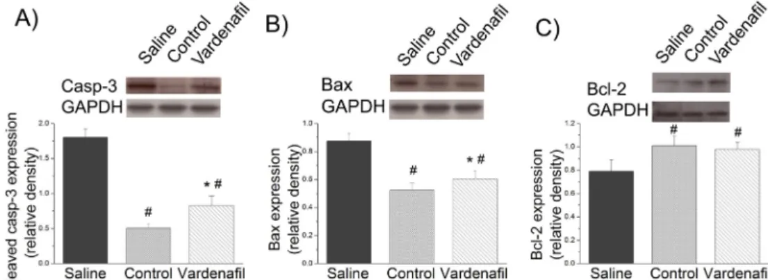
Effect of vardenafil on cleaved caspase-3 level, and Bax and Bcl-2 protein expression
Densitometric analysis of caspase-3 p17 cleavage and Bax bands showed a significant increase in the saline group and was significantly moderated by vardenafil supplement (Fig. 4A, B). Expression of Bcl-2 (Fig. 4C) was significantly decreased in saline group and maintained at the level of controls when supplemented with vardenafil.
DISCUSSION
The major finding of this study is that, with respect to function and structure, long-term storage of rat aortic segments in a vardenafil-supplemented preservation solu- tion provides significantly better protection against I/R injury than the most frequently used saline solution. The
beneficial effect of vardenafil was demonstrated by improved vasodilatatory capacity, lower grade DNA strand breaks, maintained cGMP content, and preserved mRNA and protein expression.
To simulate the whole reperfusion process the well established method of reoxygenation and rewarming with additional HOCl incubation was applied.16,17 After pro- longed cold storage and high reperfusion levels of ROS, including other less toxic oxygen species, HOCl is released from activated leucocytes and severely impairs endothelial cells and NO production, and induces apoptosis in endo- thelial cells and in the vessel wall through oxidative stress.
After prolonged cold storage and reperfusion, high levels of ROS severely impair endothelial cells and NO production.
Damaged endothelial cells are responsible for impaired vasodilatory graft function and for the developing vascul- opathy.20,21 Vascular tone (endothelium-dependent vaso- relaxation) is particularly important in cardiac, as well as in vascular surgery, as it determines postoperative bloodflow and is responsible for the early and late graft thrombosis and stenosis.22 Furthermore, the long-term benefit of revascularisation surgery depends heavily on the long-term
Figure 3.Effects of vardenafil in in vitro ischaemiaereperfusion on the gene expression of (A)endothelin-1(ET-1), (B)caspase-3(casp-3), (C)Bax, (D)Bcl-2, (E)endothelial nitric oxide synthase(eNOS), and (F)inducible nitric oxide synthase(iNOS) in isolated aortic rings. Controls were given the arbitrary value of 1. Values represent medianquartiles.Note. *p<.05 versus control.#p<.05 versus saline.
patency of bypass grafts, which is determined by several factors: the progress of heart/vascular disease, the biolog- ical properties of the implanted graft, and the degree of I/R injury.
Ingemansson et al.23 showed weak contractive ability after administration of U-46619, a TXA2 agonist, after 24 hours of ischaemic storage using University of Wisconsin solution. Another in vitro study demonstrated decreased rat coronary vasodilatory capacity after 8 hours of cold storage in HTK solution.5 Different research groups have aimed to develop individual methods to maintain vascular function during storage.17,24 In our laboratory, we demonstrated a maintained vascular function with new iron chelators after 24 hours of ischaemia.19In accordance with the literature, in the present study we also found an impaired vasodilatative capacity in aortic rings after prolonged cold storage for 24 hours followed by warm HOCl-induced reperfusion injury by using vitro measurements.16,17 The preserved contraction and endothelium-independent relaxation indicated that the impaired vascular response was not a functional deficit of vascular smooth muscle.
Recently, there has been considerable interest in the role of the NOesolube-guanylate-cyclase (sGC)ecGMPePKG pathway in myocardial and endothelial protection.9,25 Pre- vious studies have reported powerful myocardial protection by pharmacological PDE-5 inhibition,1,26by sGC activation,10 and by supplementation of an NO-donor, L-Arginin.27 Intracellular cGMP-accumulation has been shown to reduce oxidative tissue injury in conditions associated with increased free radical release and oxidative stress.10 More recently, the observed cardioprotection with pharmacolog- ically increased cGMP levels has been reported to be mediated by hydrogen sulphide signalling in a PKG- dependent fashion.28 The PKG-dependent cytoprotective mechanism of vardenafil involves phosphorylation of ERK, induction of Bcl-2, and opening of mitochondrial KþATP
channels.13,29 We also demonstrated, in the vardenafil- treated group, up-regulation in both Bcl-2 mRNA and pro- tein expression parallel to a drop in bax and caspase-3 mRNA expressions, and cleaved caspase-3 level, demon- strating inhibition of the apoptotic processes, as also evi- denced by the TUNEL staining results. In this study mRNA
expression of eNOS and iNOS was significantly decreased in the saline and vardenafil groups. Vardenafil treatment had no effect on these levels. It does not contradict the effect of the PDE-5 inhibitors, as they act through the suppression of cGMP degradation, but have no expected direct effect on NO accumulation. Based on functional improvement we propose that the reduced intracellular cGMP level plays a role in the pathogenesis of vascular dysfunction after 24 hours cold storage, which is also supported by the cGMP immunohistochemistry performed in aortic tissue in this study. Furthermore, severe DNA damage could also be effectively prevented by vardenafil, as evidenced by our TUNEL labelling.
To the best of our knowledge, it has not been previously tested whether the selective inhibition of PDE-5 would also be capable of preventing I/R injury after long-term storage.
In conclusion, we report, for thefirst time, that the PDE-5 inhibitor vardenafil added to a preservation solution restored impaired endothelial function of aortic rings after long-term cold storage followed by HOCl-induced warm reperfusion. This work supports the view that PDE5 inhibi- tion might be advantageous in the treatment of endothelial dysfunction during long-term storage.
Study limitations
The PDE-5 inhibitor vardenafil may have therapeutic po- tential to lower oxidative stress with the aim of improving clinical outcome of I/R injury in blood vessels after trans- plantation. However, in our study, aortic rings were har- vested ex vivo to examine vascular reactivity without the involvement of non-aortic tissue, with lack of blood flow and absence of leucocyte activation. Therefore, confirma- tion of these observations in vivo is essential. Also, 24 hours of cold ischaemic storage is validated as part of the well- established in vitro vascular model of I/R, but is not com- mon in clinical practice. Moreover, additional studies would be interesting to investigate the effect of vardenafil on venous grafts.
CONFLICT OF INTEREST None.
Figure 4.The effects of vardenafil in in vitro ischaemiaereperfusion on protein expression of (A)cleaved caspase-3(casp-3), (B)Bax, and (C)Bcl-2in isolated aortic rings (n¼6 per group). Values represent meanstandard error of the mean.Note. *p<.05 versus control.
#p<.05 versus saline.
FUNDING None.
APPENDIX A. SUPPLEMENTARY MATERIAL
Supplementary data related to this article can be found online athttp://dx.doi.org/10.1016/j.ejvs.2013.05.006.
REFERENCES
1 Szabo G, Radovits T, Veres G, Krieger N, Loganathan S, Sandner P, et al. Vardenafil protects against myocardial and endothelial injuries after cardiopulmonary bypass. Eur J Car- diothorac Surg2009;36:657e64.
2 Maathuis MH, Leuvenink HG, Ploeg RJ. Perspectives in organ preservation.Transplantation2007;83:1289e98.
3 Schaeffer U, Tanner B, Strohschneider T, Stadtmuller A, Hannekum A. Damage to arterial and venous endothelial cells in bypass grafts induced by several solutions used in bypass surgery.Thorac Cardiovasc Surg1997;45:168e71.
4 Wilbring M, Tugtekin SM, Zatschler B, Ebner A, Reichenspurner H, Matschke K, et al. Even short-time storage in physiological saline solution impairs endothelial vascular function of saphenous vein grafts. Eur J Cardiothorac Surg 2011;40:811e5.
5 Schroder C, Heintz A, Pexa A, Rauen U, Deussen A. Preclinical evaluation of coronary vascular function after cardioplegia with HTK and different antioxidant additives. Eur J Cardiothorac Surg2007;31:821e6.
6 Zweier JL, Talukder MA. The role of oxidants and free radicals in reperfusion injury.Cardiovasc Res2006;70:181e90.
7 Korkmaz S, Radovits T, Barnucz E, Neugebauer P, Arif R, Hirschberg K, et al. Dose-dependent effects of a selective phosphodiesterase-5-inhibitor on endothelial dysfunction induced by peroxynitrite in rat aorta. Eur J Pharmacol 2009;615:155e62.
8 Boyle Jr EM, Pohlman TH, Cornejo CJ, Verrier ED. Endothelial cell injury in cardiovascular surgery: ischemia-reperfusion.Ann Thorac Surg1996;62:1868e75.
9 Kukreja RC, Ockaili R, Salloum F, Yin C, Hawkins J, Das A, et al.
Cardioprotection with phosphodiesterase-5 inhibition e a novel preconditioning strategy. J Mol Cell Cardiol 2004;36:
165e73.
10 Korkmaz S, Radovits T, Barnucz E, Hirschberg K, Neugebauer P, Loganathan S, et al. Pharmacological activation of soluble guanylate cyclase protects the heart against ischemic injury.
Circulation2009;120:677e86.
11 Abdollahi M, Fooladian F, Emami B, Zafari K, Bahreini- Moghadam A. Protection by sildenafil and theophylline of lead acetate-induced oxidative stress in rat submandibular gland and saliva.Hum Exp Toxicol2003;22:587e92.
12 Dias-Junior CA, Souza-Costa DC, Zerbini T, da Rocha JB, Gerlach RF, Tanus-Santos JE. The effect of sildenafil on pul- monary embolism-induced oxidative stress and pulmonary hypertension.Anesth Analg2005;101:115e20.
13 Salloum FN, Ockaili RA, Wittkamp M, Marwaha VR, Kukreja RC.
Vardenafil: a novel type 5 phosphodiesterase inhibitor reduces myocardial infarct size following ischemia/reperfusion injury via opening of mitochondrial K(ATP) channels in rabbits.J Mol Cell Cardiol2006;40:405e11.
14 Mazo E, Gamidov S, Iremashvili V. The effect of vardenafil on endothelial function of brachial and cavernous arteries. Int J Impot Res2006;18:464e9.
15 Gori T, Sicuro S, Dragoni S, Donati G, Forconi S, Parker JD.
Sildenafil prevents endothelial dysfunction induced by ischemia and reperfusion via opening of adenosine triphosphate-sensitive potassium channels: a human in vivo study.Circulation2005;111:742e6.
16 Radovits T, Lin LN, Zotkina J, Gero D, Szabó C, Karck M, et al.
Poly(ADP-ribose) polymerase inhibition improves endothelial dysfunction induced by reactive hydrogen peroxide in vitro.Eur J Pharmacol2007;564:158e66.
17 Barnucz E, Veres G, Hegedus P, Klein S, Zoller R, Radovits T, et al. Prolyl-hydroxylase inhibition preserves endothelial cell function in a rat model of vascular ischemia reperfusion injury.
J Pharmacol Exp Ther2013;345:25e31.
18 Hirschberg K, Radovits T, Loganathan S, Entz L, Beller CJ, Gross ML, et al. Selective phosphodiesterase-5 inhibition re- duces neointimal hyperplasia in rat carotid arteries after sur- gical endarterectomy. J Thorac Cardiovasc Surg 2009;137:
1508e14.
19 Radovits T, Lin LN, Zotkina J, Koch A, Rauen U, Köhler G, et al.
Endothelial dysfunction after long-term cold storage in HTK organ preservation solutions: effects of iron chelators and N- alpha-acetyl-L-histidine.J Heart Lung Transplant2008;27:208e 16.
20 Chu Y, Wu YC, Chou YC, Chueh HY, Liu HP, Chu JJ, et al. Endo- thelium-dependent relaxation of canine pulmonary artery after prolonged lung graft preservation in University of Wisconsin solution: role of L-arginine supplementation. J Heart Lung Transplant2004;23:592e8.
21 Zhang RZ, Yang Q, Yim AP, He GW. Alteration of cellular elec- trophysiologic properties in porcine pulmonary microcircula- tion after preservation with University of Wisconsin and Euro- Collins solutions.Ann Thorac Surg2004;77:1944e50.
22 He GW. Endothelial function related to vascular tone in cardiac surgery.Heart Lung Circ2005;14:13e8.
23 Ingemansson R, Sjoberg T, Massa G, Steen S. Long-term pres- ervation of vascular endothelium and smooth muscle. Ann Thorac Surg1995;59:1177e81.
24 Zatschler B, Dieterich P, Muller B, Kasper M, Rauen U, Deussen A. Improved vessel preservation after 4 days of cold storage: experimental study in rat arteries. J Vasc Surg 2009;50:397e406.
25 Kukreja RC, Salloum FN, Das A. Cyclic guanosine mono- phosphate signaling and phosphodiesterase-5 inhibitors in cardioprotection.J Am Coll Cardiol2012;29:1921e7.
26 Radovits T, Bomicke T, Kokeny G, Arif R, Loganathan S, Kecsan K, et al. The phosphodiesterase-5 inhibitor vardenafil improves cardiovascular dysfunction in experimental diabetes mellitus.Br J Pharmacol2009;156:909e19.
27 Soos P, Andrasi T, Buhmann V, Kohl B, Vahl C, Hagl S, et al.
Myocardial protection after systemic application of L-arginine during reperfusion.J Cardiovasc Pharmacol2004;43:782e8.
28 Salloum FN, Das A, Samidurai A, Hoke NN, Chau VQ, Ockaili RA, et al. Cinaciguat, a novel activator of soluble guanylate cyclase, protects against ischemia/reperfusion injury: role of hydrogen sulfide.Am J Physiol Heart Circ Physiol2012;302:H1347e54.
29 Das A, Salloum FN, Xi L, Rao YJ, Kukreja RC. ERK phosphory- lation mediates sildenafil-induced myocardial protection against ischemia-reperfusion injury in mice.Am J Physiol Heart Circ Physiol2009;296:H1236e43.