ORIGINAL PRE-CLINICAL SCIENCE
Methane supplementation improves graft function in experimental heart transplantation
K alm an Benke, MD, PhD,
a,b,1D avid Kursz an J asz, MD,
c,1Agnes Lilla Szil agyi,
c,dB alint Bar ath, MD,
c,dEszter Tuboly, PhD,
c,dAnett Rox ana M arton,
c,dPetra Varga,
c,dArp ad Moh acsi, PhD,
eAnna Szab o, PhD,
eZs ofia Sz ell,
c,dMih aly Ruppert, MD,
aTam as Radovits, MD, PhD,
aG abor Szab o, MD, PhD,
bB ela Merkely, MD, PhD, DSc,
aPetra Hartmann, MD, PhD,
c,dand Mih aly Boros, MD, PhD, DSc
c,dFrom theaHeart and Vascular Centre, Semmelweis University, Budapest, Hungary;bDepartment of Cardiac Surgery, Uni- versity of Halle, Halle, Germany;cInstitute of Surgical Research, University of Szeged, Szeged, Hungary;dMTA−SZTE Research Group on Photoacoustic Spectroscopy, University of Szeged, Szeged, Hungary; and theeMTA−SZTE Research Group on Photoacoustic Spectroscopy, University of Szeged, Szeged, Hungary.
BACKGROUND: Maintenance of cell viability during cold storage is a key issue in organ transplanta- tion. Methane (CH4) bioactivity has recently been recognized in ischemia/reperfusion conditions; we therefore hypothesized that cold storage in CH4-enriched preservation solution can provide an increased defense against organ dysfunction during experimental heart transplantation (HTX).
METHODS:The hearts of donor Lewis rats were stored for 60 minutes in cold histidine-tryptophan- ketoglutarate (Custodiol [CS]) or CH4-saturated CS solution (CS-CH4) (n= 12 each). Standard het- erotopic HTX was performed, and 60 minutes later, the left ventricular (LV) pressure-volume rela- tionships LV systolic pressure (LVSP), systolic pressure increment (dP/dtmax), diastolic pressure decrement, and coronary blood flow (CBF) were measured. Tissue samples were taken to detect proinflammatory parameters, structural damage (by light microscopy), endoplasmic reticulum (ER) stress, and apoptosis markers (CCAAT/enhancer binding protein [C/EBP] homologous protein, GRP78, glycogen synthase kinase-3b, very low-density lipoprotein receptor, caspase 3 and 9, B-cell lymphoma 2, and bcl-2-like protein 4), whereas mitochondrial functional changes were analyzed by high-resolution respirometry.
RESULTS:LVSP and dP/dtmax increased significantly at the largest pre-load volumes in CS-CH4grafts as compared with the CS group (114.5§16.6 mm Hg vs 82.8§4.6 mm Hg and 3,133§430 mm Hg/
s vs 1,739§169 mm Hg/s, respectively); the diastolic function and CBF (2.4§0.4 ml/min/g vs 1.3§ 0.3 ml/min/g) also improved. Mitochondrial oxidative phosphorylation capacity was more preserved (58.5§9.4 pmol/s/ml vs 27.7§6.6 pmol/s/ml), and cytochrome c release was reduced in CS-CH4 storage. Signs of HTX-caused myocardial damage, level of ER stress, and the transcription of proapop- totic proteins were significantly lower in CS-CH4grafts.
KEYWORDS:
methane;
cold ischemia- reperfusion;
preservation solution;
experimental heart transplantation;
endoplasmic reticulum stress;
mitochondria
1These authors have contributed equally to this work.
Reprint requests : Mihaly Boros, MD, PhD, DSc/Petra Hartmann, MD, PhD, Institute of Surgical Research, University of Szeged, H-6724 Szeged, Pulz u. 1., Hungary. Telephone: +(36-62) 545-103. Fax: +(36-62) 545-743.
E-mail address:boros.mihaly@med.u-szeged.hu
1053-2498/$ - see front matterÓ2020 International Society for Heart and Lung Transplantation. All rights reserved.
https://doi.org/10.1016/j.healun.2020.11.003
CONCLUSION:The addition of CH4during 1 hour of cold storage improved early in vitro graft function and reduced mitochondrial dysfunction and activation of inflammation. Evidence shows that CH4
reduced ER stress−linked proapoptotic signaling.
J Heart Lung Transplant 2021;40:183−192
Ó2020 International Society for Heart and Lung Transplantation. All rights reserved.
Transplantation is routine medical practice for treating end-stage organ failure, but research to improve outcomes and patient safety is still ongoing.1One of the decisive fac- tors in clinical success is effective allograft protection after organ procurement. Several concepts have been attempted to date, but static storage in a cold solution is still the method of choice for organ preservation after surgical explantation.2,3 Nevertheless, currently used techniques cannot provide a complete defense against transient anoxia or reperfusion-induced tissue damage, and therefore, the search for prevention or reduction of cold storage−related organ dysfunction and injury is a priority task.4
It is recognized that enrichment of preservation solutions with biologically active gases is a conceivable option to improve graft function because gas molecules in a fluid milieu are likely to have access to membranes, channels, and cell components involved in the maintenance of organ homeostasis.5−7 Against this background, a link between methane (CH4) supplementation and organ protection seems unconventional but reasonable. CH4 is the most hydrogen-substituted form of carbon, and as a consequence of its physicochemical properties, it is distributed evenly across membrane barriers.8It is intrinsically non-toxic and widely regarded as physiologically inert.8However, various recent data have provided evidence for CH4bioactivity in various in vivo settings; most importantly, several studies have demonstrated modulator, anti-inflammatory potential for inhaled CH4-based approaches in anoxia-reoxygenation experiments.8−10These results are supported by a series of studies where anti-apoptotic properties have been demon- strated for CH4-enriched solutions, as well as an influence on the pathways involved in pyroptosis, the proinflamma- tory form of programed cell death.11−14
In this context, we set out to establish whether deteriorat- ing graft functions might be modified by the CH4content of a preservation fluid. The effects of CH4in terms of organ storage or transplantation conditions have not yet been investigated, and therefore, the main purpose was to test the feasibility of CH4enrichment of a standard storage solution in a relevant experimental setting. With this aim, we have used the generally employed histidine-tryptophan-ketoglu- tarate (HTK) solution, with or without CH4admixture, in an isogenic rat model of heterotopic heart transplantation (HTX), devoid of immunologic effects.
Our next aim was to explore the cross-sectional details of the in vivo consequences and the underlying mechanisms of CH4 action. Structural and functional mitochondrial damage and disturbed protein folding in the lumen of the rough endoplasmic reticulum (ER), defined as ER stress, are major upstream factors that govern the progression of graft dysfunction after organ procurement.15,16 It has
already been demonstrated that CH4 can protect against ischemia/reperfusion (I/R)-induced apoptosis by inhibiting the phosphoinositide 3-kinase/protein kinase B/glycogen synthase kinase-3b (GSK-3b) pathway and nuclear factor erythroid 2-related factor 2 activation.13,14,17 Other evi- dence suggests that CH4 can possibly limit ER stress as well.18,19
Therefore, we put special emphasis on the detection of the most important hemodynamic variables, together with mitochondrial respiratory parameters and myocardial ER stress−and apoptosis-associated gene expression changes, to investigate the hypothesis that a cytoprotective action of CH4 enrichment may target ER stress and its functional links to mitochondria in transplanted rat hearts.
Materials and methods
The experiments were carried out on male Lewis rats (250−350 g;
Charles River, Sulzfeld, Germany) in accordance with EU Direc- tive 2010/63 for the protection of animals used for scientific pur- poses and in compliance with criteria set down in the US National Institutes of Health Guidelines for the Care and Use of Laboratory Animals. The study was approved by the national competent authority of Hungary National Scientific Ethical Committee on Animal Experimentation (ATET) under license number PEI/001/
2374-4/2015.
Production of CH
4-enriched
HTKCommercially available Custodiol (CS) solution (Dr Franz K€ohler Chemie GmbH, Bensheim, Germany) was saturated with pure CH4(>99.9%) under 0.4 MPa for 4 hours in a high-pressure vessel (Messer, Budapest, Hungary), as described previously.20The CH4
concentration in the fluid phase was detected by gas chromatogra- phy, whereas the stability of the solution was checked by near- infrared laser-based photoacoustic spectroscopy. The solution con- taining 6.57§0.27mmol/ml CH4was freshly prepared and stored at 4˚C before use (Supplementary Material, available online at www.jhltonline.org).
Experimental protocol
Isogenic male Lewis to Lewis HTX (n= 36) was performed as described previously.21Briefly, after excision from the donors, the grafts were cold-stored in a transplantation solution for 60 minutes (cold ischemia time), which was followed by heterotopical trans- plantation and a 60-minute reperfusion period. During transplanta- tions, the aorta and the pulmonary artery of the donor heart were anastomosed end-to-side to the abdominal aorta and the inferior vena cava of the recipient rat, respectively, using microsurgical techniques. At the end of the reperfusion, in situ hemodynamic measurements were performed in the recipient to evaluate early
184 The Journal of Heart and Lung Transplantation, Vol 40, No 3, March 2021
graft functions; thereafter, biopsies were taken from the left ventri- cle of the grafts for mitochondrial functional measurements and bio- chemical assays. Tissue myeloperoxidase (MPO) and xanthine oxidoreductase (XOR) activity, reduced glutathione (GSH) and oxi- dized glutathione disulfide (GSSG) ratio, and tissue nitrite/nitrate (NOx) level were determined. Blood samples for serum biomarkers of myocardial injury were taken from the vena cava at the end of the reperfusion period (Supplementary Material online).
The animals were randomly allocated into 3 groups. In control Group 1 (n= 12), donor rats underwent the same surgical proce- dure until the explantation, but the hearts were not subjected to cold ischemia and storage and were not transplanted. In Group 2 (n= 12), the explanted grafts were stored in CS solution at 4˚C during the 60-minute cold ischemic period, whereas in Group 3 (n= 12), the grafts were stored in CH4-enriched CS during the 60- minute cold ischemic period. In this group, the cold cardioplegic CS solution used to arrest the heart was also supplemented with CH4(Figure 1).
Hemodynamic measurements in the graft
We have employed a heterotopic HTX model where the recipient aorta supplies the graft; that is, the blood flows from the recipient aorta to the aorta root of the graft. The aortic valve of the graft is competent, and therefore, the coronaries are perfused without entering the left ventricle. This virtually completely unloaded LV model is suitable for measurements of global hemodynamic parameters and the extent of functional damage of the experimen- tally transplanted hearts.22,23 The LV pressure-volume relation- ships were determined as follows. After transplantation and 60- minute reperfusion, a 3F latex balloon catheter (Edwards Life- sciences Corporation, Irvine, CA) was introduced in situ into the left ventricle via the apex. The maximal LV systolic pressure
(LVSP), the maximal slope of systolic pressure increment (dP/
dtmax), and diastolic pressure decrement (dP/dtmin) were deter- mined with a Millar micromanometer (SPR-838, Millar, Houston, TX) at different LV volumes (20−180 ml) with an injection of saline solution. The coronary blood flow (CBF) of the graft was measured indirectly with an ultrasonic flowmeter (Transonic Sys- tems Inc, Ithaca, NY) mounted on the ascending aorta of the graft, which is the only outlet for circulating blood through the coronaries.22,24
Examination of cardiac mitochondrial functions
The efficacy of the mitochondrial respiration was assessed from heart homogenates by high-resolution respirometry (Oxygraph- 2k, Oroboros Instruments, Innsbruck, Austria). Mitochondrial O2
consumption (respiratory flux), complex II−linked baseline respi- ration (succinate-fueled, in the presence of complex I inhibitor rotenone), oxidative phosphorylation (OxPhos) capacity, and cyto- chrome c release (an indicator of inner mitochondrial membrane damage) were determined as described previously.25
Quantitative real-time polymerase chain reaction analysis
Myocardial mRNA expression was analyzed by quantitative real-time polymerase chain reaction (Applied Biosystems, Fos- ter City, CA) for the following genes: caspase-3, caspase-9, DNA damage-inducible transcript 3 (Ddit3; also known as CCAAT/enhancer binding protein [C/EBP] homologous protein [CHOP]), hypoxia-inducible factor 1-alpha (HIF1a), glycogen synthase kinase-3b(GSK-3b), andvery low-density lipoprotein receptor (VLDLr).
Figure 1 Experimental protocol. The heart grafts were explanted from the donors and stored for 60 minutes in cold preservation solution before heterotopical HTX. At 60 minutes after the start of reperfusion, hemodynamic measurements were performed in the recipients to evaluate early post-transplant graft function. Thereafter, samples were taken from the left ventricle for mitochondrial functional measure- ments, biochemical assays, qPCR analysis, and histology. Hearts in the control group underwent the same surgical procedure as those of the donors but were not subjected to cold storage and transplantation. Grafts in the CS group were stored in cold (4˚C) CS solution during the cold ischemia period; in the CS-CH4group, the protocol was identical, except that CH4-enriched CS solution was used. CH4, methane; CS, Custodiol; HTX, heart transplantation; qPCR, quantitative real-time polymerase chain reaction.
Histology and immunohistochemistry
The samples were fixed in buffered paraformaldehyde solution (4%) and embedded in paraffin. Next, 5-mm thick sections were cut and stained with hematoxylin and eosin. Structural damage assessment was performed according to a previously described histological scoring system (Supplementary Material).
Sarco/endoplasmic reticulum Ca2+ ATPase (SERCA) is the major regulator of Ca2+homeostasis and contractility in cardiac and skeletal muscle.26 In addition, the ER stress response can induce an overexpression of SERCA isoforms, including SERCA1 in the post-ischemic heart. Based on this background, SERCA1 expression was detected with standard immunohistochemical staining technique (using #S1189 ab, Sigma Aldrich, St. Louis, MO) in the left ventricles of transplanted rat heart grafts.
Results
Hemodynamic parameters of the transplanted grafts
After transplantation, increasing LV balloon volumes (pre- load) resulted in elevated LVSP and dP/dtmax, which were both significantly increased at the largest pre-load values in the CS-CH4group compared with CS alone (Figure 2A and B). A similar change in diastolic function was noted at higher pre-load volumes, bringing about significantly ele- vated dP/dtmin values (p < 0.05) compared with CS, reflecting better myocardial relaxation (Figure 2C). CBF was also significantly (p<0.05) higher after 60 minutes of reperfusion in CS-CH4 storage than in the CS group (Figure 2D). There was no statistically significant differ- ence in heart rate values within and between experimental groups (Figure 2E).
Cardiac mitochondrial function
Complex II−linked basal respiration was significantly higher in the CS-CH4 grafts than in the CS group 60 minutes after reperfusion (data not shown). After adding saturating amounts of adenosine diphosphate, the OxPhos capacity was significantly higher in the CS-CH4 group (Figure 3C). Mitochondrial respiration in response to cyto- chrome c (Figure 3D) was tested to determine the ability of exogenous cytochrome c to replace the enzyme in the mito- chondrial membrane. In comparison with the CS group, the release of cytochrome c was significantly lower in the CS- CH4group.
Myocardial
ERstress
−and apoptosis-associated gene expression
The relative mRNA expression for hypoxia- and ER stress
−associated genes (HIF-1a, CHOP, GSK-3b, and Vldlr) was significantly lower in the CS-CH4 group (Figure 4).
The expression of caspase-3 and caspase-9 and the proa- poptotic bcl-2-like protein 4 (Bax) were not significantly decreased. However, the anti-apoptotic B-cell lymphoma 2 (Bcl2) and the ratio of Bax/Bcl2 expression were signifi- cantly different in the CS-CH4group, thus indicating the relative dominance of anti-apoptotic pathways (Figure 4).
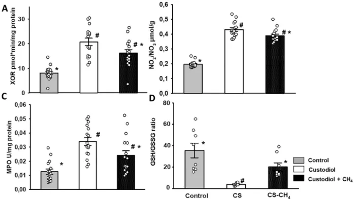
Oxidative stress markers
XOR is a key enzyme in reperfusion-induced reactive oxy- gen species production; in addition, it can catalyze the reduction of nitrates and nitrites to nitric oxide. XOR activ- ity and tissue NOx levels were both significantly decreased
Figure 2 LV pressure-volume relations and CBF changes. (A) dP/dtmax, (B) maximal LVSP, (C) dP/dtmin, (D) CBF, and (E) heart rate. White columns: CS group; black columns: CS-CH4group. Data are presented as means§SEM. #p<0.05 vs CS (one-way ANOVA, Tukey’s test). ANOVA, analysis of variance; CBF, coronary blood flow; CH4, methane; CS, Custodiol; dP/dtmax, maximal slope of the sys- tolic pressure increment; dP/dtmin, diastolic pressure decrement; LV, left ventricular; LV, left ventricular systolic pressure.
186 The Journal of Heart and Lung Transplantation, Vol 40, No 3, March 2021
Figure 3 Oxygen consumption of cardiac mitochondria (pmol/s/ml 1). (A, B) The upper charts demonstrate representative records of mitochondrial oxygen consumption of (A) CS-stored or (B) CS-CH4−stored samples measured by high-resolution respirometry. The blue line represents the instantaneous oxygen concentration in the respiration chamber, whereas the red line indicates the simultaneous oxygen consumption of the sample. (C) The lower right-hand chart shows OxPhos capacity, and (D) the lower left-hand chart demonstrates cyto- chrome c release data. Gray columns: control group; white columns: CS group; black columns: CS-CH4group. Data are presented as means§SEM, individual data points are shown (n= 7-7). Cytochrome c release measurements were made in duplicate. *p<0.05 vs CS;
#p<0.05 vs. control (one-way ANOVA, Tukey’s test). ANOVA, analysis of variance; CH4, methane; CS, Custodiol; OxPhos, oxidative phosphorylation.
Figure 4 Gene expression changes. White columns: CS group; black columns: CS-CH4group. Data are presented as means§SEM, individual data points are shown (n = 7-7). #p<0.05 vs. CS (one-way ANOVA, Tukey’s test). ANOVA, analysis of variance; Bax, bcl-2- like protein 4; Bcl-2, B-cell lymphoma 2; CH4, methane; CHOP, CCAAT/enhancer binding protein (C/EBP) homologous protein; CS, Cus- todiol; GSK-3b, glycogen synthase kinase-3b; HIF-1a, hypoxia-inducible factor-1a; VLDLr, very low-density lipoprotein receptor. The y- axis shows fold-change in gene expression.
when CS-CH4was applied during the cold ischemia period relative to the data for the CS group (Figure 5A and B). MPO is mostly produced by activated polymorphonuclear leuko- cytes. Although tissue MPO was significantly increased as compared with that of the control group, MPO activity was significantly reduced when CH4-CS was applied (Figure 5C).
The GSH/GSSG ratio is one of the most important markers of oxidoreductive stress. This ratio was significantly decreased in the CS group; however, preservation of grafts in CS-CH4resulted in a sustained GSH/GSSG ratio (Figure 5D).
Laboratory parameters of myocardium-specific enzyme changes
CH4 admixture in the CS-CH4 group resulted in signifi- cantly lower plasma lactate dehydrogenase, creatine kinase,
creatine kinase myocardial band, and troponin T levels as compared with CS storage alone (Table 1).
Histology
Hematoxylin and eosin staining showed only a mild disor- ganization of the myofibrils with loss of striations and a combination of waviness, contraction bands, and disruption of plasma membranes of myocytes in the CS group as com- pared with the controls (Figure 6A and B). The architecture of cardiac myocytes was nearly normal in the CS-CH4stor- age group (Figure 6C). These changes were not signifi- cantly different from those in the CS group, thus indicating nearly equal potential for tissue protection (Figure 6D). The number of SERCA1 immunoreactive cardiac myocytes increased significantly in sections from CS-stored grafts as compared with the controls (Figure 6E and F). In contrast, Figure 5 Biochemical assays for oxidoreductive stress parameters. (A) Tissue XOR activity, (B) NO2/NO3levels, (C) MPO activity and (D) GSH/GSSG ratio. Gray columns: control group; white columns: CS group; black columns: CS-CH4group. Data are presented as means§ SEM, individual data points are shown for the columns (n= 8-8). XOR, MPO, and NO2/NO3measurements were made in duplicate.*p<
0.05 vs CS; #p< 0.05 vs. control (one-way ANOVA, Tukey’s test). ANOVA, analysis of variance; CH4, methane; CS, Custodiol; GSH, reduced glutathione; GSSG, oxidized glutathione disulfide; MPO, myeloperoxidase; NO2, nitrite; NO3, nitrate; XOR, xanthine oxidoreductase.
Table 1 Myocardium-Specific Enzyme Changes
Group CK (U/L) CK-MB (U/L) LDH (U/L) Troponin T (ng/L)
Control (n= 12) 503§57a 206§31a 358§79a 42§9a
CS (n= 12) 2327§23b 527§43b 938§108b 172§36b
CS-CH4(n= 12) 1507§49a,b 328§52a,b 732§96a,b 110§21a,b
Abbreviations: ANOVA, analysis of variance; CH4, methane; CK, creatine kinase; CK-MB, creatine kinase myocardial band; CS, Custodiol.
Data are presented as means§SEM.
ap<0.05 vs CS
bp<0.05 vs control (one-way ANOVA, Tukey’s test).
188 The Journal of Heart and Lung Transplantation, Vol 40, No 3, March 2021
the number of immunoreactive cells was significantly reduced in the CS-CH4group (Figure 6G and H).
Discussion
This study aimed to investigate whether adding CH4to a cold preservation solution modifies the graft function in experi- mental HTX. The hemodynamic efficacy of CS-CH4storage was evidenced by increased LVSP, cardiac contractility, and coronary circulation as compared with CS-treated grafts. The sum of biochemical data showed that the CH4-containing HTK solution effectively reduced the degree of oxidoreduc- tive stress in myocardial samples and significantly influenced several components of ER stress—mitochondria-related proapoptotic signaling pathways. In addition, high-resolution respirometry confirmed that CH4supplementation preserved the respiratory mechanism of cardiac mitochondria during cold storage. These pathways together may have contributed to improved structures and functions in this HTX model.
The myocardium has particularly poor tolerance to pro- longed ischemia, and the issue of preservation is a major concern in transplantation.27The HTK solution is generally used in clinical practice; therefore, it is an appropriate testbed for alternative options.28 The gas mediators nitric oxide, carbon monoxide, and hydrogen sulfide have already been tried as additives to solutions in transplantation mod- els, assuming that a potential efficacy could be related to their tendency to react with biologically important molecules.5,6,29In contrast, CH4is intrinsically non-toxic in vivo; it is a simple asphyxiant, which means that hypoxia might occur when an increasing concentration of CH4
displaces inhaled air in a restricted area and the concentra- tion of oxygen is reduced.30 Nevertheless, there are perti- nent data that demonstrate that CH4 can modulate nitric oxide−, carbon monoxide−, and hydrogen sulfide−linked reactions in living systems.8,10In addition, higher concen- trations of exogenous CH4can lead to direct anti-cytokine effects via master switches, such as nuclear factor erythroid 2-related factor 2/Keap1 or nuclear factor-kB, and anti- inflammatory responses in experimental conditions.9,11,20,25 In the case of myocardial I/R, treatment with CH4-enriched saline significantly ameliorated the sequelae of proinflam- matory activation (evidenced by reduced tumor necrosis factor-a, interleukin-1b, MPO activity, and oxidative DNA damage) and maintained cardiac function 4 weeks after infarction.11
The immediate hemodynamic circulatory consequences of CH4-enriched graft storage included a significantly improved myocardial contractility and a parallel increase in CBF during the 60-minute reperfusion. These data suggest that, in this short time frame, exogenous CH4can restrain or counteract those mechanisms that would otherwise influ- ence the cardiac contractility negatively. This conclusion is consistent with earlier results where CH4treatment main- tained a satisfactory cardiac function measured at 4 weeks after infarction, with improved LV ejection fraction, dia- stolic volume, and contractility, among other improve- ments, compared with non−CH4-treated rats.11
Despite the wide range of research to map the biological effects, the role of CH4in cold ischemia or organ transplan- tation settings has not yet been investigated. Therefore, we manufactured a CH4-saturated HTK solution according to Figure 6 Histology and immunohistochemistry. (A−C) H&E staining of heart sections. (A) Control group, (B) CS group, and (C) CS- CH4group. (D) Histological grading of groups represents a composite of number of damaged myocytes and number of foci of damage (n= 12-12). (E−G) SERCA1 immunostaining of heart sections. (E) Control group, (F) CS group, and (G) CS-CH4group. (H) SERCA1 immunoreactivity is demonstrated as percentage of immunopositive cells quantified per field of view. Data are presented as means§SD (individual data points are shown for the columns,n= 12-12). *p<0.05 vs CS; #p<0.05 vs control (ANOVA on rank, Tukey−Kramer).
Magnification:£200. Bar = 200mm. ANOVA, analysis of variance; CH4, methane; CS, Custodiol; H&E, hematoxylin and eosin; SERCA, sarco/endoplasmic reticulum Ca2+ATPase.
reported protocols and in a concentration range that demon- strated efficacy in I/R studies in vivo.20Many details of the mechanism are still unknown, but we have shown that this approach affected the mitochondrial physiology during cold ischemia and after reperfusion, perhaps through an indirect influence on Ca2+homeostasis.11,20During circula- tory arrest, depletion of mitochondrial substrates is a major contributor to Ca2+ influx−mediated membrane dysfunc- tions. As a result of CH4 enrichment, the mitochondria were more responsive to adenosine diphosphate utilization, which contributed to the maintenance of OxPhos capacity.
Furthermore, cytochrome c release, a sign of mitochondrial inner membrane injury, was also reduced (Figure 7).
The process of cold ischemia−induced cellular damage with the dual contribution of ER and mitochondria is rela- tively well characterized. Hypoxic conditions trigger changes in cytoplasmic resting potential and, through the activation of ER-mediated Ca2+ transport, increase the expression of HIF-1a, one of the key initial factors in the cascade of events, which will finally lead to cell apoptosis or necrosis.31−34More directly, as a consequence of HIF- 1aexpression, mRNA expression of VLDLr and the proa- poptotic transcription factor CHOP are also increased, and by the end of this process, CHOP upregulates bcl-2-like protein 11 mRNA expression and activates Bax protein to translocate from the cytosol to the mitochondria.31,35 Our results demonstrate that cold ischemia and graft storage activated all these participants, starting from higher
SERCA1 protein levels reflecting an increased Ca2+pump function in the ER and elevated HIF-1aexpression in cardi- omyocytes. In addition, higher intracellular Ca2+ can acti- vate GSK-3b, the proapoptotic factor in the intrinsic mitochondrial apoptotic pathway.36 These mitochondrial changes raise the expression of Bax protein and its activa- tion, whereas modified proapoptotic Bax and anti-apoptotic Bcl2 levels lead to further proapoptotic events, such as cytochrome c release. CS-CH4storage did not influence the caspase enzyme system, but the Bax/Bcl2 ratio and the reduced cytochrome c release suggest that the intrinsic mitochondrial pathway of apoptosis was affected. More importantly, if the preservation solution was supplemented with CH4, the expression of individual genes in the pro- posed signaling pathway was also reduced (Figure 7).
Our study has several limitations. First, animal models do not predict human responses precisely, and in prioritiz- ing the interventions, we had to build on previously col- lected scientific information. Therefore, it is conceivable that other known or unknown mechanisms could also play a role in the reduction of tissue damage in this setup. Second, a rat model of heterotopic abdominal HTX provides impor- tant data on the dynamics of myocardial changes, but the unloaded reperfusion of the graft leads to relatively fast recovery. In other words, this method reduces experimental variability, but the relevant observation time is limited.
Therefore, further pre-clinical transplantation studies should evaluate whether CH4supplementation can confer Figure 7 Proposed effects of methane supplementation on cold ischemia−induced intracellular changes. (A) Cold storage, ischemic, and hypoxic states will lead to perturbations in normal ER functions. ER stress is accompanied by intracellular Ca2+overload, which modu- lates the activation of proapoptotic GSK-3band leads to caspase activation. Hypoxia increases HIF-1aexpression as well, which interacts with the promotor of VLDLr and the proapoptotic transcription factor CHOP by direct binding and by the non-classical HRE. By the end of this process, CHOP upregulates the Bcl-2 family BH3 protein Bim mRNA expression, which directly activates Bax to translocate from the cytosol to the mitochondria. These events will trigger cardiomyocyte apoptosis. (B) Phospholipid membranes of the ER and the mitochon- drion are possible targets of CH4. In the presence of a saturating amount of CH4, the expression of genes downstream of HIF-1adecreases, affecting the mitochondrial pathway of apoptosis by lowering the Bax/Bcl2 ratio, thus creating an anti-apoptotic milieu for cardiac muscle cells. Bax, bcl-2-like protein 4; Bcl-2: B-cell lymphoma 2; Bim, bcl-2-like protein 11; CH4, methane; CHOP: CCAAT/enhancer binding protein (C/EBP) homologous protein; ER, endoplasmic reticulum; GSK-3b: glycogen synthase kinase-3b; HIF-1a, hypoxia-inducible fac- tor-1a; HRE, hypoxia responsive element; VLDLr, very low-density lipoprotein receptor.
190 The Journal of Heart and Lung Transplantation, Vol 40, No 3, March 2021
in vivo tissue protection not only in rodents but also in larger animals with longer cold ischemia and prolonged reperfusion times.
In summary, our study has demonstrated that CH4 enrichment of HTK solution results in increased graft pro- tection during cold ischemia and isogenic HTX in rats. Oxi- doreductive imbalance is an inevitable consequence of ex vivo periods and a basis for a cascade of proinflammatory events following reoxygenation. Based on the totality of data, it seems that CH4 supplementation conferred increased efficacy on HTK to reduce signs of nitroxidative stress as shown by the maintained GSH/GSSG ratio, reduced MPO and XOR activity, and lower NOx level in the reperfused myocardium.
The underlying mechanism is attributed at least partly to an influence of CH4on myocardial ER stress and its link to mitochondrial structural and functional reactions. CH4 enrichment is a simple and effective option for static organ preservation and also seems feasible for dynamic graft stor- age. Future research should particularly seek to answer the question of whether this approach confers long-term protec- tion in immunologically challenged situations.
Disclosure statement
The authors declare no conflicts of interest. This study was supported by a National Research Development and Inno- vation Fund of Hungary (National Research Development and Innovation Fund Grant K120232 and NVKP_16-1- 2016-0017[National Heart Program]), an Economic Devel- opment and Innovation Operative Programme grant (GINOP-2.3.2-15-2016-00015), an Higher Educational Institutional Excellence Program grant (TUDFO/47138-1/
2019-ITM), and a Human Resources Development Opera- tional Programme grant (EFOP-3.6.2-16-2017-0006).
Supplementary materials
Supplementary material associated with this article can be found in the online version athttps://doi.org/10.1016/j.hea lun.2020.11.003.
Supplementary data
Supplementary data associated with this article can be found in the online version at www.jhltonline.org.
References
1. Braithwaite SA, van der Kaaij NP. New techniques for optimization of donor lungs/hearts. Anesthesiol Clin 2019;37:639-60.
2. Ardehali A, Esmailian F, Deng M, et al. Ex-vivo perfusion of donor hearts for human heart transplantation (PROCEED II): a prospective, open-label, multicentre, randomised non-inferiority trial. Lancet 2015;
385:2577-84.
3.Nasralla D, Coussios CC, Mergental H, et al. A randomized trial of nor- mothermic preservation in liver transplantation. Nature 2018;557:50-6.
4. Li Y, Guo S, Liu G, et al. Three preservation solutions for cold storage of heart allografts: a systematic review and meta-analysis. Artif Organs 2016;40:489-96.
5. Nakao A, Kaczorowski DJ, Wang Y, et al. Amelioration of rat cardiac cold ischemia/reperfusion injury with inhaled hydrogen or carbon monoxide, or both. J Heart Lung Transplant 2010;29:544- 53.
6. Srinivasan PK, Yagi S, Doorschodt B, et al. Impact of venous systemic oxygen persufflation supplemented with nitric oxide gas on cold- stored, warm ischemia-damaged experimental liver grafts. Liver Transpl 2012;18:219-25.
7. Tan M, Sun X, Guo L, Su C, Sun X, Xu Z. Hydrogen as additive of HTK solution fortifies myocardial preservation in grafts with pro- longed cold ischemia. Int J Cardiol 2013;167:383-90.
8. Boros M, Keppler F. Methane production and bioactivity-a link to oxido-reductive stress. Front Physiol 2019;10:1244.
9. Boros M, Ghyczy M,Erces D, et al. The anti-inflammatory effects of methane. Crit Care Med 2012;40:1269-78.
10. Poles MZ, Juhasz L, Boros M. Methane and inflammation—a review (fight fire with fire). Intensive Care Med Exp 2019;7:68.
11. Chen O, Ye Z, Cao Z, et al. Methane attenuates myocardial ischemia injury in rats through anti-oxidative, anti-apoptotic and anti-inflamma- tory actions. Free Radic Biol Med 2016;90:1-11.
12. Tong Y, Dong Y, Feng Y, et al. Methane-rich saline: a potential resus- citation fluid for hemorrhagic shock. Oxid Med Cell Longev 2019;
2019:4929107.
13. Wang L, Yao Y, He R, et al. Methane ameliorates spinal cord ische- mia-reperfusion injury in rats: antioxidant, anti-inflammatory and anti-apoptotic activity mediated by Nrf2 activation. Free Radic Biol Med 2017;103:69-86.
14. Li Z, Jia Y, Feng Y, et al. Methane alleviates sepsis-induced injury by inhibiting pyroptosis and apoptosis: in vivo and in vitro experiments.
Aging (Albany NY) 2019;11:1226-39.
15. Glembotski CC. Endoplasmic reticulum stress in the heart. Circ Res 2007;101:975-84.
16. Pallet N, Fougeray S, Beaune P, Legendre C, Thervet E, Anglicheau D. Endoplasmic reticulum stress: an unrecognized actor in solid organ transplantation. Transplantation 2009;88:605-13.
17. Zhang B, Gao M, Shen J, He D. Inhaled methane protects rats against neurological dysfunction induced by cerebral ischemia and reperfu- sion injury: PI3K/Akt/HO-1 pathway involved. Arch Med Res 2017;
48:520-5.
18. Gomez L, Thiebaut PA, Paillard M, et al. The SR/ER-mitochondria calcium crosstalk is regulated by GSK3bduring reperfusion injury.
Cell Death Differ 2015;22:1890.
19. Meszaros AT,AL Szil agyi, Juhasz L, et al. Mitochondria as sources and targets of methane. Front Med (Lausanne) 2017;4:195.
20. Ye Z, Chen O, Zhang R, et al. Methane attenuates hepatic ischemia/
reperfusion injury in rats through antiapoptotic, anti-inflammatory, and antioxidative actions. Shock 2015;44:181-7.
21. Benke K, Matyas C, Sayour AA, et al. Pharmacological precondition- ing with gemfibrozil preserves cardiac function after heart transplanta- tion. Sci Rep 2017;7:14232.
22. Benke K, Sayour AA, Matyas C, et al. Heterotopic abdominal rat heart transplantation as a model to investigate volume dependency of myo- cardial remodeling. Transplantation 2017;101:498-505.
23. Kato H, Tomita S, Yamaguchi S, Ohtake H, Watanabe G. Subzero 24- hr nonfreezing rat heart preservation: a novel preservation method in a variable magnetic field. Transplantation 2012;94:473-7.
24. Ahmed N, Farooq J, Sadiq S, et al. Fingolimod (FTY720) preserves high energy phosphates and improves cardiac function in heterotopic heart transplantation model. Int J Mol Sci 2020;21:E6548.
25. Strifler G, Tuboly E, Szel E, et al. Inhaled methane limits the mito- chondrial electron transport chain dysfunction during experimental liver ischemia-reperfusion injury. PLoS One 2016;11:e0146363.
26. Chemaly ER, Troncone L, Lebeche D. SERCA control of cell death and survival. Cell Calcium 2018;69:46-61.
27. Minasian SM, Galagudza MM, Dmitriev YV, Karpov AA, Vlasov TD.
Preservation of the donor heart: from basic science to clinical studies.
Interact Cardiovasc Thorac Surg 2015;20:510-9.
28. Rauen U, Klempt S, de Groot H. Histidine-induced injury to cultured liver cells, effects of histidine derivatives and of iron chelators. Cell Mol Life Sci 2007;64:192-205.
29. Lobb I, Mok A, Lan Z, Liu W, Garcia B, Sener A. Supplemental hydro- gen sulphide protects transplant kidney function and prolongs recipient survival after prolonged cold ischaemia-reperfusion injury by mitigating renal graft apoptosis and inflammation. BJU Int 2012;110:E1187-95.
30. Boros M, Tuboly E, Meszaros A, Amann A. The role of methane in mammalian physiology-is it a gasotransmitter? J Breath Res 2015;
9:014001.
31. Delbrel E, Soumare A, Naguez A, et al. HIF-1atriggers ER stress and CHOP-mediated apoptosis in alveolar epithelial cells, a key event in pulmonary fibrosis. Sci Rep 2018;8:17939.
32. Lopez-Hernandez B, Ce~na V, Posadas I. The endoplasmic reticulum stress and the HIF-1 signalling pathways are involved in the neuronal damage caused by chemical hypoxia. Br J Pharmacol 2015;172:2838-51.
33. Rizzuto R, Marchi S, Bonora M, et al. Ca(2+) transfer from the ER to mitochondria: when, how and why. Biochim Biophys Acta 2009;
1787:1342-51.
34. Yang D, Gao L, Wang T, Qiao Z, Liang Y, Zhang P. Hypoxia triggers endothelial endoplasmic reticulum stress and apoptosis via induction of VLDL receptor. FEBS Lett 2014;588:4448-56.
35. Huang D, Yan ML, Chen KK, et al. Cardiac-specific overexpression of silent information regulator 1 protects against heart and kidney deteri- oration in cardiorenal syndrome via inhibition of endoplasmic reticu- lum stress. Cell Physiol Biochem 2018;46:9-22.
36. Yang K, Chen Z, Gao J, et al. The key roles of GSK-3bin regu- lating mitochondrial activity. Cell Physiol Biochem 2017;44:
1445-59.
192 The Journal of Heart and Lung Transplantation, Vol 40, No 3, March 2021