Experimental investigation of myocardial hypertrophy in health and disease
PhD dissertation
Balázs Tamás Németh, MD
Semmelweis University
Doctoral School of Basic Medicine
Tutor: Tamás Radovits, M.D., Ph.D.
Opponents: Attila Borbély, M.D., Ph.D.
Levente Kiss, M.D., Ph.D.
Head of the Final Examination Committee:
Emil Monos, M.D., D.Sc.
Members of the Final Examination Committee:
Anikó Görbe, M.D., Ph.D.
Csaba Csonka, M.D., Ph.D.
Budapest
2019
Contents
1. LIST OF ABBREVIATIONS ... 3
2. INTRODUCTION ... 8
2.1.PHYSIOLOGICAL MYOCARDIAL HYPERTROPHY ... 11
2.1.1. Cardiac structural changes in physiological myocardial hypertrophy... 11
2.1.2. Functional changes associated with exercise training ... 13
2.1.3. Molecular pathways underlying physiological myocardial hypertrophy... 15
2.2.PATHOLOGICAL MYOCARDIAL HYPERTROPHY ... 19
2.2.1. Main structural changes in pathological compared with physiological myocardial hypertrophy ... 19
2.2.2. Myocardial dysfunction associated with pathological myocardial hypertrophy ... 21
2.2.3. Molecular pathways implicated in pathological myocardial hypertrophy ... 22
2.3.REDOX AND NITRIC OXIDE/CGMP SIGNALING IN CARDIOVASCULAR PHYSIOLOGY AND PATHOLOGY ... 28
2.3.1. The role of redox signaling in cardiac (patho)physiology ... 28
2.3.2. Nitric oxide and cGMP signaling in cardiovascular health and disease... 31
2.3.3. Modulation of sGC activity – a novel pharmacotherapeutic concept ... 34
3. OBJECTIVES ... 36
4. MATERIALS AND METHODS ... 37
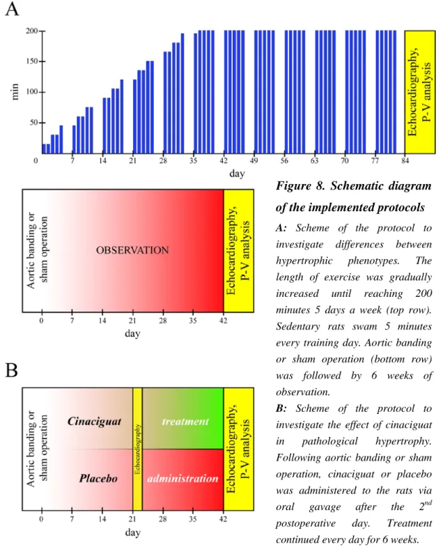
4.1.EXPERIMENTAL PROTOCOLS, TREATMENT GROUPS ... 37
4.1.1. Differences between physiological and pathological myocardial hypertrophy ... 37
4.1.2. Effects of cinaciguat in pathological myocardial hypertrophy ... 39
4.2.ECHOCARDIOGRAPHY ... 39
4.3.HEMODYNAMIC MEASUREMENTS:LVPRESSURE-VOLUME (P-V)ANALYSIS ... 40
4.4.MEASUREMENT OF ORGAN WEIGHTS AND TIBIA LENGTH ... 43
4.5.HISTOLOGY AND IMMUNOHISTOCHEMISTRY ... 43
4.5.1. Cardiomyocyte diameter measurement ... 43
4.5.2. Assessment of LV collagen content ... 43
4.5.3. cGMP immunostaining ... 44
4.5.4. Terminal deoxynucleotidyl transferase dUTP nick end labeling (TUNEL) assay . 44 4.6.BIOCHEMICAL MEASUREMENTS ... 45
4.7.CARDIAC MRNAANALYSIS ... 45
4.8.IMMUNOBLOT ANALYSIS ... 47
4.9.DRUGS ... 48
4.10.STATISTICAL ANALYSIS... 48
5. RESULTS ... 50
5.1.DIFFERENCES BETWEEN PHYSIOLOGICAL AND PATHOLOGICAL MYOCARDIAL HYPERTROPHY ... 50
5.1.1. Morphological assessment ... 50
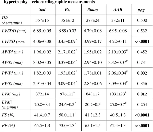
5.1.2. Echocardiographic parameters ... 50
5.1.3. Pressure-volume analysis ... 51 1
5.1.4. Histology ... 53
5.1.5. Cardiac mRNA analysis ... 53
5.2.EFFECTS OF CINACIGUAT IN PATHOLOGICAL MYOCARDIAL HYPERTROPHY ... 58
5.2.1. Morphological assessment ... 58
5.2.2. Echocardiographic parameters ... 58
5.2.3. Invasive hemodynamic measurements and P-V analysis ... 58
5.2.4. Histology ... 64
5.2.5. Molecular and biochemical measurements ... 64
6. DISCUSSION ... 69
6.1.DIFFERENCES BETWEEN PHYSIOLOGICAL AND PATHOLOGICAL MYOCARDIAL HYPERTROPHY ... 69
6.2.EFFECTS OF CINACIGUAT IN PATHOLOGICAL MYOCARDIAL HYPERTROPHY ... 76
6.3.LIMITATIONS ... 81
7. CONCLUSIONS ... 82
8. SUMMARY ... 83
9. ÖSSZEFOGLALÁS ... 84
10. REFERENCES ... 85
11. LIST OF PUBLICATIONS ... 130
11.1.PUBLICATIONS RELATED TO THE DISSERTATION ... 130
11.2.PUBLICATIONS NOT RELATED TO THE DISSERTATION ... 130
12. ACKNOWLEDGEMENTS ... 136
2
1. List of Abbreviations
AAB – abdominal aortic banding
ACEi – angiotensin converting enzyme inhibitor Akt – protein kinase B
ANOVA – analysis of variance ANP – atrial natriuretic peptide AWT – anterior wall thickness Bcl-2 – B-cell lymphoma 2 BNP – B-type natriuretic peptide BW – body weight
CaMKII – Ca2+/calmodulin dependent kinase II Cat – catalase
cDNA – complementary DNA
cGMP – cyclic guanosine monophosphate CI – cardiac index
cMRI – cardiac magnetic resonance imaging CNP – C-type natriuretic peptide
CO – cardiac output CytC – cytochrome C DAB – diaminobenzidine DNA – deoxyribonucleic acid
dP/dtmax – maximal increment of LV pressure
dP/dtmax-EDV – dP/dtmax – end-diastolic volume relationship dP/dtmin – maximal decrement of LV pressure
Ea – arterial elastance ECM – extracellular matrix
EDRF – endothelium-derived relaxing factor EDTA – ethylenediamine-tetraacetic acid Ees – end-systolic elastance
EF – ejection fraction Eff – efficiency
3
EIA – enzyme immunoassay
ERK – extracellular signal-regulated kinase Errα – estrogen-related receptor α
ESPVR – end-systolic pressure-volume relationship Ex – exercised animal
FS – fractional shortening
GAPDH – glyceraldehyde-3-phosphate dehydrogenase GPCR – G-protein coupled receptor
H&E – hematoxylin and eosin staining H2O2 – hydrogen peroxide
HDAC – histone deacetylase HF – heart failure
HFpEF – heart failure with preserved ejection fraction HFrEF – heart failure with reduced ejection fraction HHD – hypertensive heart disease
HR – heart rate
HRP – horse radish peroxidase HSP70 – heat shock protein 70 kDa HW – heart weight
IGF-1 – insulin-like growth factor-1 IGF-1R – IGF-1 receptor
IgG – immunoglobulin G IL-1β – interleukin-1β
IP3 – inositol-1,4,5-triphosphate IR – insulin receptor
JNK – c-Jun N-terminal kinase LiW – liver weight
LuW – lung weight
LV – left ventricle, left ventricular LVEDD – LV end-diastolic diameter LVEDP – LV end-diastolic pressure LVEDV – LV end-diastolic volume
4
LVESD – LV end-systolic diameter LVESP – LV end-systolic pressure LVESV – LV end-systolic volume LVH – LV hypertrophy
LVM – LV mass LVMi – LV mass index MAP – mean arterial pressure
MAPK – mitogen activated protein kinase MEF-2 – myocyte enhancer factor 2 MHC – myosin heavy chain
MHCα/β – myosin heavy chain α/β MKP-1, -4 – MAPK phosphatase 1 and 4 MLP – muscle LIM protein
mTOR – mammalian target of rapamycin
NADPH – nicotinamide adenine dinucleotide phosphate NFAT – nuclear factor of activated T cells
NIH – National Institutes of Health NO – nitric oxide
NOS1 – neuronal NO synthase NOS3 – endothelial NO synthase NOX – NADPH oxidase
Nrf1 – nuclear respiratory factor 1
NT-proBNP – N-terminal pro-B-type natriuretic peptide O2- – superoxide anion
PCR – polymerase chain reaction PDE – phosphodiesterase
pGC – particulate guanylate cyclase
PGC1α – peroxisome proliferator activated receptor γ coactivator 1α PI3K – phosphoinositide 3-kinase
PKA – cAMP dependent kinase PKG – cGMP dependent kinase PLC – phospholipase C
5
Pln – phospholamban
PPARα – peroxisome proliferator activated receptor α p-Pln – phospho-Pln
PRSW – preload recruitable stroke work P-V – pressure-volume
p-VASP – phospho-vasodilator-stimulated phosphoprotein PWT – posterior wall thickness
RIPA – radio-immunoprecipitation assay lysis buffer RNA – ribonucleic acid
RNS – reactive nitrogen species ROS – reactive oxygen species
rTDT – recombinant terminal deoxynucleotidyl transferase RV – right ventricle, right ventricular
RWT – relative wall thickness RyR2 – ryanodine receptor 2 SDS – sodium dodecyl sulphate Sed – sedentary animal
SERCA2a – sarcoplasmic and endoplasmic reticulum Ca2+-ATPase isoform 2a sGC – soluble guanylate cyclase
SOD-2 – superoxide dismutase 2 SV – stroke volume
SW – stroke work
TCAP – titin CAP protein TGFβ – tissue growth factor β TL – tibia length
TNFα – tumor necrosis factor α
Tris – 2-amino-2-(hydroxymethyl)propane-1,3-diol TRPC – transient receptor potential cation channel Trx1 – thioredoxin 1
TTBS – tris-buffered saline with Tween-20
TUNEL – terminal deoxyuridine triphosphate nick-end labeling VAC – ventriculo-arterial coupling
6
VASP – vasodilator-stimulated phosphoprotein τ – time constant of active LV relaxation
7
2. Introduction
The vast majority of adult mammalian cardiomyocytes are terminally differentiated and therefore do not proliferate under physiological conditions. The heart still retains its capability to respond to environmental demands, and cardiomyocytes can grow in reaction to various physiological or pathological stimuli. Primary triggering events for cardiac hypertrophy are mechanical stress and neurohumoral stimulation, which induce various cellular responses including changes in gene expression, protein synthesis and cell metabolism, leading to the development and progression of cardiac hypertrophy (Francis et al., 1993; Lyon et al., 2015; Maillet et al., 2013). Growth of the body, pregnancy or physical exercise induces physiological enlargement of the heart, which occurs through hypertrophy of the individual cardiomyocytes, and is characterized by normal or enhanced contractility coupled with normal architecture and organization of cardiac structure (Weeks & McMullen, 2011). Therefore, physiological myocardial hypertrophy is generally not considered to be a risk factor for heart failure. In contrast, pathological cardiac hypertrophy is associated with hemodynamic overload, injury and loss of cardiomyocytes resulting in cardiac remodeling (Sano et al., 2007; Shimizu et al., 2010). The occurring pathophysiological changes include, but are not limited to metabolic derangement, altered calcium handling, inflammation, cell death and fibrosis.
Although pathological and physiological myocardial hypertrophy might appear to be similar phenotypically, it has long been known that they differ fundamentally in the signaling pathways that drive their development (Shimizu & Minamino, 2016).
Mortality share of cardiovascular diseases has continuously been increasing for decades, now accounting for approximately 40% of deaths caused by non-communicable diseases (Figure 1.). Long standing pathological hypertrophy is a major underlying cause of heart failure (HF). Pressure overload, a highly prevalent cause of pathological myocardial hypertrophy induces adverse remodeling of the left ventricle (LV) that can result in HF with preserved ejection fraction (HFpEF). HFpEF is increasingly investigated, as its burden is similar to HF with reduced ejection fraction [HFrEF, (Kelly et al., 2015)]. Although effective pharmacological and device therapies have been developed to decrease the burden of HFrEF (Emdin et al., 2015), clinical trials targeting patients with HFpEF have had neutral results to this date (Emdin et al., 2015;
8
Kelly et al., 2015). Therefore, new therapeutic approaches might be feasible in addressing the growing public health burden of HFpEF.
A feasible option to improve outcomes of HFpEF patients might be to target and alter molecular pathways currently not involved in pharmacological therapies. Such an interesting target is the second messenger cyclic GMP (cGMP) and its downstream signaling in cardiomyocytes. cGMP generated in response to nitric oxide (NO) production is an important intracellular regulator of many physiological and pathophysiological processes in the cardiovascular system, including cardiac remodeling (Tsai & Kass, 2009). It has previously been shown that elevated cytosolic
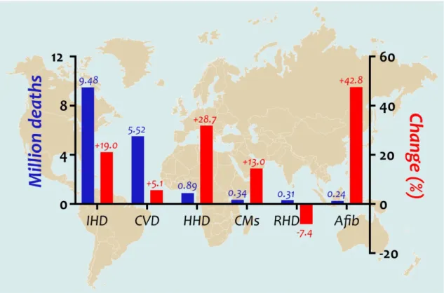
Figure 1. Leading causes of cardiovascular mortality worldwide and their change between 2006 and 2016
For the first time in human history, non-communicable diseases cause more deaths every year than epidemics worldwide, approximately 40% of which deaths are of cardiovascular origin. As such, cardiovascular conditions are the leading causes of mortality on our globe.
This figure details the most frequent cardiovascular diseases resulting in death, showing the annual number of deaths and the percent change of death rate in 2016 compared with 2006 (GBD 2016 Mortality Collaborators, 2017).
Afib – atrial fibrillation; CMs – cardiomyopathies; CVD – cerebrovascular disease; HHD – hypertensive heart disease; IHD – ischemic heart disease; RHD – rheumatic heart disease
9
levels of cGMP originated either from blockade of its degrading enzyme, phosphodiesterase type 5 (PDE-5) (Takimoto et al., 2005a) or from increasing its production by stimulating or activating its producing enzyme, soluble guanylate cyclase (sGC) (Fraccarollo et al., 2014; Frankenreiter et al., 2018; Korkmaz et al., 2009) preserved myocardial structure and function in experimental ischemia-reperfusion models. Therefore, elevating myocardial cGMP levels might prove to be an effective new method of preventing the development of pathological myocardial hypertrophy. A new group of drugs named sGC activators has been developed in order to counteract the impairment of the NO-cGMP pathway (Evgenov et al., 2006). Cinaciguat (BAY 58- 2667) is the most potent member of the sGC activators developed to this date (Stasch et al., 2002), which is capable of activating even inactive forms of sGC (Schmidt et al., 2009).
In the following brief review of the literature, I will summarize our knowledge of physiological and pathological myocardial hypertrophy, redox and NO/cGMP signaling in health and disease, as well as the concept of sGC activation, a novel therapeutic option as a preamble of my work.
10
2.1. Physiological myocardial hypertrophy
2.1.1. Cardiac structural changes in physiological myocardial hypertrophy
Physiological myocardial hypertrophy occurs naturally during growth or pregnancy, and is also the response of the heart to regular exercise. The diameter of human cardiomyocytes increase approximately 3-fold during growth from infancy to adulthood, and there is a linear relationship between body weight and cardiac weight.
Cardiomyocytes in an adult heart retain their ability to increase the amount of contractile proteins within them in response to moderate mechanical stress, and the mode of sarcomere addition, discussed below in more detail, will define the final hypertrophic phenotype. Importantly, adverse events characteristic to remodeling such as fibrosis do not develop during this type of growth, resulting in maintained cardiac structure, function, and metabolism (Gibb & Hill, 2018).
Exercise is associated with a similar enlargement of the heart that is generally thought to be harmless and even beneficial in healthy people. Different types of exercise, however, affect cardiac enlargement differently, as the heart conforms the type of hemodynamic load imposed on the cardiovascular system (Maillet et al., 2013). Dynamic sports, such as running or swimming, induce volume overload of the heart since there is an intensive dilatation of the skeletal muscle vasculature, leading to increased venous return to the heart (Pluim et al., 2000). Thus, athletes trained in sports requiring sustained isotonic movement develop eccentric cardiac hypertrophy that is characterized by chamber enlargement and proportionate thickening of the ventricular walls (Figure 2.). In this setting, cardiomyocytes grow both in length and width. In contrast, sports involving development of muscular tension against increased resistance, such as weightlifting or wrestling, induce pressure overload of the heart. As such, static exercise leads to concentric LV hypertrophy (LVH), where wall thickness increases without proportionate dilation of the LV, leading to decreased LV cavity dimensions [Figure 2.;
(Bernardo et al., 2010)]. Cardiomyocytes usually increase in thickness more than in length during concentric hypertrophy (Heineke & Molkentin, 2006; Selby et al., 2011).
There are also sports that combine dynamic and static components, such as cycling or rowing, where endurance training is completed against elevated resistance. This combined type of exercise results in the greatest degree of LV chamber dilatation and
11
increase in wall thickness (Barbier et al., 2006; Pelliccia et al., 1991; Pluim et al., 2000). Therefore, it is now widely accepted that the phenotype of hypertrophy resulting from any type of exercise is dependent upon the ratio of the static and dynamic components of that particular exercise (Mitchell et al., 2005). Furthermore, factors such as gender, ethnicity or age also influence the final observable phenotype (Colombo &
Finocchiaro, 2018; Pavlik et al., 2013). It must also be noted here, however, that Figure 2. The proposed two extremes of cardiac wall and chamber alterations in physiological myocardial hypertrophy
Classical view of cardiac hypertrophy in response to exercise was dichotomous: dynamic types of exercise were thought to induce eccentric hypertrophy due to the volume overloading of the heart, while static sports would induce pressure overload of the left ventricle, therefore causing concentric hypertrophy. More recent investigations highlighted that the final phenotype of hypertrophy is dependent upon the ratio of dynamic and static components of the exercise, and that it is more of a continuum than two easily separable extremes (Mitchell et al., 2005).
12
extreme exercise associated with competitive sport might result in a maladaptive hypertrophic response, which is still a diagnostic challenge for clinicians to differentiate from innocuous changes based on observation of phenotypic deteriorations alone (De Innocentiis et al., 2018; Gabrielli et al., 2018).
In contrast to the LV, response of the right ventricle (RV) to exercise training was not investigated in detail until relatively recently, mostly because of its more complex geometry and location inside the chest. Echocardiographic investigation of the RV is difficult at best, and many times accurate measurements are not possible due to anatomical reasons. The development of cardiac magnetic resonance imaging (cMRI) allowed for a more complex and accurate characterization of RV structural and functional changes following exercise. Similarly to LV alterations, RV volume, mass and stroke volume have all been observed to increase in athletes (Scharhag et al., 2002).
Contrary to the LV, however, RV hypertrophy and dilatation seem to occur only following dynamic training, while static exercise does not influence RV dimensions (Eijsvogels et al., 2016; Weiner & Baggish, 2012). A possible downside of endurance training might be that atrial enlargement in athletes predisposes them to an increased risk of atrial fibrillation (D'Ascenzi et al., 2015; Wilhelm et al., 2012).
2.1.2. Functional changes associated with exercise training
While structural changes of the LV have been described somewhat consistently, functional alterations are not so well characterized. This discrepancy stems from the fact that the overwhelming majority of studies investigating the athlete’s heart phenomenon have utilized conventional echocardiography to describe cardiac function (Utomi et al., 2013), and while this modality is optimal for the investigation of structural alterations, it is not as reliable when it comes to functional parameters. The value of both fractional shortening (FS) and ejection fraction (EF), which are the most common systolic indices that have been used to evaluate cardiac function in athletes, is dependent upon multiple factors including loading conditions and heart rate. cMRI, although less influenced by anatomical variations and thus capable of providing better spatial resolution, can only measure these parameters as well. Meta-analyses based on studies utilizing these non- invasive modalities concluded that systolic function is preserved or somewhat enhanced in athletes compared with sedentary controls during resting conditions (Fagard, 2003;
Fagard, 1996; Pluim et al., 2000; Scharf et al., 2010a; Scharf et al., 2010b), but a
13
significant effect of study-to-study heterogeneity was noted (Utomi et al., 2013). EF and FS investigated in these studies reflect chamber mechanics, which, especially in rest, might leave subtle, but important differences unnoticed. Investigation of intrinsic myocardial mechanics therefore might provide a better insight into the real functional alterations characterizing exercise-induced hypertrophy (Simsek et al., 2013). Our research group has published a more detailed characterization of athlete’s heart in a rat model (Radovits et al., 2013), utilizing the invasive pressure-volume (P-V) analysis that is capable of providing load independent indices of both systolic and diastolic function.
Athlete’s heart in humans, however, cannot ethically be investigated with an invasive method, given its generally harmless nature. Development of the novel echocardiographic imaging modes tissue Doppler and speckle-tracking imaging allows for a more detailed and more accurate assessment of LV systolic function that correlates well with invasive measurements (Kovacs et al., 2015). The results of studies implementing these modalities provided similar data, i.e. normal or supernormal cardiac systolic and diastolic function was found in athletes (Beaumont et al., 2017; D'Andrea et al., 2006; Kovacs et al., 2014; Richand et al., 2007).
Despite its clinical importance, LV diastolic function is an entity that has been difficult to assess (George et al., 2010). The gold standard measure of LV active relaxation, τ, is the time constant of isovolumic pressure decline in the LV from LV pressure at maximum decrement of LV pressure (dP/dtmin) to the level of LV end-diastolic pressure (LVEDP), and was first described by Weiss et al. in 1976 (Weiss et al., 1976), and later modified by Raff & Glantz (Raff & Glantz, 1981). Similarly to systolic LV parameters, however, routinely implementing invasive measurements to evaluate τ is not justifiable in healthy people. Therefore, alternative methods such as estimation of blood flow velocity through the mitral valve utilizing pulsed Doppler echocardiography are widely used to assess LV diastolic function (Nagueh et al., 2009; Rakowski et al., 1996), with the limitation that this method measures both active relaxation and myocardial stiffness, therefore incorporating passive components of diastolic function. Trans-mitral flow velocity comprises two components; peak early (E) and peak atrial (A) filling velocities.
In the normal heart, most diastolic filling occurs during the early filling phase, so that E is characteristically greater than A, and the ratio E/A is usually >1.0 (Rakowski et al., 1996). E/A was found to be normal or slightly enhanced in athletes as well (Fagard,
14
2003; Schmidt-Trucksass et al., 2001; Sharma et al., 2002). In some studies, however, the A wave was unusually lower than the E wave that can probably be ascribed to the lower heart rate, which prolongs the diastolic filling period and reduces the atrial component (Fagard et al., 1987). To conclude, studies estimating LV diastolic function are consistent that diastolic function is not compromised in athletes despite the presence of cardiac hypertrophy.
2.1.3. Molecular pathways underlying physiological myocardial hypertrophy
Several pathways have been implicated in the background of physiological myocardial hypertrophy, the most important of which being hormones such as insulin, insulin-like growth factor-1 or thyroid hormone, and also signal transduction activated by mechanical forces. These factors induce physiological myocardial hypertrophy via the activation of several signaling pathways converging on phosphoinositide 3-kinase (PI3K), Akt, AMP-activated protein kinase or mammalian target of rapamycin (mTOR).
In the following, I will briefly review the literature on these pathways.
2.1.3.1. Mechanical forces and signal transduction
Mechanotransduction enables cardiomyocytes to convert mechanical stimuli into biochemical events through the modulation of specific signaling molecules, giving them the ability to regulate hypertrophic or atrophic response depending on the extent and duration of mechanical stress imposed on them. Molecular pathways implicated in mechanotransduction and their significance in cardiac hypertrophy and failure has been extensively reviewed recently by Lyon and colleagues (Lyon et al., 2015). Key loci within cardiomyocytes in this regard are the sarcomere, the intercalated discs and the sarcolemma, which, through a plethora of proteins, are all interconnected, functioning as a complex sensor of mechanical stimuli. At the sarcomere, titin and attached proteins (such as muscle LIM protein [MLP], titin-Cap [TCAP] or calsarcin-1) serve mainly as a stretch sensor and stress response signalosome (Frey et al., 2004a; Gautel, 2011; Knoll et al., 2002; Miller et al., 2003). In the intercalated discs, N-cadherin was shown to mediate an adaptive response of the cardiomyocyte cytoskeleton to changes in mechanical stimuli (Chopra et al., 2011; Kostetskii et al., 2005). Besides the intercalated discs at the ends of cardiac myocytes, sarcolemma-associated proteins and complexes along the lateral surfaces of elongated myocytes (such as integrins) have been described as foci of force transmission. By forming a connection between the
15
extracellular matrix (ECM) and the contractile apparatus, costameric structures also facilitate the maintenance of mechanical integrity of the sarcolemma (Manso et al., 2013; Sharp et al., 1997). Furthermore, there is evidence that cardiomyocyte stress sensing might be dependent on the direction of the mechanical stimulus (Gopalan et al., 2003; Simpson et al., 1999), which may be related to different modes of hypertrophic growth (Kerckhoffs et al., 2012).
2.1.3.2. Thyroid hormone-related signaling
Thyroid hormones have significant biological effect mainly during postnatal growth (Stubbe et al., 1978), which effect is partially mediated by the activation of PI3K/Akt/mTOR signaling (Kinugawa et al., 2005) (Figure 3.). Whether this hormone promotes physiological cardiac hypertrophy in adults, however, is still controversial, but studies indicate that thyroid hormone effect convert pathological to physiological cardiac hypertrophy (Pantos et al., 2011; Pantos et al., 2007; van Rooij et al., 2007).
2.1.3.3. Insulin and insulin-like growth factor-1 signaling
Insulin and insulin-like growth factor-1 (IGF-1) signaling are the most well-known pathways in the development of physiological hypertrophy (Figure 3.). Their pathways converge on Akt (also known as protein kinase B), which is the main mediator of their downstream effects in cardiac myocytes (Catalucci et al., 2009a; Catalucci et al., 2009b;
Kemi et al., 2008; Kim et al., 2003), although IGF-1 has another canonical pathway through extracellular-signal-regulated kinase (ERK), and a non-canonical pathway through Gi/phospholipase C (PLC)/inositol-1,4,5-triphosphate (IP3)/Ca2+ signaling (Troncoso et al., 2014). Both insulin and IGF-1 are critically important in the pre- and postnatal growth of the heart, and both are involved in regulating cell proliferation, growth, differentiation, metabolism, and survival (Saltiel & Kahn, 2001; Takeda et al., 2010; Tatar et al., 2003; Ungvari & Csiszar, 2012; Vinciguerra et al., 2009; Vinciguerra et al., 2012). Genetic deletion of IGF-1 leads to a significant decrease in body weight during development and usually results in embryonic lethality or death from respiratory failure shortly after birth (Liu et al., 1993; Powell-Braxton et al., 1993; Shimizu &
Minamino, 2016). Similarly, cardio-specific deletion of insulin receptor (IR) results in a significant reduction of cardiomyocyte size and heart weight, with persisting fetal gene expression profile, mitochondrial dysfunction and reduced cardiac function (Belke et al., 2002; Boudina et al., 2009; Sena et al., 2009). Ikeda and colleagues demonstrated
16
the role of IGF-1 and IR mediated signaling in regulating the hypertrophic response induced by exercise in an elegant series of experiments. Deletion of the receptors of Figure 3. Major molecular factors governing the development of physiological hypertrophy
Physiological hypertrophic growth is induced mainly by growth factors such as insulin, IGF-1, or thyroid hormone, and also by periodical mechanical stress occurring during strenuous exercise either through pressure- or volume overload. The effects of hormonal factors are mediated mostly via the PI3K/Akt/mTOR signaling pathway, while mechanical stress exerts its effects through the sarcolemma, intercalated discs and sarcomeres (Lyon et al., 2015).
Akt – protein kinase B; IGF-1 – insulin-like growth factor-1; IGF1-R – IGF-1-receptor;
mTOR – mammalian target of rapamycin, N-CAD – N-cadherin; PI3K – phosphoinositide-3- kinase; T3 – triiodothyronine
17
insulin or IGF-1 in cardiomyocytes is associated with reduced or normal baseline cardiac growth, respectively, while the hypertrophic response to exercise is normal in both cases. When homozygous deletion of either IR or IGF-1 receptor (IGF-1R) is aggravated with the deletion of one of the alleles of the other receptor, exercise-induced cardiac hypertrophy becomes attenuated as well. The phenotypic changes in exercise- induced hypertrophy in Igf1r+/−-Ir−/− mice were found to be more severe, which suggests that insulin is more closely involved in physiological hypertrophy related to exercise than IGF-1 (Ikeda et al., 2009). Nevertheless, cardiac level of IGF-1 was shown to be higher in athletes than in sedentary controls, and exercise increased the serum level of IGF-1 (Neri Serneri et al., 2001; Poehlman et al., 1994), suggesting a significant role of IGF-1 in the development of physiological hypertrophy in humans.
Akt is the best characterized downstream effector of both insulin and IGF-1 mediated signaling, and was shown to promote cardiac hypertrophy via modulation of a variety of signaling pathways. Akt was shown to improve Ca2+-handling, enhance cardiac contractility and promote physiological cardiac hypertrophy by activating or suppressing numerous transcription factors (Figure 3.) (Catalucci et al., 2009a;
Catalucci et al., 2009b; Condorelli et al., 2002; Matsui et al., 2002; McMullen et al., 2003; Pallafacchina et al., 2002; Shioi et al., 2000; Shioi et al., 2002; Yamashita et al., 2001). Taken together, interrelated pathways of insulin and IGF-1 signaling in physiological hypertrophy, although both are known to contribute to the phenotypic changes associated with it, need to be further elucidated.
18
2.2. Pathological myocardial hypertrophy
2.2.1. Main structural changes in pathological compared with physiological myocardial hypertrophy
While physiological stimuli, such as growth, pregnancy or exercise induce cardiac hypertrophy that is associated with maintained structure and function, various pathological conditions such as hypertension, myocardial infarction, diabetes mellitus, valvular heart disease or cardiomyopathies evoke hypertrophic growth that significantly alters the composition of the myocardium. This hypertrophic response of the heart to different stressors has been believed to be, at least initially, adaptive and to have a compensatory function by diminishing wall stress and thus decreasing myocardial oxygen consumption according to the law of Laplace (Grossman et al., 1975; Hood et al., 1968; Sandler & Dodge, 1963). It was shown later, however, that the presence of LVH developed in response to pathological stimuli, is associated with a significant increase in the risk of heart failure and malignant ventricular arrhythmias (Koren et al., 1991; Levy et al., 1990).
Similarly to physiological myocardial hypertrophy, two main phenotypes of pathological myocardial hypertrophy can be distinguished: (1) concentric hypertrophy due to pressure overload originating most commonly from hypertension or stenotic valvular disease, which is characterized by parallel addition of sarcomeres and lateral growth of individual cardiomyocytes; and (2) eccentric hypertrophy due to volume overload (valvular regurgitation or arteriovenous fistulas) or prior infarction, characterized by addition of sarcomeres in series and longitudinal cell growth (Dorn et al., 2003; Frey et al., 2004b). Furthermore, pathological LVH is usually associated with an increased rate of cardiomyocyte death and fibrotic remodeling that promote systolic and diastolic dysfunction, rendering it maladaptive in the long term (Berk et al., 2007;
Spinale, 2007). The combination of LVH with increased levels of biomarkers of subclinical myocardial injury (high-sensitivity cardiac troponin T, N-terminal pro–B- type natriuretic peptide [NT-proBNP]) identifies patients at highest risk for developing symptomatic HF, especially HFrEF (Heinzel et al., 2015; Messerli et al., 2017; Seliger et al., 2015). In the following, I will summarize the consequences of longstanding hypertension on LV remodeling and hypertrophy in more detail.
19
Hypertension is a major public health problem associated with a significant and rapidly increasing mortality worldwide (Figure 1.). The bulk of patients who develop HFpEF suffer from persistent hypertension (Steinberg et al., 2012). It is well known that hypertensive heart disease (HHD) is initially characterized by compensated LVH, but there is considerable inter-individual variability in the increase of LV mass and geometry (Drazner, 2011). LV mass can increase from either wall thickening or chamber dilation in hypertensive patients; the ratio of LV wall thickness to diastolic diameter (relative wall thickness, RWT) classifies LVH as concentric (RWT is increased) or eccentric (RWT is not increased), as discussed above (Ganau et al., 1992).
A third pattern, termed concentric remodeling, occurs when RWT, but not LV mass, is increased. Interestingly, contrary to the classical view of concentric hypertrophy slowly progressing into eccentric hypertrophy and then to decompensation during the course of the disease, hypertensive patients can have any of these patterns of LV geometry as an initial hypertrophic response (Ganau et al., 1992; Sehgal & Drazner, 2007). What decides whether a patient develops one pattern or another in response to hypertension seems likely to be defined by a multitude of variables, including the extent of hypertension itself (Fagard et al., 1997; Ross, 1997), ethnicity (Drazner et al., 2005;
Kizer et al., 2004), sex (Krumholz et al., 1993), age (Chahal et al., 2010; Cheng et al., 2009), neurohumoral activation (Alderman et al., 2004; Brunner et al., 1972; Davila et al., 2008; du Cailar et al., 2000; Muscholl et al., 1998; Nakahara et al., 2007; Olsen et al., 2002; Velagaleti et al., 2008), troponin I variants and thus altered Ca2+ sensitivity of the contractile apparatus in cardiomyocytes (Davis et al., 2016) and the presence of comorbidities such as diabetes or obesity (Avelar et al., 2007; de Simone et al., 1994;
Gottdiener et al., 1994; Markus et al., 2011; Palmieri et al., 2001; Zabalgoitia et al., 2001). How these factors combine to generate the final phenotype, however, is yet to be discovered. On the molecular level, LVH in response to pathological stimuli such as hypertension is characterized by increased myocardial fibrosis (Villari et al., 1995), injury or loss of cardiomyocytes (Sano et al., 2007; Shimizu et al., 2010) and coronary microvascular rarefaction (Mohammed et al., 2015; Paulus & Tschope, 2013), all of which are ultimately contributing to the transition of LVH to HF. Lastly, there is evidence that excessive LVH in response to pressure overload might not be necessary to evade dilated cardiac failure (Esposito et al., 2002; Hill et al., 2000; Hill et al., 2002),
20
which suggests that inhibition of the development of concentric LVH might be a potential therapeutic target in pressure overload, such as hypertension (Drazner, 2011;
Frey et al., 2004b).
2.2.2. Myocardial dysfunction associated with pathological myocardial hypertrophy
Unlike physiological myocardial hypertrophy that is associated with normal cardiac performance, sustained pathological LVH results in both diastolic and systolic functional deterioration eventually progressing to HF. This transformation, however, might not present itself for extended periods of time, which is termed the compensated phase of pathological LVH. During this period, cardiac performance is normal or only mildly decreased in spite of the pathological structural alterations already existing in the myocardium. Despite the long-known maladaptive nature of pathological myocardial hypertrophy, the mechanisms that determine how longstanding hypertrophy ultimately progresses to overt heart failure are still poorly understood. Again, as a detailed review of the diverse functional consequences of different types of pathological stimuli exceeds the limitations of this dissertation, I will focus on the functional deterioration associated with longstanding hypertension.
The causality between maladaptive concentric LVH and diastolic dysfunction was established more than 30 years ago (Lorell et al., 1990). In HFpEF patients, LVH is the most frequent cardiac structural abnormality, and it is correlated with hospitalization for heart failure, cardiovascular death, and aborted cardiac arrest (Hawkins et al., 2007;
Shah et al., 2014), although the exact mechanisms linking pathological LVH to diastolic dysfunction have not been elucidated completely. Nevertheless, HFpEF patients were shown to have more pronounced concentric hypertrophy than patients with HHD but without HFpEF (Melenovsky et al., 2007). Furthermore, pathological LVH is associated with an attenuated increase or even decrease in EF during exercise and reduced exercise capacity, this reduction being worst in patients with a concentric type of LVH (Lam et al., 2010; Meyer et al., 2015; Schnell et al., 2013). There is evidence that this failure to increase EF in response to exercise is not related to an inability to increase LV end- diastolic volume (LVEDV), therefore to a restriction in LV filling, but rather to a failure in the Frank-Starling mechanism in patients with HFpEF (Abudiab et al., 2013;
Borlaug, 2014; Shibata et al., 2011). Increased LV stiffness might also play a significant
21
role in the diastolic dysfunction related to pathological LVH, as τ in HFpEF patients was shown not to decrease during exercise as it normally would in healthy subjects, but even become prolonged (Borlaug et al., 2011).
Many studies have shown that patients with HFpEF display impairments in regional deformation detected by tissue Doppler and strain-based imaging techniques, resulting in subtle but significant abnormalities in chamber and myocardial contractility, despite the overall preservation of EF (Borlaug et al., 2009; Shah & Solomon, 2012; Tan et al., 2009; Wang et al., 2008). Even this subtle impairment of contractility at rest may indicate marked limitations in reserve, which is markedly impaired in HFpEF patients compared with age-matched healthy and hypertensive controls (Borlaug et al., 2010).
Systolic dysfunction affects diastolic function as well, as the ability to contract more enhances the recoil and suction forces during early diastole (Opdahl et al., 2009), which have also been shown to be impaired in HFpEF (Ohara et al., 2012).
Animal models are crucially important tools in the discovery of the driving forces behind human pathologies. As such, many small and large animal models have been developed to model the various clinical conditions resulting in the cardiac phenotype discussed above as closely as possible. The most commonly modeled diseases of pressure overload of the LV are aortic stenosis via thoracic aortic constriction (Huss et al., 2007; Tagawa et al., 1998) and hypertension via renovascular constriction (Cangiano et al., 1979; Goldblatt et al., 1934), compression of renal parenchyma (Grollman, 1955; Hart et al., 2001), reduction of renal mass (Anderson et al., 1985; Li et al., 2009), increased salt intake (Coleman et al., 1975), genetic modifications (Molkentin et al., 1998; Okamoto & Aoki, 1963; Pfeffer et al., 1982; Rapp & Dene, 1985), endocrine stimulation (Bois & Selye, 1957; Krege et al., 1995) and aortic banding (Silver et al., 1990; Thiedemann et al., 1983).
2.2.3. Molecular pathways implicated in pathological myocardial hypertrophy
During pathological conditions, increased mechanical stress imposed on cardiomyocytes is accompanied by modulating factors that eventually shepherd actuated hypertrophic signaling pathways in the direction of generating pathological myocardial hypertrophy. Regarding the immense complexity of hypertrophic signaling, elucidation of the exact mechanism how activated pathways combine to create pathologic LVH is still in progress. The result of the processes discovered so far is generally referred to as
22
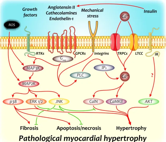
Figure 4. Key molecular pathways implicated in the development of pathological myocardial hypertrophy
Neurohumoral and growth factors together with the increased mechanical stress imposed on cardiomyocytes and other cellular components of the myocardium trigger multiple signaling pathways that bring about the characteristic phenotypical changes associated with pathological left ventricular hypertrophy. The two most important signaling cascades involved are Ca2+-related and mitogen activated protein kinase signaling, members of which can potently induce gene expression changes generating all major hallmarks of pathological myocardial hypertrophy: fibrosis, apoptosis/necrosis and cardiomyocyte hypertrophy.
CalN – calcineurin; CaMKII – Ca2+/calmodulin dependent kinase II; ERK1/2 – extracellular signal-regulated kinase 1/2; GPCR – G protein coupled receptor; IP3 – inositol-1,4,5- triphosphate; IR – insulin receptor; JNK – c-Jun N-terminal kinase; LTCC – L-type Ca2+
channel; MAP3K – MAP2K kinase; MAP2K – mitogen activated protein kinase kinase; PLC – phospholipase C; ROS – reactive oxygen species; RTK – receptor tyrosine kinase; TRPC – transient receptor cation channel
23
“reactivation of the fetal gene program”, which covers re-expression of a wide array of molecular markers that are characteristic to the fetal period of ontogeny. Among others, such marker is the isotype-switch of myosin heavy chains (MHCs), significant overexpression of atrial natriuretic peptide (ANP), or the increased expression of endothelial nitric oxide synthase (NOS3). This altered gene expression profile is the driving force in the background of pathological LVH, and in the following section, I will shortly summarize the most important pathways initiating it.
2.2.3.1. Hypertrophy-inducing signals converging on Ca2+-dependent pathways
Neurohumoral mediators such as catecholamines, angiotensin II or endothelin-1 have all been implicated in the development of pathological LVH and in the long term, HF. β- blockers and angiotensin converting enzyme inhibitors (ACEi) were shown to reduce the mortality of HF patients, hence becoming first line pharmacotherapeutic agents in HF (AIRE Study Investigators, 1993; Packer et al., 2001; Poole-Wilson et al., 2003).
These mediators bind to G protein-coupled receptors (GPCRs) that comprise seven transmembrane domains. The Gα proteins involved can either be Gαs, Gαi, Gαq or Gα12/13, Gαq being the most important for the mediators listed above. Gαq signaling activates PLC, which catalyzes the synthesis of IP3, thereby inducing intracellular Ca2+
release and thus an increase in [Ca2+]i. The excess Ca2+ can also originate from the extracellular space, e.g. through transient receptor potential cation (TRPC) channels.
TRPCs regulate Ca2+ and Na+ movement in specific microdomains, and were shown to be upregulated in pathological myocardial hypertrophy and heart failure (Kuwahara et al., 2006; Wu et al., 2010). Regardless of its origin, Ca2+ modulates the activity of various Ca2+-dependent signaling pathways, the two most important of which within cardiomyocytes being calcineurin/nuclear factor of activated T cells (NFAT) signaling and Ca2+/calmodulin-dependent kinase II (CaMKII) signaling. Either of these pathways is sufficient to induce pathological LVH alone (Hoch et al., 1999; Kirchhefer et al., 1999; Molkentin et al., 1998), but interplay between them is highly likely in various pathological conditions [Figure 4., (Zarain-Herzberg et al., 2011)].
Calcineurin is a Ca2+-activated serine/threonine protein phosphatase that induces translocation of NFAT into the nucleus by dephosphorylating it in the cytoplasm, thus increasing the expression of genes involved in pathological LVH (Figure 4.). Although cardiac-specific disruption of calcineurin expression revealed that it is necessary for
24
normal postnatal cardiac growth (Schaeffer et al., 2009), calcineurin/NFAT signaling does not seem to be involved in physiological hypertrophy (Wilkins et al., 2004).
The serine/threonine kinase CaMKII has been known to be involved in heart failure based on animal models and clinical data (Ai et al., 2005; Kirchhefer et al., 1999), and more recently was implicated in the progression of pressure overload-induced pathologic LVH to HF (Ling et al., 2009).
Forced expression or activation of CaMKII mediates cardiac hypertrophy that is phenotypically similar to that of induced by norepinephrine, phenylephrine, or endothelin-1, while inhibition of its function prevents pathological LVH and improves HF [Figure 4., (Bossuyt et al., 2008; Hoch et al., 1999; Zhu et al., 2000)]. Furthermore, class II histone deacetylase (HDAC) phosphorylation by CaMKII was shown to induce hypertrophic growth via myocyte enhancer factor-2 (MEF-2) dependent gene expression upregulation (Backs et al., 2006; Passier et al., 2000).
2.2.3.2. Mitogen activated protein kinase (MAPK) pathways in pathological hypertrophy
MAPKs are highly conserved kinases among eukaryotes that are implicated in a wide array of cellular processes. In the cardiovascular system, these include cell proliferation, cell growth, fibrotic remodeling and cellular response to different stressors. MAPK activation involves a three-tiered, phosphorylation based amplification system, during which signals originating from GPCRs, receptor tyrosine kinases, ion channels or oxidative and other types of stress, including pressure overload, activate the three main branches of MAPK signaling: ERK1/2, p38 MAPKs and c-Jun N-terminal kinases (JNKs, Figure 4.). All three branches play a role in the development of pathological hypertrophy and HF (Gutkind & Offermanns, 2009; Haq et al., 2001; Toischer et al., 2010), albeit reports so far on these roles are contradictory (Javadov et al., 2014).
ERK1/2 is implicated in promoting cardiac hypertrophy in response to activation of GPCRs by catecholamines, angiotensin II or endothelin-1 and also to increased oxidative stress (Figure 4.). Although pro-hypertrophic, ERK1/2 and its downstream signaling does not seem to be essential in cardiac hypertrophy, since the deletion of its gene (ERK1–/– and ERK2+/–) did not prevent development of LVH (Purcell et al., 2007).
It does, however, seem to play a role in determining the phenotype (i.e., eccentric or concentric) of LVH developed in response to various stimuli (Kehat et al., 2011).
25
Potential downstream targets of ERK1/2-mediated hypertrophy include members of Ca2+ homeostasis and activation of the transcription factor GATA4 [Figure 4., (Zheng et al., 2004)]. Furthermore, activation of ERK1/2 was shown to play a role in resistance to apoptosis (Yamaguchi et al., 2004).
The role of p38 in the development of pathological hypertrophy is still controversial.
Although it was shown to promote LVH in some studies (Liang & Molkentin, 2003), others concluded that even dramatic down-regulation of the kinase left cardiac hypertrophic growth unaffected (Nishida et al., 2004). This discrepancy in the results published might be related to methodological differences, such as the use of different cell types, animal models, inhibitors or agonists, and also the temporal design of a specific study (Javadov et al., 2014). Demonstration of the participation of p38 in fibrotic remodeling was more consistent: its activation (Koivisto et al., 2011; Liao et al., 2001; Wang et al., 1998) or inhibition (Liu et al., 2005; Yin et al., 2008; Zhang et al., 2003) resulted in enhanced or reduced myocardial fibrosis, respectively (Figure 4.).
Furthermore, p38 activation is associated with increased apoptosis (Kaiser et al., 2004;
Ren et al., 2005), thereby promoting the transition of pathologic LVH to overt HF.
There is, similarly to p38, contradiction in what role JNK plays in the development of pathologic LVH. Neonatal cardiomyocytes were shown to respond with hypertrophy to targeted activation of JNK signaling (Wang et al., 1998), whereas adult hearts exhibited increased hypertrophy, fibrosis and apoptosis when JNK or members of its signaling were inhibited (Hilfiker-Kleiner et al., 2005; Liang et al., 2003), suggesting an anti- hypertrophic role for JNK in adult hearts via interference with calcineurin/NFAT signaling (Figure 4.). On the other hand, JNK was shown to induce cardiac dysfunction as well via (1) decreasing intercellular communication within the myocardium due to the downregulation of connexin-43, and thus loss of gap junctions (Petrich et al., 2002), and (2) the activation of matrix metalloproteinase-2 resulting in detrimental cardiac remodeling (Krishnamurthy et al., 2007).
An endogenous negative regulator of MAPK cascades is MAPK phosphatase 1 (MKP- 1), constitutive expression of which in the heart downregulates all three major pathways discussed above, and also prevents induction of hypertrophy by catecholamines or aortic banding (Bueno et al., 2001). MKP-1 and MKP-4 were shown to have a cardioprotective role; MKP-1 and -4 knockout mice express elevated amounts of
26
p38MAPK with no change of JNK or ERK1/2 levels, and have a low survival rate associated with systolic dysfunction and cardiac dilatation (Auger-Messier et al., 2013).
2.2.3.3. Excessive activation of insulin signaling contributes to pathological myocardial hypertrophy
Although insulin/IR/Akt signaling is widely accepted to promote physiological myocardial hypertrophy, neither animal studies nor large clinical trials have provided conclusive evidence whether insulin signaling is cardioprotective in adults. Instead, insulin resistance, and thus hyperinsulinemia, has been reported to increase the risk of developing heart failure in patients with systolic dysfunction, which observation gains epidemiological importance if one considers the high prevalence of this condition (Ashrafian et al., 2007; Ingelsson et al., 2005; Witteles et al., 2004). Also, contrary to what might be expected, intensive glycemic control with insulin increased cardiovascular events in diabetic patients instead of reducing these complications (Action to Control Cardiovascular Risk in Diabetes Study Group, 2008).
Experimental evidence shows that chronic hyperinsulinemia might induce pathological hypertrophy via an angiotensin II-dependent manner [Figure 4., (Samuelsson et al., 2006)], and that excessive insulin signaling exacerbates the transition of hypertrophy to overt HF (Shimizu et al., 2010). Furthermore, pressure overload was shown to induce adipose tissue inflammation and lipolysis, resulting in insulin resistance (Shimizu et al., 2012). Suppression of adipose tissue inflammation in the same study decreased insulin resistance, therefore, possibly, hyperinsulinemia, and lead to improved systolic function in mice subjected to chronic pressure overload (Shimizu et al., 2012).
27
2.3. Redox and nitric oxide/cGMP signaling in cardiovascular physiology and pathology
Redox signaling, like many other signaling pathways implicated in cardiovascular physiology, is Janus-faced. Whether physiological processes or detrimental effects are mediated by reactive oxygen (ROS) and nitrogen species (RNS), depends on the local concentration and compartmentation of the reactive species involved, and also on the capacity of antioxidant mechanisms present. Oxidative and nitrosative stress, in fact, cannot be meaningfully separated, as generation of either ROS or RNS will bring about the genesis of the other, hence the term nitro-oxidative stress.
Previously known as the elusive endothelial-derived relaxing factor [EDRF, (Furchgott
& Zawadzki, 1980)], the discovery of nitric oxide (NO) and its involvement in many physiological processes and virtually all cardiovascular pathologies reestablished our thinking concerning cardiovascular health and disease. NO, though often referred to as a toxic agent when present in higher concentrations and thus considered a major player in nitrosative stress, is not particularly reactive. There is, however, one reactive species that seems to stand out from the rest, especially in terms of biological importance:
peroxynitrite, a powerful oxidant that is formed in a spontaneous reaction between NO and superoxide (Pacher et al., 2007).
The cardioprotective effects of NO, mediated intracellularly via cGMP signaling, are severely reduced in disease states, including hypertension, associated with increased nitro-oxidative stress. Therefore, novel pharmacological approaches specifically targeting the upregulation of this physiologically important pathway are increasingly investigated. In the following section, I will briefly review the most important aspects of redox- and NO signaling in cardiovascular homeostasis and pathology, and also give a short introduction into the concept of sGC activation as a novel pharmacological interventional option.
2.3.1. The role of redox signaling in cardiac (patho)physiology
2.3.1.1. Forms, sources, targets and elimination of reactive oxygen species in the heart ROS are powerful oxidants that contain at least one oxygen atom with an unpaired electron. The most important ROS in vivo are superoxide anion (O2-) and hydrogen peroxide (H2O2), but other species such as the highly reactive hydroxyl radical or
28
singlet oxygen also play a significant role. ROS can either be generated by nicotinamide adenine dinucleotide phosphate (NADPH) oxidases (Noxs) that are specialized enzymes having no known biological role other than forming ROS, or can originate from the functioning of the mitochondrial electron transport chain, oxidases involved in metabolism (e.g. xanthine oxidase and monoamine oxidases) and uncoupled NO synthases as by-products [Figures 5. and 6., (Burgoyne et al., 2012)]. The compartmentation and local concentration of ROS produced have a significant role in their effect, as these define the targets they can reach; in cardiomyocytes, these redox sensitive targets include CaMKII (Erickson et al., 2008), cAMP-dependent protein kinase (PKA, Brennan et al., 2006), cGMP-dependent protein kinase (PKG, Burgoyne et al., 2007), MAPKs (Valko et al., 2007) and members of Ca2+ homeostasis such as ryanodine receptor 2 (RyR2) and sarcoplasmic and endoplasmic reticulum Ca2+-ATPase 2a (SERCA2a, Zima & Blatter, 2006). An efficient endogenous antioxidant system comprising enzymatic and non-enzymatic components exists to scavenge and thus limit the effects of ROS both spatially and temporally. Superoxide dismutase (SOD) catalyzes the conversion of O2- to H2O2, which is then further converted by catalase (Cat) and glutathione peroxidase to water and molecular oxygen. Furthermore, non- enzymatic defense is represented by ascorbic acid (vitamin C), α-tocopherol (vitamin E), glutathione, carotenoids, flavonoids and other antioxidants such as thioredoxin (Burgoyne et al., 2012; Valko et al., 2007).
2.3.1.2. Oxidative stress and its role in pathological myocardial hypertrophy
Reviewing all the known ROS-related mechanisms in the background of cardiovascular pathologies would significantly exceed the possibilities of this dissertation; therefore, I will focus on pathological myocardial hypertrophy and relevant mechanisms including alterations in Ca2+ handling and induction of apoptosis/necrosis.
Oxidative modification of CaMKII (Wagner et al., 2011), RyR2 (Terentyev et al., 2008) and SERCA2a (Lancel et al., 2010) all contribute to the imbalance of cellular Ca2+
homeostasis in pathological LVH (Figure 5.). Diastolic Ca2+ leakage through oxidized RyR2 and SERCA2a results in increased [Ca2+]i and thus impaired relaxation, contractile dysfunction and increased probability of arrhythmias. Oxidation-enhanced activation of PKA could also potentially contribute to Ryr2 dysfunction via hyper- phosphorylation.
29
Mitochondria play a central role in orchestrating apoptotic/necrotic cell death in response to ROS and Ca2+ overload (Sack et al., 2017). Mitochondrial permeability transition pore opening or translocation of pro-apoptotic proteins Bax/Bad to the mitochondria are both central processes in the intrinsic pathway of apoptotic cell death
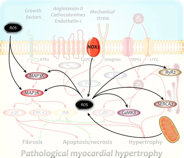
Figure 5. Major redox-sensitive targets with a significant role in the development of pathological myocardial hypertrophy
Reactive oxygen species are capable of activating key players of pathological hypertrophic growth independently of their original activators. ROS, therefore, further enhance the over- activation of hypertrophic signaling, resulting in an aggravated hypertrophic phenotype.
CaMKII – Ca2+/calmodulin dependent kinase II; MAP2K – mitogen activated kinase kinase;
MAP3K – MAP2K kinase; NOX2 – nicotinamide adenine dinucleotide phosphate oxidase 2;
ROS – reactive oxygen species; RyR2 – ryanodine receptor 2; SERCA2 – sarcoplasmic and endoplasmic reticulum Ca2+-ATPase isoform 2
30
facilitated by ROS (Donath et al., 2006). Excessive GPCR activation also leads to ROS production and activation of JNK and p38 MAPK, leading to activation of apoptotic pathways (Yamaguchi et al., 2003).
As discussed above, both Ca2+-dependent and MAPK signaling involved in cardiac hypertrophy might also be activated by ROS; in fact, due to the interdependence of the effects of vasoactive mediators and ROS, they cannot meaningfully be separated in pressure overload-induced LVH. Nox2 is of central importance in generating the reactive species activating/interfering with hypertrophic signaling actuated by other factors [Figure 5., (Bendall et al., 2002; Grieve et al., 2006)], but activation of Nox2 is certainly not obligatory for the development of pressure overload-induced LVH (Maytin et al., 2004; Touyz et al., 2005). Furthermore, mechanical stretch has also been associated with ROS-mediated induction of hypertrophy, also via the activation of Nox2 (Pimentel et al., 2001).
2.3.2. Nitric oxide and cGMP signaling in cardiovascular health and disease
NO, despite its simple structure comprising only two atoms, is among the most important intercellular messengers in all vertebrates, playing a significant role in the modulation of thrombosis, neural activity and cardiovascular homeostasis (Pacher et al., 2007). NO mediates its effects mainly through increasing the generation of cGMP, but also has cGMP-independent effects via protein nitrosylation (Lima et al., 2010), here not discussed in more detail. Certain disease conditions, especially those associated with oxidative stress result in the dysfunction of this important pathway, which will be reviewed briefly in this section.
2.3.2.1. cGMP generation and degradation
NO in the cardiovascular system is predominantly produced by NOS3 located in the endothelium, but cardiac myocytes are also capable of producing it – mainly for tightly localized regulatory purposes – by neuronal NOS (NOS1). NOS3-derived NO then diffuses to its target cells, including cardiomyocytes, to activate sGC. The enzyme binds NO with its heme moiety (Derbyshire & Marletta, 2009), resulting in an up to 800-fold increase in its activity and a production of cGMP from GTP in a similar magnitude.
sGC, however, is not the only source of cGMP within the cell. Membrane-bound enzymes, particulate GCs (pGCs) also produce it in response to natriuretic peptides ANP, B-type (BNP) and C-type NP (CNP) (Kuhn, 2009). Compartmentation of the
31
cGMP generated in response to these peptides differ from that of derived from sGC, therefore cardiomyocytes exhibit distinct responses as well (Castro et al., 2006;
Fischmeister et al., 2006); ANP has anti-hypertrophic properties (Patel et al., 2005), while BNP mediates anti-fibrotic effects (Tamura et al., 2000). CNP does not play a significant role in the cGMP-generation of cardiomyocytes (Yasoda et al., 2004).
cGMP is removed from the cell by either enzymatic degradation or extrusion via multi drug resistance proteins. Enzymatic degradation is responsible for the bulk of cGMP elimination, and is catalyzed by cyclic nucleotide-specific phosphodiesterases (PDEs),
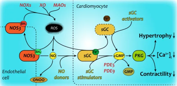
Figure 6. NO-cGMP signaling in health and disease
Physiological signaling through the NO/cGMP/PKG pathway deteriorates at multiple levels under pathological conditions. Oxidative stress decreases the generation, amount and sensing of nitric oxide by target cells, therefore disrupting cGMP generation and signaling within these cells as well. There have been various attempts at restoring cGMP signaling in cardiovascular pathologies, these are marked with yellow color. The novel sGC stimulators require reduced, functional sGC to mediate their effects, while sGC activators are capable of inducing cGMP production of dysfunctional sGC in absence of its heme prosthetic group (Evgenov et al., 2006)
BH4/2 – reduced/oxidized tetrahydrobiopterin; cGMP – cyclic GMP; GMP – guanosine monophosphate; MAO – monoamine oxidase; NO – nitric oxide; NOS3 – endothelial NO synthase; NOX – nicotinamide adenine dinucleotide phosphate oxidase; ONOO- – peroxynitrite; PDE5/9 – phosphodiesterase 5/9; PKG – cGMP dependent kinase; ROS;
reactive oxygen species; sGC – soluble guanylate cyclase; XO – xanthine oxidase
32
which are members of a superfamily comprising eleven smaller families (PDE1-11).
PDEs have distinct specificities for cAMP and cGMP, among which PDE5 and PDE9 are significant in the cardiovascular system regarding the hydrolysis of cGMP (Lee et al., 2015; Takimoto et al., 2005a).
2.3.2.2. cGMP-dependent effects of nitric oxide signaling
cGMP mediates its effects through a number of mechanisms. Predominantly it activates PKGs and thus regulates phosphorylation state of various protein targets, but it can also modulate the activity of distinct PDEs and cAMP-dependent protein kinases either directly or indirectly (termed as cAMP-cGMP crosstalk). Most cells contain at least one of three PKGs: PKGIα, PKGIβ, or PKGII that are targeted by their distinct amino termini to different substrates (Hofmann et al., 2009). Cardiomyocytes express PKGIα, which resides within the cell in a soluble form. PKGIα has been established to be a powerful brake on the cellular stress response signaling [Figure 6., (Hofmann, 2018;
Rainer & Kass, 2016)]: it can suppress selective GPCR agonism through regulator of G protein signaling subtypes (Takimoto et al., 2009), phosphorylate titin and troponin I (Kruger et al., 2009; Lee et al., 2010), increase the activity of late rectifier K+-channels (Bai et al., 2005) and antagonize members of the TRPC channel family (Koitabashi et al., 2010; Wu et al., 2010). More recently it was discovered that PKG might also modulate mechanosensing (Seo et al., 2014) and protein quality control (Ranek et al., 2013). In addition, as described above, oxidation of the PKG1α isoform in cardiomyocytes impairs its brake-like function; therefore oxidative stress adversely impacts stress-response signaling (Nakamura et al., 2015).
2.3.2.3. Oxidative stress results in an imbalance in NO-cGMP signaling
Oxidative stress impairs NO signaling at multiple levels (Figure 6.).
Tetrahydrobiopterin, which serves as a prosthetic group in NOS3, becomes oxidized, which results in uncoupling of the enzyme. Uncoupled NOS3 cease to produce NO, and instead, starts production of O2-, thereby aggravating oxidative stress [Figure 6., (Stasch et al., 2011)]. NO bioavailability is further reduced by its instantaneous conversion to peroxynitrite by O2-. Peroxynitrite is a strong oxidant itself, mediating nitro-oxidative stress by nitrating tyrosine residues of various proteins and damaging other targets (Pacher et al., 2007). Oxidative stress results in damaged sGC via the oxidation of its heme group (Figure 6.). Oxidized heme of sGC is unable to bind NO, therefore
33
rendering sGC incapable of generating cGMP (Evgenov et al., 2006). Furthermore, cGMP degradation is increased in oxidative stress mainly because of upregulation of PDE5 (Das et al., 2015). As a result, cGMP level within cardiomyocytes becomes significantly decreased, resulting in the loss of its cardioprotective effects.
2.3.3. Modulation of sGC activity – a novel pharmacotherapeutic concept
Long before any knowledge regarding the mechanism of their action, glyceryl trinitrate and other organic nitrates, as well as nitro-vasodilators had been in clinical use to treat angina pectoris and hypertensive crises for decades (Megson & Miller, 2009). Most of these drugs, however, have an important limitation: they lose their efficacy during prolonged use, termed as nitrate tolerance (Munzel et al., 2005). Furthermore, NO resistance is an important feature of ischemic heart disease and hypertension, and is also present in a significant portion of patients with pulmonary hypertension. The discovery that cGMP generated by sGC, as discussed above, mediates the cardioprotective effects associated with NO, provided rationale for development of new pharmaceuticals acting at different steps of the NOS3-NO-sGC-cGMP-PKG axis, potentially downstream of NO (Figure 6.).
sGC stimulation and activation as a highly effective method to increase intracellular cGMP-production, have been subject to extensive preclinical and clinical research (Boerrigter et al., 2007; Schmidt et al., 2009; Stasch et al., 2015; Stasch & Hobbs, 2009). sGC stimulators require sGC to be functional (i.e., sGC having an intact, reduced heme prosthetic group) to increase cGMP production, and synergize with NO in activation of the enzyme [Figure 6., (Evgenov et al., 2006)]. A sGC stimulator, riociguat under the trade name Adempas®, has recently been approved by the Food and Drug Administration for clinical use in pulmonary hypertension based on data from a large, randomized, Phase III clinical trial (Ghofrani et al., 2013). sGC activators, in contrast, are capable of activating sGC independently of both NO and heme, therefore might be more potent in cardiovascular conditions with oxidative stress where sGC activity is severely diminished (Figure 6.).
Cinaciguat (also known as BAY 58-2667) was the first sGC activator to be developed, and remains the most potent such drug to this day. Due to its strong and selective binding to heme-free and thus inactive sGC (Figure 7.) prevalent mainly in conditions associated with oxidative stress, makes this drug especially interesting because of the
34