Placenta 99 (2020) 197–207
Available online 22 June 2020
0143-4004/© 2020 The Authors. Published by Elsevier Ltd. This is an open access article under the CC BY license (http://creativecommons.org/licenses/by/4.0/).
Proteomic identification of Placental Protein 1 (PP1), PP8, and PP22 and characterization of their placental expression in healthy pregnancies and in preeclampsia
Szilvia Szabo
a,b,1, Katalin Karaszi
a,c,1, Roberto Romero
d,e,f,g,h,i, Eszter Toth
a, Andras Szilagyi
a, Zsolt Gelencser
a, Yi Xu
d,j, Andrea Balogh
a, Gabor Szalai
a, Petronella Hupuczi
k,
Beata Hargitai
l, Tibor Krenacs
c, Eva Hunyadi-Gulyas
m, Zsuzsanna Darula
m,
Katalin A. Kekesi
n,o, Adi L. Tarca
d,j,p, Offer Erez
d,j,q, Gabor Juhasz
o,r, Ilona Kovalszky
c, Zoltan Papp
k, Nandor Gabor Than
a,c,k,*aSystems Biology of Reproduction Lendulet Research Group, Institute of Enzymology, Research Centre for Natural Sciences, Budapest, Hungary
bDepartment of Morphology and Physiology, Faculty of Health Sciences, Semmelweis University, Budapest, Hungary
cFirst Department of Pathology and Experimental Cancer Research, Semmelweis University, Budapest, Hungary
dPerinatology Research Branch, Eunice Kennedy Shriver National Institute of Child Health and Human Development, National Institutes of Health, United States Department of Health and Human Services, Bethesda, Maryland, and Detroit, MI, USA
eDepartment of Obstetrics and Gynecology, University of Michigan, Ann Arbor, MI, USA
fDepartment of Epidemiology and Biostatistics, Michigan State University, East Lansing, MI, USA
gCenter for Molecular Medicine and Genetics, Wayne State University, Detroit, MI, USA
hDetroit Medical Center, Detroit, MI, USA
iDepartment of Obstetrics and Gynecology, Florida International University, Miami, FL, USA
jDepartment of Obstetrics and Gynecology, Wayne State University School of Medicine, Detroit, MI, USA
kMaternity Private Clinic of Obstetrics and Gynecology, Budapest, Hungary
lWest Midlands Perinatal Pathology Centre, Cellular Pathology Department, Birmingham Women’s and Children’s NHS FT, Birmingham, United Kingdom
mInstitute of Biochemistry, Biological Research Centre, Szeged, Hungary
nDepartment of Physiology and Neurobiology, ELTE Eotvos Lorand University, Budapest, Hungary
oLaboratory of Proteomics, Institute of Biology, ELTE Eotvos Lorand University, Budapest, Hungary
pDepartment of Computer Science, Wayne State University College of Engineering, Detroit, MI, USA
qMaternity Department “D,” Division of Obstetrics and Gynecology, Soroka University Medical Center, School of Medicine, Faculty of Health Sciences, Ben Gurion University of the Negev, Beer-Sheva, Israel
rCRU Hungary Ltd., God, Hungary
A R T I C L E I N F O Keywords:
HELLP syndrome Placenta Preeclampsia Pregnancy Tissue microarray Trophoblast Syncytiotrophoblast Small for gestational age Liver dysfunction Thrombocytopenia Biomarkers Immunohistochemistry Mass spectrometry
A B S T R A C T
Introduction: Placental Protein 1 (PP1), PP8, and PP22 were isolated from the placenta. Herein, we aimed to identify PP1, PP8, and PP22 proteins and their placental and trophoblastic expression patterns to reveal potential involvement in pregnancy complications.
Methods: We analyzed PP1, PP8, and PP22 proteins with LC-MS. We compared the placental behaviors of PP1, PP8, and PP22 to the predominantly placenta-expressed PP5/TFPI-2. Placenta-specificity scores were generated from microarray data. Trophoblasts were isolated from healthy placentas and differentiated; total RNA was isolated and subjected to microarray analysis. We assigned the placentas to the following groups: preterm controls, early-onset preeclampsia, early-onset preeclampsia with HELLP syndrome, term controls, and late-onset preeclampsia. After histopathologic examination, placentas were used for tissue microarray construction, im- munostaining with anti-PP1, anti-PP5, anti-PP8, or anti-PP22 antibodies, and immunoscoring.
Results: PP1, PP8, and PP22 were identified as ‘nicotinate-nucleotide pyrophosphorylase’, ‘serpin B6’, and
‘protein disulfide-isomerase’, respectively. Genes encoding PP1, PP8, and PP22 are not predominantly placenta-
* Corresponding author. Systems Biology of Reproduction Lendulet Research Group, Institute of Enzymology, Research Centre for Natural Sciences, Magyar tudosok korutja 2, H-1117, Budapest, Hungary.
E-mail addresses: csorgo.szilvi@gmail.com (S. Szabo), tika0604@gmail.com (K. Karaszi), than.gabor@ttk.hu (N.G. Than).
1 Equal contribution to the study.
Contents lists available at ScienceDirect
Placenta
journal homepage: http://www.elsevier.com/locate/placenta
https://doi.org/10.1016/j.placenta.2020.05.013
Received 27 March 2020; Received in revised form 28 May 2020; Accepted 29 May 2020
expressed, in contrast with PP5. PP1, PP8, and PP22 mRNA expression levels did not increase during trophoblast differentiation, in contrast with PP5. PP1, PP8, and PP22 immunostaining were detected primarily in tropho- blasts, while PP5 expression was restricted to the syncytiotrophoblast. The PP1 immunoscore was higher in late- onset preeclampsia, while the PP5 immunoscore was higher in early-onset preeclampsia.
Discussion: PP1, PP8, and PP22 are expressed primarily in trophoblasts but do not have trophoblast-specific regulation or functions. The distinct dysregulation of PP1 and PP5 expression in either late-onset or early- onset preeclampsia reflects different pathophysiological pathways in these preeclampsia subsets.
1. Introduction
Preeclampsia (PE) is one of the most severe obstetrical syndromes that leads to maternal and perinatal morbidity and mortality [1,2].
According to gestational age at the time of the clinical presentation of the disease, preeclampsia can be subdivided into early-onset (<34 weeks of gestation) and late-onset (≥34 weeks of gestation) subforms [2,3].
Early-onset preeclampsia is the more severe form, affecting the placenta and fetus, and it is more often associated with HELLP (Hemolysis, Elevated Liver enzymes, and Low Platelet count) syndrome, intrauterine growth restriction (IUGR) or the delivery of a small-for-gestational-age (SGA) neonate than late-onset preeclampsia [2,4].
Several mechanisms contribute to the development of preeclampsia.
In early-onset preeclampsia, abnormal placentation and defective spiral artery remodeling by invasive trophoblasts are associated with placental lesions consistent with maternal vascular malperfusion (MVMP) and oxidative stress, ultimately leading to the release of syncytiotrophoblast debris and pro-inflammatory and anti-angiogenic molecules into the maternal circulation [5–18]. Compared to the physiological state at the maternal-fetal interface [19,20], these conditions will result in endo- thelial dysfunction, cardiovascular changes and an exaggerated maternal systemic inflammatory response, which also includes leuko- cyte and complement activation as well as thrombin generation [7,9, 21–25]. Although its complex pathophysiology and triggering condi- tions are still not fully understood, it is evident that the placenta plays an important role in the development of early-onset preeclampsia, further substantiated by the fact that the only current, effective therapy for preeclampsia is the delivery of the placenta [1].
Uteroplacental vascular insufficiency, placental lesions consistent with MVMP, an anti-angiogenic state and increased shedding of syncy- tiotrophoblast microparticles are less characteristic of late-onset pre- eclampsia [16,26]. Indeed, this late-onset subform may be induced by stress factors distinct from ischemic stress of the placenta [27]. Based on these observations, it was proposed that early-onset preeclampsia is principally a placental disease, while late-onset preeclampsia is pre- dominantly a maternal disease. For example, pro-inflammatory and metabolic conditions in various maternal diseases, e.g., diabetes melli- tus, kidney disease, and autoimmune diseases, may lead to the devel- opment of late-onset preeclampsia [28,29]. Nevertheless, our recent study suggested that these predisposing maternal disease conditions also eventually lead to placental malfunction in late-onset preeclampsia [16]. Therefore, there is an increased interest in the identification of placental factors involved in the pathogenesis of early- or late-onset subforms of preeclampsia in order to develop new diagnostic and ther- apeutic tools for this deadly syndrome.
Placental proteins (PPs) may play a role in the pathogenesis of these placental diseases [13,16,30–34]. Placental Protein 1 (PP1), PP8, and PP22 were originally isolated from the human placenta and character- ized by Hans Bohn and his colleagues in the 1970’s, among the 26 sol- uble placental proteins they isolated and named sequentially [35,36].
Data on their physico-chemical properties and expression patterns in human tissues and body fluids were reported; however, nothing has been revealed on their identity, functional properties, or placental expression in either healthy or diseased conditions in pregnancy [36].
During our earlier studies, we isolated and sequenced the cDNAs for a set of placental proteins isolated by Dr. Bohn and explored their structure
and function as well as placental and maternal expression in pregnancy [37–44].
Herein, we aimed to identify PP1, PP8, and PP22 amino acid se- quences, expression patterns in the villous placenta in healthy and diseased conditions, and villous trophoblastic differentiation in order to reveal their involvement in severe pregnancy complications of placental origin.
2. Materials and methods
2.1. Protein identification with liquid chromatography–tandem mass spectrometry
Lyophilized PP1, PP8, and PP22 protein samples (the kind gift from Dr. Hans Bohn, Behringwerke AG, Marburg-Lahn, Germany) were analyzed at the Biological Research Centre of the Hungarian Academy of Sciences (Szeged, Hungary). Briefly, samples were dissolved in guani- dine HCl, disulfide bridges were reduced with dithiothreitol, and the resulting free sulfhydryls were alkylated with iodoacetamide. Digestion with trypsin (Promega, Madison, WI, USA) proceeded at 37 ◦C for 4 h.
Sample clean-up was performed using C18 pipette tips.
Samples were analyzed on an Eldex nanoHPLC system (Eldex Labo- ratories, Inc., Napa, CA, USA) online coupled to a 3D ion trap mass spectrometer (MS) (LCQ Fleet, Thermo Fisher Scientific, Waltham, MA, USA) in “triple play” data-dependent acquisition mode, where MS ac- quisitions were followed by collision-induced dissociation (CID) ana- lyses on computer-selected multiply charged ions. The HPLC conditions were as follows: 0–10 min: 5% of solvent B followed by a linear gradient of 5–60% solvent B in the next 35 min (flow rate was 300 nL/min, sol- vent A was 0.1% formic acid in water, and solvent B was 0.1% formic acid in acetonitrile). A reversed phase C18 nano column was used (Waters Atlantis C18 Column, 3 μm particle size, 75 μm ×100 mm, Waters Corporation, Milford, MA, USA). Raw data were converted to mascot generic file format using Mascot Distiller software v2.1.1.0.
(Matrix Science Inc, London, UK) and searched against the human en- tries of the NCBInr 20080718 protein database (186,569 sequences were searched) on an in-house Mascot server (v2.2.04) using standard pa- rameters. (Searching parameters: trypsin was specified as an enzyme allowing only fully specific cleavages with maximum two missed cleavage sites, carbamidomethylation of Cys was set as fixed; and acetylation of protein N-termini, pyroglutamic acid formation of peptide N-terminal Gln, and oxidation of Met were set as variable modifications.
Mass tolerance was set to ±0.6 Da and ±1 Da for the precursor and the fragment ions, respectively.) Acceptance parameters were minimum two unique peptides per protein (Mascot peptide score ≥40, p <0.05). In the manuscript hereafter proteins are referred to as their “Uniprot names” from Uniprot database.
2.2. Primary villous trophoblast differentiation
As described in Szilagyi et al. [45], placentas (n =6) were collected at the Perinatology Research Branch (NICHD/NIH/DHHS, Detroit, MI, USA) from normal pregnant women at term who delivered an appropriate-for-gestational-age (AGA) neonate by Cesarean section.
Villous cytotrophoblasts were isolated from these placentas utilizing the modified method of Kliman et al. [46]. Briefly, 100 g of villous tissues
were cut and rinsed in PBS and then digested with Trypsin (0.25%; Life Technologies, Grand Island, NY, USA) and DNAse I (60 U/mL; Sig- ma-Aldrich) for 90 min at 37 ◦C. Dispersed cells were filtered through Falcon nylon mesh cell strainers (100 μm, BD Biosciences, San Jose, CA, USA). Erythrocytes were lysed with 5 mL of NH4Cl solution (Stemcell Technologies, Vancouver, BC, Canada), and then washed, and the resuspended cells were layered over 20–50% Percoll gradients and centrifuged for 20 min at 1,200g. Bands containing trophoblasts were collected and non-trophoblastic cells were excluded by negative selec- tion using anti-CD9 (20 μg/mL) and anti-CD14 (20 μg/mL) mouse monoclonal antibodies (R&D Systems, Minneapolis, MN, USA), MACS anti-mouse IgG microbeads (Miltenyi Biotec, Auburn, CA, USA), and MS columns (Miltenyi Biotec). Primary villous trophoblasts were plated on a collagen-coated 12-well plate (BD Biosciences; 3 ×10⁶ cells/well) in Iscove’s modified Dulbecco’s medium (IMDM; Life Technologies), sup- plemented with 10% fetal bovine serum (FBS) and 1% pen- icillin/streptomycin (P/S). Primary trophoblasts were kept in IMDM containing 5% non-pregnant human serum (SeraCare, Milford, MA, USA) and 1% P/S. The medium was replenished every 24 h, and cells were harvested for total RNA every 24 h between Day 0 and Day 7.
2.3. Total RNA isolation and microarray
As described in Szilagyi et al. [45], total RNA was isolated from primary villous trophoblasts from Day 0 to Day 7 of differentiation with TRIzol reagent (Life Technologies) and the RNeasy kit (QIAGEN, Dus- seldorf, Germany), according to the manufacturers’ recommendations.
The 28S/18S ratios and the RNA integrity numbers were assessed using an Agilent Bioanalyzer 2100 (Agilent Technologies, Santa Clara, CA, USA); RNA concentrations were measured with NanoDrop 1000 (Thermo Fisher Scientific). Five hundred micrograms of DNase-treated RNA samples (n =3) were amplified and biotin-labeled with the Illu- mina TotalPrep RNA Amplification Kit (Ambion, LifeTechnologies, Foster City, CA, USA), and then the labeled cRNAs were hybridized to HumanHT-12v4 Expression BeadChips (Illumina Inc., San Diego, CA, USA). Subsequently, BeadChips were imaged using an Illumina Bea- dArray Reader, and raw data were obtained with Illumina BeadStudio Software V.3.4.0.
2.4. Clinical samples and definitions
The study was approved by the Health Science Board of Hungary (ETT-TUKEB approval number: 22–164/2007-1018EKU). Samples were
collected after written informed consent was obtained from all women enrolled into the study. Specimens and data were stored anonymously.
All placental samples were obtained from Cesarean sections, and maternal blood specimens were collected from the same women at de- livery at the First Department of Obstetrics and Gynecology, Semmel- weis University (Budapest, Hungary). Pregnancies were determined within 8–12 weeks of gestation according to first-trimester ultrasound scans. Cases with multiple births, fetal congenital abnormalities or chromosomal disorders were excluded from this study, after the enrolled women were classified into the following groups: 1) early-onset pre- eclampsia (n =7); 2) early-onset preeclampsia with HELLP syndrome (n
=8); 3) late-onset preeclampsia (n =8); 4) preterm controls (n =5); and 5) term controls (n =9) (Table 1).
Term controls delivered an AGA neonate without medical or obstetrical complications. Preterm control placentas were obtained from women who had episode of preterm labor leading to preterm birth without clinical or histological signs of chorioamnionitis. Preterm con- trols had no other medical complications and delivered an AGA neonate [47].
Preeclampsia was defined by the presence of new-onset hypertension (systolic/diastolic blood pressure ≥140/90 mmHg, respectively, measured at two time points, more than 4 h apart) and proteinuria (24 h urine protein ≥300 mg, 2 dipsticks more than 4 h apart ≥1+ or 1 dipstick ≥2+protein in random urine specimens) that developed after the 20th week of gestation [1,2]. HELLP syndrome was characterized by hemolysis (serum LDH >600 IU/L; bilirubin >1.2 mg/dL; presence of schistocytes in peripheral blood), elevated liver enzymes (serum ALT and/or AST >70IU/L), and thrombocytopenia (platelet count <100,000 /mm3) [4,48]. All women diagnosed with preeclampsia received anti- hypertensive treatment before surgery. Cesarean section was performed in all cases due to severe, conservatively untreatable maternal symptoms and/or non-reassuring fetal condition as well as in all controls due to previous Cesarean section or malpresentation.
2.5. Sample collection and histopathologic examination
Immediately after delivery, the placentas were examined according to a standard perinatal pathologic protocol that describes the topog- raphy and size of macroscopic lesions [49]. Six-eight tissue samples were taken from central and peripheral cotyledons and the maternal side of the placenta, fixed in formalin and embedded in paraffin (FFPE) or were deep-frozen and stored at − 80 ◦C. We completed tissue sampling and processing within one half-hour after Cesarean delivery of all Table 1
Demographic and clinical characteristics.
Groups Preterm control Early-onset preeclampsia Term control Late-onset preeclampsia
without HELLP sy. with HELLP sy.
Number of casesa 5 7 8 9 8
Maternal age (years)b 31.6 (31.1–34.3) 34.0 (27.6–35) 29.4 (27.1–30.1) 30.8 (30.1–34.2) 31.3 (26–34.2) Gestational age at delivery (weeks)b 31.7 (31–34) 32.6 (31.2–34.4) 29.4 (28.4–32.3) 38.9 (38.7–39.7) 37.4 (36.8–38)# Systolic blood pressure (mmHg)b 120 (120–133) 160 (156.3–160)* 165 (147.5–170)* 130 (125–135) 156.5 (150.8–167.5)##
Diastolic blood pressure (mmHg)b 80 (70–80) 100 (100-100)* 100 (97.5–110)* 80 (78–85) 95 (90–100)##
Maternal BMI (kg/m2)b 23.4 (20.1–24.6) 24.4 (23.4–25.2) 24.7 (21.3–26.8) 26.7 (23.1–28) 21.9 (19.6–23.1) Birth weight (g)b 1,990 (910–2,210) 1,100 (1,010–1,280) 965 (885–1,512.5) 3,470 (3,400–4,030) 2,955 (2,587.5–3,162.5)##
Placental weight (g)b 294 (290–301) 217 (211–227)* 185 (141–279)* 518 (481–650) 470 (431–486)#
MVMP scoreb 1 (0–2) 8 (5–8.5)* 6.5 (5–8)** 2 (1–3) 2 (1.75–4.5)
Proteinuriac 0 100** 100** 0 100##
All women were Caucasian.
MVMP: maternal vascular malperfusion.
**p <0.01.
*p <0.05 compared to gestational-age-matched preterm controls.
##p <0.01.
#p <0.05 compared to gestational-age-matched term controls.
aValues are presented as number.
b Values are presented as median (interquartile (IQR) range).
cValues are presented as percentage.
placentas to minimize sampling and sample processing bias according to international standards [50].
For microscopic examinations, 4 μm sections were cut from FFPE tissue blocks and mounted on SuperFrost/Plus slides (Gerhard Menzel GmbH, Braunschweig, Germany). After deparaffinization, slides were rehydrated, stained with hematoxylin-eosin, and evaluated in 10 randomly selected microscopic fields. Macroscopic and microscopic le- sions were defined according to published criteria [49,51,52]. Histo- pathological signs of MVMP were summarized and a composite MVMP score was generated for each placenta, as described earlier [16,33].
2.6. Tissue microarrays and immunostaining
FFPE tissue sections were stained with hematoxylin-eosin for histo- pathologic evaluation at the First Department of Pathology and Exper- imental Cancer Research, Semmelweis University (Budapest, Hungary).
Representative areas were selected for the construction of tissue microarrays (TMAs), as described earlier [13,15,31], which contained 2 mm cores in diameter from five tissue blocks from each of the cases (n = 37).
After inhibition of endogenous peroxidases with 10% H2O2 in methanol for 20 min at room temperature, antigen retrieval was carried out by incubating slides at 100 ◦C in TRS (10 mM Tris; 1 mM EDTA;
0.05% Tween 20; pH =9; 3 min). Immunostaining was performed using the NovoLink Polymer Detection System (Peroxidase/DAB+, Rabbit, Novocastra Laboratories, Newcastle, UK). Unspecific antibody binding was blocked for 10 min at room temperature using Novocastra Protein Block. Slides were incubated with anti-PP1, anti-PP8, anti-PP22, and anti-PP5 antibodies overnight at 4 ◦C as described in Suppl. Table 1.
Subsequently, the NovoLink Polymer was applied for 30 min at room temperature. The primary antibody binding was visualized using DAB, and counterstained with hematoxylin. Primary antibodies were omitted for negative controls.
2.7. Evaluation of the immunostainings
Placental TMAs immunostained for PPs were digitally scanned by a high-resolution bright field slide scanner (Pannoramic Scan, 3DHistech Ltd., Budapest, Hungary). Immunostainings on virtual slides were evaluated by two examiners blinded to the clinical information using the Pannoramic Viewer 1.15.4 (3DHistech Ltd.). At least 25 terminal or intermediate villi (diameter of 20–150 μm) were scored semi- quantitatively in each core, using a scoring system modified from that previously published [30]. The intensity of the immunoscores was graded as 0-3+. The average intensity was determined for each core as the representative data for that core. By averaging immunoscores of the cores, the overall intensity score was assigned to each patient group.
2.8. Data analysis
Demographic data were analyzed using Microsoft Excel v.2016 (Microsoft Corp., Redmond, WA, USA). Comparisons among the groups were performed using Chi-square and Fisher’s exact tests for pro- portions, Kruskal–Wallis and Mann–Whitney tests for non-normally distributed continuous variables, and the Student’s t-test for normally distributed continuous variables. Statistical significance was considered as p <0.05.
The placenta-specificity score (expression level in the placenta/me- dian expression level in 78 other tissues and cell sources) of placental proteins was determined from BioGPS microarray data similar to that described in Than et al. [16]. Herein, we considered a gene to be pre- dominantly expressed by the placenta if its highest expression level was found to be in the placenta and its placental expression was ≥6-fold higher than its median expression values in 78 other tissues and cell sources.
The expression changes of genes encoding for these placental
proteins during trophoblast differentiation were determined from our microarray data (GEO accession number: GSE130339). Microarray data analysis of villous trophoblastic gene expression was performed in the R statistical language and environment as described in Than et al. [16] and Szilagyi et al. [45].
3. Results
3.1. Demographic and clinical data
Demographic and clinical characteristics are displayed in Table 1.
Systolic and diastolic blood pressures were higher in all disease groups than in the control groups. Proteinuria was detected in all cases but not in the controls. Although controls were matched to cases within two weeks of gestational age, the median gestational age of term controls was slightly higher than that of cases with late-onset preeclampsia.
Placental weights were lower in all disease groups compared to controls.
Birth weights were lower in late-onset preeclampsia than in controls, and they tended to be lower in early-onset preeclampsia with or without HELLP syndrome than in controls. Early-onset preeclampsia patients who were also diagnosed with HELLP syndrome tended to have higher systolic blood pressure and to deliver earlier a neonate with lower birthweight and placental weight than those patients who had early- onset preeclampsia without HELLP syndrome.
3.2. Proteomic identification of placental proteins
First, PP1, PP8 and PP22 protein fractions were analyzed using mass spectrometry-based proteomics. The name of the identified proteins (NCBI, Uniprot), protein identifiers (NCBI, Uniprot), Mascot scores and other details are listed in Table 2. PP1 was identified as ‘nicotinate- nucleotide pyrophosphorylase’, hereinafter referred to as the protein’s short name QPRTase. Twelve peptides were identified covering 59% of the sequence of QPRTase. In Suppl. Fig. 1a, the identified peptides are shown in red. PP8 was found to be ‘serpin B6’. Sequence coverage of
‘serpin B6’ was 71% with 31 identified unique peptides (Suppl. Fig. 1b).
PP22 was identified as ‘protein disulfide-isomerase’. Twenty-five pep- tides were found and 50% sequence coverage was achieved (Suppl. Fig.
1c).
3.3. Gene expression profiles of placental proteins in the human placenta compared to other tissues and in villous trophoblasts during differentiation Next, we were interested in the placental expression of genes that encode PPs. The placenta-specificity score of TFPI2, the gene encoding PP5 (tissue factor pathway inhibitor-2, TFPI-2), was 3,304.1 (Fig. 1a).
TFPI2 also had the highest expression in the placenta, suggesting a predominantly placenta-expressed gene. By contrast, genes encoding for PP1, PP8, and PP22 had placenta-specificity scores of only 21.6, 1.9, and 4.4, respectively (Fig. 1a), and their highest tissue expression was not in the placenta, showing that these are not predominantly placenta- expressed genes.
To investigate whether these genes are developmentally regulated in the placenta, villous trophoblast differentiation-related expression changes were determined. We found that the PP5 mRNA level contin- uously increased from Day 1 until Day 6 of differentiation similar to placenta-specific genes [45]. On the contrary, PP1, PP8, and PP22 mRNA expression levels did not show any increase during differentia- tion (Fig. 1b).
3.4. Placental expression of placental proteins in normal placentas We then investigated the placental expression pattern of the three PPs. The immunostaining of TMAs with specific antibodies, raised against PP1, PP8, and PP22, allowed the simultaneous examination of these proteins’ expression in 37 placentas. We also immunostained for
PP5 as a control for syncytiotrophoblastic protein expression.
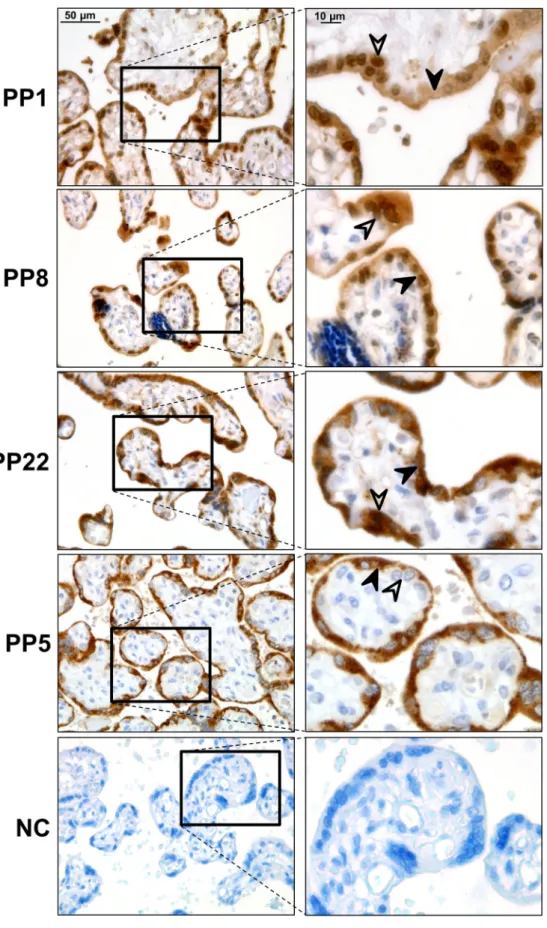
First, we examined the expression pattern of these proteins in normal, healthy term placentas. PP1, PP8, and PP22 immunostaining was detected primarily in the trophoblasts. The results are as follows: 1) Strong PP1 immunostaining was detected in the cytotrophoblasts and syncytiotrophoblast, and faint PP1 immunostaining was also detected in the villous mesenchymal and endothelial cells. 2) Strong PP8 immuno- staining was detected in the cytotrophoblasts and syncytiotrophoblast, while other cell types of the chorionic villi did not stain for PP8. 3) Strong PP22 immunostaining was detected in the cytotrophoblasts and syncytiotrophoblast, and faint PP22 immunostaining was also detected
in the villous mesenchymal and endothelial cells. 4) PP5 immuno- staining was detected only in the syncytiotrophoblast but not in other cell types of the chorionic villi (Fig. 2).
3.5. Placental expression of placental proteins in preeclampsia and HELLP syndrome
Subsequently, expression patterns of the PPs were investigated in placentas taken from cases with preeclampsia and HELLP syndrome compared to gestational age-matched controls. Given that PPs are mainly expressed by the villous trophoblasts, we evaluated trophoblastic Table 2
Identified placental proteins in lyophilized protein fractions.
Protein
name Identified protein’s Uniprot accession number
Identified protein’s Uniprot
name Identified
protein’s NCBI identifier
Identified protein’s NCBI
name MW
(Da) Mascot
Score Number of unique peptides
Sequence coverage (%)
PP1 Q15274 nicotinate-nucleotide
pyrophosphorylase [carboxylating]
45269149 quinolinate phosphoribosyl-
transferase 31,138 1,075 12 59
PP8 P35237 serpin B6 41152086 serine (or cysteine) proteinase
inhibitor, clade B (ovalbumin), member 6
42,936 2,227 31 71
PP22 P07237 protein disulfide-isomerase 20070125 prolyl 4-hydroxylase, beta
subunit precursor 57,480 954 25 50
Fig. 1. Placental and trophoblastic expression of placental proteins. Based on placenta-specificity scores and tissue expression levels, Placental Protein 5 (PP5) is predominantly expressed in the placenta, while PP1, PP8, and PP22 are not (a). PP5 mRNA expression level increased from the first day of villous trophoblast differentiation, while PP1, PP8, and PP22 mRNA level did not (b).
immunoscores. Because PP8 and PP22 immunoscores did not show any changes between normal and preeclamptic placentas (data not shown), further investigations were focused on only PP1.
The trophoblastic PP1 immunoscore decreased with gestational age as it was lower in term controls compared to preterm controls (mean ±
SE: preterm controls: 2.00 ±0.11 vs. term controls: 1.63 ±0.06, p = 0.003) (Figs. 3 and 4). There was increased PP1 immunostaining in late- onset preeclampsia (1.98 ±0.08, p =0.0008) compared to term controls (1.63 ±0.06) (Figs. 3 and 4). There was no difference in PP1 immu- noscores between early-onset preeclampsia (1.91 ±0.09, p =0.52) and
Fig. 2. Placental expression of placental proteins in normal pregnancies. Strong Placental Protein 1 (PP1) immunostaining was detected in cytotrophoblasts and the syncytiotrophoblast. Faint PP1 immuno- staining was also detected in villous mesen- chymal and endothelial cells. Strong PP8 immunostaining was detected in cyto- trophoblasts and the syncytiotrophoblast, while other cell types of the chorionic villi did not stain for PP8. Strong PP22 immu- nostaining was detected in cytotrophoblasts and the syncytiotrophoblast. Faint PP22 immunostaining was also detected in villous mesenchymal and endothelial cells. PP5 immunostaining was detected only in the syncytiotrophoblast but not in other cell types of the chorionic villi. No immuno- staining was detected on negative controls (NC). Representative images, hematoxylin counterstain, 400 x and 1,000 x magnifica- tions, scale bar 50 μm and 10 μm. Black ar- rows depict syncytiotrophoblasts while white arrows depict cytotrophoblasts.
preterm controls (2.00 ±0.11) or between early-onset HELLP syndrome (2.08 ±0.1, p =0.57) and preterm controls (2.00 ±0.11) (Figs. 3 and 4).
3.6. The correlation of Placental Protein 1 expression with placental weight and birth weight
The PP1 immunoscores negatively correlated with placental weight among controls (R = − 0.54, p =0.04), but this was not significant among patients with preeclampsia (R = − 0.25, p =0.14). Also, PP1 immunoscores negatively correlated with birth weight among controls (R = − 0.49, p =0.04), but this was not significant among patients with preeclampsia (R = − 0.22, p = 0.16). The PP1 immunoscore did not correlate with the MVMP scores of the placenta neither among patients with preeclampsia (R =0.17, p =0.22) nor among controls (R =0.07, p
=0.4) (Suppl. Table 2).
4. Discussion
4.1. Principal findings of this study
1) PP1, PP8, and PP22 were identified by mass spectrometry as
‘nicotinate-nucleotide pyrophosphorylase’, ‘serpin B6’ and ‘protein di- sulfide-isomerase’. 2) Genes encoding PP1, PP8, and PP22 are not pre- dominantly expressed by the placenta. 3) PP1, PP8, and PP22 mRNA expression does not increase during villous trophoblast differentiation.
4) PP1, PP8, and PP22 proteins are expressed primarily in villous tro- phoblasts in the placenta. 5) Trophoblastic PP1 immunoscores decreased with gestational age. 6) Trophoblastic PP1 immunoscores
were higher in late-onset preeclampsia compared to term controls.
4.2. Identification of placental proteins
We utilized mass spectrometry to identify the uncharacterized placental proteins that had been isolated from the human placenta de- cades ago.
We identified PP1 as ‘nicotinate-nucleotide pyrophosphorylase’ (QPRTase), a key enzyme in the catabolism of quinolinic acid and the biosynthesis of NAD(+) in the kynurenine pathway [53], also important in tryptophan metabolism in the placenta [54]. QPRTase was described in cells of the human central nervous system [55], liver [56], and blood [57]. The molecular weight (MW) of PP1 was originally described to be 160 kDa as a composite of identical subunits by Bohn et al. [35,36]. In good accordance, the MW of QPRTase is 180 kDa, and it is a hexamer formed by three homodimers. Based on the Uniprot database, a signif- icant amount of glycans or other post-translational modifications cannot be found on QPRTase, which is consistent with the minimal carbohy- drate content of the PP1 extracted by Bohn et al. [35].
We identified PP8 as ‘serpin B6’, a serine protease inhibitor, also referred to as a ‘placental thrombin inhibitor’ given its presence in placental tissue and its interaction with thrombin [58]. PP8 was described as a single-chain protein of 45 kDa that contains 4.1% car- bohydrate by Bohn et al. [35,36]. In accordance, we found that the single-chain ‘serpin B6’ is 43 kDa.
We identified PP22 as ‘protein disulfide isomerase’, a multifunc- tional protein that 1) has an important role in the formation of disulfide bonds; 2) at high concentrations inhibits aggregation (chaperone Fig. 3. Placental expression of Placental Pro- tein 1 in normal pregnancy and in pre- eclampsia and HELLP syndrome. Placental Protein 1 (PP1) immunostaining was lower in term controls (d) compared to preterm controls (a). PP1 immunostaining was stronger in late- onset preeclampsia (e) compared to term con- trols (d). PP1 immunostaining was similar be- tween early-onset preeclampsia with (c) or without HELLP syndrome (b) and preterm con- trols (a). No immunostaining was detected on negative controls (f). Representative images, he- matoxylin counterstain, 400 x magnifications, scale bar 50 μm.
function); 3) at low concentrations facilitates aggregation (anti-chap- erone function); 4) is a subunit of different enzymes, like prolyl 4-hy- droxylase; and 5) is a receptor for galectin-9: the interaction retains the protein at the cell surface of Th2 T helper cells, at the plasma membrane increases the activity of disulfide reductase and alters redox state as well as influences cell migration [59,60]. Using a placental cDNA library, earlier PP22 was found to be identical to ‘cellular thyroid hormone-binding protein’ by Bohn et al. [35,36]. This is an alternative name of ‘protein disulfide-isomerase’; therefore, our study confirms former results. PP22 was described as a single-chain protein of 50–65 kDa without a detectable amount of carbohydrates by Bohn et al. [35].
This shows good agreement with the MW of ‘protein disulfide-isomer- ase’, which is 57 kDa.
4.3. Placental expression and function of placental proteins in healthy conditions
When analyzing the expression pattern of PPs in the placenta, we took into account that, in normal physiologic conditions, there is a complex interaction between trophoblasts and other cells, including maternal immune cells, that influences the expression of placental proteins, and that unique condition is changed after delivering the placenta. To better preserve the placental protein expression pattern reflecting the normal behavior of placental cells, we completed placental sample collection and processing within one half-hour after Cesarean delivery in case of all samples according to international standards [50].
It was interesting to find that neither of the identified PPs have placenta- or trophoblast-specific functions; rather, they are involved in general cellular functions. This observation is also reflected by the fact
that these PPs are not expressed predominantly by the placenta as we found in this study. Moreover, this result contrasts with another placental protein, PP5/TFPI-2, whose expression occurs predominantly in the placenta where it exerts placenta-specific functions in the coag- ulation cascade. Indeed, quinolinic acid catabolism and NAD(+) biosynthesis in the kynurenine pathway, thrombin inhibition in the coagulation cascade, and the catalysis of disulfide bond rearrangements are processes widely utilized by various cells and tissues in the human body.
Still, the placenta produces large amounts of these PPs as supported by past and present RNA/protein evidence; therefore, their functions must be pivotal for placental physiology. Indeed, the kynurenine pathway is important for fetal development, maternal-fetal immune tolerance, and the prevention of placental oxidative stress [59,61–63];
thrombin inhibition is critical for placental hemostasis [24]; and chap- erone functions and redox defense mediated by protein disulfide isom- erase are critical in placental physiology and defense [10–12,64].
It was also interesting to note that these PPs have predominant and strong expression in villous trophoblasts; however, we did not find PP1, PP8, or PP22 expression to be developmentally regulated in villous trophoblasts in contrast to PP5/TFPI-2, which had increasing expression during villous trophoblast differentiation. Given that we used a well- established in vitro trophoblast culturing and differentiation system [16,44–46], we are confident that the detected PP expression patterns reflected the trophoblastic developmental program and were only minimally affected by the applied in vitro conditions. Overall, this finding underscores that these PPs exert their functions, important for all villous trophoblastic cells, at various stages of their development.
Fig. 4. Trophoblastic Placental Protein 1 immu- noscores. The number of placentas on TMAs and the number of immunoscored villi in each study group are shown on the first panel (a). Placental Protein 1 (PP1) immunoscores decreased in term controls (1.63 ±0.06, p =0.003) compared to preterm con- trols (2.00 ±0.11). PP1 immunostaining increased in late-onset preeclampsia (1.98 ±0.08, p =0.0008) compared to term controls (1.63 ± 0.06). PP1 immunoscores did not change between early-onset preeclampsia (1.91 ±0.09, p =0.52) and preterm controls (2.00 ±0.11) or between early-onset HELLP syndrome (2.08 ±0.1, p =0.57) and preterm controls (2.00 ±0.11) (b,c). PE: preeclampsia; HELLP: HELLP syndrome; TMA: tissue microarray; *p <0.05, **p
<0.01.
4.4. Placental expression and function of placental proteins in preeclampsia and HELLP syndrome
While examining the potential changes in placental expression of PPs in various subsets of preeclampsia and HELLP syndrome, we docu- mented the up-regulation of PP1 protein quantities in late-onset pre- eclampsia but not in other preeclampsia subsets when compared to gestational age-matched controls. We also did not find any correlation of PP1 immunoscores with placental and fetal weight, which are the proxies of placental functions. When we combined the early-onset pre- eclampsia with or without HELLP syndrome groups into one and repeated the analyses, there was still no difference between the com- bined preeclampsia group and preterm controls regarding PP1 immu- noscores. This substantiates that the lack of difference is a result of biological and not statistical reasons. The up-regulation of PP1 only in late-onset preeclampsia is a rather interesting finding: similar to other investigators, we earlier found gene- or protein-level dysregulations in the placenta only in early-onset but not in late-onset preeclampsia, which was associated with placental dysfunction. As the placenta is severely ill in early-onset preeclampsia, proteins predominantly or specifically expressed by the placenta and/or trophoblast are generally dysregulated in this disease subset, which is also the case for PP5 [13,16, 30,31,33,34,44]. However, late-onset preeclampsia is thought to be a maternal disease in which the placenta and fetal weights are affected less or not at all [7,16,28,65–69].
In this context, it is interesting why PP1 expression is up-regulated in late-onset preeclampsia. To understand this phenomenon, we need to take into account the multi-etiologic nature of preeclampsia and the molecular subsets and pathways of preeclampsia that have been iden- tified by us and others in recent omics and systems biological studies [16,70,71]. According to these observations, early-onset preeclampsia is mainly associated with placental disease involving the dysregulation of trophoblastic functions and gene modules, while late-onset preeclamp- sia is rather associated with maternal immune and metabolic changes.
Since PP1/QPRTase is active in the kynurenine pathway involved in sustaining placental immune privilege [61,72–75], the dysregulation of PP1/QPRTase may be involved in the molecular mechanisms leading to a disturbed immune balance at the maternal-fetal interface. In fact, the disturbance of the placental kynurenine pathway in preeclampsia has been documented by recent studies [73,75] and by other studies con- nected to inflammatory processes promoting placental dysregulation of the kynurenine pathway [54,76]. Nevertheless, our descriptive study can only infer an association, and future studies are necessary to investigate the functional role of placental PP1/QPRTase in the disease pathways of preeclampsia.
5. Conclusions
PP1, PP8, and PP22 are expressed primarily in trophoblasts but do not have trophoblast-specific regulation or functions. The distinct dys- regulation of PP1 and PP5 expression in late-onset or early-onset pre- eclampsia reflects different pathophysiological pathways in these preeclampsia subsets. PP5 dysregulation in early-onset preeclampsia is associated with placental disease, while PP1 dysregulation in late-onset preeclampsia may represent maternal inflammation inducing placental functional alterations in late-onset preeclampsia.
Author contributions
NGT, SzSz, KK conceptualized study and designed research; SzSz, KK, ET, YX, BH, EHG, ZsD, KAK, NGT performed research; RR, PH, TK, IK, ZP, NGT contributed new reagents/analytic tools/clinical specimens;
SzSz, KK, ET, ASz, ZsG, AB, GSz, ALT, OE, GJ, NGT analyzed and interpreted data; all authors contributed to the writing of the paper.
Declaration of competing interest The authors report no conflict of interest.
Acknowledgements
The authors thank Tibor Fule, Prof. Andras Matolcsy, Prof. Balint Nagy, Edit Parsch, and Tibor Varkonyi (Semmelweis University) for their support or assistance. This work was supported by grants from the Hungarian National Science Fund (OTKA-K119283 to IK; OTKA- K124862 to NGT; OTKA-K128262 to ASz), Hungarian Academy of Sci- ences (Momentum Grant LP2014-7/2014 to NGT, Premium_2019-436 grant to AB), Hungarian National Research, Development and Innova- tion Office (FIEK-16-1-2016-0005 to NGT, KAK, GJ), Hungarian Minis- try for National Economy (VEKOP-2.3.3-15-2017-0014 Grant to NGT, EFOP-3.6.3-VEKOP-16-2017-00009 scholarship to KK) and from the European Union (FP6 Grant Pregenesys-037244 to NGT).
This research was also supported, in part, by the Perinatology Research Branch, Division of Obstetrics and Maternal-Fetal Medicine, Division of Intramural Research, Eunice Kennedy Shriver National Insti- tute of Child Health and Human Development, National Institutes of Health, U.S. Department of Health and Human Services (NICHD/NIH/
DHHS); and, in part, with Federal funds from NICHD/NIH/DHHS under Contract No. HHSN275201300006C.
Dr. Romero has contributed to this work as part of his official duties as an employee of the United States Federal Government.
Appendix A. Supplementary data
Supplementary data to this article can be found online at https://doi.
org/10.1016/j.placenta.2020.05.013.
References
[1] ACOG Practice Bulletin, Diagnosis and management of preeclampsia and eclampsia. Number 33, January 2002, Obstet. Gynecol. 99 (1) (2002) 159–167.
[2] B. Sibai, G. Dekker, M. Kupferminc, Pre-eclampsia, Lancet 365 (9461) (2005) 785–799.
[3] P. von Dadelszen, L.A. Magee, J.M. Roberts, Subclassification of preeclampsia, Hypertens. Pregnancy 22 (2) (2003) 143–148.
[4] L. Weinstein, Syndrome of hemolysis, elevated liver enzymes, and low platelet count: a severe consequence of hypertension in pregnancy, Am. J. Obstet. Gynecol.
142 (2) (1982) 159–167.
[5] J.S. Moldenhauer, J. Stanek, C. Warshak, J. Khoury, B. Sibai, The frequency and severity of placental findings in women with preeclampsia are gestational age dependent, Am. J. Obstet. Gynecol. 189 (4) (2003) 1173–1177.
[6] S. Nagy, M. Bush, J. Stone, R.H. Lapinski, S. Gardo, Clinical significance of subchorionic and retroplacental hematomas detected in the first trimester of pregnancy, Obstet. Gynecol. 102 (1) (2003) 94–100.
[7] C.W. Redman, I.L. Sargent, Latest advances in understanding preeclampsia, Science 308 (5728) (2005) 1592–1594.
[8] I. Crocker, Gabor Than Award Lecture 2006: pre-eclampsia and villous trophoblast turnover: perspectives and possibilities, Placenta 28 (Suppl A) (2007) S4–S13.
[9] S. Maynard, F.H. Epstein, S.A. Karumanchi, Preeclampsia and angiogenic imbalance, Annu. Rev. Med. 59 (2008) 61–78, 61-78.
[10] G.J. Burton, A.W. Woods, E. Jauniaux, J.C. Kingdom, Rheological and physiological consequences of conversion of the maternal spiral arteries for uteroplacental blood flow during human pregnancy, Placenta 30 (6) (2009) 473–482.
[11] G.J. Burton, H.W. Yung, T. Cindrova-Davies, D.S. Charnock-Jones, Placental endoplasmic reticulum stress and oxidative stress in the pathophysiology of unexplained intrauterine growth restriction and early onset preeclampsia, Placenta 30 (Suppl A) (2009) S43–S48.
[12] T. Cindrova-Davies, Gabor Than Award Lecture 2008: pre-eclampsia - from placental oxidative stress to maternal endothelial dysfunction, Placenta 30 (Suppl A) (2009) S55–S65.
[13] T. Varkonyi, B. Nagy, T. Fule, A.L. Tarca, K. Karaszi, J. Schonleber, P. Hupuczi, N. Mihalik, I. Kovalszky, J. Rigo Jr., H. Meiri, Z. Papp, R. Romero, N.G. Than, Microarray profiling reveals that placental transcriptomes of early-onset HELLP syndrome and preeclampsia are similar, Placenta 32 (Suppl) (2011) S21–S29.
[14] O. Lapaire, S. Grill, S. Lalevee, V. Kolla, I. Hosli, S. Hahn, Microarray screening for novel preeclampsia biomarker candidates, Fetal Diagn. Ther. 31 (3) (2012) 147–153.
[15] S. Szabo, M. Mody, R. Romero, Y. Xu, K. Karaszi, N. Mihalik, Z. Xu, G. Bhatti, T. Fule, P. Hupuczi, T. Krenacs, J. Rigo Jr., A.L. Tarca, S.S. Hassan, T. Chaiworapongsa, I. Kovalszky, Z. Papp, N.G. Than, Activation of villous
trophoblastic p38 and ERK1/2 signaling pathways in preterm preeclampsia and HELLP syndrome, Pathol. Oncol. Res. 21 (3) (2015) 659–668.
[16] N.G. Than, R. Romero, A.L. Tarca, K.A. Kekesi, Y. Xu, Z. Xu, K. Juhasz, G. Bhatti, R.
J. Leavitt, Z. Gelencser, J. Palhalmi, T.H. Chung, B.A. Gyorffy, L. Orosz, A. Demeter, A. Szecsi, E. Hunyadi-Gulyas, Z. Darula, A. Simor, K. Eder, S. Szabo, V. Topping, H. El-Azzamy, C. LaJeunesse, A. Balogh, G. Szalai, S. Land, O. Torok, Z. Dong, I. Kovalszky, A. Falus, H. Meiri, S. Draghici, S.S. Hassan,
T. Chaiworapongsa, M. Krispin, M. Knofler, O. Erez, G.J. Burton, C.J. Kim, G. Juhasz, Z. Papp, Integrated systems biology approach identifies novel maternal and placental pathways of preeclampsia, Front. Immunol. 9 (2018) 1661.
[17] S. Haider, G. Meinhardt, L. Saleh, C. Fiala, J. Pollheimer, M. Knofler, Notch1 controls development of the extravillous trophoblast lineage in the human placenta, Proc. Natl. Acad. Sci. U. S. A. 113 (48) (2016) E7710–E7719.
[18] J.E. Davies, J. Pollheimer, H.E. Yong, M.I. Kokkinos, B. Kalionis, M. Knofler, P. Murthi, Epithelial-mesenchymal transition during extravillous trophoblast differentiation, Cell Adhes. Migrat. 10 (3) (2016) 310–321.
[19] V. Fulop, G. Vermes, J. Demeter, The relationship between inflammatory and immunological processes during pregnancy. Practical aspects, Orv. Hetil. 160 (32) (2019) 1247–1259.
[20] K. Kovacs, B. Vasarhelyi, B. Gyarmati, G. Karvaly, Estrogen metabolism during pregnancy, Orv. Hetil. 160 (26) (2019) 1007–1014.
[21] J.M. Roberts, K.Y. Lain, Recent Insights into the pathogenesis of pre-eclampsia, Placenta 23 (5) (2002) 359–372.
[22] P. Tamas, E. Hantosi, B. Farkas, Z. Ifi, J. Betlehem, J. Bodis, Preliminary study of the effects of furosemide on blood pressure during late-onset pre-eclampsia in patients with high cardiac output, Int. J. Gynaecol. Obstet. 136 (1) (2017) 87–90.
[23] L. Vokalova, S.V. van Breda, X.L. Ye, E.A. Huhn, N.G. Than, P. Hasler, O. Lapaire, I. Hoesli, S.W. Rossi, S. Hahn, Excessive neutrophil activity in gestational diabetes mellitus: could it contribute to the development of preeclampsia? Front.
Endocrinol. 9 (2018) 542.
[24] O. Erez, R. Romero, E. Vaisbuch, N.G. Than, J.P. Kusanovic, S. Mazaki-Tovi, F. Gotsch, P. Mittal, Z. Dong, T. Chaiworapongsa, C.J. Kim, C.L. Nhan-Chang, S.
K. Kim, L. Yeo, M. Mazor, S.S. Hassan, Tissue factor activity in women with preeclampsia or SGA: a potential explanation for the excessive thrombin generation in these syndromes, J. Matern. Fetal Neonatal Med. 31 (12) (2018) 1568–1577.
[25] P. Murthi, A.A. Pinar, E. Dimitriadis, C.S. Samuel, Inflammasomes-A molecular link for altered immunoregulation and inflammation mediated vascular dysfunction in preeclampsia, Int. J. Mol. Sci. 21 (4) (2020).
[26] S. Hahn, O. Lapaire, N.G. Than, Biomarker development for presymptomatic molecular diagnosis of preeclampsia: feasible, useful or even unnecessary? Expert Rev. Mol. Diagn 15 (5) (2015) 617–629.
[27] C.W. Redman, I.L. Sargent, A.C. Staff, IFPA Senior Award Lecture: making sense of pre-eclampsia - two placental causes of preeclampsia? Placenta 35 (Suppl) (2014) S20–S25.
[28] R.B. Ness, J.M. Roberts, Heterogeneous causes constituting the single syndrome of preeclampsia: a hypothesis and its implications, Am. J. Obstet. Gynecol. 175 (5) (1996) 1365–1370.
[29] G. Eastabrook, M. Brown, I. Sargent, The origins and end-organ consequence of pre-eclampsia, Best Pract. Res. Clin. Obstet. Gynaecol. 25 (4) (2011) 435–447.
[30] N.G. Than, O. Abdul Rahman, R. Magenheim, B. Nagy, T. Fule, B. Hargitai, M. Sammar, P. Hupuczi, A.L. Tarca, G. Szabo, I. Kovalszky, H. Meiri, I. Sziller, J. Rigo Jr., R. Romero, Z. Papp, Placental protein 13 (galectin-13) has decreased placental expression but increased shedding and maternal serum concentrations in patients presenting with preterm pre-eclampsia and HELLP syndrome, Virchows Arch. 453 (4) (2008) 387–400.
[31] S. Szabo, Y. Xu, R. Romero, T. Fule, K. Karaszi, G. Bhatti, T. Varkonyi, I. Varkonyi, T. Krenacs, Z. Dong, A.L. Tarca, T. Chaiworapongsa, S.S. Hassan, Z. Papp, I. Kovalszky, N.G. Than, Changes of placental syndecan-1 expression in preeclampsia and HELLP syndrome, Virchows Arch. 463 (3) (2013) 445–458.
[32] A. Balogh, E. Toth, R. Romero, K. Parej, D. Csala, N.L. Szenasi, I. Hajdu, K. Juhasz, A.F. Kovacs, H. Meiri, P. Hupuczi, A.L. Tarca, S.S. Hassan, O. Erez, P. Zavodszky, J. Matko, Z. Papp, S.W. Rossi, S. Hahn, E. Pallinger, N.G. Than, Placental galectins are key players in regulating the maternal adaptive immune response, Front.
Immunol. 10 (2019) 1240.
[33] K. Karaszi, S. Szabo, K. Juhasz, P. Kiraly, B. Kocsis-Deak, B. Hargitai, T. Krenacs, P. Hupuczi, O. Erez, Z. Papp, I. Kovalszky, N.G. Than, Increased placental expression of Placental Protein 5 (PP5)/Tissue Factor Pathway Inhibitor-2 (TFPI-2) in women with preeclampsia and HELLP syndrome: relevance to impaired trophoblast invasion? Placenta 76 (2019) 30–39.
[34] N.L. Szenasi, E. Toth, A. Balogh, K. Juhasz, K. Karaszi, O. Ozohanics, Z. Gelencser, P. Kiraly, B. Hargitai, L. Drahos, P. Hupuczi, I. Kovalszky, Z. Papp, N.G. Than, Proteomic identification of membrane-associated placental protein 4 (MP4) as perlecan and characterization of its placental expression in normal and pathologic pregnancies, PeerJ 7 (2019), e6982.
[35] H. Bohn, W. Kraus, W. Winkler, New soluble placental tissue proteins: their isolation, characterization, localization and quantification, in: A. Klopper (Ed.), Immunology of Human Placental Proteins, Praeger Publishers, East Sussex, 1982.
[36] G.N. Than, H. Bohn, D.G. Szabo, Advances in Pregnancy-Related Protein Research, CRC Press, Boca Raton, 1993.
[37] N.G. Than, B. Sumegi, G.N. Than, G. Kispal, H. Bohn, Cloning and sequence analysis of cDNAs encoding human placental tissue protein 17 (PP17) variants, Eur. J. Biochem. 258 (2) (1998) 752–757.
[38] N.G. Than, B. Sumegi, G.N. Than, Z. Berente, H. Bohn, Isolation and sequence analysis of a cDNA encoding human placental tissue protein 13 (PP13), a new
lysophospholipase, homologue of human eosinophil Charcot-Leyden Crystal protein, Placenta 20 (8) (1999) 703–710.
[39] N.G. Than, B. Sumegi, G.N. Than, S. Bellyei, H. Bohn, Molecular cloning and characterization of placental tissue protein 18 (PP18a)/human mitochondrial branched-chain aminotransferase (BCATm) and its novel alternatively spliced PP18b variant, Placenta 22 (2-3) (2001) 235–243.
[40] B. Visegrady, N.G. Than, F. Kilar, B. Sumegi, G.N. Than, H. Bohn, Homology modelling and molecular dynamics studies of human placental tissue protein 13 (galectin-13), Protein Eng. 14 (11) (2001) 875–880.
[41] N.G. Than, E. Pick, S. Bellyei, A. Szigeti, O. Burger, Z. Berente, T. Janaky, A. Boronkai, H. Kliman, H. Meiri, H. Bohn, G.N. Than, B. Sumegi, Functional analyses of placental protein 13/galectin-13, Eur. J. Biochem. 271 (6) (2004) 1065–1078.
[42] S. Bellyei, A. Szigeti, A. Boronkai, Z. Szabo, J. Bene, T. Janaky, L. Barna, K. Sipos, O. Minik, A. Kravjak, R. Ohmacht, B. Melegh, P. Zavodszky, G.N. Than, B. Sumegi, H. Bohn, N.G. Than, Cloning, sequencing, structural and molecular biological characterization of placental protein 20 (PP20)/human thiamin
pyrophosphokinase (hTPK), Placenta 26 (1) (2005) 34–46.
[43] A. Balogh, J. Pozsgay, J. Matko, Z. Dong, C.J. Kim, T. Varkonyi, M. Sammar, J. Rigo Jr., H. Meiri, R. Romero, Z. Papp, N.G. Than, Placental protein 13 (PP13/
galectin-13) undergoes lipid raft-associated subcellular redistribution in the syncytiotrophoblast in preterm preeclampsia and HELLP syndrome, Am. J. Obstet.
Gynecol. 205 (2) (2011), 156e1-e14.
[44] N.G. Than, R. Romero, Y. Xu, O. Erez, Z. Xu, G. Bhatti, R. Leavitt, T.H. Chung, H. El-Azzamy, C. LaJeunesse, B. Wang, A. Balogh, G. Szalai, S. Land, Z. Dong, S.
S. Hassan, T. Chaiworapongsa, M. Krispin, C.J. Kim, A.L. Tarca, Z. Papp, H. Bohn, Evolutionary origins of the placental expression of chromosome 19 cluster galectins and their complex dysregulation in preeclampsia, Placenta 35 (11) (2014) 855–865.
[45] A. Szilagyi, Z. Gelencser, R. Romero, Y. Xu, P. Kiraly, A. Demeter, J. Palhalmi, B.
A. Gyorffy, K. Juhasz, P. Hupuczi, K.A. Kekesi, G. Meinhardt, Z. Papp, S. Draghici, O. Erez, A.L. Tarca, M. Knofler, N.G. Than, Placenta-specific genes, their regulation during villous trophoblast differentiation and dysregulation in preterm preeclampsia, Int. J. Mol. Sci. 21 (2) (2020).
[46] H.J. Kliman, J.E. Nestler, E. Sermasi, J.M. Sanger, J.F. Strauss III, Purification, characterization, and in vitro differentiation of cytotrophoblasts from human term placentae, Endocrinology 118 (4) (1986) 1567–1582.
[47] C. Papp, G. Szabo, E. Toth-Pal, Z. Papp, Fetal growth rate and its variations 1988/
89, Orv. Hetil. 132 (34) (1991) 1865–1870.
[48] J.R. Barton, B.M. Sibai, Diagnosis and management of hemolysis, elevated liver enzymes, and low platelets syndrome, Clin. Perinatol. 31 (4) (2004) 807–833 (vii).
[49] R.W. Redline, Placental pathology: a systematic approach with clinical correlations, Placenta 29 (Suppl A) (2008) S86–S91.
[50] G.J. Burton, N.J. Sebire, L. Myatt, D. Tannetta, Y.L. Wang, Y. Sadovsky, A.C. Staff, C.W. Redman, Optimising sample collection for placental research, Placenta 35 (1) (2014) 9–22.
[51] B. Hargitai, T. Marton, P.M. Cox, Best practice no 178. Examination of the human placenta, J. Clin. Pathol. 57 (8) (2004) 785–792.
[52] T.Y. Khong, E.E. Mooney, I. Ariel, N.C. Balmus, T.K. Boyd, M.A. Brundler, H. Derricott, M.J. Evans, O.M. Faye-Petersen, J.E. Gillan, A.E. Heazell, D.S. Heller, S.M. Jacques, S. Keating, P. Kelehan, A. Maes, E.M. McKay, T.K. Morgan, P.
G. Nikkels, W.T. Parks, R.W. Redline, I. Scheimberg, M.H. Schoots, N.J. Sebire, A. Timmer, G. Turowski, J.P. van der Voorn, I. van Lijnschoten, S.J. Gordijn, Sampling and definitions of placental lesions: amsterdam placental workshop group consensus statement, Arch. Pathol. Lab Med. 140 (7) (2016) 698–713.
[53] D. Moore, T. Bice, L. Jin, A. Grandhi, L.J. DeLucas, S.V. Narayana, Crystallization and preliminary crystallographic investigation of porcine quinolinate
phosphoribosyltransferase, Acta Crystallogr. D Biol. Crystallogr. 54 (Pt 1) (1998) 119–120.
[54] P. Murthi, E.M. Wallace, D.W. Walker, Altered placental tryptophan metabolic pathway in human fetal growth restriction, Placenta 52 (2017) 62–70.
[55] C. Kohler, E. Okuno, P.R. Flood, R. Schwarcz, Quinolinic acid
phosphoribosyltransferase: preferential glial localization in the rat brain visualized by immunocytochemistry, Proc. Natl. Acad. Sci. U. S. A. 84 (10) (1987) 3491–3495.
[56] E. Okuno, R.J. White, R. Schwarcz, Quinolinic acid phosphoribosyltransferase:
purification and partial characterization from human liver and brain, J. Biochem.
103 (6) (1988) 1054–1059.
[57] A.C. Foster, R. Schwarcz, Characterization of quinolinic acid
phosphoribosyltransferase in human blood and observations in Huntington’s disease, J. Neurochem. 45 (1) (1985) 199–205.
[58] P. Coughlin, J. Sun, L. Cerruti, H.H. Salem, P. Bird, Cloning and molecular characterization of a human intracellular serine proteinase inhibitor, Proc. Natl.
Acad. Sci. U. S. A. 90 (20) (1993) 9417–9421.
[59] S. Bi, P.W. Hong, B. Lee, L.G. Baum, Galectin-9 binding to cell surface protein disulfide isomerase regulates the redox environment to enhance T-cell migration and HIV entry, Proc. Natl. Acad. Sci. U. S. A. 108 (26) (2011) 10650–10655.
[60] C.T. Kwok, A.G. Morris, J. Frampton, B. Smith, C.E. Shaw, J. de Belleroche, Association studies indicate that protein disulfide isomerase is a risk factor in amyotrophic lateral sclerosis, Free Radic. Biol. Med. 58 (2013) 81–86.
[61] P. Sedlmayr, A. Blaschitz, R. Stocker, The role of placental tryptophan catabolism, Front. Immunol. 5 (2014) 230.
[62] H.J. Kliman, S.B. Quaratella, A.C. Setaro, E.C. Siegman, Z.T. Subha, R. Tal, K.
M. Milano, T.L. Steck, Pathway of maternal serotonin to the human embryo and fetus, Endocrinology 159 (4) (2018) 1609–1629.
[63] K. Xu, G. Liu, C. Fu, The tryptophan pathway targeting antioxidant capacity in the placenta, Oxid Med. Cell Longev. 2018 (2018) 1054797.
[64] J.H. Veerbeek, M.C. Tissot Van Patot, G.J. Burton, H.W. Yung, Endoplasmic reticulum stress is induced in the human placenta during labour, Placenta 36 (1) (2015) 88–92.
[65] L.J. Vatten, R. Skjaerven, Is pre-eclampsia more than one disease? BJOG 111 (4) (2004) 298–302.
[66] N.J. Sebire, R.D. Goldin, L. Regan, Term preeclampsia is associated with minimal histopathological placental features regardless of clinical severity, J. Obstet.
Gynaecol. 25 (2) (2005) 117–118.
[67] H. Valensise, B. Vasapollo, G. Gagliardi, G.P. Novelli, Early and late preeclampsia:
two different maternal hemodynamic states in the latent phase of the disease, Hypertension 52 (5) (2008) 873–880.
[68] T. Chaiworapongsa, P. Chaemsaithong, L. Yeo, R. Romero, Pre-eclampsia part 1:
current understanding of its pathophysiology, Nat. Rev. Nephrol. 10 (8) (2014) 466–480.
[69] L. Myatt, J.M. Roberts, Preeclampsia: syndrome or disease? Curr. Hypertens. Rep.
17 (11) (2015) 83.
[70] K. Leavey, S.A. Bainbridge, B.J. Cox, Large scale aggregate microarray analysis reveals three distinct molecular subclasses of human preeclampsia, PloS One 10 (2) (2015), e0116508.
[71] K. Leavey, S.J. Benton, D. Grynspan, J.C. Kingdom, S.A. Bainbridge, B.J. Cox, Unsupervised placental gene expression profiling identifies clinically relevant subclasses of human preeclampsia, Hypertension 68 (1) (2016) 137–147.
[72] P. Ligam, U. Manuelpillai, E.M. Wallace, D. Walker, Localisation of indoleamine 2,3-dioxygenase and kynurenine hydroxylase in the human placenta and decidua:
implications for role of the kynurenine pathway in pregnancy, Placenta 26 (6) (2005) 498–504.
[73] H. Nishizawa, M. Suzuki, K. Pryor-Koishi, T. Sekiya, S. Tada, H. Kurahashi, Y. Udagawa, Impact of indoleamine 2,3-dioxygenase on the antioxidant system in the placentas of severely pre-eclamptic patients, Syst. Biol. Reprod. Med. 57 (4) (2011) 174–178.
[74] J.P. Routy, B. Routy, G.M. Graziani, V. Mehraj, The kynurenine pathway is a double-edged sword in immune-privileged sites and in cancer: implications for immunotherapy, Int. J. Tryptophan Res. 9 (2016) 67–77.
[75] S.A. Keaton, P. Heilman, E.Y. Bryleva, Z. Madaj, S. Krzyzanowski, J. Grit, E.
S. Miller, M. Jalmby, G. Kalapotharakos, K. Racicot, A. Fazleabas, S.R. Hansson, L. Brundin, Altered tryptophan catabolism in placentas from women with pre- eclampsia, Int. J. Tryptophan Res. 12 (2019), 1178646919840321.
[76] U. Manuelpillai, T. Nicholls, E.M. Wallace, D.J. Phillips, G. Guillemin, D. Walker, Increased mRNA expression of kynurenine pathway enzymes in human placentae exposed to bacterial endotoxin, Adv. Exp. Med. Biol. 527 (2003) 85–89.