DEVELOPMENT OF NOVEL SURGICAL, IMAGING AND MOLECULAR METHODS FOR THE IN VIVO MODELING OF PREECLAMPSIA
PhD thesis Gábor Szalai, MD
Doctoral School of Clinical Medicine Semmelweis University
Supervisor: Nándor Gábor Than, MD, PhD
Official Reviewers: Péter Tamás, MD, PhD Sándor Valent, MD, PhD
Head of the Final Examination Committee:
Ferenc Perner, MD, DSc
Members of the Final Examination Committee:
Artúr Beke, MD, PhD Zorán Belics, MD, PhD
Budapest 2015
DOI:10.14753/SE.2015.1828
TABLE OF CONTENTS
LIST OF ABBREVIATIONS ... 5
1. INTRODUCTION ... 7
1.1. Preeclampsia is a ‘Great Obstetrical Syndrome’ ... 7
1.2. Pathophysiology and molecular mechanisms ... 10
1.2.1. Placental pathophysiology ... 10
1.2.2. The terminal pathway of preeclampsia ... 11
1.2.3. Angiogenic – anti-angiogenic imbalance ... 11
1.2.4. Alterations in the renin-angiotensin system ... 12
1.2.5. Systemic inflammatory response, endothelial dysfunction and coagulopathy12 1.3. sFlt-1 isoforms ... 13
1.4. Animal models of preeclampsia ... 16
1.4.1. General overview ... 16
1.4.2. Anti-angiogenic animal models with the overexpression of sFlt-1 ... 17
2. OBJECTIVES ... 18
3. METHODS ... 19
3.1. Study I ... 19
3.1.1. Ethics statement ... 19
3.1.2. Animals and husbandry ... 19
3.1.3. Determination of pregnancy status with ultrasound ... 20
3.1.4. Implantation of the telemetric blood pressure monitoring system ... 21
3.1.5. Determination of the telemetry catheter position with high-frequency ultrasound... 23
3.1.6. Telemetric blood pressure monitoring ... 23
3.1.7. Adenoviral gene delivery ... 24
3.1.8. Ultrasound-guided bladder puncture (cystocentesis) ... 25
3.1.9. Cesarean section ... 27
3.1.10. Tissue collection ... 29
3.1.11. Total RNA isolation, cDNA generation, quantitative real-time RT-PCR .... 29
3.1.12. Histopathological evaluation of tissues ... 30
3.1.13. Immunohistochemistry ... 30
3.1.14. Aortic ring assays ... 30
3.1.15. Albumin-creatinine immunoassays ... 31 DOI:10.14753/SE.2015.1828
3.1.16. Adenoviral infection of BeWo cells ... 31
3.1.17. Protein isolation and Western blot... 31
3.1.18. Confocal microscopy ... 32
3.1.19. Data and statistical analyses... 32
3.2. Study II. ... 34
3.2.1. Ethics statement... 34
3.2.2. Animals and husbandry ... 34
3.2.3. Evaluation of pregnancy status with high-frequency ultrasound... 34
3.2.4. Telemetric blood-pressure catheter implantation and data acquisition ... 35
3.2.5. Determination of the telemetry catheter position with high-frequency ultrasound... 35
3.2.6. Telemetric blood pressure monitoring ... 35
3.2.7. Adenoviral gene delivery ... 35
3.2.8. Minimal invasive survival cesarean section... 37
3.2.9. Tissue collection ... 37
3.2.10. Total RNA isolation, cDNA generation, quantitative real-time RT-PCR .... 37
3.2.11. Histopathological evaluation ... 37
3.2.12. Immunohistochemistry ... 38
3.2.13. Aortic ring assays ... 38
3.2.14. Urine collection and albumin-creatinine immunoassays ... 38
3.2.15. Primary human trophoblast isolation and cultures ... 38
3.2.16. Confocal microscopy ... 39
3.2.17. Data and statistical analyses... 39
4. RESULTS ... 42
4.1. Study I ... 42
4.1.1. Pregnancy status determination with high-resolution ultrasound ... 42
4.1.2. Implantation and evaluation of telemetry devices... 42
4.1.3. Survival cesarean surgery ... 42
4.1.4. Overexpression of human placental sFlt-1-e15a isoform and GFP with adenoviral vectors ... 43
4.1.5. Blood pressure telemetry monitoring ... 45
4.1.6. Evaluation of endothelial and kidney functions ... 48
4.1.7. Evaluation of fetal survival rate, placental and fetal weights ... 50
4.1.8. Evaluation of a mouse with early-onset preeclampsia-like symptoms ... 50 DOI:10.14753/SE.2015.1828
4.2. Study II ... 53
4.2.1. The development of various transgene delivery systems ... 53
4.2.2. Unique placental expression of msFlt-1... 53
4.2.3. Expression patterns of various viral transgenes ... 54
4.2.4. Blood pressure telemetry monitoring ... 56
4.2.5. Morphological and functional changes in the kidneys in sFlt-1 treated mice57 4.2.6. Aortic endothelial dysfunction caused by hsFlt-1-e15a and msFlt-1 ... 60
4.2.7. Human sFlt-1-e15a but not msFlt-1(1-3) increases litter sizes ... 61
5. DISCUSSION ... 63
5.1. Principal findings ... 63
5.2. Pregnancy status was accurately determined with high-resolution ultrasound ... 64
5.3. Telemetric blood pressure monitoring provided accurate data acquisition in pregnant mice ... 64
5.4. A 100% survival rate was obtained with cesarean surgery... 66
5.5. A full-length, primate-specific sFlt-1-e15a isoform was selected to induce preeclampsia in mice ... 67
5.6. Human placental hsFlt-1-e15a increased blood pressure that normalized after delivery ... 69
5.7. Human placental hsFlt-1-e15a impaired endothelial and kidney functions... 70
5.8. Human placental hsFlt-1-e15a induced distinct preeclampsia phenotypes in CD-1 mice ... 71
5.9. The preeclampsia-inducing effects of the full-length hsFlt-1-e15a are similar to those of the mouse truncated sFlt-1(1-3) ... 74
5.10. sFlt-1 promotes the development of chronic disease following preeclampsia .. 76
5.11. sFlt-1 supports embryonic survival in early pregnancy ... 77
6. CONCLUSIONS ... 80
7. SUMMARY ... 81
8. ÖSSZEFOGLALÁS ... 82
9. BIBLIOGRAPHY ... 83
10. BIBLIOGRAPHY OF THE CANDIDATE’S PUBLICATIONS ... 120
10.1. Publications related to the PhD Thesis ... 120
10.2. Publications unrelated to the PhD Thesis ... 120
11. ACKNOWLEDGEMENTS ... 122 DOI:10.14753/SE.2015.1828
LIST OF ABBREVIATIONS
Ad: Adenovirus vector
Ad-RGD: RGD fiber-mutant adenovirus vector AT1: Angiotensin II receptor 1
cDNA: Complementary deoxyribonucleic acid
CMV: Cytomegalovirus
DHHS: Department of Health and Human Services DIC: Disseminated intravascular coagulation DLAR: Division of Laboratory Animal Resources DSI: Data Sciences International Inc.
EM: Experimental mouse
FLT1: Fms-like tyrosine kinase-1 gene
FW: Fetal weight
GFP: Green fluorescent protein
GD: Gestational day
HELLP syndrome: Hemolysis, elevated liver enzymes and low platelets syndrome
H&E: Hematoxylin and eosin sHLA: Human leukocyte antigen
hsFlt-1: Human soluble fms-like tyrosine kinase-1 IACUC: Institutional Animal Care and Use Committee IUFD: Intrauterine fetal demise
IUGR: Intrauterine growth restriction
KIR: Killer-cell immunoglobulin-like receptors
LME: Linear mixed effects
MAP: Mean arterial blood pressure mRNA: Messenger ribonucleic acid
msFlt-1: Mouse soluble fms-like tyrosine kinase-1 NET: Neutrophil extracellular trap
NICHD: National Institute of Child Health and Human Development
NIH: National Institutes of Health of the USA DOI:10.14753/SE.2015.1828
NO: Nitric oxide NK cell: Natural killer cell PAS: Periodic acid Schiff PBS: Phosphate-buffered saline
PE: Preeclampsia
PFU: Plaque-forming units PFR: Placental/fetal weight ratio PlGF: Placental growth factor
PPD: Postpartum day
PW: Placental weight
qRT-PCR: Quantitative real-time reverse transcription polymerase chain reaction
RDS: Respiratory distress syndrome SGA: Small-for-gestational age
sEng: Soluble endoglin
sFlt-1: Soluble fms-like tyrosine kinase-1
SMA: Smooth muscle actin
STBM: Syncytiotrophoblast microparticle
TMA: Tissue microarray
VEGF:
v
ascular endothelial growth factorVEGFR1: Vascular endothelial growth factor receptor 1 DOI:10.14753/SE.2015.1828
1. INTRODUCTION
1.1. Preeclampsia is a ‘Great Obstetrical Syndrome’
The ‘Great Obstetrical Syndromes’ are major causes of maternal and fetal morbidity and mortality with various effects on the mother and her fetus. These syndromes are characterized by multiple etiologies, frequent fetal involvement, clinical manifestations which are often adaptive in nature, and gene–environment interactions that may predispose to the syndromes [1-3]. Among the ‘Great Obstetrical Syndromes’, the most severe maternal and fetal consequences are often observed in preeclampsia, preterm labor, preterm premature rupture of membranes, intrauterine growth restriction (IUGR), and stillbirth [1-3].
Preeclampsia is one of the most severe obstetrical syndrome [2,3], which complicates 3-5% of pregnancies, and has a high incidence of maternal and fetal morbidity and mortality [4-11]. Preeclampsia is a leading cause of maternal (10-15%), perinatal and neonatal (10%) death worldwide [8,9,11]. The clinical diagnosis of preeclampsia is based on new-onset hypertension and proteinuria in formerly normotensive women developing after the 20th week of gestation. The complications of preeclampsia could include multi-organ failure mostly affecting the central nervous system, the kidneys and the liver. The general endothelial damage and multisystem failure caused by preeclampsia is characteristic to human pregnancies, since preeclampsia has not been detected in any other species except some anecdotal cases in a few anthropoid primates (i.e. gorillas and chimpanzees) [12-14]. The central role of the placenta in the pathogenesis of preeclampsia is unquestionable [11], and the definitive treatment of preeclampsia is still the delivery of the placenta [7,9,15,16].
Preeclampsia may be characterized by severe maternal and fetal complications like IUGR, intrauterine fetal demise (IUFD), preterm parturition, low birth weight, hemolysis elevated liver enzymes and low platelets (HELLP) syndrome, and with a very higher incidence of maternal and fetal morbidity and mortality [11]. It should also be emphasized that the long term consequences of preeclampsia may include chronic hypertension, ischemic cardiovascular disease, stroke, metabolic syndrome and diabetes, which could be developed among growth-restricted fetuses and their mothers affected by severe preeclampsia [11,17,18].
DOI:10.14753/SE.2015.1828
1.1.1. Diagnostic criteria for preeclampsia
In our studies we used the diagnostic criteria for preeclampsia proposed by the United States National High Blood Pressure Education Program Working Group on High Blood Pressure in Pregnancy, which has been standardly used in clinical studies for more than a decade [5,9,11,19]. According to this, the clinical definition of preeclampsia included new-onset hypertension and proteinuria after 20 weeks of gestation in women with previously normal blood pressures. Elevated blood pressures were diagnosed in case of systolic and/or diastolic blood pressures of ≥140 and/or ≥90 mm Hg measured at two or more different time points, at least 4 hours but not more than 1 week apart. Proteinuria was diagnosed in case of ≥300 mg protein measured in a 24 hour urine sample, or two random urine specimens obtained at least 4 hours but not more than 1 week apart containing ≥1+ protein on a dipstick [11].
1.1.2. Preeclampsia subforms
Based on the time of the onset of the clinical symptoms, preeclampsia can be divided into early-onset (<34 weeks of gestation) and late-onset (>34 weeks) disease [9,11,20- 22]. Most cases are late-onset, with 75% developing after 37 weeks of gestation [9,23,24], and ~6% of cases occurring postpartum [11,25]. Late-onset preeclampsia is typically mild and occurs with less severe maternal and fetal complications than early- onset preeclampsia [20,26,27]. Early-onset preeclampsia has a higher incidence of perinatal and maternal morbidity and mortality, and a more frequent association with HELLP syndrome, IUGR, preterm birth, and stillbirth [6,9,11,20,26,28-40].
1.1.3. Epidemiology
Preeclampsia is one of the leading cause of perinatal morbidity and mortality, as this syndrome has a prevalence of 3-5% worldwide and in developing countries the problem is even more complicated, since there are limited resources for perinatal care which could also have a strong negative effect on perinatal outcomes [9,11,19]. In certain geographic areas (e.g. Africa, Asia) and among certain ethnic or social groups (e.g. African-American women), the incidence of preeclampsia could be three times higher than in the rest of the world [11,41,42]. Moreover, severe seizures – so-called eclampsia – develop in 2-3 cases among every 10,000 patient in Europe, while eclampsia is 10-30 times more frequent in
DOI:10.14753/SE.2015.1828
developing countries [19,43]. Because of these reasons, maternal mortality due to preeclampsia is higher in developing countries than in high-income developed countries [10,19]. The increase in the incidence of pre-existing diseases including chronic hypertension, diabetes and obesity might be the reason why the incidence of preeclampsia has been increasing in developed countries such as the USA [19,44,45].
1.1.4. Risk factors
Several factors could augment the risk of preeclampsia and escalate the symptoms and severity, such as younger or older maternal age (<20 years and >40 years), nulliparity, infections (e.g. urinary tract infection), chronic inflammation, metabolic dysfunctions (e.g. diabetes mellitus), high body mass index, thrombophilias, chronic renal failure or preeclampsia in a previous pregnancy. A new partner may also increase the risk because of the short term exposure of sperm which could hamper the maternal immune tolerance to paternal antigens. Interestingly, “dangerous fathers” who have already fathered a preeclamptic pregnancy have high risk of fathering another pregnancy complicated by preeclampsia with the same woman or with a new partner. Of note, the inhibition of NK cell functions by the combination of maternal inhibitory KIRs (AA genotype) on uterine NK cells and HLA-C2 antigens on trophoblasts also increases the risk of preeclampsia.
On the other hand, smoking reduces the risk of preeclampsia [7,9,11,30,31,34,46-48].
1.1.5. Maternal and fetal outcomes
Preeclampsia is a major cause of maternal and perinatal morbidity and mortality, and numerous factors have influence on the outcomes of preeclampsia such as preexisting maternal diseases, the severity and the time of the onset of preeclampsia, and the treatment of the clinical symptoms and complications. The clinical outcomes of preeclampsia are more benign among women with late-onset preeclampsia who have milder symptoms, and these are more severe among women with early-onset preeclampsia with severe symptoms and with pre-existing multisystem disorders like diabetes mellitus, hypertension or renal failure [9,32,37]. The 10-15% of the half million maternal deaths worldwide is due to the severe complications of preeclampsia such as seizures (eclampsia), cerebrovascular hemorrhages, renal and liver failure, liver rupture, lunge edema, and disseminated intravascular coagulation (DIC) [11,16,23,49].
DOI:10.14753/SE.2015.1828
Preeclampsia is also the cause of 500,000 perinatal and neonatal deaths worldwide in every year. Perinatal mortality mainly results from placental abruption, asphyxia, IUFD, IUGR, preterm birth [9,11]. The incidence of perinatal and neonatal mortality caused by preeclampsia is higher when associated with HELLP syndrome and in cases of early- onset syndrome [11]. Maternal morbidity is multifarious due to multi-organ failure of the kidneys, brain and liver. The generalized endothelial damage may typically affect the glomeruli of the kidneys, leading to proteinuria, oliguria or anuria. The effects on the brain may range from headache and visual disturbances to a rapid progress to eclampsia, cerebral hemorrhage or encephalopathy. The epigastric pain draws attention to liver failure or could be a sign of subcapsular liver hematoma. Also, generalized endothelial damage may lead to hypertension and development of intravascular coagulation activation [9,11,19,50]. In preeclampsia, perinatal and neonatal morbidity primarily depends on the gestational age of the fetus at delivery, the severity of maternal symptoms, and the infrastructure of the neonatal care unit. Preterm birth is the biggest problem in early-onset preeclampsia, particularly the outcome is very poor before 25 weeks of gestation and below a birth weight of 700g. The risk of asphyxia, respiratory distress syndrome (RDS) and neonatal thrombocytopenia associated with HELLP syndrome increases with prematurity and low birth-weight [9,11,31,46,50-52].
1.2. Pathophysiology and molecular mechanisms 1.2.1. Placental pathophysiology
The origins of preeclampsia lie in the placenta, as the clinical symptoms of this syndrome usually diminish within 24-48 hours after the delivery of the placenta, and the presence of the fetus is not required for the development of this obstetrical syndrome [19,46,53-57]. Preeclampsia is associated with placental histopathological lesions consistent with defective invasion of extravillous trophoblasts into the decidua and myometrium [58-66], a phenomenon that may be linked to changes in placental gene expression [67-75] and immune maladaptation [73,76-83]. As a consequence, the failure of physiologic transformation of the spiral arteries by these trophoblasts into low resistance vessels impairs the continuous blood supply of the placenta, leading to hemorheological changes, as well as endoplasmic, nitrosative, and oxidative stress of the placenta [19,56,84-88]. Because of the extensive placental damage and loss of
DOI:10.14753/SE.2015.1828
function, early-onset preeclampsia is frequently associated with IUGR [9,11,20,28,30,47]. On the contrary, placental histopathological changes are less frequent in late-onset preeclampsia [11,30,68,89-93], where IUGR occurs infrequently or even overgrowth of the fetus can be seen [20,27,93,94]. It has recently been suggested that in these late-onset preeclampsia cases the overcrowding of the placental villi may cause placental stress, leading to the “terminal pathway” of preeclampsia [95].
1.2.2. The terminal pathway of preeclampsia
The current hypothesis about the development of preeclampsia implies that the placental stage is followed by a maternal stage, which includes the activation of this terminal pathway [9,16,19,30,33,46,47,50,56,96-104]. It has been shown that the injured placenta can excessively release various factors, such as apoptotic-necrotic trophoblast debris called syncytiotrophoblast microparticles (STBMs), soluble anti-angiogenic molecules, cytokines, and yet unknown factors [16,46-48,68,69,71,74,97,99,104-119], which, in turn, lead to exaggerated maternal immune activation [46,47,118-125] and generalized endothelial cell dysfunction, including the damage of the glomeruli [15,46,47,49,101,103,126-133]. This terminal pathway, characterized by an imbalance of of angiogenic [e.g. placental growth factor (PlGF)] and anti-angiogenic [e.g. soluble fms-like tyrosine kinase-1 (sFlt-1) and soluble endoglin (sEng)] factors has been extensively investigated [15,48,49,75,96,97,104-109,112,117,134-177].
1.2.3. Angiogenic – anti-angiogenic imbalance
Of major interest, the overactivation of anti-angiogenic pathways and inhibited placental angiogenesis in the second half of pregnancy have been identified as central to the pathogenesis of preeclampsia and some others among the ‘Great Obstetrical Syndromes’
[15,49,96,97,100,102,104-109,112,115,134,137-139,141-147,149-157,159-167,169- 174,178,179]. Preeclampsia is characterized by the increased placental expression and maternal systemic concentrations of anti-angiogenic molecules, and decreased maternal concentrations of angiogenic molecules [15,48,49,75,96,97,104-109,112,117,134- 177,180,181]. Longitudinal studies have shown that the increase in sFlt-1 and the decrease in PlGF concentrations precede the clinical onset of preeclampsia by several weeks [96,97,145,147,153,157,159,162]. Some studies have even provided evidence for
DOI:10.14753/SE.2015.1828
the placental overexpression of sFlt-1 in preeclampsia, suggesting that the major source of circulating sFlt-1 is the placenta [112,182-184], which would explain why the delivery of the placenta relieves the clinical symptoms of preeclampsia [19,46,53- 57,61,109,185]. Since sFlt-1 is a decoy receptor for PlGF and VEGF, its increased concentrations in maternal blood decrease the bioavailability of angiogenic factors, severely inhibiting placental angiogenesis and maternal endothelial functions in the kidneys and in the systemic circulation, leading to an anti-angiogenic state, generalized endothelial dysfunction, hypertension and proteinuria in the second half of pregnancy, preceding the onset of clinical symptoms [15,16,49,56,96,97,104-108,112,135,137- 141,143-147,149,150,152-155,157,159-164,166-174,185].
1.2.4. Alterations in the renin-angiotensin system
Besides the anti-angiogenic imbalance, the renin-angiotensin system (RAS) has also been implicated in the development of preeclampsia. Angiotensin II elevates blood pressure due to generalized vasoconstriction by binding to the angiotensin II receptor (AT1) [56,186]. Of note, women with preeclampsia are more sensitive to angiotensin II [56,187], and autoantibodies against AT1, which could elevate blood pressure, were found frequently in women with preeclampsia [188-190]. As a functional evidence of their pathophysiological role, AT1 autoantibodies isolated from sera of women with preeclampsia could induce placental sFlt-1 production, angiogenic imbalance and maternal endothelial dysfunction in pregnant mice [190,191]. Moreover, if persisting beyond pregnancy, AT1 autoantibodies could increase the risk of chronic cardiovascular disease in women with preeclampsia [16,50,192-194].
1.2.5. Systemic inflammatory response, endothelial dysfunction and coagulopathy Besides the imbalance in angiogenic factors and the role of the RAS, preeclampsia is characterized by an excessive activation of the maternal innate immune system [65,195], including the complement [117,195-200] and neutrophils [195,201-203]. In fact, the extent of neutrophil activation is larger in preeclampsia than in sepsis [66,204- 206]. This is due to the excessive release of placental microparticles (STBMs) [67,120], which can trigger the release of neutrophil extracellular traps (NETs) [19,202,203].
Since NETs promote coagulation [68,207], the excessive presence of NETs in the DOI:10.14753/SE.2015.1828
intervillous space [19,202] could lead to maternal underperfusion of the placenta [20,203], further contributing to ischemic placental stress [48,86,208]. As a result of the exaggerated systemic inflammatory response, increased platelet activation, generalized endothelial damage, and the imbalance of vasodilatative (e.g. NO) and vasoconstrictive (e.g. thromboxane A2) factors, the increased intravascular coagulation may lead to thrombotic microangiopathy. Due to the microangiopathy of the small vessels, the damaged endothelial and intimal layers cause fragmentation of erythrocytes, triggering microangiopathic hemolytic anaemia. Thrombocytopenia is the result of enhanced local platelet consumption at damage sites of the endothelium. Disseminated intravascular coagulation (DIC), hemorrhage and multi-organ failure could be the eventual result of intravascular coagulopathy and endothelial dysfunction [11,32,35,37,46,47,118- 125,209].
1.3. sFlt-1 isoforms
Since the role of sFlt-1 is unquestionable in the terminal pathway of preeclampsia, it has gained much attention in the past decade. Flt-1 was first characterized in 1990, when Shibuya et al. determined the nucleotide sequence of its encoding cDNA [210- 212]. It was revealed that Flt-1 contains seven extracellular Ig-like domains and an intracellular tyrosine kinase domain [210-212] (Figure 1A). Later, Flt-1 was shown to bind VEGF and PlGF [211-213] and important for embryonic vascular development [212,214]. It was also revealed that the first three extracellular Ig-like domains of Flt-1 are essential for ligand-binding, while the 4-7th extracellular Ig- like domains for receptor dimerization [211,215-217]. In 1993, a soluble isoform of Flt-1 was identified, which is encoded by the first 13 out of 30 exons of FLT1, and is generated by skipped splicing of the Flt-1 mRNA and its premature termination due to intron 13 polyadenylation, hence it is denoted as sFlt-1-i13 (Figure 1B) [211,212,218]. sFlt-1-i13 lacks the tyrosine kinase domain of Flt-1, since it only contains the first six extracellular Ig-like domains, corresponding to 1-657 amino acids in Flt-1, along with a unique 31-amino-acid tail which is encoded by Intron 13 (Figure 1A) [211,212,219]. Since this unique tail of sFlt-1-i13 is evolutionarily highly conserved among mammals [211,220], it is thought to have an important biological role [212] (Figure 1A). Of importance, sFlt-1-i13 is more abundantly
DOI:10.14753/SE.2015.1828
expressed in the placenta than the transmembrane Flt-1 receptor in the second half of the pregnancy in mice [221] and in term gestation in humans [183].
The exact molecular mechanism how the shift from Flt-1 to sFlt-1 production occurs in the placenta is not yet understood [212]. Since sFlt-1-i13 acts as a potent VEGF and PlGF antagonist and a dominant negative inhibitor of angiogenesis [222], it has been suggested to maintain a barrier against extreme VEGF-signaling and vascular hyperpermeability in the placenta [112,212,221].
As an important expansion in the research area, the heterogeneity of human sFlt-1 was described by Thomas et al. in 2007, who discovered a new alternatively spliced sFlt-1 mRNA, which contains the first 14 exon of FLT1 as well as an alternatively spliced exon (Exon 15a) within an AluSeq retrotransposon, hence it was denoted as sFlt-1-e15a [218,223] (Figure 1B). In 2008, Sela et al. published their results on this same sFlt-1 isoform; however, they named it as “sFlt1-14" [112].
Although since then a new terminology was introduced for the sFlt-1 isoforms [182], in our studies we kept with the one described by Thomas et al. reflecting the alternative splicing events during sFlt-1 translation [218,223]. Interestingly, the sFlt- 1-e15a mRNA is primate specific, since AluSeq retrotransposons appeared in the primate genome ~40 million years ago [218,223]. The sFlt-1-e15a isoform diverges from Flt-1 after amino acid 706, and contains a unique 28-amino-acid tail (Figure 1A) [182]. HsFlt-1-e15a is predominantly expressed in the placenta in humans, and it has a dominant abundance over the hsFlt-1-i13 isoform in the placenta after the first trimester [112,182,183,218,223]. Two additional sFlt-1 isoforms (hsFlt-1-e15b and hsFlt-1-i14) have also recently been described in humans. These are alternatively spliced after exon 14, and contain 13 and 31-amino-acid unique C- termini compared to hsFlt-1-e15a (Figure 1B) [182]. These newly described sFlt-1 isoforms have much lower expression in the placenta compared to the two most abundant sFlt-1 isoforms [182,183]. Strikingly, although the transmembrane Flt-1 receptor is the major FLT1 transcript in most tissues, these four sFlt-1 isoforms account for 95% of all FLT1 transcripts in the placenta in healthy, term pregnancies [183].
DOI:10.14753/SE.2015.1828
Figure 1. FLT1 protein isoforms and mRNA transcript variants. (A) Flt-1 contains seven extracellular Ig-like domains and an intracellular tyrosine kinase. The first three extracellular Ig- like domains are essential for ligand-binding, while the 4-7th extracellular Ig-like domains for receptor dimerization. The truncated mouse sFlt-1 mutant [msFlt-1(1-3)] contains only 1-329 amino acids of Flt-1, corresponding to the first three Ig-like domains. Mouse and human sFlt-1- i13 contains the first six Ig-like domains corresponding to 1-657 amino acids of Flt-1, as well as a unique 31-amino-acid tail. This unique C-terminus is evolutionarily highly conserved among mammals; the mouse and human amino acid sequences of this tail are only different in two positions (shown with blue letters). Among the human placental expressed sFlt-1 isoforms, hsFlt-1-i14, hsFlt-1-e15a and hsFlt-1-e15b diverge from Flt-1 after amino acid 706, and contain a 31-, 28- and 13-amino-acid unique tails, respectively. (B) Among the placental expressed FLT1 transcripts, the mRNA encoding for the transmembrane receptor is about 2.5% in preeclampsia. FLT1 transcript expression data was retrieved from Jebbink et al. and is shown as transcript level divided by total FLT1 transcript level [183]. HsFlt-1-i13, the second most abundant placental FLT1 transcript in preeclampsia, is generated by skipped splicing and extension of exon 13. Similarly, hsFlt-1-i14 is generated by skipped splicing and extension of exon 14. HsFlt-1-e15a and hsFlt-1-e15b contain alternatively spliced exons derived from intronic sequences (exon 15a and exon 15b, respectively). The most abundant placental transcript, hsFlt-1-e15a contains exon 15a, which is located within a primate-specific AluSeq retrotransposon. The cartoons were adapted with permission from figures in publications of Heydarian et al. [182] and Shibuya M. [212]. Permissions for reuse of these original figures were obtained from Elsevier Ltd. and from the Proceedings of the Japan Academy, Series B, respectively.
DOI:10.14753/SE.2015.1828
In spite of its important role during pregnancy, sFlt-1-i13 is overexpressed in the human placenta in preeclampsia [105,224], and it induces hypertension, proteinuria and glomerular endotheliosis in vivo in rats [105]. Interestingly, hsFlt-1-e15a expression was found to be up-regulated in the trophoblast by hypoxia [223], and hsFlt-1-e15a to be the most abundant sFlt-1 isoform in the placenta in healthy pregnant women and in patients with preeclampsia, corresponding to 81.69% of the placental FLT1 transcripts [112,182-184,211,212]. These findings suggest that hsFlt-1-e15a may have important functions in normal pregnancy;
however, its overexpression may promote preeclampsia. It is important in this context that preeclampsia was considered to be a human disease, since only a few cases presenting with preeclampsia-like symptoms have been reported among other primates (pregnant gorillas and chimpanzees), and preeclampsia has not been observed in any other species [12,13,225,226]. Since preeclampsia is specific to primates and sFlt-1-e15a is a primate- specific isoform, it was speculated that hsFlt-1-e15a may have yet unidentified properties which may be critical in the development of preeclampsia [112,223]. Indeed, the unique C- terminus of hsFlt-1-e15a includes a polyserine stretch [112,218], and hsFlt-1-e15a has strong VEGF inhibitor properties [112]. Although it is possible that this primate-specific sFlt-1 isoform has an important role in the development of preeclampsia in humans and in anthropoid primates, the observations on non-human primate pregnancies are limited [227- 229]. Although a plethora of studies have implicated placental and maternal blood sFlt-1 overexpression in preeclampsia pathogenesis [96,97,105,107,108,112,139,141,143- 147,150,152,153,157,159,163,164,166,167,169,172], none of these had investigated the in vivo effects of the hsFlt-1-e15a isoform.
1.4. Animal models of preeclampsia 1.4.1. General overview
Humans have hemochorial placentation with uniquely deep trophoblast invasion that is in certain extent similar to chimpanzees and gorillas, which is not the case in other species [226,230,231]. Because of this and other anatomical and physiological uniqueness of human placentation, it has been impossible to develop adequate animal models to mimic the early placental stages of preeclampsia, especially for early-onset preeclampsia associated with severely impaired placental developm ent.
On the other hand, various animal models of preeclampsia could model the terminal DOI:10.14753/SE.2015.1828
pathway of preeclampsia [232,233]. Most of these models either utilized hypertensive mouse strains [234,235] or were based on impaired uterine perfusion [236-247], nitric oxide synthase function [248-251], metabolic functions [252-254], oxidative and nitrosative stress [255,256], or altered renin-angiotensin system functions [190,194,257,258]. Other preeclampsia models generated systemic maternal inflammatory response [259,260] or utilized the overexpression of anti- angiogenic molecules [105,175,261-267]. Among the various species used in these studies, mice turned to be a good animal model for the study of late-onset preeclampsia, since they have hemochorial placentation similar to humans [268,269]. Although trophoblast invasion is limited in mice, placentation events can be somewhat comparable to those in humans [14,268-272].
1.4.2. Anti-angiogenic animal models with the overexpression of sFlt-1
Most anti-angiogenic preeclampsia models utilized the overexpression of an artificially truncated sFlt-1 mutant [sFlt-1(1-3)] [105,108,177,261,262,264,266,273- 277], which is not expressed in any species, lacks the highly conserved sFlt -1 domains important in dimerization, bioavailability and yet unknown functions [211,263], and may induce a stronger preeclampsia phenotype than the full-length sFlt-1-i13 [263]. There was only a recent study that utilized the overexpression of the full-length hsFlt-1-i13, the second most abundant sFlt-1 variant in the human placenta and generated late-onset preeclampsia in mice [265]. However, no study was previously conducted to investigate whether the overexpression of full -length hsFlt-1-e15a may have milder effects in inducing hypertension and proteinuria in mice than the truncated sFlt-1(1-3) [7].
In addition to this limitation, many previous models of preeclampsia in rodents had several technical constraints, which limited follow-up during pregnancy and postpartum, including the: 1) lack of appropriate imaging techniques to determine pregnancy status in early gestation posed by the small size of these rodents; 2) lack of urine protein measurements due to difficulties with urine collection techniques; 3) limitations in continuous and/or non-stressed blood pressure monitoring; and 4) lack of postpartum monitoring.
DOI:10.14753/SE.2015.1828
2. OBJECTIVES
Because of the above listed limitations of earlier animal models of preeclampsia, we designed our studies to overcome these limitations. Accordingly, we set up our aims to:
1. develop a biologically more relevant anti-angiogenic mouse model of preeclampsia by overexpressing the most abundant human placental hsFlt-1-e15a isoform in preeclampsia in order to detect its presumed in vivo pathologic effects;
2. use high-frequency ultrasound imaging in early pregnancy determination;
3. utilize high-frequency ultrasound for the detection of telemetric catheter positioning in the aortic arch in order to promote more accurate blood pressure monitoring;
4. utilize non-stressed blood pressure monitoring during pregnancy and postpartum;
5. develop a novel survival cesarean section for enabling postpartum monitoring;
6. develop a novel method for cystocentesis with the use of high-frequency ultrasound to enable accurate urine protein analysis;
7. investigate the tissue distribution of hsFlt-1-e15a viral transgene expression and its relation to the induced clinical symptoms;
8. utilize histopathological, cell- and molecular biological as well as immunological methods for the in vitro investigations of the functional effects of hsFlt-1-e15a;
9. compare the biological effects of the full-length hsFlt-1-e15a with that of the truncated msFlt-1(1-3) on the development of preeclampsia in mice;
10. examine the biological effects of full-length hsFlt-1-e15a with that of the truncated msFlt-1(1-3) on the fetus and the placenta.
Aims 1 to 8 were addressed in Study I, while Aims 9 to 10 were addressed in Study II.
DOI:10.14753/SE.2015.1828
3. METHODS
We conducted two interconnected but separate studies under the same study protocol.
In spite of the similar methodologies, certain important differences also existed, and therefore, I separately describe the methods and results of these studies in my Thesis.
3.1. Study I
3.1.1. Ethics statement
The study protocol (A#11-03-11) was approved by the Institutional Animal Care and Use Committee (IACUC) of Wayne State University (Detroit, MI, USA). Animal handling and care followed all standards in strict accordance with the recommendations in the “Guide for the Care and Use of Laboratory Animals” of the National Institutes of Health (NIH) [278]. All surgeries were performed under isoflurane anesthesia, and all efforts were made to minimize suffering. Mice were euthanized in accordance with the “Guidelines on Euthanasia” of the American Veterinary Medical Association, and the IACUC guidelines at Wayne State University.
3.1.2. Animals and husbandry
Timed-pregnant CD-1 mice arrived from Charles River Laboratories (Wilmington, MA, USA) on gestational day (GD) 5, and then were acclimated for two days before the experiments. Mice were kept separately in standard-size filter top rodent cages and fed with ad libitum water and food. Constant temperature (24±1ºC) and humidity (50±5%) were maintained in the animal room with a daily regular 12:12 hour light- dark period. Mice were monitored daily for food and water intake, vital signs, activity, and behavior. Incision sites were examined daily to detect any signs of infection and/or inflammation, and genital regions for signs of vaginal discharge or preterm labor. Animals were excluded from the study in case of miscarriage, surgical complications, or any condition that a veterinarian deemed severe enough to warrant exclusion. (Figure 2) shows the experimental procedures performed at certain time- points during Study I.
DOI:10.14753/SE.2015.1828
Figure 2. Experimental procedures. The flow-chart shows the experimental procedures performed at certain time-points during the study. GD, gestational day; GFP, green fluorescent protein; PPD, postpartum day; PFU, plaque forming unit.
3.1.3. Determination of pregnancy status with ultrasound
Timed-pregnant CD-1 mice arrived on GD5 (Figure 2), when the vendor guarantees only a 75% pregnancy rate. As a methodological development by our study, ultrasound scans were performed on GD6 (n=12) or GD7 (n=35) to determine pregnancy status. Anesthesia was induced by inhalation of 4-5% isoflurane (Aerrane, Baxter Healthcare Corporation, Deerfield, IL, USA) and 1 -2 L/min of oxygen in an induction chamber. Anesthesia was maintained with a mixture of 2%
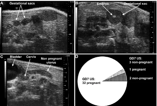
isoflurane and 1-1.5 L/min of oxygen. Expired gas from mice and leaking gas from the anesthesia mask were scavenged by a ventilation system connected to a charcoal filter canister. Mice were positioned on a heating pad and stabilized with adhesive tape, and then fur was shaved from the abdomen and neck. Body temperature was supported in the range of 37±1˚C and detected with a rectal probe. Respiratory and heart rates were monitored throughout the ultrasound scans (Vevo Imaging Station, Visual Sonics Inc., Toronto, ON, Canada). After the 55MHz linear ultrasound probe (Vevo 2010, Visual Sonics Inc.) was fixed and mobilized with a mechanical holder, pregnancy status was evaluated while looking for signs of a gestational sac (GD6, Figure 3A), as well as an embryo and an advanced endometrial reaction (GD7, Figure 3B) [279-287].
DOI:10.14753/SE.2015.1828
Figure 3. Determination of mouse pregnancy status with a 55MHz ultrasound probe. (A) Pregnant uterus on GD6. Gestational sacs of 1.8mm–2.7mm were observed in the proximity of the abdominal surface without visible signs of an embryo. (B) Pregnant uterus on GD7.
Advanced endometrial reaction and the presence of an embryo were visible in the gestational sacs. (C) Non-pregnant uterus of a mouse seven days after mating, equivalent to GD7. (D) The pie chart shows that pregnancy could be diagnosed in 32 (white) of the 35 mice with high- frequency ultrasound on GD7. Of three mice diagnosed as non-pregnant on GD7 (shading), two were non-pregnant (grey shading), while one carried a pregnancy to term (white shading).
3.1.4. Implantation of the telemetric blood pressure monitoring system
Only mice with confirmed pregnancies underwent telemetric blood pressure monitoring system implantation on GD7. Isoflurane anesthesia was induced and maintained, and gas was scavenged as previously described. Mice were laid on their backs on the surgical platform, and their upper incisors and limbs were stabilized. Body temperature was controlled by a T/Pump warm-water circulating blanket (Gaymar Industries, Inc. Orchard Park, NY, USA).
The incision site was scrubbed with Betadine (Purdue Pharma L.P., Stamford, CT, USA); and 2% lidocaine (0.5 mg/kg, Vedco Inc., St. Joseph, MO, USA) and 0.5% bupivacaine (1.5mg/kg, Hospira Inc., Lake Forest, IL, USA) were injected subcutaneously (s.c.) before the incision, following the rules of aseptic surgery [288]. An approximate 1.5cm midline incision was made on the neck, and the salivary glands were gently dissected and retracted laterally with elastic plastic stay hooks. An approximate space of 1cm of the left common carotid
DOI:10.14753/SE.2015.1828
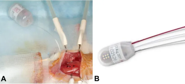
artery was exposed from the bifurcation in the direction of the heart, with the intention not to injure the vagal nerve. After carotid artery ligation at the level of bifurcation, arteriotomy and cannulation were prepared with the assistance of a 25G tip needle (Figure 5A), and the blood pressure monitoring catheter (TA11PA-C10 or HD-X11, Data Sciences International, St.
Paul, MN, USA) (Figure 4) was positioned into the aortic arch (Figure 5B). The catheter was fixed with 6/0 non-absorbable braided surgical silk sutures (Teleflex Medical, Coventry, CT, USA), and the transmitter was placed in a subcutaneous pocket in the left flank, preformed with blind dissection (Figure 5C). After repositioning the salivary glands over the catheter, the skin was closed with a continuous 6/0 non-absorbable monofilament polypropylene suture (CP Medical, Portland, OR, USA). Postoperative pain was reduced with s.c. injection of carprofen (5 mg/kg/24h, Rymadil, Pfizer Inc., New York, NY, USA), and with the administration of lidocaine and bupivacaine adjacent to the surgical incision site. In order to avoid post-surgical dehydration, 0.5ml of 0.9% saline solution was s.c. injected. During the postoperative period, mice were kept in their cages, with one-half of each cage placed on a warm water circulating blanket, and vital signs were regularly checked.
Figure 4. TA11PA-C10 and HDX-11 telemetry blood pressure monitoring device. (A) The TA11PA-C10 telemetric blood pressure monitoring device with left carotid artery cannulation in mice. The weight of the TA11PA-C10 device is 1.4 grams, the volume is 1.1 cc, and the battery life is approximately 1.5 months. The photo is courtesy of the Perinatology Research Branch, NICHD, NIH, DHHS. (B) The HDX-11 telemetric device can simultaneously record blood pressure, ECG, temperature and activity data from a single mouse. The weight of the HDX-11 device is 2.2 grams, the volume is 1.4 cc, and the battery life is approximately 1 month. The image was downloaded form http://www.datasci.com/products/implantable- telemetry/mouse-(miniature)/hd-x11.
DOI:10.14753/SE.2015.1828
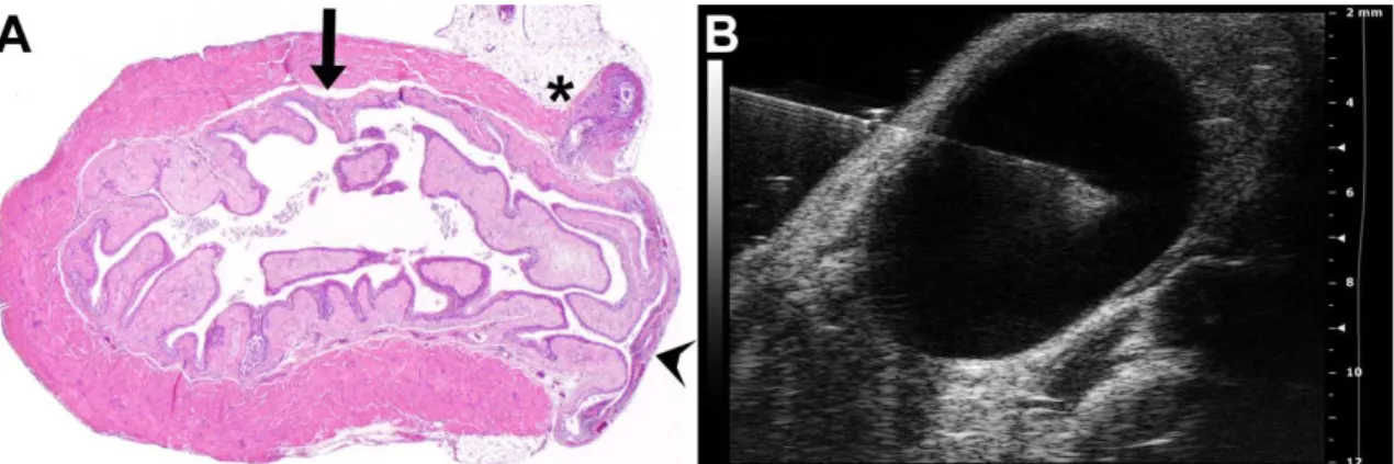
3.1.5. Determination of the telemetry catheter position with high-frequency ultrasound As another methodological development by our study, transmitter catheter tip positions were examined with the 55MHz linear ultrasound probe (Visual Sonics Inc.) during the routine GD13 ultrasound scans. The ultrasound probe was fixed and mobilized with a mechanical holder, and the transducer was moved downward toward the chest. The left carotid artery, aortic arch, and ascending aorta were visualized, and the position of the catheter tip was determined (Figure 5D).
Figure 5. Implantation with a telemetric blood pressure monitoring system. (A) On GD8, after isolation and ligation of the left common carotid artery at the level of bifurcation, a small arteriotomy was prepared with a 25G tip needle, (B) and the blood pressure monitoring catheter was positioned into the aortic arch with the assistance of the vessel cannulation forceps. (C) The transmitter was placed into a subcutaneous pocket in the left flank and preformed with blind dissection. (D) On GD13, the position of the telemetry catheter was determined with a 55MHz ultrasound probe. The catheter tip is situated in the aortic arch, and the intra-aortic part of the catheter reaches the optimal 2mm length. (E) On GD18, a pregnant mouse is shown before a cesarean section. The incision line of the telemetry surgery healed completely. The projected graph illustrates the position of the intra-aortic catheter and the subcutaneous telemetric blood pressure transmitter. (F) On PPD8, the catheter position, aortic arch, and main arterial branches were visualized after autopsy in the mouse mediastinum and chest. The dotted lines show the heart and main arteries of the mediastinum. (A-F) Head orientations are shown with asterisks.
3.1.6. Telemetric blood pressure monitoring
As postoperative pain has a strong effect on blood pressure, telemetry monitoring was started on GD10, three days after the catheter implantations on conscious, unrestrained animals, and was continued until postpartum day (PPD)7 using the Dataquest A.R.T.
4.31 acquisition and analysis system (Data Sciences International) (Figure 6). Blood DOI:10.14753/SE.2015.1828
pressures were recorded for 10s every five minutes for at least 8-12 hours a day during both the light and dark cycles.
Figure 6. Telemetric blood pressure monitoring system. Mice were kept separately in standard-size filter top rodent cages. Blood pressure monitoring was started three days after the catheter implantations on conscious, unrestrained animals, and was continued until postpartum day (PPD) 7 using the Dataquest A.R.T. 4.31 acquisition and analysis system (Data Sciences International). Freely moving mice housed in plastic cages were placed on the top of the receiver, and the implanted telemetric devices transmitted the data to the receiver with radio frequency. The photo is courtesy of the Perinatology Research Branch, NICHD, NIH, DHHS.
3.1.7. Adenoviral gene delivery
As a methodological development by our study, recombinant adenoviruses expressing enhanced green fluorescent protein (GFP) or hsFlt-1-e15a (NP_001153502.1) under the control of a cytomegalovirus promoter (Ad-CMV-GFP and Ad-CMV-hsFlt-1-e15a, respectively) were constructed and titered by Vector BioLabs (Philadelphia, PA, USA). Mice were divided into four groups [hsFlt-1-e15a 1x (n=6), hsFlt-1-e15a 2x (n=5), GFP 1x (n=4), and GFP 2x (n=5)] according to the number of viral construct injections. All mice were injected with adenovirus constructs [1x10⁹ plaque-forming units (PFU) in 100μl] via the tail vein on GD8, and a subset of mice (GFP 2x and hsFlt-1-e15a 2x) was repeatedly injected with 1x10⁹ PFU adenoviral constructs on GD11. All of these mice underwent the subsequently described procedures.
DOI:10.14753/SE.2015.1828
3.1.8. Ultrasound-guided bladder puncture (cystocentesis)
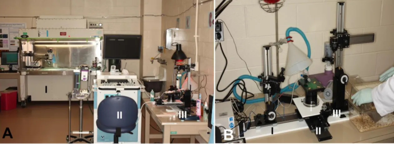
As another methodological development by our study, ultrasound-guided cystocentesis was performed on GD7, GD13, GD18, and PPD8 under isoflurane anesthesia. Urine samples were obtained using a micro-injection system and a linear 55 MHz high- frequency ultrasound probe (Visual Sonics Inc.) (Figure 7).
Figure 7. High frequency ultrasound and micro-injection system. (A) I. Telemetry implantation was done under surgical microscope in a biosafety hood. II. 55 MHz high- frequency ultrasound (Vevo 2010, Visual Sonics Inc., Toronto, ON, Canada). III. Micro- injection unit. (B) I. A 1ml insulin syringe with a 30 gauge, 12.7mm long needle was mounted on the micro-injection system, and orientated to target the bladder. II. Anesthetized mice were positioned on a heating pad of the imaging unit. III. The transcutaneous bladder puncture was performed under continuous ultrasound guidance using the mechanical holder of the micro- injection system. The photos are courtesy of the Perinatology Research Branch, NICHD, NIH, DHHS.
Specifically, the transcutaneous bladder puncture was performed under continuous ultrasound guidance using the mechanical holder of the micro-injection system. The ultrasound probe was aligned and adjusted to obtain a clear view of the maternal bladder; the skin was disinfected with a sterile pad saturated with 70% isopropyl alcohol. A 1ml insulin syringe with a 30 gauge, 12.7mm long needle was mounted on the micro-injection system, and orientated to target the bladder. The procedure was attempted when urine was visualized, disregarding bladder size. The needle was then stopped before entering the skin, and the angle between the needle and the bladder was corroborated. The angle of the scanning pad and the microinjection system was adjusted to allow for a paramedial entrance of the needle through the dome of the bladder in order to avoid damage to the abdominal and pelvic organs. After defining the optimal
DOI:10.14753/SE.2015.1828
site for puncture, color Doppler ultrasound was activated for the identification of vascular structures. If vessels were detected, then an alternative site for the bladder puncture was selected. After obtaining the urine sample, a small amount of urine was left in the bladder. Slow forward movements in the micro-injection system allowed the visualization of the tip of the needle in the ultrasound screen. With a fast forward movement of the injection system, the needle was introduced in the bladder without transposing into the posterior wall (Figures 8). After obtaining the urine sample, a small amount of urine was left in the bladder. The needle was then retired under continuous ultrasound visualization. After the procedure, the fluid loss was supplemented by subcutaneous injection of pre-warmed 0.9% NaCl (10-15 microliters/g/hour) into the midscapular region of the mice according to the recommendation of the IACUC and the Division of Laboratory Animal Resources (DLAR) of Wayne State University.
Figure 8. Ultrasound guided cystocentesis. (A) A cross-section of a mouse urinary bladder.
The urinary bladder can be anatomically divided into two parts: the dome consists of three layers of smooth muscle (arrow), and the thin bladder base (arrowhead) consists of the trigone extending from the urethra (star) to the two ureters. (B) An ultrasound image of a filled urinary bladder positioned just underneath the abdominal wall. After image optimization, the urinary bladder is punctured by a 30G needle guided by a high 55MHz ultrasound probe. The images are courtesy of the Perinatology Research Branch, NICHD, NIH, DHHS.
Five µl of urine samples were evaluated for blood contamination using a highly sensitive Urine Chemstrip 5 OB (Roche Diagnostics, Indianapolis, IN, USA). This assay can detect a minimum of 5 erythrocytes/µl [289-291] based on the pseudoperoxidase reaction of erythrocytes and hemoglobin [290]. Then urine samples were stored at -80oC until analysis.
DOI:10.14753/SE.2015.1828
3.1.9. Cesarean section
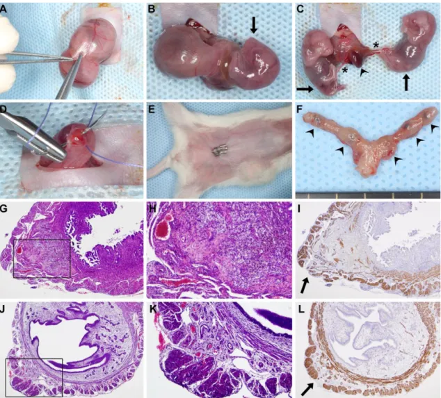
Utilizing another methodological development, mice underwent survival cesarean section on GD18. Pre- and intra-operative preparation of the mice (i.e. isoflurane anesthesia induction and maintenance, eye protection, surgical stabilization and body temperature control, skin disinfection, and local analgesia) were performed as in the case of the telemetry device implantation. After a short (1-1.5cm) midline abdominal incision in the area where fur had been previously removed, a short segment of one uterine horn was exteriorized at once, and kept moisturized with sterile 0.9% saline.
According to the number of pups, two to three exteriorizations and minimal (3-5mm) longitudinal midline hysterectomies were made on each horn, on the opposite side of the mesometrial arterial arcade, while keeping the residual parts of the uterine horn inside the abdominal cavity to avoid contamination (Figure 9A). After delivering pups and placentas (Figure 9B,C), minimal incisions were closed with a single 4/0 absorbable multifilament polyglycolic acid suture (CP Medical, Portland, OR, USA) (Figure 9D).
Then, lavage was applied to the abdominal cavity with 2-3ml of 0.9% sterile saline. The abdominal wall was closed with a continuous 4/0 absorbable multifilament polyglycolic acid suture (CP Medical), and the skin was closed with 7mm staples (Braintree Scientific Inc., Braintree, MA, USA) (Figure 9E). Body fluids were replenished by an injection of 0.5ml of 0.9% sterile saline subcutaneously. Postoperative pain was reduced with s.c. carprofen (Pfizer Inc.), as well as with the injection of lidocaine and bupivacaine adjacent to the incision line. Postoperative care was similar to that which followed the telemetry system implantation.
DOI:10.14753/SE.2015.1828
Figure 9. Survival cesarean section. (A) After a 1cm–1.5cm midline abdominal incision, a short segment of one uterine horn was exteriorized, and a 3mm–5mm longitudinal hysterectomy was performed on the opposite side of the mesometrial arterial arcade. (B-C) As the uterine wall could be easily dilated, this minimal incision enabled the delivery of two to three fetuses and their placentas using gentle fingertip pressure. An arrowhead depicts a placenta, stars depict umbilical cords, and arrows point to the fetuses. (D) Hysterectomies were closed with a single 4/0 absorbable multifilament suture. (E) After abdominal lavage with 0.9% sterile saline, the abdominal wall was closed with an absorbable multifilament continuous suture, and the skin was closed with 7mm-wide staples. The image shows a surgical field before euthanization on PPD8. (F) One uterus harvested after euthanization on PPD8. Arrowheads show hysterectomy sutures. (G) H&E staining of a uterine cross-section of a control mouse euthanized on PPD8 shows granulation tissue with enlarged vessels, foam cells, myofibroblasts, macrophages, and hemosiderin deposition. The image inside the black box is magnified in Subfigure H. (I) SMA immunostaining of the same uterus. The arrow depicts the cesarean section incision site with the disruption of the two-layered myometrium. (J) H&E staining of a uterine cross-section of a control mouse euthanized on PPD77 shows granulation tissue and the complete healing of the two-layered myometrium. The image inside the black box is magnified in Subfigure K. (L) SMA immunostaining of the same uterus. The arrow depicts a suture site. (G, I, J and L: 40x magnifications, H and K: 100x magnifications).
DOI:10.14753/SE.2015.1828
3.1.10. Tissue collection
Following cesarean section on GD18, each fetus was separated from the placenta and umbilical cord. All fetuses and placentas were weighed with a Scout Pro SP402 digital scale (Ohaus Corp., Pine Brook, NJ, USA). The first placentas adjacent to the uterine cervix in both uterine horns were fixed in 4% paraformaldehyde (PFA) diluted with phosphate buffered saline (PBS, Gibco, Life Technologies Corporation, Grand Island, NY, USA) for 24h, then dehydrated in 70% graded ethanol (Richard-Allan Scientific Dehydrant, Thermo Fisher Scientific Inc., Waltham, MA, USA), and embedded in paraffin for histopathological examinations. The second placentas adjacent to the uterine cervix were collected and homogenized in TRIzol (Invitrogen, Life Technologies Corporation, Carlsbad, CA, USA) and stored at -80˚C until gene expression analyses.
After the euthanization of dams on PPD8, tissues from several organs (spleen, uterus, liver, kidney, and brain) were dissected and sectioned. Tissues were fixed in 4%
PFA for 24h, then dehydrated in 70% graded ethanol, and embedded in paraffin for histopathological examinations, or homogenized in TRIzol reagent and stored at -80˚C until gene expression analyses. To evaluate the changes in uterine histology with time after the cesarean sections, additional untreated mice were euthanized on PPD38 (n=3), PPD50 (n=3), and PPD77 (n=3), respectively, and uteri were processed as those on PPD8.
3.1.11. Total RNA isolation, cDNA generation, quantitative real-time RT-PCR
Tissues were homogenized in TRIzol reagent with a homogenizer (Pro Scientific Inc., Oxford, CT, USA) immediately after tissue collection. Total RNAs were isolated using the QIAshredder (Qiagen, Valencia, CA, USA) and the RNeasy Mini Kit (Qiagen), according to the manufacturer’s instructions. Complementary DNAs were generated with SuperScript III First-Strand Synthesis System (Invitrogen). Quantitative real-time RT-PCR assays were performed on the Biomark System (Fluidigm, San Francisco, CA, USA) using TaqMan assays (Applied Biosystems, Life Technologies Corporation, Foster City, CA, USA) for GFP (Mr04097229_mr) and human FLT1 (Hs01052961_m1).
DOI:10.14753/SE.2015.1828
3.1.12. Histopathological evaluation of tissues
Five-µm-thick sections of paraffin embedded placenta, kidney, and uterus tissue blocks were serially cut, mounted on silanized slides, deparaffinized, and rehydrated in descending grades of ethanol. Selected levels of all tissues were then stained with hematoxylin and eosin (H&E) to evaluate general morphology, and selected levels of all kidneys were stained with periodic acid Schiff (PAS) reagent for the visualization of basement membranes of glomerular capillary loops and tubular epithelium.
Histopathological examination of these tissue sections was performed on an Olympus BX50F light microscope (Olympus America Inc., Melville, NY, USA) by a pathologist (FQ). Kidney sections were evaluated for glomerular endotheliosis (e.g. ballooning of tips of capillary loops, capillary endothelial swelling, occlusion of glomerular capillaries) in at least 20 glomeruli in the inner cortex of one kidney in each animal.
3.1.13. Immunohistochemistry
Selected layers of uteri were immunostained for CD68 and smooth muscle actin (SMA).
Immunostainings were performed using a rabbit anti-mouse SMA polyclonal antibody (1:300 dilution; Abcam Inc., Cambridge, MA, USA) and the Bond Polymer Refine Detection Kit (Leica Microsystems, Wetzlar, Germany) on a Leica Bond Max automatic staining system (Leica Microsystems), or using a rabbit anti-mouse CD68 polyclonal antibody (1:150 dilution; Abcam Inc.) and the DAB Map Detection Kit (Ventana Medical Systems, Inc., Tucson, AZ, USA) on a Ventana automatic staining system (Ventana Medical Systems, Inc.).
3.1.14. Aortic ring assays
Aortic ring assays were performed as previously described [292,293]. Briefly, thoracic aortas were dissected from euthanized mice and placed in a Petri dish containing DMEM+GlutaMAX low glucose medium (Gibco, Life Technologies Corporation). The peri-adventitial fibro-adipose tissue was removed, then aortas were sectioned into 1mm- long rings, and incubated in 12-well plates at 37oC in Opti-MEM+GlutaMAX reduced serum medium (Gibco, Life Technologies Corporation) overnight for serum starvation.
The serum-starved aortic rings were then placed into 96-well tissue culture plates pre- coated with 50µL of Growth Factor Reduced BD Matrigel Matrix (BD Biosciences,
DOI:10.14753/SE.2015.1828
Bedford, MA, USA). Then, aortic rings were covered with an additional 50µL of Matrigel and 100µL of Opti-MEM medium supplemented with 1% Penicillin–
Streptomycin (Gibco, Life Technologies Corporation), 2.5% fetal bovine serum (FBS;
Atlanta Biologicals, Lawrenceville, GA, USA), and 30ng/mL of vascular endothelial growth factor (VEGF-A; ProSpec, East Brunswick, NJ, USA). Plates were incubated at 37oC for six days with a change of medium every second day. After six days of incubation, aortic rings were fixed with 4% PFA diluted with PBS, and images were obtained with an Olympus 1X51 inverted microscope equipped with an Olympus DP25 digital camera (Olympus, Tokyo, Japan).
3.1.15. Albumin-creatinine immunoassays
Urine specimens collected on GD6/GD7, GD13, GD18 and PPD8 were examined for murine urinary albumin with the Albuwell kit (Exocell Inc., Philadelphia, PA, USA), and for creatinine with the Creatinine Companion assay employing the alkaline picrate method (Exocell Inc.).
3.1.16. Adenoviral infection of BeWo cells
BeWo cells (American Type Culture Collection, Manassas, VA, USA) were cultured with F12 medium supplemented with 10% FBS and 1% Penicillin/Streptomycin (all from Life Technologies Corporation). Cells were plated on either 6-well plates (0.5x10⁶/well) or 35mm cell culture dishes, and infected with Ad-CMV-GFP or Ad- CMV-hsFlt-1-e15a at a multiplicity of infection of 100. After 16h of infection, cell supernatants were removed, and BeWo cells were washed with PBS and used for Western blotting or confocal imaging.
3.1.17. Protein isolation and Western blot
Total protein from BeWo cell samples was extracted with the RIPA lysis buffer (Sigma-Aldrich Co., St Louis, MO, USA) containing Complete Mini Protease Inhibitor Cocktail Tablets (Roche, Indianapolis, IN, USA). Protein concentrations were determined with the Quick Start Bradford Protein Assay (Bio-Rad, Hercules, CA, USA). Twenty micrograms of total protein from each sample were electrophoresed on 4-12% SDS-PAGE gels (Life Technologies Corporation), and
DOI:10.14753/SE.2015.1828
electro-transferred onto nitrocellulose membranes (Bio-Rad). Membranes were probed with goat anti-human Flt-1 polyclonal antibody (AF321, R&D Systems, Inc., Minneapolis, MN, USA) in a 1:2,000 dilution at 4Co for 16h, and then with peroxidase- conjugated anti-goat IgG (Vector Laboratories, Burlingame, CA, USA) in a 1:5,000 dilution at room temperature for 1h. Protein bands were developed using the ChemiGlow Western Blotting Detection Reagents (Protein Simple, Santa Clara, CA, USA), and then scanned and imaged with a Fujifilm LAS-4000 Image Reader (GE Healthcare, Piscataway, NJ, USA).
3.1.18. Confocal microscopy
BeWo cells infected with adenovirus constructs and cultured in 35mm cell culture dishes were mounted with ProLong Gold Antifade Reagent and 4',6-diamidino-2- phenylindole (DAPI; Invitrogen) followed by confocal microscope imaging using a Leica TCS SP5 spectral confocal system (Leica Microsystems CMS GmbH, Mannheim, Germany). Confocal imaging was performed at the Microscopy, Imaging and Cytometry Resources Core of the Wayne State University School of Medicine.
3.1.19. Data and statistical analyses
Blood pressures: Mean arterial blood pressures (MAP) were calculated from systolic and diastolic blood pressures for each time point and animal, and then were averaged. The mean MAP values on GD10 were subtracted from all MAP values to obtain normalized ∆MAP values. We fitted the MAP and the ∆MAP data with a Linear Mixed Effects (LME) models [294] for the time intervals before and after cesarean delivery, respectively. These models included explanatory variables such as the treatment (GFP or hsFlt-1-e15a) or the dose (1x or 2x), and a continuous measure of time (gestational day or postpartum day) while allowing a random intercept for each animal. An interaction was allowed between the treatment and time, and therefore, we could test if the slope of the MAP or ∆MAP over time was different between the treatments. We relaxed the linear fixed effect patterns to quadratic for the analysis of time intervals after cesarean delivery. Since blood
DOI:10.14753/SE.2015.1828
pressure has a circadian daily rhythm in mice, we also examined blood pressures in 12-hour light and dark cycles separately.
Urine albumin/creatinine ratios: Albumin/creatinine ratios between the hsFlt-1-e15a and GFP groups on different time points were compared with the Student's t-test.
Gene expression profiling: Relative gene expressions were quantified by averaging target (FLT1 or GFP) and reference (Gapdh) gene Ct values over technical replicates, and then by subtracting mean target gene Ct values from mean reference gene Ct values within the same sample. The Student's t-test was used to compare gene expression levels between treatments in a given tissue. To examine the dose effect on gene expression for each tissue, we computed the percentage of samples expressing a given gene when over-expressed with a given dose of that gene. Statistical comparison on the percentages across all tissues between the two doses was performed with the one-tailed paired Student's t-test.
Aortic ring assays: The angiogenic response of the aortic rings was analyzed by quantifying the microvessel outgrowth. A ruleset was developed using Definiens Developer XD2 (Definiens, Munich, Germany) to analyze the transmitted light images.
A new image layer (or channel) with enhanced local contrast was produced using
“contrast to neighbor pixels” to distinguish the newly formed microvessels sprouting from the aortic ring. Next, a series of segmentation and classification operations was performed on the channel to exclude the ring from the area measurements, and the total area of the objects determined to be “outgrowth” was reported. Data were averaged on the picture level for the same ring, then further averaged on the ring level for the same animal, followed by a Student's t-test for group comparisons.
Fetal survival rates, fetal and placental weights: The fetal survival rate (number of live fetuses / number of total fetuses) for each mouse was computed, and the non-parametric Kruskal-Wallis test was used for multiple group comparisons. Fetal weights, placental weights, and placental/fetal weight ratios were compared with the two-way ANOVA test and with a linear mixed effects model.
DOI:10.14753/SE.2015.1828