R E S E A R C H A R T I C L E Open Access
The effect of ranibizumab and aflibercept treatment on the prevalence of outer
retinal tubulation and its influence on retreatment in neovascular age-related macular degeneration
Attila Kovacs1, Timea Kiss1, Ferenc Rarosi2, Gabor M. Somfai3,4, Andrea Facsko1and Rozsa Degi1*
Abstract
Background:We aimed to analyze the differences in the prevalence of outer retinal tubulation (ORT) in neovascular age-related macular degeneration (AMD) treated with anti-vascular endothelial growth factor (anti- VEGF) agents, either aflibercept or ranibizumab. Our further aim was to examine the changes in the frequency of injections of ranibizumab before and after ORT appearance.
Methods:Two hundred thirty six eyes of 230 patients were included in the study (184 eyes treated with ranibizumab by pro re nata regimen (PRN), 52 eyes with aflibercept bimonthly) and followed for 6–24 months.
Using optical coherence tomography (OCT), the first appearance of ORT was documented, and fixed time point evaluations were also made every six months to determine the existence of ORT. The number of injections, the presence or absence of subretinal hyperreflective material (SHRM) at treatment initiation and visual acuity were also noted.
Results:The survival analysis with Cox proportional hazard model showed no significant difference between the ranibizumab and aflibercept groups in relation to the development of ORT (p= 0.79, hazard ratio 0.92). In the PRN treated ranibizumab group the number of injections showed significant decrease after ORT development (p= 0.004).
When SHRM was present at treatment initiation the chance of developing ORT was 2.75 and 11.14 times higher in the ranibizumab and aflibercept groups, respectively.
Conclusions:The prevalence of ORT increased over time independently from the chosen anti-VEGF drug. Our results suggest that upon the appearance of ORT a decrease in retreatments can be expected.
Keywords:Outer retinal tubulation, Prevalence, Anti-VEGF, Aflibercept, Retreatment, Subretinal hyperreflective material
Background
Outer retinal tubulation (ORT) is a spectral-domain op- tical coherence tomography (SD-OCT) biomarker [1], first described by Zweifel et al. [2]. They defined ORTs as hyporeflective, branching tubular structures with hyperre- flective borders within the outer nuclear layer of the retina [2]. The “en face” OCT technique can help map these
branching networks [3]. ORTs have been observed in many retinal diseases, including exudative age-related macular degeneration (AMD) [2]. Based on histological reports, the border of the outer retinal tubulation consists of photoreceptor inner segment mitochondria and exter- nal limiting membrane (ELM), with fluid and photorecep- tor outer segments being potentially present in the ovoid hyporeflective lumen of the ORT [4–6]. Adaptive optics scanning laser ophthalmoscopy findings are in correlation with histology reports and show lack of ORT cone
* Correspondence:degirozsa57@gmail.com
1Department of Ophthalmology, Faculty of Medicine, University of Szeged, 10-11 Koranyi fasor, Szeged 6720, Hungary
Full list of author information is available at the end of the article
© The Author(s). 2018Open Access This article is distributed under the terms of the Creative Commons Attribution 4.0 International License (http://creativecommons.org/licenses/by/4.0/), which permits unrestricted use, distribution, and reproduction in any medium, provided you give appropriate credit to the original author(s) and the source, provide a link to the Creative Commons license, and indicate if changes were made. The Creative Commons Public Domain Dedication waiver (http://creativecommons.org/publicdomain/zero/1.0/) applies to the data made available in this article, unless otherwise stated.
reflectivity which can be due to the loss of cone outer seg- ments and subsequent retinal remodeling [7].
Schaal et al. classified outer retinal tubulations as either open (incomplete closure with curving external limiting membrane at the ends, horizontally elongated shape in cross-section) or closed (completely encircled, oval shape in cross-section) ORTs [4].
ORT can be mistaken for intraretinal cysts, or subretinal fluid but with the recognition of its hyperreflective border and special occurrence in the outer nuclear layer these mistakes can be reduced, leading to a reduction in the rate of anti-VEGF overtreatment in exudative AMD [2].
The ORT prevalence in exudative AMD is low at the time of first diagnosis but over time during anti-VEGF ther- apy its prevalence increases [8,9]. The importance of ORT as an OCT biomarker for photoreceptor degeneration is due to its connection with reduced visual acuity [1,8–10].
It has been also reported that ORTs develop above areas of subretinal hyperreflective material (SHRM) or atrophy [8, 9]. SHRM is a medium- to hyperreflective mass be- tween the neurosensory retinal layers and retinal pigment epithelium on OCT [11]. It usually represents either a type II choroidal neovascular complex or is the consequence of an active choroidal neovascularisation, including subretinal haemorrhage and lipid or fluid exudation [1,11,12].
The aim of the present study was to investigate the prevalence of ORTs in eyes with neovascular AMD undergoing treatment either with ranibizumab or afliber- cept. Our further aim was to examine the changes in the frequency of injections before and after ORT appearance.
We also assessed the presence of subretinal hyperreflec- tive material and its relationship with ORT.
Methods Ethics, consent
This retrospective study was performed at the Medical Retina Unit of the Ophthalmology Department of University of Szeged, in Hungary. The study was ap- proved by the Institutional Review Board of University of Szeged Albert Szent-Györgyi Clinical Centre (reference number: 3650) and was in accordance with the ethical standards of the Declaration of Helsinki. Since this was a retrospective review of patient data, informed consent was not required. The need for a consent was formally waived by the ethics committee, and this was also in line with the national regulations.
Patients
Treatment-naïve exudative AMD patients were enrolled in the study. For the ranibizumab group enrollment took place between October 2014 to April 2016 while patients in the aflibercept group were enrolled between April 2015 to April 2016.
All patients were over 50 years of age, the mean follow-up period was 16.3 months and 9.2 months (range 6–24 months and 6–12 months) in the ranibizumab and aflibercept groups, respectively.
During each visit a comprehensive ophthalmic examin- ation was carried out including best-corrected visual acuity (BCVA, Early Treatment Diabetic Retinopathy Study (ETDRS) score) assessment, slit-lamp biomicroscopy, dilated funduscopy and SD-OCT examination of the retina (Heidelberg Spectralis, Heidelberg Engineering, Heidelberg, Germany). Eyes with poor quality SD-OCT scans (Q index below 20) or with poor compliance were excluded from the study (14 eyes from the ranibizumab and 3 eyes from the aflibercept group).
Treatment regimen for both ranibizumab (0.5 mg) and aflibercept (2 mg) started with 3 monthly injections.
After this initiation phase the ranibizumab group was treated by a pro re nata (PRN) regimen with follow-up visits scheduled monthly. The retreatment criteria for ranibizumab patients consisted of any subretinal or intraretinal fluid on OCT, or new haemorrhage on fun- duscopy. In the aflibercept group follow-up after the loading phase was scheduled every two months, treat- ment was given at each follow-up. The above regimens were in accordance with the available treatment guide- lines in Hungary at the time of the study.
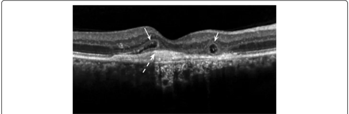
For SD-OCT imaging a pattern size of 5.8 × 5.8 mm, 20° × 20° was applied with 25 B-scans, using the “fol- low-up” mode. By manual review of the scan volumes we determined the first appearance of the ORT in both groups. We also assessed the presence of ORT at fixed time points at baseline, month 6 and 12 in both groups and at months 18 and 24 in the ranibizumab group. Im- ages were assessed by two independent retina specialists, in case of incongruity the images were referred to a third retina specialist to make a decision. During the evalu- ation of OCT scans we did not differentiate between the above described open (incomplete hyperreflective ring) and closed (complete hyperreflective ring) forms of ORT according to Schaal [4]. Thus, both types of ORT detected on the images were considered an ORT positive case. The criterion of ORT was a hyperreflective ovoid-elongated structure in the outer nuclear layer of the retina with lower reflective content (Fig.1).
The readers also identified the presence or absence of subretinal hyperreflective material on SD-OCT images at treatment initiation. The criterion for SHRM was a medium- to hyperreflective mass between the neurosen- sory retinal layers and retinal pigment epithelium, as described by Keane et al. [11] (Fig.1).
Statistical methods
The BCVA was compared across the two groups using the Mann-Whitney U-test. The survival analysis for
ORT development was analyzed by a Cox proportional hazard model. We analyzed the correlation between the presence of SHRM at treatment initiation with the de- velopment of ORT by Chi-square test and calculated relative risks. Where zeros were involved for the compu- tation of relative risk, 0.5 was added to all cells, accord- ing to the previous suggestions [13].
The injection rate was calculated only in the PRN treated ranibizumab group due to the fixed 2 month therapeutic regimen of aflibercept. We assessed the in- jection rate only before and after the appearance of outer retinal tubulation and compared using the the Mann-Whitney U-test. In order to correct bias due to the unequal follow-up time (some patients had a higher number of injections due to the longer follow-up), we divided the follow-up time with the number of injections and calculated with monthly injections.
Ap-value ofp< 0.05 was taken as statistically significant.
For the analyses the IBM SPSS Software (Version 22) was used.
Results
In the ranibizumab group we evaluated 184 eyes of 179 patients, with a median age of 74 years (range 51 to 88), while in the aflibercept group there were 52 eyes of 51 patients with a median age of 75 years (range 58 to 87).
The mean baseline best corrected visual acuities in the two groups were (mean ± SD) 59.16 ± 13.9 (median 61) and 53.96 ± 13.54 (median 55.5) ETDRS letters in the ranibizumab and aflibercept group, respectively. There was no significant difference between the two groups (Mann-Whitney U-test p= 0.083). The BCVA at the end of the follow-up was 57.19 ± 20.19 (median 63) and 59.46 ± 15.54 (median 64) ETDRS letters in the ranibizumab and aflibercept group, respectively. There
was no significant difference between the two groups (Mann-Whitney U-test p= 0.69).
Table1 shows the number of eyes during the follow-up in the two groups. The number of eyes was reduced over time due to gradual enrollment in the study, thereby not every patient reached the same follow-up time. In the ranibizumab group outer retinal tubulation was observed in 17.4% of cases at baseline, in 33.7% of cases at month 6, in 45.3% of cases at month 12, and in 55.3% and in 60.8%
of cases at months 18 and 24, respectively. The ORT prevalence in the aflibercept group was 23.1% at baseline, 40.4% at month 6, and 50% at month 12.
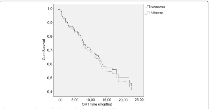
The survival analysis showed no significant difference between the ranibizumab and aflibercept treated groups in terms of ORT development. (p= 0.79, hazard ratio 0.92, 95% confidence interval 0.500–1.693) (Fig.2).
The injection rates showed that the mean injection num- ber per month before the ORT appearance was 0.37 ± 0.17 while after the ORT development it decreased to 0.21 ± 0.17 (Mann-Whitney U-test p= 0.004).
The presence of subretinal hyperreflective material at treatment initiation in the two subgroups was 75.5% in the ranibizumab, and 80.8% in the aflibercept group. In
Fig. 1Outer retinal tubulations and subretinal hyperreflective material on an SD-OCT single B-scan. Open and closed ORTs in cross section (left and right solid arrows) above subretinal hyperreflective material (dash arrow). The definition of ORT was a hyperreflective ovoid-elongated structure in the outer nuclear layer of the retina with lower reflective content
Table 1Number of eyes reaching the follow-up in the ranibizumab and aflibercept treated groups
Ranibizumab Aflibercept
Time point n(eyes) n(eyes)
Baseline 184 52
at 6 months 184 52
at 12 months 161 28
at 18 months 103 0
at 24 months 51 0
Table legend: The column with“n”corresponds to the number of eyes reaching the follow-up
the ranibizumab treated group ORT developed in 85 eyes of 139 eyes with SHRM (61.15%), while without SHRM (45 eyes) ORTs were found merely in 10 eyes (22.2%) corresponding to a relative risk of 2.75. (p< 0.01). In the aflibercept treated group 55.81%
of eyes with SHRM developed ORT (24 eyes of 43). No ORT developed in the eyes without SHRM (out of 9 eyes), consistent with a relative risk of 11.14 (p< 0.01).
Discussion
Our study found no significant difference between the ranibizumab and aflibercept treated groups according to ORT development. There was a statistically significant reduction in the frequency of injections in the ranibizu- mab treated group before and after ORT appearance.
The presence of SHRM at treatment initiation as a bio- marker had a statistically significant correlation with the development of ORT in both groups.
Zweifel first described outer retinal tubulation in 2009 [2]. Later the authors reported ORTs in various retinal dis- orders like neovascular AMD, geographic atrophy, polypoi- dal choroidal vasculopathy, non-AMD associated choroidal neovascularisation, and other degenerative retinal disorders (e.g. retinitis pigmentosa, gyrate atrophy, choroideremia, Stargardt disease, pattern dystrophy) [2,3,14–19].
The pathogenesis of ORT formation is still not com- pletely clear, though recent reports helped a lot explor- ing it. Dolz-Marco et al. called the attention on the role of Müller cells in the pathomechanism of ORT develop- ment, namely the progressive photoreceptor damage
that can result in Müller cell activation which thereby starts to produce glial fibrillary acidic protein, facilitating the formation of ORT [20]. Based on histological exami- nations it seems that the evolution of ORT starts with ELM and ellipsoid zone disruption [4]. ELM starts to scroll inward at its free edges, representing an initial form of ORT, leading to the development of a formed open ORT. With time the large, open ORTs split, their margins beginning to scroll ending in multiple smaller closed ORTs. During the scrolling process a downward displacement of adjacent inner nuclear layer and outer plexiform layer happens separating each ORT, and caus- ing the appearance of microcystic lesions in the inner nuclear layer. The downward displacement of these layers might be due to the involvement of Müller cells in this scrolling/dragging process as Müller cells are con- tributing to the constitution of ELM with the inner segments of photoreceptors [4,15,20,21].
Most of the studies in the field focused on exudative AMD and its relationship with ORT. From these reports it is known that ORT is an SD-OCT biomarker, the prevalence of which increases with time and is associ- ated with decreased visual acuity [1, 8, 9]. It has been also reported that ORTs develop adjacent to areas of subretinal hyperreflective material or atrophy [8,9]. The differential diagnostic importance of outer retinal tubu- lation lies in the avoidance of overtreatment due to its similarity to intraretinal fluid [2,8].
In the present study, we evaluated treatment-naïve ex- udative AMD patients treated with ranibizumab and
Fig. 2Cox proportional hazard model of ORT survival in the ranibizumab and aflibercept groups
aflibercept regarding the presence of outer retinal tubula- tion. Altogether 236 eyes were followed in both groups with no statistical difference between the baseline charac- teristics of the two groups considering age and BCVA.
The prevalence of ORT continuously increased during the follow-up period, in both groups. In the ranibizumab group its prevalence almost quadrupled at the 24-month follow-up, while there was a doubling in the aflibercept group in 12 months. It is important to note, that in the ranibizumab group the baseline prevalence was lower (17.4% versus 23.1%). Dirani et al. found a similar in- creasing trend in their study starting with 2.5% at pres- entation, reaching 41.6% at 4 years of follow-up [8]. In our study the baseline ORT prevalence was higher com- pared to other reports [8, 9]. The reason behind this could be the more advanced disease state at the time of presentation (due to the real life nature of our retro- spective study) and a relative delay in therapy initiation due to country-specific financial difficulties. The poorer baseline BCVA in both groups also supports this idea.
There is one previous article known, describing ORT development in 24 non-treatment-naïve eyes, treated with aflibercept only after receiving at least 6 ranibizu- mab injection, reporting an initial 97% ORT prevalence which later decreased to 75% [22].
To our knowledge, our study is the first to report re- sults in treatment naïve patients treated with aflibercept and its connection with ORT development. The Cox proportional hazard model analysis suggested that there was no difference between the two in-label therapies ranibizumab and aflibercept in regard to the prevalence of outer retinal tubulation. Lee et al. in the Comparison of AMD Treatment Trials (CATT) study group evalu- ated the prevalence of ORTs in ranibizumab and bevaci- zumab treated neovascular AMD patients, and found no difference between the two drugs related to the preva- lence of outer retinal tubulation [9].
In the present study we found a statistically significant difference in the monthly injection rate before and after the appearance of outer retinal tubulation in the ranibizu- mab treated group. Our results suggest that in patients who develop ORT a decrease in the retreatment rate can be expected which may be a very important clinical marker. Although we had a PRN regimen according to the Hungarian guidelines, Lee et al. found no difference be- tween the fixed monthly regimen versus PRN regimen in regards to ORT development in patients treated either with ranibizumab or bevacizumab [9].
We found a statistically significant connection between ORT development and the presence of subretinal hyper- reflective material at treatment initiation. When subret- inal hyperreflective material was present the chance of developing ORT was 2.75., and 11.14 higher in the ranibizumab and aflibercept groups, respectively, in
accordance with the results of Lee et al. in ranibizumab and bevacizumab treated patients [9].
Our findings, in concordance with the above mentioned study results suggest that ORT is independent of the chosen anti-VEGF drug or the dosing regimen of intravit- real anti-VEGF treatment. The appearance of ORT suggests that the clinicians can expect a decrease in the number of injections when following a pro re nata ranibizumab regi- men. Our fixed bimonthly treatment with aflibercept did not allow us to analyze the injection rate before and after ORT development in this group. Our study also supports the previously reported higher prevalence of ORT develop- ment in the presence of subretinal hyperreflective material at treatment initiation [9].
There is a number of limitations of our study. Namely, the relatively small sample size in the aflibercept group compared to the ranibizumab group, along with the bi- monthly follow-up in the aflibercept group. We believe that the number of subjects involved in both groups is comparable with other studies published in the field, while the bimonthly treatment regimen with aflibercept was fixed due to the country-specific guideline regulations.
The decreasing number of eyes during the follow-up could also bias the analysis by including patients with increasing disease severity. However, we believe these fac- tors were similar to those in similar studies available in the field. The strength of this report is the comparison of present in-label therapies, ranibizumab and aflibercept in exudative AMD patients in relation to ORT besides the evaluation of the injection rate in association with outer retinal tubulation. We used real life data that makes the study more relevant in the daily clinical practice.
Conclusions
The development of ORT could be a potential biomarker for the treatment prognosis in patients with wet AMD. Its clin- ical significance lies also in its similarity to activity-related intraretinal fluid. Further studies are needed to explore the nature and development of ORTs employing a comparable dosing and follow-up regimen of all three currently available anti-VEGF drugs ranibizumab, bevacizumab and aflibercept.
Abbreviations
AMD:Age-related macular degeneration; anti-VEGF: Anti-vascular endothelial growth factor; BCVA: Best-corrected visual acuity; CATT: Comparison of AMD Treatment Trials; CNV: Choroidal neovascularisation; ELM: External limiting membrane; ETDRS: Early Treatment Diabetic Retinopathy Study; OCT: Optical coherence tomography; ORT: Outer retinal tubulation; PRN: Pro re nata; SD- OCT: Spectral-domain optical coherence tomography; SHRM: Subretinal hyperreflective material
Acknowledgments
This research received no specific grant from any funding agency in the public, commercial, or not-for-profit sectors.
Funding
No funding was received for this research.
Availability of data and materials
The datasets used and analyzed during the current study are available from the corresponding author on reasonable request.
All data generated or analyzed during this study are included in this published article [and its supplementary information files].
Authors’contributions
AK, RD made the conception and design of the study and analyzed the OCT images, in case of incongruity the final decision was made by AF. AK and TK collected and arranged the data (demographics, visual acuity, injection number and date). FR made the statistical analyzes and also the interpretation of data together with GMS. AK, TK drafted the manuscript.
GMS was a major contributor editing it. AF, GMS and RD revised the manuscript. All authors read and approved the final manuscript.
Ethics approval and consent to participate
This study was approved by the the Human Investigation Review Board of the University of Szeged Albert Szent-Györgyi Clinical Centre (reference number: 3650) and was performed in accordance with the tenets of the Declaration of Helsinki.
In the study we used a retrospective review of patient data; therefore consent was not collected from participants. The need for a consent was formally waived by the ethics committee, and this was also in line with the national regulations (a reference to the relevant legislation: 23/2002. (V. 9.) Eüm 20/Q. §).
Consent for publication
The attached OCT image is entirely unidentifiable and there are no details on individuals reported within the manuscript.
Competing interests
The authors declare that they have no competing interests.
Publisher’s Note
Springer Nature remains neutral with regard to jurisdictional claims in published maps and institutional affiliations.
Author details
1Department of Ophthalmology, Faculty of Medicine, University of Szeged, 10-11 Koranyi fasor, Szeged 6720, Hungary.2Department of Medical Physics and Informatics, Faculty of Medicine, University of Szeged, Szeged, Hungary.
3Augenzentrum Pallas Kliniken, Olten, Switzerland.4Department of Ophthalmology, Faculty of Medicine, Semmelweis University, Budapest, Hungary.
Received: 2 November 2017 Accepted: 26 October 2018
References
1. Schmidt-Erfurth U, Waldstein SM. A paradigm shift in imaging biomarkers in neovascular age-related macular degeneration. Prog Retin Eye Res. 2016;50:
1–24.https://doi.org/10.1016/j.preteyeres.2015.07.007.
2. Zweifel SA, Engelbert M, Laud K, Margolis R, Spaide RF, Freund KB. Outer retinal tubulation: a novel optical coherence tomography finding. Arch Ophthalmol. 2009;127(12):1596–602.https://doi.org/10.1001/archophthalmol.
2009.326.
3. Wolff B, Matet A, Vasseur V, Sahel JA, Mauget-Faÿsse M. En face OCT imaging for the diagnosis of outer retinal tubulations in age-related macular degeneration. J Ophthalmol. 2012;2012:542417.https://doi.org/10.1155/
2012/542417.
4. Schaal KB, Freund KB, Litts KM, Zhang Y, Messinger JD, Curcio CA. Outer retinal tubulation in advanced age-related macular degeneration: optical coherence tomographic findings correspond to histology. Retina. 2015;35(7):
1339–50.https://doi.org/10.1097/IAE.0000000000000471.
5. Litts KM, Messinger JD, Freund KB, Zhang Y, Curcio CA. Inner segment remodeling and mitochondrial translocation in cone photoreceptors in age- related macular degeneration with outer retinal Tubulation. Invest Ophthalmol Vis Sci. 2015;56(4):2243–53.https://doi.org/10.1167/iovs.14-15838.
6. Litts KM, Messinger JD, Dellatorre K, Yannuzzi LA, Freund KB, Curcio CA.
Clinicopathological correlation of outer retinal tubulation in age-related macular degeneration. JAMA Ophthalmol. 2015;133(5):609–12.https://doi.
org/10.1001/jamaophthalmol.2015.126.
7. Litts KM, Wang X, Clark ME, Owsley C, Freund KB, Curcio CA, Zhang Y.
Exploring photoreceptor reflectivity through multimodal imaging of outer retinal tubulation in advanced age-related macular degeneration. Retina.
2017;37(5):978–88.https://doi.org/10.1097/IAE.0000000000001265.
8. Dirani A, Gianniou C, Marchionno L, Decugis D, Mantel I. Incidence of outer retinal tubulation in ranibizumab-treated age-related macular degeneration.
Retina. 2015;35(6):1166–72.https://doi.org/10.1097/IAE.0000000000000439.
9. Lee JY, Folgar FA, Maguire MG, Ying GS, Toth CA, Martin DF, Jaffe GJ, CATT Research Group. Outer retinal tubulation in the comparison of age-related macular degeneration treatments trials (CATT). Ophthalmology. 2014;
121(12):2423–31.https://doi.org/10.1016/j.ophtha.2014.06.013.
10. Faria-Correia F, Barros-Pereira R, Queirós-Mendanha L, Fonseca S, Mendonça L, Falcão MS, Brandão E, Falcão-Reis F, Carneiro AM. Characterization of neovascular age-related macular degeneration patients with outer retinal tubulations. Ophthalmologica. 2013;229(3):147–51.https://doi.org/10.1159/
000346854.
11. Keane PA, Patel PJ, Liakopoulos S, Heussen FM, Sadda SR, Tufail A.
Evaluation of age-related macular degeneration with optical coherence tomography. Surv Ophthalmol. 2012;57(5):389–414.https://doi.org/10.1016/j.
survophthal.2012.01.006.
12. Shah VP, Shah SA, Mrejen S, Freund KB. Subretinal hyperreflective exudation associated with neovascular age-related macular degeneration. Retina. 2014;
34(7):1281–8.https://doi.org/10.1097/IAE.0000000000000166.
13. Agresti A. Categorical data analysis. 2nd ed. Gainesville: Wiley; 2002.
14. Hariri A, Nittala MG, Sadda SR. Outer retinal tubulation as a predictor of the enlargement amount of geographic atrophy in age-related macular degeneration. Ophthalmology. 2015;122(2):407–13.https://doi.org/10.1016/j.
ophtha.2014.08.035.
15. Hua R, Liu L, Hu Y, Chen L. The occurrence and progression of outer retinal tubulation in Chinese patients after intravitreal injections of ranibizumab. Sci Rep. 2015;7(5):7661.https://doi.org/10.1038/srep07661.
16. Goldberg NR, Greenberg JP, Laud K, Tsang S, Freund KB. Outer retinal tubulation in degenerative retinal disorders. Retina. 2013;33(9):1871–6.
https://doi.org/10.1097/IAE.0b013e318296b12f.
17. Iriyama A, Aihara Y, Yanagi Y. Outer retinal tubulation in inherited retinal degenerative disease. Retina. 2013;33(7):1462–5.https://doi.org/10.1097/IAE.
0b013e31828221ae.
18. Giachetti Filho RG, Zacharias LC, Monteiro TV, Preti RC, Pimentel SG.
Prevalence of outer retinal tubulation in eyes with choroidal
neovascularization. Int J Retina Vitreous. 2016;2(1):6.https://doi.org/10.1186/
s40942-016-0029-8.
19. Jung JJ, Freund KB. Long-term follow-up of outer retinal tubulation documented by eye-tracked and en face spectral-domainoptical coherence tomography. Arch Ophthalmol. 2012;130(12):1618–9.https://doi.org/10.
1001/archophthalmol.2012.1902.
20. Dolz-Marco R, Litts KM, Tan ACS, Freund KB, Curcio CA. The evolution of outer retinal tubulation, a neurodegeneration and gliosis prominent in macular diseases. Ophthalmology. 2017;124(9):1353–67.https://doi.org/10.
1016/j.ophtha.2017.03.043.
21. Preti RC, Govetto A, Filho RGA, Cabral Zacharias L, Gianotti Pimentel S, Takahashi WY, Monteiro MLR, Hubschman JP, Sarraf D. Optical coherence tomography analysis of outer retinal tubulations: sequential evolution and pathophysiological insights. Retina. 2018;38(8):1518–25.https://doi.org/10.
1097/IAE.0000000000001810.
22. Massamba N, Dirani A, Butel N, Fardeau C, Bodaghi B, Ingram A, Lehoang P.
Evaluation of outer retinal tubulations in eyes switched from intravitreal ranibizumab to aflibercept for treatment of exudative age-related macular degeneration. Graefes Arch Clin Exp Ophthalmol. 2017;255(1):61–7.https://
doi.org/10.1007/s00417-016-3423-x.