Predictive factors for response to neoadjuvant therapy in patients with oesophageal cancer
A. Imdahl
a,*, G. Bognar
b, J. Schulte-Mo¨nting
c, U. Scho¨ffel
a, E.H. Farthmann
a, C. Ihling
daDepartment of Surgery, Division of General Surgery, University of Freiburg, Hugstetterstrasse 55, 79106 Freiburg, Germany
bII. Department of Surgery, Semmelweis University of Budapest, Budapest, Hungary
cDepartment of Biomedicine and Statistics, University of Freiburg, Freiburg, Germany
dDepartment of Pathology, University of Freiburg, Freiburg, Germany
Received 11 September 2001; received in revised form 11 January 2002; accepted 11 January 2002
Abstract
Background: Preoperative radio-chemotherapy (RCX) was introduced to improve the outcome of patients with oesophageal cancer (EC), but conflicting results have been released. Some 20–30% of patients show a complete pathological response, however, the perioperative morbidity and mortality is increased. To search for factors indicating response prior to the onset of RCX we investigated the proliferative activity (MIB-1), the expression of vascular endothelial growth factor (VEGF), and the capillary density (CD34) in samples of EC obtained by endoscopy prior to the start of the treatment.Methods: Forty-six (MIB-1) and 21 (VEGF, CD34) tissue specimens of ECs were available from 56 patients undergoing pretherapeutic endoscopy, RCX and surgery. Perioperative morbidity was divided into surgery and non-surgery related morbidity. MIB-1, VEGF and CD34 expression were investigated immunohistochemically. Multivariate analysis was carried out to prove independence of investigated variables.Results: Postoperative morbidity was noticed in 54 of 56 operated patients. Eight of 56 patients who received RCX died in hospital. Survival was significantly different between the group of complete responders (n¼14) and non- responders (n¼23;P¼0:0026). None of the investigated tumour samples from patients with a complete response (CR) had a proliferation index of less than 45. Tumour samples from patients with a CR showed a VEGF expression of 10.7 compared with 36.58 of tumours with no response (P¼0:035). CD34 expression showed a correlation with VEGF expression. The relation of mean indices of VEGF expression and proliferative activity in tumours from patients with complete, partial or no response was 10.7:58.8, 18.3:53.8 and 36.6:43.5, respectively.
Conclusions: According to these results, it may be expected that tumours with a VEGF/MIB-1 ratio of 1:6 or less prior to RCX will respond to this therapy.q2002 Elsevier Science B.V. All rights reserved.
Keywords: Oesophageal cancer; Neoadjuvant therapy; Predictive factors
1. Introduction
The prognosis for patients with oesophageal cancer (EC) remains poor with 5-year survival rates of 10–20% only. In some selected patients, 5-year survival rates of 40% have been reported after extended lymph node dissection [1].
Introduction of preoperative radio-chemotherapy (RCX) may improve the overall survival rate, but conflicting results have been released [2,3]. From a statistical point of view, at least 450 patients need to be included in a controlled study to demonstrate a 5-year survival benefit of 10–15% [4].
Most of the published studies include less patients. Never- theless, an improvement of survival rates has been consis- tently reported in many different studies in patients in whom RCX led to a complete tumour response, defined as the absence of vital tumour cells within the resected specimen [5,6]. Three-year survival rates of.60% have been reported in these patients. According to many phase II- and some randomized phase III studies, a complete tumour response can be expected in 20–30% [5–8] regardless of the applied protocol, type of histology and tumour stage. On the other hand, perioperative morbidity and mortality are increased after RCX [8]. This affects all patients including those who show only a partial or no response at all after preoperative RCX. Therefore, identification of factors which predict a response to RCX prior to the onset of this therapy are urgently required.
Vascular endothelial growth factor (VEGF) is a potent
1010-7940/02/$ - see front matterq2002 Elsevier Science B.V. All rights reserved.
PII: S 1 0 1 0 - 7 9 4 0 ( 0 2 ) 0 0 0 4 4 - 1
www.elsevier.com/locate/ejcts
* Corresponding author. Tel.: 149-761-2702401; fax: 149-761- 2702804.
E-mail address:imdahl@chir.ukl.uni-freiburg.de (A. Imdahl).
Abbreviations: RCX, radio-chemotherapy; EC, oesophageal cancer; VEGF, vascular endothelial growth factor; CR, complete response; PR, partial response; NR, no response/tumour progress
Downloaded from https://academic.oup.com/ejcts/article-abstract/21/4/657/486254 by Hungary EISZ Consortium user on 29 August 2019
mitogen for vascular endothelial cells derived from small and large vessels but it is devoid of appreciable mitogenic activity for other cell types. It is also able to induce angio- genesis in a variety of in vivo models. In situ hybridization studies have shown that the VEGF mRNA is markedly upre- gulated in many human tumours including gastrointestinal tract carcinomas. In all these tumours, VEGF mRNA is expressed by tumour cells but not by endothelial cells. On the other hand, the VEGF protein is detectable not only in the tumour cells but also in blood vessels indicating an accumulation of the tumour secreted VEGF within the target cells. A correlation of VEGF expression with the T-stage was reported in patients with EC [13]. The expression of VEGF was inversely correlated with the 5-year survival rate in squamous cell carcinoma of the oesophagus [14]. A vascular network serves as a prerequisite for nutritional supply for a growing tumour, otherwise the tumour becomes necrotic or remains in a state of dormancy. Moreover, angiogenesis provides a vascular route for haematogenous spread of cancer cells to distant sites. The role of tumour angiogenesis in the response to RCX is not yet defined.
Intramural microvessel density has been reported in EC with conflicting results concerning the prognosis [10–12].
The aim of this study was to investigate the relation of VEGF expression and vascular density in pretherapeutic tumour biopsies with respect to preoperative RCX. In a previous investigation, we have shown a correlation between proliferative activity of tumour cells and histologi- cally proven response in a small number of patients [9].
Extended data on proliferation indices expressed as a percentage of MIB-1 positive tumour cells are presented now using multivariate analysis for statistics. To our knowl- edge this is the first study which examines the correlation of the expression of VEGF, microvascular density and the proliferative activity in EC with respect to outcome after preoperative RCX. The results provide evidence that tumours with a quotient of MIB-1/VEGF positive tumour cells of.6:1 are likely to respond to preoperative RCX.
2. Materials and methods
From 1994 to 1998, 56 patients with EC were treated with preoperative RCX and resection at the University Hospitals of Freiburg. Fourteen patients had adenocarcinoma and 42 squamous cell carcinoma of the oesophagus (Table 1).
According to the treatment protocol, tumours invaded at least the muscularis propria without evidence of distant metastasis (T.1, M0, UICC 1997). Clinical tumour staging before the start of treatment consisted of endoscopy with biopsy, endosonography, computed tomography, and upper gastrointestinal contrast series. Lymph node enlarge- ment of.1 cm was considered as malignant. All patients were assessed clinically for operability as a precondition for RCX. Pulmonary function was evaluated by spirometry, cardiac function by echocardiography, cardiac stress tests,
and coronary angiography in patients with abnormal find- ings. Liver and renal functions were assessed by laboratory tests (aspartate aminotransferase, alkaline phosphatase, serum bilirubin, blood urea and creatinine).
2.1. RCX
Radiotherapy was administered concomitantly with chemotherapy for 4 weeks at 5 days/week. A total dose of 36 Gy was applied at daily fractions of 1.8 Gy on days 1–5 for 4 weeks. In addition, patients received 500 mg/m2 5- fluorouracil on days 1–5 for 4 weeks and 20 mg/m2cisplatin on days 1–5 in the first and fourth week. After a break of at least 4 weeks, restaging and resection were performed.
Response to preoperative RCX was determined by compar- ing clinical tumour stage before the onset of treatment with the pathological tumour stage as determined in the resected specimen.
2.2. Surgical procedure
In most cases, a transthoracic approach (n¼48) was used for oesophageal resection including en-bloc lymphadenect- omy. In some patients, a transhiatal approach (n¼8) was chosen mainly because of impaired lung function. In almost all cases, reconstruction was performed by the stomach with a cervical anastomosis in all patients. Only in a one patient a colonic interposition graft was placed in the posterior mediastinum. Details of operative procedures are given in Table 1. Perioperative morbidity was divided into surgery related and non-surgery related morbidity (Table 2).
In patients with no evidence of vital malignant cells within the surgical specimen after RCX, a complete
Table 1
Variables of patients treated with preoperative RCX and surgerya RCX
N 56
Men/women 51:5
Histology: squamous/adenocarcinoma 42:14
Age (range) 58 (39–74)
Surgical approach: Transthoracic/transhiatal 48:8
Stage 3 28
Localization: distal/middle/upper 35:17:4
a n¼56.
Table 2
Morbidity and mortality after preoperative RCX and resectiona
Morbidity RCX (%) Mortality (%)
Leakage 33.9 3.5
Recurrent nerve palsy/paresis 39.2 1.7
Other surgery related 14.3 5.3
Pneumonia 75.0 14.0
Mortality 14.3
a n¼56.
Downloaded from https://academic.oup.com/ejcts/article-abstract/21/4/657/486254 by Hungary EISZ Consortium user on 29 August 2019
response (CR) was assumed. Tumour down-staging (either change of lymph node involvement or reduction of tumour infiltration) was classified as partial response (PR). No change or tumour progress was classified as no response (NR).
2.3. Biopsy specimens
Usually 4–6 samples/patient were obtained endoscopi- cally to prove malignancy. Samples of 46 (MIB-1) and 21 (VEGF, CD34) patients with an EC were available under- going pretherapeutic endoscopy. For control experiments, sections from a follicular hyperplasia of a tonsil served as positive control. The specimens were immediately immersed in 4% unbuffered formalin and then prepared according to standard methods. Serial sections of one obtained sample/patient demonstrating the malignancy were stained with hematoxylin and eosin (H&E) and peri- odic acid Schiff and used for immunohistochemistry. After quenching of endogenous peroxidase with 1% H2O2for 30 min, serial sections were incubated with 0.5% normal bovine serum to reduce non-specific background staining.
Thereafter, the slides were incubated with monoclonal anti- bodies directed against MIF (dilution 1:600; DIANOVA, Hamburg, Germany), CD34 (dilution 1:200, DAKO, Hamburg, Germany), and polyclonal antibodies against VEGF (dilution 1:1000; 1 mg/ml, Santa Cruz Biotechnol- ogy, Santa Cruz, CA), respectively. For immunostaining all slides were unmasked by pressure cooking in 10 mmol/l citric acid, pH 6, for 5 min. Negative control experiments were performed by replacing the primary antibodies with preimmune serum of the corresponding species (mouse, rabbit). All slides were then incubated with biotinylated secondary antibody at room temperature, followed by incu- bation with avidin and biotinylated horseradish peroxidase complex (ABC-method, Vector, Burlingame, CA) as previously described [25]. Peroxidase activity was visua- lized by 3-amino-9-ethylcarbazole (AEC; Sigma, Mu¨nchen, Germany) to yield a brown reaction product. The nuclei were slightly counterstained with hematoxylin.
2.4. Image processing
Five high-power fields (£400)/slide (three slides/
patient) from each tumour specimen were analyzed by light microscopy using a morphometric software (Analysis, Softimaging Software GmbH, Mu¨nster, Germany). On aver- age, 230 tumour cells/slide were counted. Tumour cells were scored as positive or negative. Proliferating cells were detected by positive staining for MIB-1. Calculation of the proliferation index (PI) was performed as a percen- tage of all tumour cells (Index¼positive tumour cells£100/(positive1negative tumour cells)) [9]. All results were reported as means^SD. Calculation of VEGF index was performed accordingly.
Microvessel density was assessed in tumour areas show- ing the strongest density of staining as determined by an
initial scan with low magnification [15]. For determination of vessel density, five vascular areas/slide were counted (£200) using the SIS-computer assisted analyzing software package (Soft-Imaging Software GmbH, Mu¨nster, Germany). The average counts were recorded.
2.5. Statistics
Survival rates (actuarial survival) were calculated by Kaplan–Meier’s procedure, statistical differences were calculated by Wilcoxon’s test, respectively;P,0:05 was regarded as significant. Differences in Chi square test of numbers of stained cells were evaluated by the Student’s t-test for independent samples. Multivariate analysis (Cox procedure) was performed to prove independence of inves- tigated variables. Sixteen variables were included (Table 3).
Stepwise forward and backward procedures were used to strengthen the used models.
3. Results
Fifty-six patients were operated on for EC between 1994 and 1998 (51 men, five women) following RCX. The mean age was 58 years (range, 39–75 years). Fourteen patients had adenocarcinoma (25%) and 42 squamous cell carci- noma (75%). In 35 patients, cancer was localized within the lower third of the oesophagus. Preoperative staging revealed stage 2 or 3 disease in most of the patients (Table 1). Postoperative morbidity occurred in 54 of 56 patients, 22 patients suffered from a recurrent nerve palsy/
paresis combined with pneumonia. Eight of 56 patients died in hospital after resection (14.3%; Table 2).
Fourteen patients (25%) showed a complete pathological
Table 3
Variables tested for multivariate analysis
Variable Defined as:
Age Sex
Localization Distal, middle, upper oesophagus Histology Squamous, adenocarcinoma Grading
Response NR, PR, CR
R R0, R112 (UICC 1987)
Morbidity Leakage, recurrent nerve palsy, other surgery related, pneumonia
Mortality In hospital
Proliferative index Expressed as positive*100/total numbers of tumour cells
VEGF index Expressed as positive*100/total numbers of tumour cells
CD34 index Expressed as number of vessels/five fields in the tumour area with strongest staining
CT Pretherapeutically
CN Pretherapeutically
PT Pathological after resection PN Pathological after resection
Downloaded from https://academic.oup.com/ejcts/article-abstract/21/4/657/486254 by Hungary EISZ Consortium user on 29 August 2019
response (3/14 adenocarcinoma, 11/42 squamous cell carci- noma). In 23 of 56 patients (41%), NR or even tumour progress was observed (8/14 adenocarcinoma, 15/41 squa- mous cell). There was no correlation between clinical staging and response (Table 4). The calculated 5-year survi- val rate of all 56 patients was 25% (median, 26 months). At the last survey (10/2001), 26.8% of patients with a squa- mous cell cancer were still alive (median, 18 months) and 50% of those with adenocarcinoma (median, 17 months;
P¼0:5).
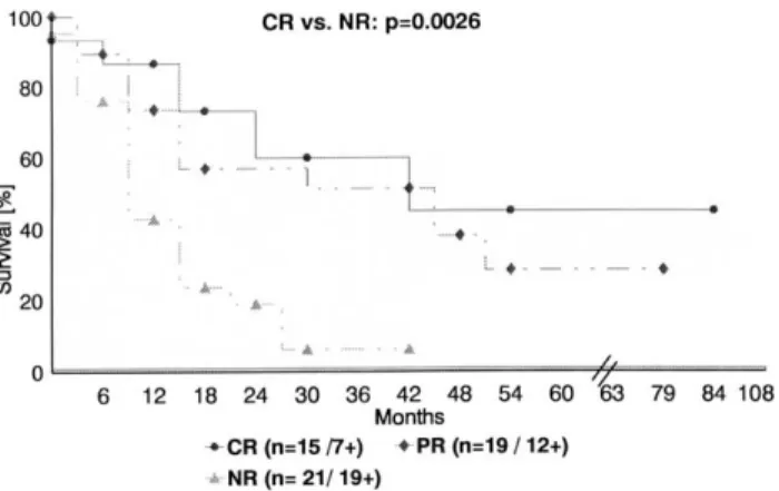
A significant difference (P¼0:0026) in survival was observed with regard to tumour response (including hospital death, Fig. 1): 53.3% of patients were still alive after complete pathological response (median, 54 months) compared with 36.8% of patients with a PR (median, 40 months) and with 9.5% of those with no change/tumour progress (median, 11 months). As numbers were too small, no survival analysis was performed according to histological tumour differentiation.
3.1. Predictive factors 3.1.1. MIB-1
In 46 of 56 patients who received preoperative RCX, proliferative activity of tumour samples obtained prior to RCX could be related to the response after resection.
Tumour samples of 13 patients with a complete pathological response showed a mean PI of 58.81 compared with 53.98 and 45.9 of tumours with PR or NR (P¼0:012). None of the tumours with a CR had an index of less than 45 (Table 5). Correlation with survival was statistically significant: an increased PI led to a better long-term survival (P¼0:0114).
3.1.2. VEGF expression
Tumour cells exhibited a strong cytoplasmatic VEGF- immunoreactivity (IR). In addition, capillaries within the tumour revealed positive VEGF-IR staining of the endothe- lial cells. VEGF expression could be determined in 21 biop- sies prior to RCX. Tumour samples of five patients with a complete pathological response showed a VEGF expression index of 10.7 compared with 18.34 (n¼8) and 36.58 (n¼8) of tumours with PR or NR (P¼0:035; Table 5).
Correlation with survival was statistically significant: a decreased VEGF expression led to a better long-term survi- val (P¼0:0205).
3.1.3. CD34
Endothelial cells of the capillaries exhibited a strong reaction with the CD34 antibody. In contrast, there was no positive reaction at all with tumour cells. Tumour samples of five patients with a complete pathological response showed a CD34 index of 10.92 compared with 18.97 (n¼8) and 18.16 (n¼8) of tumours with PR or NR (P¼0:036). None of the tumours with a CR had an index of more than 20 (Table 5). CD34 expression showed a corre- lation with VEGF expression (r¼0:4). Both vascular density and VEGF expression were higher in tumour samples which showed only PR or NR to preoperative RCX compared with tumours from complete responders.
There was no correlation of CD34 expression with long- term survival (P¼0:63).
3.2. Correlation of proliferative activity and VEGF expression
There was no direct correlation between the PI and the VEGF expression of a respective specimen. In tumours which showed a CR to chemoradiation, the relation of mean indices between VEGF expression and proliferative activity was 10.7–58.8 (Table 5). The relation of mean indices of VEGF expression and proliferative activity in tumours with PR or NR was 18.3–53.8 and 36.6–43.5, respectively. According to these results, it may be expected that tumours with a VEGF/MIB-1 quotient of 1:6 or less prior to RCX will respond to this therapy.
Multivariate analysis revealed PI (P¼0:0203), response (P¼0:0017) and tumour free resection (R0;P¼0:023) as independent variables for survival in patients who received preoperative RCX. All other tested variables (sex, pT, pN, histology, grading) were not significant. Logistic regression defined PI (P¼0:0193) and pN (P¼0:0002) as indepen- dent variables for tumour response in these patients.
Table 4
Pretherapeutic, clinical stagingaand CR after RCX and surgeryb
n CR Percentage
T2 18 8 44
T3 35 6 17
T4 3 0 0
N0 20 6 30
N1 36 8 22
a Clinical staging: T, N.
bNo patient with clinically distant metastases (M1) received RCX.
Fig. 1. Actuarial survival rates of patients after preoperative RCX and resection with respect to tumour response (n¼56; last survey 10/2001).
CR, complete response after resection; PR, partial response; NR, no response/tumor progress (1, dead).
Downloaded from https://academic.oup.com/ejcts/article-abstract/21/4/657/486254 by Hungary EISZ Consortium user on 29 August 2019
4. Discussion
It is still uncertain whether preoperative RCX leads to an overall improvement of survival in patients with EC. Most studies indicate that there may be a survival advantage for patients with a complete pathological response which can be expected in 20–30% of patients regardless of the applied protocol [5,7].
Most studies published so far lack a sufficient patient number, as from a statistical point of view, at least 450 patients need to be included in a controlled study to demon- strate a 5-year survival benefit of 10–15% [4]. In addition, it remains speculative whether variation of protocols may increase the rate of complete responders.
The rate of CR in this study must be assessed carefully as in eight patients a transhiatal approach was chosen for reasons of impaired lung function. Lymph node clearance in these patients was not as radical as in the transthoracic approach. One of these eight patients revealed a complete pathological response after the resection.
As CR after resection is clearly defined it is difficult to assess PR. Tumour shrinkage of more than 50% is widely regarded as major response, but assessment of lymph node involvement is more difficult with the present staging proce- dures. In most studies, lymph node enlargement of.1 cm is regarded as malignancy which is not necessarily the case [21]. Due to the unreliability of clinical staging and resta- ging after RCX, PR is not further addressed in this paper.
Our data add further evidence that preoperative RCX
raises perioperative morbidity compared with a historical control (data yet unpublished). It is our opinion that post- operative complications after RCX are much more difficult to treat than those occurring after resection without preo- perative RCX, which is possibly related to an impaired cellular immunity [22]. The increased perioperative morbid- ity affects all patients who receive preoperative RCX, including patients without any benefit from this therapy.
Moreover, RCX is time consuming and expensive. There- fore, identification of factors are urgently required prether- apeutically indicating response to RCX prior to the onset of therapy.
Our data revealed great differences in the relation of mean indices of VEGF expression and proliferative activity in tumours with respect to the tumour response. Therefore, we expect that tumours with a VEGF/MIB-1 ratio of 1:6 or less prior to RCX will respond to preoperative RCX.
A decrease of the standard uptake value (SUV) of posi- tron emission tomography performed before the onset and after RCX was correlated with response [24]. However, whether changes in SUV can indicate response during preo- perative RCX remains open. Conventional staging proce- dures, clinical tumour stages or patient related factors could not be correlated with tumour response [22].
On a molecular tumour related basis, there are only few studies available which usually include only small numbers of patients. Immunohistochemical staining for metallothio- nein prior to the onset of RXC was inversely correlated with tumour response after resection in patients with EC [23]. C-
Table 5
PIa, VEGF expressionband CD34 expressionbin tumour samples obtained prior start of RCX and correlated with response after resectionc,d
NR PR CR
MIB-1 VEGF CD34 MIB-1 VEGF CD34 MIB-1 VEGF CD34
23.53 n.t. n.t. 38.4 4.1 8.3 46.3 n.t. n.t.
23.64 n.t. n.t. 39.9 28.7 9.2 48.8 n.t. n.t.
25.57 18.1 13.3 40.3 10 10.6 51.0 n.t. n.t.
26.1 22.8 13.4 41.2 n.t. n.t. 52.4 6.1 5.2
27.18 45.3 14.2 46.2 32.3 13.1 53.2 n.t. n.t.
40.2 37.4 14.7 47 11.4 20.8 55.3 n.t. n.t.
44.9 35.3 15.7 47.2 22.3 191 59.4 n.t. n.t.
45.7 n.t. n.t. 50.9 19.3 34.7 61.4 32 8.3
51.8 n.t. n.t. 51.9 18.6 35.7 62.3 3.5 8.4
52.3 32.3 21.5 57.6 n.t. n.t. 66.4 8.2 14.5
52.8 n.t. n.t. 58.6 n.t. n.t. 69 n.t. n.t.
56.3 51.3 24.0 61.3 n.t. n.t. 69.2 n.t. n.t.
58.2 50.2 28.5 73.7 n.t. n.t. 70 3.7 18.2
60.4 n.t. n.t. 77.0 n.t. n.t.
62.9 n.t. n.t. 78.4 n.t. n.t.
63.1 n.t. n.t.
63.9 n.t. n.t.
Mean^SD Mean^SD Mean^SD Mean^SD Mean^SD Mean^SD Mean^SD Mean^SD Mean^SD
45.9^12.1 36.58^12.5 18.16^7.3 53.98^14.8 18.34^11.6 18.97^8.3 58.81^9.0 10.7^12.1 10.92^5.3
a MIB-1;n¼46.
b n¼21.
c Indices calculated as positive/total number of counted tumour cells (described in Section 2).
d NR, no response/tumour progression; PR, partial response; CR, complete response; n.t., no tissue available.
Downloaded from https://academic.oup.com/ejcts/article-abstract/21/4/657/486254 by Hungary EISZ Consortium user on 29 August 2019
erb 2 expression was inversely correlated with outcome after preoperative RCX in patients with an EC [5].
Previously, we reported that in the CR group, none of the investigated tumours showed a PI below 40% [9]. Apoptosis investigated by terminal dUTP nick-end labelling (TUNEL) did not correlate with response. We concluded, therefore, that an imbalance of apoptosis and/or proliferation rate by cells may eventually lead to tumour formation in cells which become malignant for various reasons. Highly proliferative cells with decreased activity of apoptosis grow faster than cells with low proliferative activity and unchanged apopto- sis. Tumours with an increased cell turnover seem to respond more rapidly to RCX than tumours growing slowly.
We now present extended data on proliferation indices of tumour samples prior to RCX thus confirming our earlier results. In addition, multivariate analysis revealed MIB-1 as an independent prognostic marker for tumour response.
Intratumoural microvessel density has been investigated in EC with conflicting results concerning the prognosis [10–
12]. Angiogenetic factors such as VEGF are secreted by tumour cells, thus regulating angiogenesis in tumours.
VEGF has been shown to be hypoxia-inducible in vitro in different cultured cells and in vivo in a range of tissues [16,17]. Moreover, expression of VEGF increased tumour growth and angiogenesis in vivo in a nude mouse model [18]. Also, anti-VEGF antibodies have the potential to reduce growth of several tumour cell lines in nude mice [19]. Whether the response to RCX in EC is influenced by the expression of VEGF is unclear. The results of our study suggest that tumours with low vessel density respond more than those with high vessel density. Vessel density is depen- dent on the expression of angiogenetic growth factors such as VEGF. However, it is uncertain whether the expression of VEGF by itself determines the reaction of a tumour to RCX or whether expression of VEGF is indirectly related to tumour response.
Interestingly, in a previous report, a correlation of PI expressed as KI-67 labelling index with vessel density could not be observed in patients with squamous cell carci- noma [12]. Whilst this supports our findings, it may be speculated that highly proliferative cells are likely to form fast growing tumours and therefore need a vascular network for nutritional supply. Probably anti-angiogenetic factors keep the tumours in a state of dormancy [20] independent of the proliferative activity of single tumour cells.
In conclusion, the results of our study confirm an increased perioperative morbidity following preoperative RCX in patients with EC. The CR rate was 25%, which resulted in a significantly improved survival compared with non-responders and with a historical control group.
The results obtained by immunohistochemistry suggest that proliferative activity is positively while expression of VEGF is inversely correlated with complete tumour response. A relation of 1:6 or less of VEGF expression and proliferative activity was predictive for a complete tumour response in this retrospective study. However, data
have to be confirmed by a prospective investigation which is currently performed.
References
[1] Altorki NK, Girardi L, Skinner DB. En bloc esophagectomy improves survival for stage III esophageal cancer. J Thorac Cardiovasc Surg 1997;114:948–956.
[2] Vogel SB, Medenhall WM, Sombeck MD, Marsh R, Woodward ER.
Downstaging of esophageal cancer after preoperative radiation and chemotherapy. Ann Surg 1995;221:685–695.
[3] Walsh TN, Noonan N, Hollywood D, Kelly A, Keeling N, Hennessy TP. A comparison of multimodal therapy and surgery for esophageal adenocarcinoma. N Engl J Med 1996;335:462–467.
[4] Kelsen D. Preoperative chemoradiotherapy for esophageal cancer. J Clin Oncol 2001;19:283–285.
[5] Duhaylongsod FG, Gottfried MR, Iglehart JD, Vaughn AL, Wolfe WG. The significance of c-erb B2 and p53 immunoreactivity in patients with adenocarcinoma of the esophagus. Ann Surg 1995;221:677–684.
[6] Lackey VL, Reagan MT, Smith RA, Anderson WJ. Neoadjuvant therapy in squamous cell carcinoma of the esophagus: role of resec- tion and benefits in partial responders. Ann Thorac Surg 1989;48:218.
[7] Urba S, Orringer M, Turrisi A, Iannettoni M, Forastiere A, Strawder- man M. A randomized trial of preoperative chemoradiation versus surgery alone in patients with locoregional esophageal cancer. J Clin Oncol 2001;19:303–313.
[8] Bosset JF, Gignoux M, Triboulet JP, Tiret E, Mantion G, Elias D, Lozach P, Ollier JC, Pavy JJ, Mercier M, Sahmoud T. Chemora- diotherapy followed by surgery compared with surgery alone in squa- mous cell carcinoma of the esophagus. N Engl J Med 1997;337:161–
167.
[9] Imdahl A, Jenkner J, Ihling C, Ru¨ckauer KD, Farthmann EH. Is MIB- 1 proliferation index a predictor for response of neoadjuvant therapy in patients with esophageal cancer? Am J Surg 2000;179:514–520.
[10] Torres C, Wang H, Turner J, Shahsafaei A, Odze RD. Prognostic significance and effect of chemoradiotherapy on microvessel density (angiogenesis) in esophageal Barrett’s esophageal-associated adeno- carcinoma and squamous cell carcinoma. Hum Pathol 1999;30:753–
758.
[11] Sarbia M, Bittinger F, Porschen R, Dutkowski P, Willers R, Gabbert HE. Tumour vascularization and prognosis in squamous cell carcino- mas of the esophagus. Anticancer Res 1996;16:2117–2122.
[12] Tanigawa N, Matsumura M, Amaya H, Kitaoka A, Shimomatsuya T, Lu C, Muraoka R, Tanaka T. Tumour vascularization correlates with the prognosis in patients with esophageal squamous cell carcinoma.
Cancer 1997;79:220–225.
[13] Ogata Y, Harada Y, Fujii T, Yamana H, Fujita H, Shirouzu K. Immu- nohistochemical localization of vascular endothelial growth factor in esophageal cancer. Kurume Med J 1996;43:157–163.
[14] Koide N, Nishio A, Kono T, Yazawa K, Igarashi J, Watanabe H, Nimura Y, Hanazaki K, Adachi W, Amano J. Histochemical study of vascular endothelial growth factor in squamous cell carcinoma of the esophagus. Hepato-gastroenterology 1999;46:952–958.
[15] Vermeulen PB, Gasparini G, Fox SB, Toi M, Martin L, McCulloch P, Pezzella F, Viale G, Weidner N, Harris AL, Dirix LY. Quantification of angiogenesis in solid human tumours: an international consensus of the methodology and criteria of evaluation. Eur J Cancer 1996;32A:2474–2484.
[16] Shweiki D, Itin A, Soffer D, Keshet E. Vascular endothelial growth factor induced by hypoxia may mediate hypoxia-initiated angiogen- esis. Nature 1992;359:843–845.
[17] Banai S, Shweiki D, Pinson A, Chandra M, Lazarovici G, Keshet E.
Upregulation of vascular endothelial growth factor expression
Downloaded from https://academic.oup.com/ejcts/article-abstract/21/4/657/486254 by Hungary EISZ Consortium user on 29 August 2019
induced by myocardial ischaemia: implications for coronary angio- genesis. Cardiovasc Res 1994;28:1176–1179.
[18] Zhang HT, Craft P, Scott PA, Ziche M, Weich HA, Harris AL, Bick- nell R. Enhancement of tumour growth and vascular density by trans- fection of vascular endothelial growth factor into MCF-7 human breast carcinoma cells. J Natl Cancer Inst 1995;87:213–219.
[19] Asano M, Yukita A, Matsumoto T, Kondo S, Suzuki H. Inhibition of tumour growth and metastasis by an immunizing monoclonal anti- body to human vascular endothelial growth factor/vascular perme- ability factor 121. Cancer Res 1995;55:5296–5301.
[20] Holmgren L, O’Reilly MS, Folkman J. Dormancy of micrometastasis:
balanced proliferation and apoptosis in the presence of angiogenesis suppression. Nat Med 1995;1:149–153.
[21] Mallery S, DeCamp M, Bueno R, Mentzer SJ, Sugarbaker DJ, Swan- son SJ, Van Dam J. Pretreatment staging by endoscopic ultrasono- graphy does not predict complete response to neoadjuvant chemoradiation in patients with esophageal carcinoma. Cancer 1999;86:764–769.
[22] Mafune K, Tanaka Y. Influence of multimodal therapy on the cellular immunity of patients with esophageal cancer. Ann Surg Oncol 2000;7:609–616.
[23] Yamamoto M, Tsujinaka T, Shiozaki H, Doki Y, Tamura S, Inoue M, Hirao M, Monden M. Metallothionein expression correlates with the pathological response of patients with esophageal cancer under- going preoperative chemoradiation therapy. Oncology 1999;56:332–
337.
[24] Brucher BL, Weber W, Bauer M, Fink U, Avril N, Stein HJ, Werner M, Zimmerman F, Siewert JR, Schwaiger M. Neoadjuvant therapy of esophageal squamous cell carcinoma: response evaluation by positron emission tomography. Ann Surg 2001;233(3):300–309.
[25] Ihling C, Menzel G, Wellens E, Monting JS, Schaefer HE, Zeiher AM. Topographical association between the cyclin-dependent kinases inhibitor P21, p53 accumulation, and cellular proliferation in human atherosclerotic tissue. Arterioscler Thromb Vasc Biol 1997;17:2218–
2224.
Downloaded from https://academic.oup.com/ejcts/article-abstract/21/4/657/486254 by Hungary EISZ Consortium user on 29 August 2019