Edited by:
Nicole Thielens, UMR5075 Institut de Biologie Structurale (IBS), France
Reviewed by:
Cordula M. Stover, University of Leicester, United Kingdom Maciej Cedzynski, Institute for Medical Biology (PAN), Poland Christian Drouet, Université Grenoble Alpes, France
*Correspondence:
Péter Gál gal.peter@ttk.mta.hu
Specialty section:
This article was submitted to Molecular Innate Immunity, a section of the journal Frontiers in Immunology Received: 29 May 2018 Accepted: 26 July 2018 Published: 08 August 2018 Citation:
Dobó J, Kocsis A and Gál P (2018) Be on Target: Strategies of Targeting Alternative and Lectin Pathway Components in Complement-Mediated Diseases.
Front. Immunol. 9:1851.
doi: 10.3389/fimmu.2018.01851
Be on Target: Strategies of Targeting Alternative and Lectin Pathway
Components in Complement- Mediated Diseases
József Dobó, Andrea Kocsis and Péter Gál*
Institute of Enzymology, Research Centre for Natural Sciences, Hungarian Academy of Sciences, Budapest, Hungary
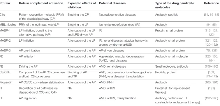
The complement system has moved into the focus of drug development efforts in the last decade, since its inappropriate or uncontrolled activation has been recognized in many diseases. Some of them are primarily complement-mediated rare diseases, such as paroxysmal nocturnal hemoglobinuria, C3 glomerulonephritis, and atypical hemolytic uremic syndrome. Complement also plays a role in various multifactorial diseases that affect millions of people worldwide, such as ischemia reperfusion injury (myocardial infarction, stroke), age-related macular degeneration, and several neurodegenerative disorders. In this review, we summarize the potential advantages of targeting various complement proteins with special emphasis on the components of the lectin (LP) and the alternative pathways (AP). The serine proteases (MASP-1/2/3, factor D, factor B), which are responsible for the activation of the cascade, are straightforward targets of inhibition, but the pattern recognition molecules (mannose-binding lectin, other collectins, and ficolins), the regulatory components (factor H, factor I, properdin), and C3 are also subjects of drug development. Recent discoveries about cross-talks between the LP and AP offer new approaches for clinical intervention. Mannan-binding lectin-associated serine proteases (MASPs) are not just responsible for LP activation, but they are also indispensable for efficient AP activation. Activated MASP-3 has recently been shown to be the enzyme that continuously supplies factor D (FD) for the AP by cleaving pro-factor D (pro-FD). In this aspect, MASP-3 emerges as a novel feasible target for the regulation of AP activity. MASP-1 was shown to be required for AP activity on various surfaces, first of all on LPS of Gram-negative bacteria.
Keywords: complement system, lectin pathway, alternative pathway, complement inhibitors, complement-related diseases
Abbreviations: 3MC syndrome, Malpuech–Michels–Mingarelli–Carnevale syndrome; aHUS, atypical hemolytic uremic syndrome; AMD, age-related macular degeneration; AP, alternative pathway; C4BP, C4b-binding protein; CARPA, complement activation-related pseudoallergy; CCP, complement control protein; CP, classical pathway; CNS, central nervous system; CR1, complement receptor 1; CR2, complement receptor 2; CRP, C-reactive protein; CUB, C1r/C1s, the sea urchin protein Uegf, and the human bone morphogenetic protein 1; CVF, cobra venom factor; EGF, epidermal growth factor; DAF, decay-accelerating factor; DAMP, damage-associated molecular pattern; FHL, FH-like protein; FHR, FH-related protein; FIMAC, FI membrane attack complex; GPI, glycosyl phosphatidylinositol; HAE, hereditary angioedema; HUVEC, human umbilical vein endothelial cell; LDLr, low-density lipoprotein receptor; LP, lectin pathway; MAC, membrane attack complex; MBL, mannose-binding lectin; MASP, MBL-associated serine protease; MCP, membrane cofactor protein; IGFBP-5, insulin-like growth factor-binding protein 5; IRI, ischemia–reperfusion injury; LPS, lipopolysaccharide; PAMP, pathogen-associated molecular pattern; PNH, paroxysmal nocturnal hemoglobinuria; PRM, pattern recognition molecule; SIRS, systemic inflammatory response syndrome;
SP domain, serine protease domain; SLE, systemic lupus erythematosus; TAFI, thrombin-activatable fibrinolysis inhibitor; TSR, thrombospondin type repeat; TCC, terminal complement complex; VWA, Von Willebrand factor type-A.
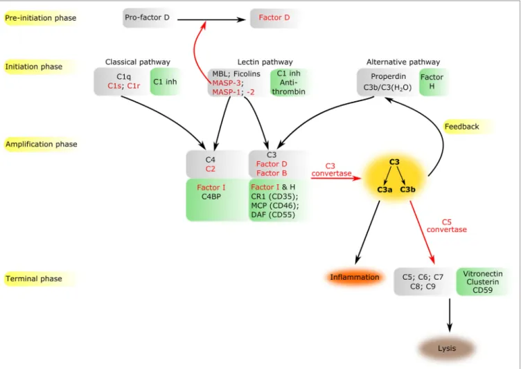
FiGURe 1 | Overview of the complement system. The components of all activation phases are listed in gray boxes, while their inhibitors in green boxes nearby.
Serine proteases are indicated by red letters. Black arrows indicate the direction of the cascade. Certain enzymatic cleavages are emphasized by red arrows.
The three activation routes merge at the cleavage of C3 highlighted by dark yellow background.
BRieF OveRview OF THe COMPLeMeNT SYSTeM
initiation Phase
The complement system is a sophisticated network of serum pro- teins (recognition molecules, proteases, modulators, inhibitors) as well as cell-surface regulators and receptors that constitute a key part of the host defense machinery. The complement system is a powerful effector component of the innate immunity and a vital modulator of the adaptive immune response (1–3). The complement system recognizes, tags, and eliminates microbial intruders and other dangerous particles such as immune com- plexes, damaged, and altered self cells. The complement system is inactive (or at least shows a very low basic activity: “tickover”) until it is activated by various danger signals. There are three canonical pathways through which the complement system can be activated: the classical pathway (CP), the lectin (LP), and the alternative pathways (AP) (Figure 1). CP and LP have several features in common. In both cases, pattern recognition molecules (PRMs) bind to the danger-associated structures. The PRMs,
like the other complement components, are modular proteins, consisting of multiple structural domains (Figure 2). C1q, the single PRM of the CP binds primarily to immune complexes containing IgG or IgM, and to C-reactive protein (CRP) via its C-terminal globular domains (4). These globular domains are fused to N-terminal collagen-like arms forming the characteristic
“bunch-of-six-tulips” structure. The structure of the PRMs of the LP resembles that of C1q; globular heads and collagen-like arms.
However, the recognition domains of mannose-binding lectin (MBL), other collectins, and ficolins bind to different structures.
The C-type lectin domains of MBL recognize the carbohydrate pattern of the bacterial surfaces. Ficolins (ficolin 1, 2, and 3) bind to acetylated compounds, typically to acetylated sugars of bacteria, via their fibrinogen-like domains (5). Collectins (CL- K1 and CL-L1) also recognize sugars and other potential danger signals. Unlike C1q, which has the well-established hexamer structure, MBL, ficolins, CL-K1, and CL-L1 exist in different oligomerization states, from dimer to hexamer; the tetramer being the dominant form at least for MBL. These PRMs circulate in complex with serine protease (SP) zymogens and monitor
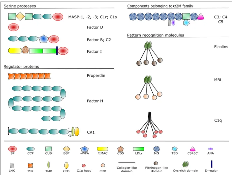
FiGURe 2 | Domain structures of the complement components discussed in the article. Each domain type is represented by a different symbol listed at the bottom.
Domain abbreviations are as follows: SP, serine protease; CCP, complement control protein; CUB, complement C1r/C1s, sea urchin Uegf and bone morphogenetic protein-1; EGF, epidermal growth factor-like; vWFA, von Willebrand factor type A; FIMAC, factor I/membrane attack complex; CD5, scavenger receptor cysteine-rich domain; LDLr, low-density lipoprotein receptor; MG, α-macroglobulin; TED, thioester domain; ANA, anaphylatoxin; LNK, linker; TSR, thrombospondin-type 1 repeat;
TMD, transmembrane domain; CPD, cytoplasmic domain; CRD, carbohydrate recognition domain.
continuously for dangerous particles the bloodstream. When the PRMs bind to the target surface, the associated SPs become activated and initiate a proteolytic cascade system, which ampli- fies the initial signal tremendously. C1q is associated with two C1r and two C1s proteases (the so-called “tetramer”) to form the C1 complex of the CP (6). MBL/ficolin-associated serine protease 1 and 2 (MASP-1 and MASP-2) are the initial proteases of the LP (7, 8). These SPs, together with the third MBL/ficolin- associated SP (MASP-3) form a protease family with the same domain structure (Figure 2) and similar function. The activation of the CP and LP results in the formation of the same enzyme complex, a C3 convertase (C4b2a) that cleaves C3, the central component of the complement system. The first enzymatic step in the CP activation is the autoactivation of C1r. Activated C1r then cleaves zymogen C1s, which in turn cleaves C4 and C2. In the LP, MASP-1 autoactivates first and then cleaves MASP-2 (9).
MASP-2 is the enzyme of the LP that cleaves C4 (10, 11), while C2
is cleaved by both MASP-1 and MASP-2. C3 and C4 are closely related thioester-containing proteins that form the basis of the convertase complexes (12, 13). Their function is to covalently attach the convertase to the activation surface and to capture the SP components of the enzyme complex. C2 is the SP component of the C3 convertase of the CP and LP. Activation of the AP is quite different from that of the CP and LP (14). When the CP/LP C3 convertase (C4b2a) cleaves C3, a smaller fragment is released (C3a). The larger fragment (C3b) covalently binds to the activa- tion surface preferably through an ester or, less likely, through an amide bond due to the reaction of the exposed thioester bond (15, 16). The nascent C3b component binds factor B (FB), the SP component of the AP C3 convertase. FB is cleaved by FD, a SP which circulates predominantly in its cleaved form in the blood.
The resulting C3bBb is the AP C3 convertase, which converts more C3 into C3b. The new C3b molecules serve as platforms for new C3 convertase complexes. In this way, a positive feedback
loop amplifies the initial signal tremendously generated either by the CP or the LP (17). According to the C3 tickover hypothesis, the AP can also initiate on its own without involvement of CP or LP (18). The circulating C3 molecules hydrolyze slowly and spontaneously in the bloodstream. The resulting C3(H2O) is a C3b-like molecule; it can bind FB and then form an “initiation”
C3 convertase (C3(H2O)Bb). If this fluid-phase C3 convertase emerges near a surface, the nascent C3b molecules can bind to the surface and initiate the positive feedback loop. In this way, the AP continuously monitors the different surfaces and if it finds an activator surface, it launches efficient complement activation.
The self-tissues are protected from AP-mediated damage by cell-bound and fluid-phase inhibitors (Figure 1). These inhibi- tors dissociate the C3bBb complex and serve as cofactors for the serine protease factor I (FI) in the degradation of C3b (19).
Decay-accelerating factor (DAF, CD55), membrane cofactor protein (MCP) (CD46), complement receptor 1 (CR1, CD35) are cell-surface-bound while the master regulator of the AP is the fluid-phase protein, factor H (FH). FH binds to cell-surface- deposited C3b and facilitates its degradation to iC3b, C3c, and C3dg by FI. On endogenous cell membranes, which expose sialic acid, binding of FH is tight and the degradation of C3b is rapid, while on the so-called “protected surfaces” (e.g., bacteria, fungi) binding is weak and the amplification loop of AP can build up.
There is a positive regulator of the AP, properdin, which increases the half-life of the C3bBb complex. Originally, at its discovery, properdin was regarded as a pattern recognition-like initiator molecule of the AP (20). Later, it was considered as a positive regulator (21); however, recently, the pattern recognition func- tion of properdin has been reconsidered (22, 23).
From the Central Phase to the Terminal Pathway
The cleavage of C3 by the C3 convertases is the turning point of complement activation. At this point, the three activation pathways (CP, LP, and AP) merge into a unified terminal pathway (Figures 1 and 3). When the density of surface-deposited C3b reaches a certain level, the substrate specificity of the C3 con- vertases switches to cleave the C5 component. The C4b2a(C3b)n and C3bBb(C3b)n convertases cleave the C5 component into a smaller (C5a) and larger (C5b) fragments. The C5a fragment, like the C3a fragment, is an anaphylatoxin. The anaphylatoxins bind to their receptors (C3aR and C5aR1/2) on leukocytes and endothe- lial cells and initiate inflammatory reactions (24). Structurally, C5 is similar to C3 and C4 (although it does not contain thioester bond) (25). The cleavage of C5 is the last enzymatic step in the complement cascade. From this point, conformational changes drive the formation of a self-organizing protein complex that damages the membrane of the attacked cells [membrane attack complex (MAC)]. After cleavage, the nascent C5b undergoes a conformational change that enables it to bind the C6 and C7 components (26). The resulting C5b–C7 complex binds to the cell membrane and captures C8. After conformational changes, C8 integrates into the membrane and pave the way to the integra- tion of multiple copies of C9 molecules. The C9 molecules form a pore in the membrane, which results in the disintegration and destruction of the cell (27).
COMPLeMeNT-MeDiATeD DiSeASeS
The complement system is an extremely effective cell-killing and inflammation provoking machinery. To prevent excessive activa- tion, the complement system is kept under strict control by the different inhibitory mechanisms. A delicate equilibrium between activation and inhibition is necessary to maintain the inflam- matory homeostasis in the human body. When this equilibrium is disrupted by any reason, the self-tissues can be damaged and severe disease conditions can occur. There are many clinical disorders in which uncontrolled (or sometimes the insufficient) complement activation is involved. Usually, the etiology of these diseases is complex, and the unwanted complement activation is only one of the pathological factors. However, evidences obtained by using various disease models suggest that preventing or inhib- iting the pathological complement activation can be a promising therapeutic approach.
insufficient Complement Activation
Since the complement system provides a first line of defense against invading pathogen microorganisms, deficiency of a complement component can lead to severe infections. The consequences could be more severe during childhood, when the adaptive immune system is not developed enough. Deficiency of the initial SPs of CP and LP (C1r, C1s, MASP-2) can result in pyogenic infections (28). Deficiency of MBL is the most common immunodeficiency in humans, affecting approximately 30% of the human population (29). It predisposes to recurrent infections in infancy; however, it is not a major risk factor in the adult population. Deficiency of the alternative and the terminal pathway components can severely compromise the defense against Gram-negative bacte- rial infections (30). Deficiency of properdin or deficiencies in the components of MAC are associated with infections of Neisseria species causing meningococcal meningitis or sepsis (31–33).
A very important function of the CP is the continuous removal of immune complexes and apoptotic cells. If this pathway is compromised in systemic lupus erythematosus (SLE) due to deficiency of C1q, or C4, or C1r/s, severe autoimmune reactions occur resulting in tissue injury in the kidneys.
excess Complement Activation
The majority of complement-related diseases are associated with overwhelming complement activation due to inappropriate con- trol. The kidney is especially vulnerable for complement-mediated attacks. In the case of C3 glomerulopathy, C3 deposition occurs in the glomeruli without immunoglobulin deposition (34, 35). C3 deposition in this case is likely the consequence of uncontrolled AP activation. In contrast to that, in the case of membranoprolifera- tive glomerulonephritis, CP activation elicits C3 deposition, since immunoglobulins and C1q are also deposited (36, 37). The IgA nephropathy is characterized by deposition of polymeric IgA1, which triggers complement activation through the AP and the LP (38, 39). Atypical hemolytic uremic syndrome (aHUS) is also a complement-related disease, which can lead to end-stage renal failure (37, 40). The driving force behind aHUS is the inappropriate AP acti- vation often due to variants of (41) or autoantibodies against (42) FH, the master regulator of the AP. aHUS is a form of thrombotic
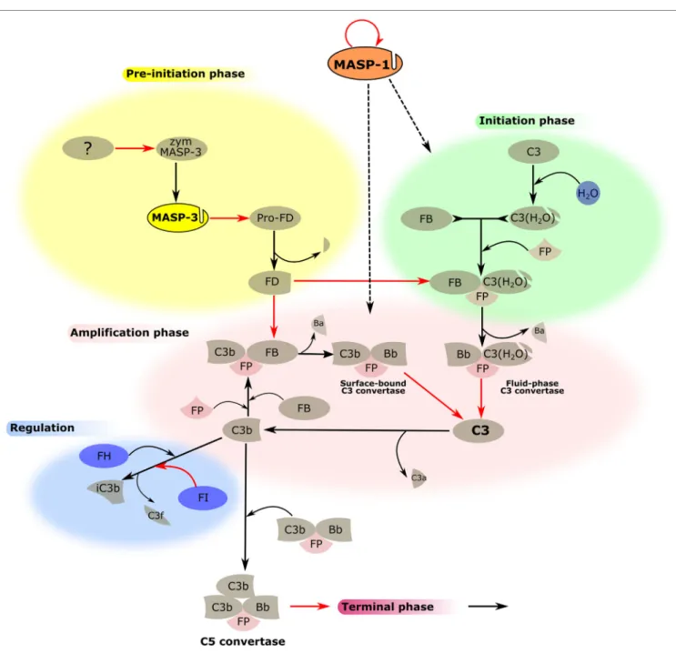
FiGURe 3 | MASP-1 and MASP-3 play roles in the activation of the alternative pathway (AP). The activation process of the AP is divided into several phases, which are indicated by differently colored backgrounds. MASP-3 is proved to be the professional activator of pro-factor D in blood, therefore, plays an important role in the pre-initiation phase. The physiological activator of zymogen MASP-3 (zym MASP-3) is hitherto undiscovered (shown by the question mark). MASP-1 is indispensable for efficient initiation and amplification of the AP on certain surfaces, although the mechanism is yet unknown (shown by dashed arrows). Black arrows represent conversion processes, while red arrows stand for enzymatic reactions pointing from the enzyme toward its substrate. The circle-shaped red arrow symbolizes the autoactivation of MASP-1.
microangiopathy accompanied with thrombocytopenia, hemolytic anemia, vascular damage, and thrombosis.
Another rare clinical condition associated with uncontrolled AP activation is paroxysmal nocturnal hemoglobinuria (PNH) (43, 44). In PNH patients, red blood cells are particularly prone to complement-mediated lysis due to the lack of two membrane- bound regulator proteins: DAF (CD55) and CD59. This is the consequence of the defect in glycosyl phosphatidylinositol (GPI)
synthesis in the cells. GPI is responsible for anchoring various proteins to the cell membrane including these inhibitors that regulate the activation of the AP (CD55) and the formation of MAC (CD59).
Age-related macular degeneration (AMD) is a complement- related disease, which affects a large population (about 100 mil- lion AMD cases) worldwide. It is the leading cause of blindness among the elderly in the developed world (45). Genetic analyses
strongly suggest that uncontrolled complement activation, espe- ci ally that of the AP, plays a major role in the pathogenesis of AMD. Genetic variants of FH, C3, FB, FI, and C9 have been associated with AMD (46). In the center of the retina of AMD patients, the photoreceptor cells are gradually degraded due to a chronic inflammation, which manifests in the accumulation of immune deposits called drusen underneath the retinal pigment epithelium. The drusens (that contain activated complement components) compromise the transport of oxygen and nutrients to the photoreceptors facilitating their degeneration. Numerous attempts have been made to curb the unwanted AP activation in the eye with limited success (47). In order to efficiently influence complement activation in the eye, we have to reveal its exact mechanism, which could be different in the periphery than in the bloodstream.
Ischemia–reperfusion injury (IRI) can be considered as a severe autoimmune reaction (48), which plays a major role in a number of clinical conditions. When the blood flow in an organ stops temporarily for any reason, the deprivation of oxygen (hypoxia) induces changes in the tissues, which predisposes them for complement-mediated attack after reperfusion. The affected cells and tissues are recognized by the immune system as damaged self [damage-associated molecular pattern (DAMP)], and a complex inflammatory reaction is launched, in which the complement system plays a decisive role. IRI significantly contributes to the tissue damage in the case of myocardial infarc- tion, stroke, transplant-induced inflammation, and it can cause a complication during coronary artery bypass graft surgery.
Although the exact mechanism of complement activation in case of IRI is not fully clarified yet, a number of evidences suggest that inhibition of the LP could be therapeutically advantageous (49–53).
Artificial materials used in modern medicine, such as poly- mer plastics and metal alloys, can also activate the complement system and trigger inflammation (54). Nanoparticles used as contrast agents or drug carriers can also activate the comple- ment system, sometimes causing a severe adverse reaction, called complement activation-related pseudoallergy (55, 56).
In this type of hypersensitivity reaction, IgE is not involved.
Liposomal drugs directly activate the complement system lib- erating C3a and C5a anaphylatoxins, which trigger mast cells and basophils.
If the immune system is exposed to an overwhelming amount of danger signals [pathogen-associated molecular patterns (PAMPs) or DAMPs], a systemic inflammatory reaction can occur, which could be more devastating than the original danger source. In the case of systemic inflammatory response syndrome, such as sepsis or polytrauma, the massive and systemic complement activation fuels a vicious cycle of hyperinflammatory events that can results in fatal tissue damage (57).
A growing number of evidences indicate that the complement system plays an important role in fundamental developmental processes. The lack of functional LP components (MASP-3, CL-K1, CL-L1) during embryogenesis results in the Malpuech–
Michels–Mingarelli–Carnevale (3MC) syndrome that manifest in characteristic craniofacial dysmorphism and multiple other anomalies (58, 59). It was shown that an intact CP is essential
for postnatal brain development. Contribution of C1, C4, and C3 was demonstrated to synaptic pruning essential for proper neuron circuit formation (60). These complement components tag synapses and mediate their elimination during a discrete window of postnatal brain development. C1q or C3 deficiency in mice results in improper central nervous system (CNS) synapse elimination. If these processes, essential during normal brain development, are pathologically upregulated during adulthood, they can contribute to the development of neurodegenerative diseases, such as Alzheimer disease and frontal temporal demen- tia (61). In addition to that, uncontrolled CP and LP activation in the CNS can also contribute to psychiatric disorders such as schizophrenia (62, 63).
CROSS-TALK BeTweeN THe AP AND THe LP
As described above, there are three canonical activation routes of the complement system. It is also obvious that the CP and the LP would not work efficiently without the amplification loop pro- vided by the AP, hence, the three pathways are naturally intercon- nected. It is also possible that homologous proteins C4 and C3, or C2 and FB can substitute each other to a certain degree; at least in vitro experiments indicate a weak cross-reactivity between CP/LP an AP C3 convertase components (64). MASP-1 (65) and MASP-2 (66) have both been implicated to be able to directly cleave C3; however, the physiological relevance of these reactions is uncertain. MBL was also shown to be involved in AP activation without the requirement of C2, C4, and MASPs (67), but in the light of our recent results, the observed effect could be mediated by MASP-1 (68). In summary, the involvement of the LP or LP components in AP activation has been demonstrated in the lit- erature before; however, some results still remain controversial.
In the subsequent two sections, recent discoveries are presented regarding the role of MASP-3 during the very early stage of AP activation, and the requirement of MASP-1 for AP activation on various surfaces.
Active MASP-3 is the Professional Pro-FD Maturase in Blood
The first evidence that MASP-1 or MASP-3 might have an essential role in AP function came from the group of T. Fujita (69). They created MASP1 knock-out mice by replacing the second exon (8). Since this region encodes a common part of both MASP-1 and MASP-3, the final homozygous mouse strain lacked both proteins. Surprisingly, these mice had pro-FD in their sera and had no AP activity (69). They suggested that MASP-1 acts as an essential enzyme for pro-FD maturation. At the time, it seemed like a logical assumption to favor MASP-1 over MASP-3 since MASP-1 is a more active enzyme in general with a relatively broad substrate specificity (70). Later, the same group suggested that MASP-3 might be more important than MASP-1 in pro-FD acti- vation and suggested that even the proenzyme form of MASP-3 can act as the activator (71).
Subsequent publications questioned the requirement of either MASP-1 or MASP-3 for AP activity. In the serum of a 3MC
syndrome patient lacking both proteins, functional AP was observed (72), and in mice deficient for MASP-1, MASP-3, and FH extensive, AP activation was observed, just like in mice defi- cient for FH only (73).
To clarify the roles of MASPs in pro-FD activation, we set up a series of experiments. In vitro all active MASPs were shown to be able to cleave pro-FD efficiently to produce FD, whereas the MASP zymogens lacked such activity (74). We prepared fluorescently labeled pro-FD, added it to different types of human plasma and serum preparations and followed the conversion of pro-FD to FD. Pro-FD was efficiently cleaved in all types of blood preparations, even in citrated and EDTA plasma, where neither the complement nor the coagulation cascade is expected to be activated. This experiment established that at least one protease is present in normal human blood capable of converting pro-D to FD without the prior activation of the abovementioned proteolytic cascades. Using a MASP-1-specific and a MASP-2-selective inhibitor, these two enzymes could be excluded. After adding recombinant active catalytic fragments of MASPs to normal human plasma samples, the half-life of labeled pro-FD was mark- edly reduced upon the addition MASP-3, whereas the other two enzymes had no effect (74).
The final “killer” experiment that established MASP-3 as the professional (near exclusive) activator of pro-FD came using a MASP-3 specific inhibitor, TFMI-3 (75). TFMI-3 blocked the conversion of labeled pro-FD to FD in citrated plasma, EDTA plasma, and hirudin plasma completely, while in serum, the half- life was markedly increased. Another conclusion of our studies was that active MASP-3 must be present in the blood, since only the activated form of MASP-3 can convert pro-FD to FD. Later, we provided direct evidence for the extensive basal-level activa- tion of MASP-3 in human blood by an unknown mechanism (76).
Finally, the debate seems to have settled. A recent paper showed that in 3MC syndrome patients lacking MASP-3, predominantly pro-FD is present in their sera, and moreover, in healthy indi- viduals, some pro-FD is also present beside the dominant active form (77).
The picture is now clear. Under normal circumstances, active MASP-3 is present in the blood, which activates pro-FD, therefore, continuously supplying active FD for the AP (Figure 3). However, MASP-3-deficient individuals are not completely defenseless. At least one coagulation enzyme can probably also provide low levels of FD for the AP, or when the LP is activated, MASP-1 or MASP-2 might also contribute. These backup mechanisms also need some consideration when targeting MASP-3 to control AP activity, as it will be discussed later.
MASP-1 is Required for AP initiation on Certain Activating Surfaces
Despite the fact that molecular mechanisms of the complement system have been thoroughly examined in the past decades, still many questions remained about the early steps of the activa- tion. MASP-1 as a promiscuous enzyme with broad substrate specificity (70) has the potential to replace other SPs or amplify enzymatic reactions. It has been reported that MASP-1 is indeed involved in biological processes beyond LP or even the comple- ment system.
Recently, we have found a novel function of MASP-1 in AP activation apart from its role in the LP (Figure 3). This func- tion suggests an unexpected linkage between the two pathways and also highlights the differences between various activation surfaces (68). Previously, specific and highly selective small- protein inhibitors against all MASPs were developed from canonical inhibitor scaffolds using the phage-display technique.
SGMI-1 is a specific MASP-1 inhibitor, whereas SGMI-2 inhibits MASP-2, and TFMI-3 is a specific inhibitor of MASP-3 (9, 75). Activation of the AP in vitro can be carried out on ELISA microtiter plates coated by bacterial lipopolysaccharide (LPS) or yeast zymosan. These materials serve as models of the pathogenic surfaces. In the absence of Ca2+ using Mg2+-EGTA buffer, AP activation can be initiated without the involvement of CP and LP. Administration of the specific MASP-1 inhibitor, SGMI-1 in this system lead to surprising results. The activity of the AP was attenuated significantly through the inhibition of MASP-1. However, this effect was only seen on the bacterial surface represented by LPS, while zymosan-induced AP acti- vation was not compromised. To rule out the possibility that SGMI-1 may impede other SPs, inhibitors of MASP-1 possess- ing different mode of action were also tested. Anti-MASP-1-SP antibody, N-terminal domains of MASP-1 (M1_D1-3) (78), and serpin domain of C1 inhibitor resulted in the same, consider- able reduction of AP activity but only on LPS. The activity of AP in MASP-1-depleted serum remarkably decreased on LPS-coated surface while on zymosan-coated surface, it was only moderately affected. The mechanism of C3b deposition, which is followed in our assay, can be divided to initiation and amplification phases. Time-course measurement of C3b deposi- tion in the presence of subsequently added SGMI-1 inhibitor indicated that MASP-1 contributes to both phases of C3b gen- eration. Although we proved unambiguously that MASP-1 has an effect on AP activation, there are still many puzzling details to be solved. First, we tested the known components of AP as possible reaction partners of MASP-1. It was clarified earlier that MASP-1 cleaves C3 only at a very low rate (11) and now we confirmed that MASP-1 does not react significantly with C3b-bound FB. The contribution of FD was also excluded since SGMI-1 does not inhibit FD, and MASP-1 is not the physiologi- cal activator of pro-FD (74). Our results lead to the conclusion that the key player of MASP-1-driven AP activation is probably not among the core components of the AP.
Differences between the activating surfaces also need fur- ther investigations since it seems to be likely that AP initiation occurs by various mechanisms. Using specific antibodies in the ELISA system, neither MASP-1 nor MBL could be detected on LPS surface in Mg2+-EGTA buffer (68). MASP-1 may be presented by some other PRMs, which do not necessarily require Ca2+ for binding (possibly ficolins). Another possible scenario is that MASP-1 forms a labile and transient complex with its reaction partners. One clue arises from the literature that properdin, which stabilizes C3bBb complex, is crucial for LPS-induced but not for zymosan-induced AP activation (79). Another coincidence is that LPS, rather than zymosan, binds FH with high affinity, which enhances the decay of C3 by FI activity. The ratio of these factors, which play a role in the
regulation of AP, may be influenced by MASP-1 through a yet unknown mechanism.
These new findings draw attention to MASP-1 in the promo- tion of LPS-induced AP and, therefore, its role in the defense against Gram-negative bacteria.
COMPONeNTS OF AP AND LP AS POTeNTiAL DRUG TARGeTS Pattern Recognition Molecules
The PRM of the CP and LP recognize danger signals (PAMPs and DAMPs) and provide the framework of the initiation multi- molecular complexes (the C1 complex, MBL–MASP complexes, ficolin–MASP complexes). They have similar overall domain architecture: N-terminal collagen-like domains and C-terminal globular domains. The structure of C1q is different from that of the other PRMs, since the basic trimeric subunit of C1q is composed of three different polypeptide chains (A, B, and C chains), while that of MBL and ficolins consist of only one kind of polypeptide chain. In the case of collectins, collectin kidney 1 (CL-K1) and collectin liver 1 (CL-L1), a heterotrimeric subunit was observed in human blood composed of one CL-L1 and two CL-K1 polypeptide chains (called CL-LK) (80). Another structural difference between C1q and the proteins of the col- lectin/ficolin family is that the latter contain a short N-terminal cysteine-rich region and an α-helical coiled-coil neck region between the collagen-like and the globular domains facilitating trimerization of the polypeptide chains. The collagen sequences (Gly-Xaa-Yaa repeats) are interrupted at one point in C1q and MBL generating a flexible kink region that may play an important role in binding to the danger patterns and activating the associ- ated SPs. The PRMs bind the associated SPs in a Ca2+-dependent manner. C1q binds the C1s-C1r-C1r-C1s tetramer, while MBL, ficolins, and CL-LK bind MASP dimers. It is probable that low oligomeric MBL and ficolins bind a single MASP dimer, while higher oligomers (pentamers, hexamers) can bind two MASP dimers simultaneously (81). There is a cross-interaction between the components of the CP and LP; MBL can bind the C1r2s2
tetramer and C1q can bind the MASP dimers, although with reduced affinity compared to the cognate pairs (82). It is unlikely that these interactions have a physiological relevance (except maybe in deficiencies); however, the existence of these cross- bindings proves that the interactions are analogous between the PRMs and the associated SPs among the components of the CP and LP. The binding of MBL and ficolins to their targets is Ca2+-dependent. The affinity of a single carbohydrate-binding domain to its target sugar is low (Kd in the millimolar range), whereas the avidity of the whole oligomeric molecule is high with a Kd in the low nanomolar range. It is very likely that the PRMs of higher oligomeric state activate the LP more efficiently than the low oligomeric PRMs due to the stronger binding to both the target surface and to the MASPs (83). The mechanism of activation of the C1 and MBL–MASP complexes is not fully clarified yet.
Theoretically, if we want to prevent or inhibit improper complement activation in a pathological situation PRMs of the
initiation complexes are ideal targets; since by inhibiting the PRMs, we can shut off the entire amplification machinery of the complement system at the very first step. There are three pos- sibilities to inhibit the function of the PRMs: (1) to prevent the binding of the PRMs to their target; (2) to prevent the binding of the associated SPs to the PRMs; (3) to prevent the conformational changes of the PRMs that are necessary for the activation of the SPs. Monoclonal antibodies that bind to the globular domains of the PRMs can efficiently interfere with the ligand binding.
Anti-C1q and anti-MBL antibodies were successfully used to block the CP and LP activation, respectively (84). An anti-MBL monoclonal antibody (3F8) attenuated myocardial IRI in mouse expressing human MBL (85).
Anti-C1q antibodies have been recently reported to greatly reduce the inflammatory demyelinating lesions in a mouse model of neuromyelitis optica (86) and also to attenuate injury with a consequent neuroprotective effect in acute Guillain–Barré syndrome mouse models (87). A peptide agent (called 2J) was selected from a peptide library on the basis of C1q binding (88).
This peptide was shown to bind to the globular domain of C1q and prevented the binding of C1q to IgG. The 2J peptide efficiently inhibited CP-mediated C4 and C3 deposition and MAC forma- tion in vitro. Although this peptide was a promising candidate for therapeutic complement inhibition, no further studies were reported about its in vivo application.
Another possibility for inhibiting the CP and the LP is to disassemble the initiation complexes. In this respect, it is worth noting that in in vitro experiments the C1 complex dissociates in the absence of Ca2+ (in the presence of EDTA), or at high ionic strength (1 M NaCl), whereas in the case of the MBL–MASP complexes, both conditions should apply at the same time (89).
Moreover, C1 inhibitor, which makes covalent complexes with the SPs, dislodges C1r and C1s from C1q, while it cannot disas- semble the MBL–MASP complexes (90). Nevertheless, it was shown that there is a dynamic equilibrium between the different MBL/ficolin–MASP complexes in human serum, in other words, MASPs can migrate between the complexes (91). Recently, it was shown that asparaginase, which is used in oncological treat- ments, inhibits the LP by reducing the amount of MBL–MASP complexes, very likely through dissociating the complexes (92).
In this case, it is an adverse effect of the oncological treatment, but it indicates that a similar approach can be feasible in anticomple- ment therapy. An anti-MASP-2 monoclonal antibody (OMS721, Omeros), which binds to a non-catalytic complement control protein (CCP) domain of MASP-2, successfully inhibited the LP in in vivo experiments, and also it could disassemble the MBL- MASP-2 complexes.
There is a report about a viral-derived peptide (PIC1), which inhibits the classical pathway through binding to the collagen- like region of C1q in the C1 complex (93). This peptide might lock the conformation of C1q and/or displace the tetramer.
There is no other report in the literature about an agent, which can block the conformational change necessary for the proper function of the initiation complexes. A deeper understanding of the activation mechanism of the C1 and MBL/ficolin–
MASP complexes is needed to harness this possibility in the therapy.
MASP-1
MASP-1 is the most abundant protease of the LP, and it plays a central role in complement activation. Its average serum con- centration is 143 nM (11 µg/mL), which is 24-times higher than the serum concentration of MASP-2 (6 nM, 0.4 µg/mL) (94). The members of the C1r/C1s/MASP protease family share the same domain organization (Figure 2). At the N-terminus, there is a CUB domain (initially recognized in C1r/C1s, sea urchin protein Uegf, and human bone morphogenetic protein 1), followed by an epidermal growth factor-like (EGF) module and a second CUB domain. The MASPs are present as dimers in the circulation, and the N-terminal CUB1-EGF-CUB2 region is responsible for the dimerization. Another important function of this region is that it mediates the binding to the PRMs. The CUB and the EGF domains bind Ca2+, and both the dimerization and the PRM binding are Ca2+-dependent. The C-terminal region, which pos- sesses the catalytic activity, consists of two CCP domains and a SP domain. The SP domain belongs to the chymotrypsin family (Family S1, MEROPS) and shows trypsin-like specificity cleaving after basic amino acids (Arg, Lys) in the polypeptide chain. The two CCP domains have at least two functions: they serve as spac- ers between the CUB1-EGF-CUB2 region and the SP domain and they provide additional binding sites (exosites) for the substrates (13). Both functions have essential roles in the activation of the PRM–MASP complexes and in the cleavage of the subsequent components (C2 and C4).
MASP-1 has multiple roles in the innate immune response.
Zymogen MASP-1 has a high autoactivation capacity, which plays a key role in the activation of the lectin pathway (95). When PRM–MASP complexes bind to the activation surface, zymogen MASP-1 autoactivates and the active MASP-1 activates zymogen MASP-2 (9, 72). In this way, MASP-1 is the initiator protease of the LP. Recently, it has been demonstrated that MASP-1 significantly contributes to AP activation on LPS surface through an unknown mechanism (68). MASP-1 is also capable of activating endothe- lial cells by cleaving protease-activating receptor 4 (91, 96).
The activated endothelial cells secrete cytokines (IL-6 and IL-8), and these cytokines promote the chemotaxis of neutrophil granulocytes (97). Moreover, MASP-1 treatment increased adhe- sion between neutrophils and endothelial cells by upregulating E-selectin expression in human umbilical vein endothelial cells (HUVECs) (98). A genome-wide gene expression profiling study on HUVECs corroborated the role of MASP-1 in triggering inflammation (99). The analysis showed that MASP-1 up- and downregulated numerous inflammation-related genes bridging complement activation and endothelial-cell-related inflamma- tory processes. It was also demonstrated that MASP-1 is able to cleave high-molecular-weight kininogen and liberate bradykinin (100). Bradykinin is a potent vasoactive, pro-inflammatory pep- tide, which is responsible for the swelling attacks in hereditary angioedema (HAE), a disease associated with C1 inhibitor defi- ciency (101). Uncontrolled activation of MASP-1 may contribute to the development of HAE attacks and worsening the symptoms of HAE patients. It was also recognized that MASP-1 serves as a link between the complement and the coagulation cascades.
MASP-1 promotes coagulation by activating prothrombin, fXIII, and thrombin-activatable fibrinolysis inhibitor (102–104).
The effect of MASP-1 on blood coagulation was confirmed by using a microvascular whole-blood-flow model (105). The physio- logical relevance of this phenomenon is not quite clear; however, it is very likely that the proteolytic activity of MASP-1 contributes to pro-inflammatory and pro-thrombotic events facilitating the development of thrombotic complications under pathological conditions (106). As the above examples highlight, MASP-1 has a relatively broad substrate specificity (it has about 10 known substrates), which is quite unusual among complement proteases.
It should be noted, however, that all the known substrates of MASP-1 are related to the innate immune response. Evolutionary considerations indicate that MASP-1 is an ancient enzyme of the complement system compared to the other members of the MASP/C1r/C1s family (107). The relaxed substrate specificity of MASP-1 is reflected in its 3D structure (70). The substrate-binding groove of MASP-1 is broad and accessible resembling that of trypsin, rather than those of other early complement proteases.
The physiological inhibitors of MASP-1 are serpins. C1 inhibi- tor, and in the presence of heparin antithrombin, attenuate very efficiently the activity of MASP-1 (108). Alpha2-macroglubulin, a pan-specific protease inhibitor in the blood was suggested to inhibit MASP-1 and consequently the LP (109), but this issue is controversial (108, 110). Another potential physiological inhibi- tor of the LP is MAp44 (aka MAP-1), an alternative splice product of the MASP1 gene (111, 112). MAp44 contains the CUB1-EGF- CUB2-CCP1 domains of MASP-1/3 plus a 17 amino-acid-long C-terminal peptide. Since MAp44 lacks the SP domain, it does not have proteolytic activity to initiate the LP, but it can dimerize and bind to the PRMs like the MASPs. MAp44 attenuates LP activity by competing with MASP-1 and MASP-2 for the PRMs and displacing them from the complexes. Recombinant MAp44 was shown to protect against myocardial IRI in mouse models, preserving cardiac function, decreasing the infarct size, and pre- venting thrombogenesis (113). Recombinant chimeric inhibitors were also designed and constructed by fusing MAp44 and the complement regulatory domains (1–5) of FH (114). One of these inhibitors showed simultaneous inhibition of the LP and AP.
Theoretically, the SPs are the most druggable targets in the complement system (115). The active sites of these enzymes can be easily targeted by small-molecule protease inhibitors. The main problem with this approach is the lack of specificity, since all the complement proteases and also the proteases of the other plasma cascade systems contain chymotrypsin-like SP domains (Figure 4). A small-molecule SP inhibitor, which blocks the activ- ity of a particular complement SP, very likely will inhibit other complement proteases, as well as proteases of the coagulation, fibrinolysis, and kallikrein–kinin systems to some extent. For example, nafamostat mesilate (FUT-175 or Futhan) is a powerful inhibitor of the complement cascade, but it has a broad specificity.
It was shown to attenuate renal and myocardial IRI (116, 117), but it also attenuates pancreatitis by inhibiting trypsin and other pan- creatic enzymes (118), and also coagulation by inhibiting throm- bin and other clotting enzymes (119). To enhance the specificity, the number of interactions should be increased between the SP and the inhibitor. A promising approach could be the fragment- based drug discovery, which generates highly specific molecules via linking small chemical fragments (Mw < 300 Da) together that
FiGURe 5 | Structure of factor D (FD) in complex with a selective small-molecule inhibitor. The figure is based on the structure of FD in complex with “inhibitor 6”
described by Maibaum et al. (120) (PDB entry 5FCK). Inhibitor 6 has multiple polar and hydrophobic interactions with the protein body. It is notable that inhibitor 6 interacts with the self-inhibited conformation of FD, probably stabilizing FD in this form. Asp177 (blue) of the S1 pocket forms a salt bridge with Arg202 of the self-inhibitory loop (red). The catalytic triad is colored magenta. Numbers indicate amino acid positions in mature FD, while numbers in parenthesis reflect the traditional chymotrypsinogen numbering. Hydrogen bonds are indicated by yellow dashed lines. (A) FD shown by ribbon representation. (B) FD shown by surface representation.
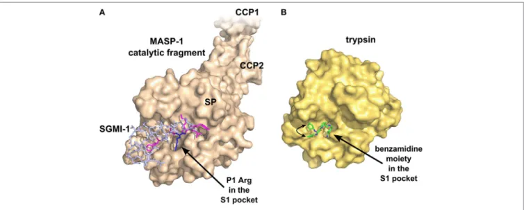
FiGURe 4 | Specific inhibition of proteases requires multiple favorable contacts in a large contact area with an inhibitor. (A) The structure of MASP-1 in complex with a specific small-protein inhibitor, SGMI-1 (PDB entry 4DJZ). SGMI-1 was developed by phage-display (122). Amino acid residues in the randomized positions are colored magenta (P4, P2, P1′, P2′, P4′) and blue (P1). All amino acid residues in the randomized positions have contacts with the protease body; moreover, SGMI-1 has other contact areas with MASP-1 in the non-randomized positions as well. (B) The structure of trypsin in complex with a non-specific small-molecule inhibitor (123) (PDB entry 3LJJ). The inhibitor is based on benzamidine. A two-headed arrow indicates the movement of the terminal cyclopentane moiety, which has two equivalent binding sites.
bind only weakly on their own to the target. This approach was successfully used to develop specific small-molecule inhibitors against FD (120) (Figure 5), but there is no report about similar molecules against MASPs. Monoclonal antibodies and other biologics can also meet the specificity criterion. Highly selective MASP inhibitors were developed by the in vitro evolution of the
interacting loop of canonical SP inhibitors. Sunflower trypsin inhibitor (SFTI) is a 14-amino acid-long cyclic peptide, which mimics the protease-interacting loop of the inhibitor scaffold of the Bowman–Birk inhibitor family. SFMI-1, an LP-selective peptide inhibitor was developed by phage-display selection of SFTI variants using MASP-1 as target (121). SFMI-1 proved to
be a strong MASP-1 inhibitor (Ki = 65 nM), and a weak MASP-2 inhibitor (Ki = 1,030 nM). In order to further increase the speci- ficity, a larger inhibitor scaffold was used in the phage-display selection. SGPI-2 (S. gregaria protease inhibitor-2) is a single domain small-protein inhibitor (35-amino acid-long) belonging to the Pacifastin family of canonical inhibitors. After randomiz- ing six positions in the protease-interacting loop (P4, P2, P1, P1′, P2′, and P4′), a highly specific MASP-1 inhibitor (SGMI-1) was selected (122) (Figure 4). SGMI-1 inhibits MASP-1 very effec- tively (Ki = 7 nM), and very selectively. This inhibitor was used to clarify the function of MASP-1 in the innate immune response using numerous in vitro and ex vivo assays. Although MASP-1 is a tempting target to halt unwanted LP activation and to prevent various pro-inflammatory processes, no pharmaceutical develop- ment of a MASP-1 inhibitor has been reported to date.
MASP-2
MASP-2 is the only protease in the LP that can cleave C4. Its serum concentration is rather low (6 nM, 0.4 µg/mL), compared to the other complement proteases. These characteristics make MASP-2 an ideal target to inhibit pathological LP activation.
MASP-2 has identical domain organization with MASP-1 and MASP-3 (Figure 2). Isolated MASP-2 has a tendency to autoactivate in a concentrated solution (11, 124). This autoac- tivation capacity, however, cannot manifest in normal human serum, where the MASP-2 concentration is low, and each MASP-2 molecule is surrounded by MASP-1 molecules on the target surface. Under these circumstances, MASP-1 is the exclusive activator of MASP-2. The autoactivation ability of MASP-2 might be important in situations, where there is no MASP-1 present (e.g., MASP-1 deficiency). It should be noted, however, that in the serum of a 3MC syndrome patient, where there was neither MASP-1 nor MASP-3 present due to a muta- tion in the MASP1 gene, no LP activity could be detected (72).
On the other hand, birds lack MASP-1, but have functional LP, suggesting that MASP-2 can independently drive LP activation (125). In the sera of these animals, however, the autoactivation capacity of MASP-2 must be much higher than that of human MASP-2 in normal human serum. A recent publication shows that MASP-2 can directly cleave C3 in the absence of C4 and/or C2 on LP-activating surfaces (66). MASP-2 was also suggested to promote fibrin polymerization by cleaving prothrombin (126).
The MASP2 gene, like the MASP1 gene, has an alternative splice product MAp19 (aka sMAP, MAP-2) (127, 128). This truncated gene product contains only the CUB1 and EGF domains plus 4 unique C-terminal residues. Since MAp19 can bind to the PRMs, it may regulate LP activity through displacing the MASPs from the complexes. Theoretically, the recombinant form of MAp19 could be suitable to attenuate LP activity, in practice, the larger MAp44 was used for this purpose since it binds to the PRMs with higher affinity.
The pathological relevance of MASP-2 was demonstrated in MASP-2 knock-out mice, where the animals were significantly protected against myocardial and gastrointestinal IRI (50). In the hearts of MASP-2-deficient mice, the infarct volume was signifi- cantly smaller than in those of the wild-type animals. Moreover, a recent study demonstrated that an inhibitory monoclonal
anti-MASP-2 antibody successfully attenuated myocardial IRI in wild-type mice (129). An anti-MASP-2 antibody, OMS721, developed by Omeros Corporation, is under clinical trial for treating aHUS (130) and other thrombotic microangiopa- thies (131), IgA nephropathy, lupus nephritis, membranous nephropathy, and C3 glomerulopathy (132). The mechanism of the protecting effect of MASP-2 inhibition in these diseases is not clear, since AP activation is believed to be the main driver of these conditions. Selective canonical inhibitors against MASP-2 were also selected by phage-display using the SFTI and SGPI scaffolds (121, 122). Both inhibitors were highly specific: SFMI-2 (Ki = 180 nM) and SGMI-2 (Ki = 6 nM) prevented LP activation efficiently, while they did not compromise the activity of the other two pathways.
MASP-3
MASP-3 was discovered as the third SP component of the LP (133). It has the same domain organization (Figure 2) as MASP-1 and MASP-2, as described above; moreover, the amino acid sequence of its first five domains is identical with that of MASP- 1. This feature is the consequence of the fact that MASP-1 and MASP-3 are the alternative splice products of the same MASP1 gene, along with a third protein MAp44 (111, 112). Variants of the MASP1 gene, resulting in the loss of the activity of MASP-3, cause the 3MC syndrome, characterized by serious craniofacial, genital, and often mental defects (58, 59, 134). The results indicate that MASP-3 is involved in neural crest cell migration in early embry- onic development. Interestingly, the same phenotype is observed in patients carrying mutations in the COLEC11 gene (58) or the COLEC10 gene (135), both encoding LP components CL-K1 (aka collectin-11) and CL-L1 (aka collectin-10). It is possible that MASP-3 is in complex with CL-K1/L1 when it exerts its function during embryogenesis, and it is likely that the proteolytic activity of MASP-3 plays an important role.
MASP-3 is different from MASP-1 and MASP-2 in several ways. MASP-3 does not autoactivate, does not cleave downstream LP/CP components, C4 and C2, and the active form has very low activity on most synthetic substrates (136). In vitro, it was shown to cleave insulin-like growth factor-binding protein 5; however, the relevance of this reaction is uncertain (137). It has also no natural inhibitor in the blood; therefore, control of its activity is prob- ably achieved simply by its very restricted substrate specificity.
MASP-3 is present in the blood as the mixture of the proenzymic and the activated forms; moreover, the activated form seems to be the more dominant variant (76). In this aspect, it also differs from MASP-1 and MASP-2, which are proenzymic. On the other hand, in many regards, MASP-3 has similarities to FD, which circulates predominantly in the active form, has no natural inhibitor, and has very restricted substrate specificity.
The function of MASP-3 in the blood had been mysterious until recently. Initially, it was considered simply as a negative reg- ulator of the LP since it competes with MASP-1 and MASP-2 for binding to PRMs (133). This function may still be valid; however, now, strong evidences exist that the active form of MASP-3 is the primary physiological activator of pro-FD, producing FD, a key enzyme of the AP. The story was detailed in a previous section;
therefore, we jump to the functional consequences of this activity.
It seems logical that the activity of the AP can be downregulated by the inhibition of MASP-3. Inhibition of MASP-3 would result in the accumulation of pro-FD in the blood with only very low levels of active FD, hence greatly attenuating AP activity. A study presented at 16th European Meeting on Complement in Human Disease (138) provided a strong evidence for this assumption.
A single dose of a monoclonal antibody inhibiting the activity of MASP-3 suppressed the activity of the AP and shifted the active to zymogen ratio of FD toward the proenzyme, pro-FD, both in mice and in cynomolgus monkey. So far, two specific inhibitors against MASP-3 were developed. One of them is a canonical Kunitz-type recombinant protein, which is based on the second domain of tissue factor pathway inhibitor (TFPI) and developed by phage-display (75). The other is the abovementioned mono- clonal antibody by Omeros Corporation (138).
What are the potential advantages of MASP-3 inhibition over FD inhibition? Inhibition of both proteins is expected to result in similar systemic effects in the blood. The plasma concentration of both proteins is similar, around 60 nM. This relatively low value is attractive for drug development. On the other hand, FD has a very high turnover rate. Its half-life in humans is less than 1 h (139).
The turnover rate of MASP-3 is not yet known, but because of its size, it is most certainly lower compared to FD. This could mean that a lower daily dose of a drug candidate inhibitor of MASP-3 would be required compared to a FD inhibitor.
Deficiency in the AP can result in potentially life-threatening meningococcal infections, and AP inhibition carries the same risk. Another potential benefit of MASP-3 inhibition would be that in this case, a pro-FD pool is still available. In case of a bacte- rial infection, the LP can be activated, and the resulting active MASP-1 and MASP-2 molecules could locally convert pro-FD to FD, making the AP amplification possible. Nonetheless, this mechanism needs experimental validation.
In all, based on MASP-3’s requirement for the maturation of pro-FD, MASP-3 presents itself as a good target to attenuate the complement system, with several potential benefits over FD inhibition.
Factor D
Factor D (FD) is a single domain SP, which circulates in the blood predominantly in the active form (77, 140). It is synthesized mainly by adipocytes, hence the alternative name adipsin. In the 1970s, it was debated whether it is produced as a proenzyme or secreted in the active form (140, 141). Since only the active form could be isolated from blood (142), it was assumed that it might be activated even before secretion (143). Nevertheless, at the DNA level, after the signal sequence, an additional 5 to 7 amino acid long propeptide is encoded. Now the consensus is that active MASP-3 converts the pro from of FD to the active form constitutively (74, 75, 77).
Although it is an active SP, FD has an extremely restricted substrate specificity. It has very low activity toward synthetic substrates, basically, it cleaves only certain thioester compounds;
however, its natural substrate, FB in complex with C3b or C3b- like molecules, is cleaved very efficiently (144). The free enzyme’s very low activity is due to a unique self-inhibitory loop (145), which is displaced when FD binds to C3bB (146).
It has a relatively low mass concentration of 1–4 µg/mL in humans (147–149), which in combination with early reports showing that FD is the bottleneck of AP activity (140), led to the assumption that FD could be the best target to achieve AP inhibi- tion. However, FD is a small protein of only 25 kDa, so, its molar concentration of 40–160 nM combined with its high turnover (139) suggest that high daily doses of a FD inhibitor would be required to achieve complete sustained inhibition. Recent results also suggest that FD may not even be the bottleneck of AP activity.
In the serum of FD-deficient mice, the addition of FD correspond- ing to only about 1–2% of the normal FD level was sufficient for normal AP activity in vitro (147). In a 3MC syndrome patient, whose serum contained mostly pro-FD, some AP activity was still present (72), although lower than the normal level (134). These data together suggest that even if a potent and specific inhibitor is used, at least equimolar amount is required for AP inhibition, and even higher doses are necessary for sustained inhibition.
A study with lampalizumab, a humanized monoclonal anti-FD IgG Fab fragment, showed similar observations (150).
One must also consider that, at least in vitro, plasma kal- likrein was shown to be able to cleave the C3bB pro-convertase (151), hence a residual, low-level AP activity might still be present even during complete FD inhibition, or FD deficiency;
however, the in vivo relevance of this cleavage needs further validation.
Nevertheless, FD remains a prime target within the comple- ment system. Several FD inhibitor molecules are under develop- ment, or in the clinical trial phase (152, 153) for PNH, aHUS, and AMD. Achillion developed several small molecule FD inhibitors that may be orally administered. A dose of 200 mg/kg of ACH-4471 per every 12 h resulted in complete AP inhibition in primates (154). An example of the combination of structure- based and fragment-based drug development targeting FD was published recently. Modifying the structure of a small-molecule kallikrein inhibitor several compounds were developed that selectively inhibit FD (120). Figure 5 shows FD in complex with one of the compounds as an example.
Near complete inhibition of FD is expected to have a similar outcome as inhibition of MASP-3. Neisserial infection or other bacterial infections constitute a possible threat, which requires prophylactic treatment or treatment with antibiotics. This is actu- ally valid for nearly all kinds of anticomplement drugs.
Factor B
Factor B (FB), a five-domain, 90 kDa glycoprotein, is composed of three CCP modules, a short connecting segment, a von Willebrand factor type A (vWFA) domain, and an SP domain (Figure 2). It circulates as a proenzyme, and its activation site (Arg234-Lys235) is hindered in the free enzyme from the cleavage by FD. FB can form a complex with C3b, or C3b-like molecules, to generate the AP pro-convertase, C3bB. The pro-convertase probably exists in two, closed and open conformations, in the latter, FB being accessible for FD cleavage (155). The FD-C3bB interaction facilitates both a shift toward the open conformation of C3bB, and a structural rearrangement in FD displacing its self-inhibitory loop (146). FD cleaves FB in the pro-convertase to release the Ba fragment. The other fragment, the catalytic
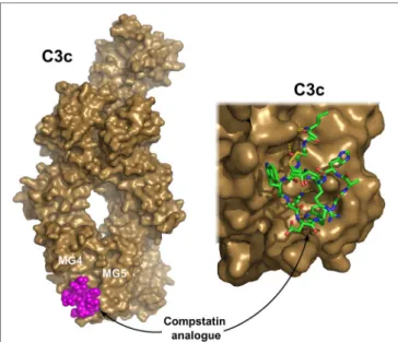
FiGURe 6 | C3c in complex with a compstatin analog. Compstatin, a cyclic peptide, was developed by phage-display. Since its discovery, several modified compstatin analogs have been developed. Compstatin and its analogs bind to C3, C3b, or C3c between the MG4 and MG5 domains.
Compstatin sterically prevents the C3-convertase (C3bBb) to access its substrate C3. The depicted structure was determined using the Ac-V4W/
H9A-NH2 variant of the original peptide. The figure was prepared based on the structure by Janssen et al. (170) (PDB entry 2QKI). On the left, the whole structure is shown with C3c (brown) in surface representation and compstatin (magenta) with spheres. On the right, a close-up of the binding site is shown with compstatin represented by sticks. Hydrogen bonds are indicated by yellow dashed lines.
Bb itself is still just a marginally active enzyme (156), it has full activity only as part of labile C3bBb complex (157). Once Bb dissociates from the convertase complex, it cannot re-associate with C3b (157).
FB is absolutely essential for the AP; therefore, it is a prime target for AP inhibition, but because of its high concentration (about 200–250 µg/mL, or 2–3 µM), it might not seem to be ideal at first sight. On the other hand, in order to prevent AP activa- tion, only the newly formed C3bBb complexes may have to be inhibited. Using a potent inhibitor with low Kd toward C3bBb could completely block the amplification phase, thereby halting the activation process. While C3bBb might be a difficult target for testing small-molecule inhibitors, because of the transient nature of this complex, the cobra venom factor (CVF)-Bb complex is more stable; therefore, it presents itself as a viable target for the development of such molecules. It is also notable that at high pH (proenzymic), FB alone has significant, easily detectable activity toward C3 and certain para-nitroanilide substrates (158). Several substrate-analog aldehyde FB inhibitors were developed along the way (158).
Inhibitory antibodies might be more easily obtained. They only need to prevent access to C3, the very large substrate of C3bBb, which is attainable by a bulky antibody molecule binding near the catalytic site. However, it is possible that such antibody would also bind to free FB; therefore, higher doses might be required. Optimally, a small-molecule inhibitor or an inhibitory antibody should only bind to Bb, or even better only to the C3bBb complex, so that a relatively low dose of the molecule be sufficient for complement inhibition. A blocking antibody, binding to free FB, which prevents the formation of the pro-convertase complex, is also a feasible option. In this case, also high doses would be required for optimal effect.
A set of small-molecule inhibitors are under development by Novartis against FB (CVF-Bb) for indications such as AMD and other complement-mediated diseases (159, 160). Neutralizing monoclonal antibodies against Bb by Novelmed Therapeutics are under development for various indications (161, 162). A mono- clonal antibody to mouse FB has been shown to be protective in a mouse model of renal IRI (163). Other approaches using antisense oligonucleotides (164), or a phage-display selected cyclopeptides (165) are other feasible options to control AP activation through FB.
While FB is a promising target, so far, no therapeutic agent hit the market, or is in the advanced state in clinical trials. As with FD or MASP-3 inhibitors, bacterial infections manifest a potential risk when patients are treated with FB inhibitors.
C3 and CvF
C3 is the central molecule of complement; the three activa- tion routes are merged at the generation of C3b and continue together as the terminal phase (Figures 1 and 3). C3 circulates in the serum at high concentration (4–7 µM; 0.75–1.35 mg/mL).
Native C3 is a 185 kDa protein containing 13 domains (Figure 2).
C3 is composed of two chains, α and β. The core is built up of 8 domains belonging to the α2-macroglobulin family. The thioester domain carries a buried thioester bond, which is prone to suffer hydrolysis or other nucleophilic attack.
Primary C3 deficiencies were described in a few families over the world. Mutation in C3 gene caused impaired C3 synthesis or secretion, which produced a low C3 level in the blood. These indi- viduals are extremely susceptible to recurrent pyogenic bacterial infections, especially to Gram-negative but also to Gram-positive bacteria (166). Moreover, C3 deficiency impairs maturation of immune cells (dendritic cells, memory B cells, certain T cells) (166, 167). Furthermore, SLE and various renal diseases were also observed; however, their mechanism is not fully understood.
Secondary C3 deficiency is due to malfunctioning of the comple- ment regulatory proteins, typically FI and FH (168).
Complement activation can be blocked completely at the level of C3. On the other hand, C3 is the most abundant protein in the complement cascade; therefore, a large amount of an inhibitor would be needed to achieve a substantial effect. Compstatin, a promising complement-based therapeutic agent, was developed against C3 by phage-display using naïve library in 1996 (169).
Compstatin is a cyclic peptide of 13 amino acids with a single disulfide bond. It blocks the access of the convertase to C3 through steric hindrance. Crystal structure with C3c showed that compstatin forms expansive H-bonds with its partner (170) (Figure 6). Neither complement regulator proteins nor other structurally related proteins (C4, C5) bind compstatin.
In the past 20 years, the compstatin family has been constantly developed. New generations of compstatin analogs possess better