Utilization of complement receptors in immune cell – microbe interaction
Szilvia Lukacsi1 , Bernadett Macsik-Valent1, Zsuzsa Nagy-Balo2, Kristof G. Kovacs2, Kristof Kliment3, Zsuzsa Bajtay1,2and Anna Erdei1,2
1 MTA-ELTE Immunology Research Group, E€otv€os Lorand University, Budapest, Hungary 2 Department of Immunology, Eotv€ €os Lorand University, Budapest, Hungary
3 EK-MFA Nanobiosensorics Group, Budapest, Hungary
Correspondence
A. Erdei, Department of Immunology, E€otv€os Lorand University, Pazmany Peter s.
1/C, Budapest H-1117, Hungary Tel:+36 1 3812 175
E-mail: anna.erdei@ttk.elte.hu or anna.erdei@freemail.hu
(Received 6 December 2019, revised 15 January 2020, accepted 16 January 2020, available online 12 February 2020) doi:10.1002/1873-3468.13743 Edited by Hanna Jarva
The complement system is a major humoral component of immunity and is essential for the fast elimination of pathogens invading the body. In addition to its indispensable role in innate immunity, the complement system is also involved in pathogen clearance during the effector phase of adaptive immu- nity. The fastest way of killing the invader is lysis by the membrane attack complex, which is formed by the terminal components of the complement cas- cade. Not all pathogens are lysed however and, if opsonized by a variety of molecules, they undergo phagocytosis and disposal inside immune cells. The most important complement-derived opsonins are C1q, the first component of the classical pathway, MBL, the initiator of the lectin pathway and C3-derived activation fragments, including C3b, iC3b and C3d, which all serve as ligands for their corresponding receptors. In this review, we discuss how complement receptors are utilized by various immune cells to tackle invading microbes, or by pathogens to evade host response.
Keywords:complement activation; complement receptors; complement- derived ligands; pathogen clearance; pathogen escape
The complement system consists of approximately 50 soluble, membrane-bound and regulatory proteins.
Most of the circulating, inactive complement compo- nents are synthesized in the liver, although the local production of complement proteins in tissues has also
been proven to play an important role in several immune processes[1–3].
Activation of the complement cascade can occurvia the classical, the lectin-dependent or the alternative pathway (Box 1).
Abbreviations
AIDS, acquired immune deficiency syndrome; BAD1, blastomyces adhesin 1; cC1q, collagen-like tail region of C1q; CCP, complement con- trol protein; CLR, C-type lectin receptors; CR, complement receptor; CRD, carbohydrate recognition domain; CRIg, complement receptor of the immunoglobulin family; CRT, calreticulin; EBV, Epstein–Barr virus; Ecb, extracellular complement binding protein; GAS, group AStrepto- coccus; gC1q, globular head region of C1q; GPI, glycosylphosphatidylinositol; HBV, hepatitis B virus; HCV, hepatitis C virus; HIV, human immunodeficiency virus; HSV, herpes simplex virus; HTLV-1, human T-lymphotropic virus 1; ICAM-1, intercellular adhesion molecule 1; LAD, leucocyte adhesion deficiency; LPS, lipopolysaccharide; LTA, lipoteichoic acid; Mac, GAS Mac-1-like protein; MAC, membrane attack com- plex; MASP, MBL-associated serine protease; MBL, mannose-binding lectin; MyD88, myeloid differentiation primary response 88; NLR, NOD-like receptor; PfRh4,Plasmodium falciparumreticulocyte-binding-homologue-4; PMN, polymorphonuclear cells; PspA, pneumococcal surface protein A; RCA, regulators of complement activation; Sap, secreted aspartic protease; SARS, severe acute respiratory syndrome;
SCR, short consensus repeat; SLE, systemic lupus erythematosus; TLR, Toll-like receptor; TRIF, TIR-domain-containing adapter-inducing interferon-b;uPAR, urokinase-type plasminogen activator receptor; VSIG4, V-set and Ig domain-containing 4.
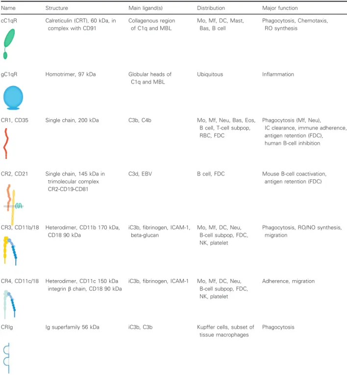
The central event in the activation cascade is the cleav- age of the third component, C3. Its first split products are C3a and C3b. The small, soluble C3a peptide may bind to G-protein-coupled C3a receptors, while the lar- ger activation fragment C3b, containing the exposed, reactive thioester group, has the capacity to bind cova- lently to the activating surface on the pathogen. Cova- lently fixed C3b is the main ligand of CR1 (CD35) and CRIg. C3b can be further processed to generate iC3b and C3d(g), which remain covalently attached to the antigen and interact with complement receptors CR3 (CD11b/CD18), CR4 (CD11c/CD18), CRIg and CR2 (CD21) expressed on several cell types. In addition to the covalently fixed C3- and C4-derived fragments, C1q and MBL are further opsonins (Table1, Fig.1).
Complement receptors are essential to empower immune cells to clear the circulation from invaders[4].
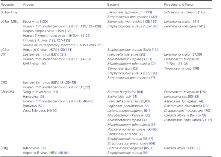
Here, we review data describing the interaction of these receptors with different pathogens – bacteria, viruses, fungi and parasites – mentioning also exam- ples of how these receptors are utilized by pathogens to evade host responses (Table2).
Complement receptor type 1 (CR1, CD35)
Human complement receptor type 1 (CR1, CD35) is expressed on the surface of various myeloid and lym- phoid cells and also on erythrocytes [5]. It is an approximately 200 kDa single-chain transmembrane glycoprotein with a short cytoplasmic tail. In the most common human allotype, the extracellular portion of the molecule is composed of 30 SCRs (Short Consen- sus Repeat), each having 60–70 amino acids. CR1 binds activated fragments of C3 and C4, such as C3b and C4b and with lower affinity, iC3b [6]. It is also an important regulator of the complement cascade, since it possesses decay-accelerating activity for the C3/C5 convertases of both the classical and alternative path- ways. Additionally, it serves as a cofactor for Factor I-mediated cleavage of C3b, thus blocks further activa- tion of the complement cascade[7].
CR1 expressed by phagocytes – macrophages and granulocytes–plays an important role in the phagocy- tosis of C3-opsonized antigens. Its further roles include Box 1. Activation of the complement system
The classical pathway is triggered by C1q, a subunit of the trimolecular C1 complex, upon its reaction with the Fc portion of IgM or IgG antibodies bound to the antigen, or directly, upon recognizing pathogen surfaces and apop- totic cells. This step is followed by the activation of C1r then C1s, the two C1q-associated serine proteases. After cleavage of C2 and C4, the larger split products, C2b and C4b form the classical pathway’s C3 convertase on the sur- face of the activating substance. This, in turn, activates C3, the major and central component of this cascade on which all three complement activation pathways converge.
The lectin pathway is activated by mannose-binding lectin (MBL) and ficolins. These pattern recognition molecules bind to microbial surface oligosaccharides and acetylated residues, and form complexes with MASP1 and MASP2, the MBL-associated serine proteases, which cleave C2 and C4 to generate C2bC4b, the classical C3 convertase.
The alternative pathway is constitutively activated at a low level by the spontaneous hydrolysis of C3 (‘tick-over mechanism’) in body fluids generating C3(H2O). This C3b-like molecule binds factor B, which is cleaved by the serine protease factor D, giving rise to the formation of the initial, fluid-phase C3 convertase (C3(H2O)Bb) of the alternative pathway. This enzyme produces further C3b fragments, resulting in a surface-bound C3bBb, the C3 convertase of the alternative pathway. Several substances can initiate this pathway, including bacterial LPS, zymosan and biomaterials.
Of note, the alternative pathway serves as a strong amplification loop, as it can be triggered also by C3b generated by either the classical or the lectin pathways.
The three pathways converge at the level of the central complement component, C3. Activation of this major com- ponent leads to the generation of several biologically important C3 fragments. C3b, the first, larger cleavage product binds covalently to the activating surface and initiates the assembly of C5 convertase enzymes of either the classical (C4bC2bC3b) or the alternative (C3bBbC3b) pathway. Upon the cleavage of C5 by the C5 convertases, the generated C5b binds C6 followed by the formation of the trimeric C5b-7 complex, which associates with the cell membrane. In the next steps, the tetrameric C5b-8 complex is formed, which allows binding and polymerization of C9 molecules. In the final step, the C5b-9 complex (MAC) inserts into the plasma membrane, which causes cell death by lysis.
Since complement activation can potentially be destructive, fluid-phase and cell membrane-bound regulators con- trol this system to protect host tissues. These regulators of complement activation (RCA) show structural homology, characterized by repeats of complement control protein (CCP) or sushi domains.
transport of soluble antigen–antibody complexes from blood to the liver [8] and inhibition of human B-cell responses[9–12].
The expression of CR1 on human erythrocytes was shown to vary among healthy individuals. The CR1 gene is highly polymorphic, and an RFLP was
identified which correlates with CR1 expression levels on RBCs. A combination of the alleles linked to high (H) and low (L) expression levels – HH, HL and LL, respectively – gives rise to three distinct genotypes with high, intermediate and low expression of CR1 [13].
Table 1.Complement receptors (For references see the text). Mo, monocyte; Mf, macrophage; DC, dendritic cell; Mast, mast cell; Bas, basophil granulocyte; Neu, neutrophil granulocyte; Eos, eosinophil granulocyte; RBC, red blood cell; FDC, follicular dendritic cell; NK, natural killer cell.
Name Structure Main ligand(s) Distribution Major function
cC1qR Calreticulin (CRT), 60 kDa, in complex with CD91
Collagenous region of C1q and MBL
Mo, Mf, DC, Mast, Bas, B cell
Phagocytosis, Chemotaxis, RO synthesis
gC1qR Homotrimer, 97 kDa Globular heads of
C1q and MBL
Ubiquitous Inflammation
CR1, CD35 Single chain, 200 kDa C3b, C4b Mo, Mf, Neu, Bas, Eos,
B cell, T-cell subpop, RBC, FDC
Phagocytosis (Mf, Neu),
IC clearance, immune adherence, antigen retention (FDC), human B-cell inhibition
CR2, CD21 Single chain, 145 kDa in trimolecular complex CR2-CD19-CD81
C3d, EBV B cell, FDC Mouse B-cell coactivation,
antigen retention (FDC)
CR3, CD11b/18 Heterodimer, CD11b 170 kDa, CD18 90 kDa
iC3b, fibrinogen, ICAM-1, beta-glucan
Mo, Mf, DC, Neu, B-cell subpop, FDC, NK, platelet
Phagocytosis, RO/NO synthesis, migration
CR4, CD11c/18 Heterodimer, CD11c 150 kDa integrinbchain, CD18 90 kDa
iC3b, fibrinogen, ICAM-1 Mo, Mf, DC, Neu, B-cell subpop, FDC, NK, platelet
Adherence, migration
CRIg Ig superfamily 56 kDa iC3b, C3b Kupffer cells, subset of
tissue macrophages
Phagocytosis
The abundance of the receptor on the surface of various leucocytes makes these cells a potential tar- get for intracellular pathogens. Phagocytosis – a cru- cial step in pathogen neutralization, regulating inflammation and antigen presentation – can be hijacked by microbes. Pathogens are able to adhere to or enter monocytes, macrophages and neutrophils via phagocytic receptors, while avoiding intracellular enzymatic digestion and subsequently contributing to disease development. Polymorphisms of CR1 have been linked to greater susceptibility to certain infec- tions as well.
Viruses
Interaction between HIV (human immunodeficiency virus) and various immunocytes, as well as red blood cells, suggests a complex pathomechanism. Comple- ment alone can target the virus to erythrocyte CR1, and antibodies only enhanced this effect [14]. Viral adherence was shown to be inhibited in the absence of complement, while undisturbed without IgG. Further- more, blocking CR1 significantly diminished HIV adhesion. These experiments indicate that adherence of the virus to red blood cells is a complement-dependent phenomenon[15].
Fig. 1.Ligand specificity and microbial utilization of complement receptors. C1q may opsonize microbial surfaces directly or indirectly,via binding to the Fc region of bound antibodies. The globular head of C1q may interact with gC1qR, while its collagen-like tail binds to cC1qR.
MBL directly bound to microbial surfaces presumably interacts with cC1qR. Complement activation leads to the deposition of complement fragments that are potent ligands for several complement receptors. The larger C3b fragment is recognized by CR1 and CRIg, the inactivated iC3b fragment interacts with CRIg, CR3 and CR4, whereas C3d is a ligand for CR2. Complement receptors interact with pathogens through the opsonins or by direct binding of microbial components. The viruses, bacteria, fungi and parasites utilizing these receptors are listed below the illustration.
Monocytes and macrophages also contribute to the progression of AIDS, as they express CD4 that HIV uses to enter these cells. In addition, it has been shown that the opsonization of HIV-1 and HIV-2 strains with complement results in higher and earlier productive infection – independently of CD4 – while blocking of CR1 (or CR3) attenuates the enhancing effect of com- plement [16]. The immune complex formation of virus envelope protein-specific antibodies and HIV-1 leads to opsonization and binding to CR1 on K562 leukae- mia-derived cell line[14].
Certain human T lymphocyte subsets express CR1 (and CR2) receptors [17,18], making them suitable for complement-induced immune responses, but on the other hand, they become potential targets for infec- tion. Similarly to monocytes and macrophages, opsonization of HIV-1 with complement leads to ear- lier and enhanced infection of CD4-expressing human T-cell lines in a CR1- or CR2-dependent manner, as blocking either of these receptors diminished the
positive effect of complement[19]. Certain HIV strains require no involvement of CD4 in the process of CR1- and CR2-mediated contagion of T cells. In addition to its crucial role in linking opsonized HIV to T lympho- cytes, CR1 presumably facilitates viral attachment to CR2 by its cofactor activity during cleavage of C3b into smaller fragments that could interact with CR2 [19].
The genotype of CR1 could be a major factor in viral infections. Namely, severe acute respiratory syn- drome (SARS) disease progression differs in patients with or without the high expression genomic type (HH) [20]. In the early stages of SARS disease, the number of CR1 receptors on RBCs decreases signifi- cantly but later returns close to physiological values.
This phenomenon may have an impact on disease pro- gression. While patients bearing HH and HL geno- types showed temporary reduction of CR1 levels, SARS patients with LL genotype had no change in their CR1 expression on erythrocytes, although this
Table 2. List of pathogens binding to various complement receptors.
Receptor Viruses Bacteria Parasites and Fungi
cC1qr–C1q Salmonella typhimurium[133]
Streptococcus pneumoniae[132]
Schistosoma mansoni[140]
cC1qr–MBL Ebola virus[120]
Human immunodeficiency virus (HIV)[119,124–126]
Herpes simplex virus (HSV)[123]
Human T-lymphotropic virus 1 (HTLV-1)[125]
Influenza A virus[122,127–129]
Severe acute respiratory syndrome (SARS-CoV)[121]
Salmonella montevideo[138,139]
Staphylococcus aureus[135–137]
Leishmania major[141]
Leishmania mexicana[141]
gC1qr Hepatitis C virus (HCV)[130,131] Staphylococcus aureus(SpA)[134]
CR1 Epstein–Barr virus (EBV)[21]
Human immunodeficiency virus (HIV)[14–16]
SARS-virus[20]
Francisella tularensis[25]
Mycobacterium leprae[30,31]
Mycobacterium tuberculosis[29]
Salmonella typhi[28]
Staphylococcus aureus(Ecb)[26]
Streptococcus pneumoniae[27]
Leishmania major[37,38]
Plasmodium falciparum (PfRh4)[32–35]
Trypanosoma cruzi[36]
CR2 Epstein–Barr virus (EBV)[21,39–43]
Human immunodeficiency virus (HIV)[19,22]
CR3/CR4 Dengue fever virus[51]
Hantavirus[53]
Human immunodeficiency virus (HIV-1)[46–48]
Rotavirus[52]
West Nile virus[49,50]
Borrelia burgdorferi[59]
Escherichia coli[54]
Francisella tularensis[25,63]
Legionella pneumophila[65]
Listeria monocytogenes[61]
Mycobacterium kansasii[60]
Mycobacterium leprae[30]
Mycobacterium tuberculosis[55]
Porphyromonas gingivalis[66–69]
Salmonella enterica[58]
Staphylococcus aureus[56,57]
Streptococcus pneumoniae[64]
Plasmodium falciparum[78]
Leishmaniassp.[80–82]
Aspergillus fumigatus[74]
Blastomyces dermatitidis[73]
Cryptococcus neoformans[70]
Candida albicans[54,75–78]
Histoplasma capsulatum[71,72]
CRIg Adenovirus[89]
Hepatitis B virus (HBV)[95,96]
Listeria monocytogenes[85,99]
Staphylococcus aureus[85]
Candida albicans[97,98]
might be a result of the small sample size, according to the authors. Thus, the correlation between the geno- type of CR1 and progression of the SARS disease is not clear yet, but the dynamic change in the CR1 numbers during infection suggests its involvement in the process[20].
CR2 (CD21) was formerly considered the sole recep- tor for the major Epstein–Barr virus (EBV) glycopro- tein gp350/220 [21]; however, there are data on EBV patients deficient in CR2 indicating an alternative CR2-independent process for viral entry. Experiments with CD21- B-cell lines showed gp350/220 binding to CR1. In the presence of HLA type II complexes in the membrane, CR1 could mediate infection[22].
Bacteria
It is long known that CR1, in cooperation with CR3, has a major role in the phagocytosis of opsonized par- ticles[23,24]. For instance, Schwartzet al.[25]demon- strated that neutrophils mediate the internalization of opsonizedFrancisella tularensis viaCR1 and CR3.
During bacterial colonization, different ‘strategies’
allow survival and spreading depending on whether the species is extra- or intracellular. For example, eva- sion of phagocytosis can be beneficial for extracellular pathogens, while intracellular organisms can utilize phagocytic receptors for invasion, simultaneously inhibiting cytoplasmic killing mechanisms.
During Staphylococcus aureus infection, bacterial ligand Ecb (extracellular complement binding protein) has been shown to bind C3b and block direct interac- tion between the soluble form of CR1 (sCR1) and C3b. Thus, Ecb reduces cofactor activity of sCR1 in the process of proteolytic inactivation of C3b [26].
CR1 expressed in the membrane of neutrophils can also be utilized by S. aureus where Ecb binds to C3b and prevents CR1 engagement, resulting in impaired phagocytosis[26].
Pneumococcal surface protein A (PspA) has been shown to interfere with complement deposition onto Streptococcus pneumoniae. Results of a mouse study revealed that PspA impairs mouse CR1/2-, as well as CR3-, and CR4-mediated protection against pneumo- coccal infection, since the bacterial protein modulates opsonization by the components of the alternative pathway[27].
Intracellular survival of Salmonella typhi has been observed in human macrophages, and CR1 – along with CR3–was found to be involved in its internaliza- tion. Correlation between CR1 mediated recognition of the bacteria and survival rate has been found as well[28].
Patients with Mycobacterium tuberculosis infection were shown to have more circulating immune com- plexes, a lower expression of CR1 on erythrocytes and a higher prevalence of the HH genotype than in healthy donors. The presence of the H allele of the Cr1 gene may contribute to higher susceptibility to pulmonary tuberculosis[29].
CR1 and CR3 blocking reduced adherence and phagocytosis of Mycobacterium leprae by monocytes [30], while CR1 polymorphisms have also been associ- ated with the infection. Susceptibility to leprosy is affected by the amount of soluble CR1 in blood which competes with cell membrane receptors for pathogen binding, subsequently affecting phagocytosis [31]. The uptake of opsonized M. lepraeis enhanced in the case of a CR1 variant with a hidden cleavage site that results in a lower concentration of sCR1 or a polymor- phism that leads to an elevated level of CR1 produc- tion[31].
Parasites
In the case of malaria, caused by the intracellular par- asite Plasmodium falciparum, CR1 receptors expressed on infected red blood cells can presumably facilitate the infection by rosetting with uninfected erythrocytes [32]. This process correlates with the severity of the disease [33]. CR1 possibly serves as a point of entry for the obligate intracellular pathogen during erythro- cyte invasion. PfRh4 (P. falciparum reticulocyte-bind- ing-homologue-4) is a major Plasmodium ligand associated with sialic acid-independent invasion of red blood cells. Its recognition site has been located at the distal amino terminus of CR1 [34]. Simultaneous engagement of CR1 by C4b and PfRh4 specifically inhibits the receptor’s convertase decay-accelerating activity [34]. Moreover, sCR1 was also shown to bind PfRh4, resulting in the blocking ofPlasmodiumattach- ment to erythrocyte CR1[35], demonstrating that CR1 has a complex role in disease progression.
Soluble CR1 can also be associated with infections with intracellular parasiteTrypanosoma cruzi, responsi- ble for Chagas disease (CD). Patients with chronic CD have a decreased amount of sCR1 compared to healthy controls. Certain haplotypes of CR1 have been linked to augmented risk of T. cruzi infection and developing chronic chagasic cardiomyopathy [36].
CR1 is a major receptor on macrophages for the obligate intracellular protozoa Leishmania major.
Simultaneously blocking CR1 and CR3 significantly decreases pathogen attachment, and CR1 inhibition alone leads to attenuated binding. Data suggest higher survival rate when pathogens enterviaCR1[37,38].
Complement receptor type 2 (CR2, CD21)
In humans, CR2 (CD21) is composed of 16 SCRs, a transmembrane domain and a short cytoplasmic tail.
The protein is encoded by theCR2gene, which is sep- arate from the CR1 receptor coding gene,CR1. Con- trarily, in mice both CR1 and CR2 are encoded by the same gene (Cr2) and alternative splicing gives rise to the two protein products. Thus, mouse CR2 is nearly identical to CR1 both in function and structure. CR2 binds fragment C3d, the final cleavage product of C3.
CR2 has also been shown to serve as a receptor for viruses.
Viruses
A large body of evidence indicates that CR2 mediates EBV infection of human B lymphocytesvia binding of a virus outer membrane glycoprotein, gp350/220 [21,39–43].
C3b-opsonized HIV was shown to bind to erythro- cyte CR1 [14,15] that is followed by the cleavage of C3b to iC3b and C3d. This way C3d-bearing HIV tar- gets CR2-expressing B cells and facilitates the B cell- mediated transmission of opsonized HIV to T cells [19,44].
Complement receptors CR3 (CD11b/
CD18) and CR4 (CD11c/CD18)
Complement receptors CR3 (aMb2, CD11b/CD18, Mac-1) and CR4 (aXb2, CD11c/CD18, p150,95) are members of the b2-integrin family, expressed on most white blood cells. Both receptors bind multiple ligands –for example iC3b, fibrinogen, ICAM-1 or pathogen- related ligands like LPS– and thereby play an impor- tant role in phagocytosis, adherence and migration.
Although it is clear that the two receptors exhibit nonoverlapping functions, comparative studies are barely available [45]. CR3 and CR4 are important phagocytic receptors, leading either effective antimicro- bial responses against pathogens or noninflammatory phagocytosis of apoptotic cells under physiological conditions of a healthy individual. Some pathogens however evolved to hijack these functions of CR3 and CR4. Extracellular pathogens are likely to avoid phagocytosis by these receptors, by blocking their function or even cleaving them from the surface of phagocytes. In contrast, intracellular pathogens often use them as an effective entry route into host cells, thereby causing a more severe infection.
Viruses
As viruses are obligate intracellular pathogens, they can benefit from their ability to exploit complement- mediated phagocytosis. For instance, the opsonization of HIV-1 by complement causes an up to 10-fold higher productive infection of human dendritic cells compared to nonopsonized or only antibody-opsonized virus particles. Complement-dependent HIV infection is mediated by CR3[46], which also modulates the sig- nal transduction of Toll-like receptor 8 (TLR8).
Modulated TLR8 signalling resulted in a lower expres- sion of antiviral and inflammatory factors such as IL- 1b, IL-6, TNF-a, IFN-b, myxovirus resistance protein A and IFN-stimulated genes, leading to enhanced infection[47,48]. In addition, CR3 has been shown to mediate the complement-dependent enhancement of West Nile virus replication in mouse macrophages [49,50], and the infection of human monocytes with the Dengue fever virus [51]. The capsid of Rotavirus contains integrin ligand motif bearing viral peptides, which are shown to bind tob1andb2integrins includ- ing CR4, and thereby promoting viral entry to host cells [52]. A recent experiment suggests that comple- ment receptors CR3 and CR4 also act as Hantavirus entry receptors[53].
Bacteria
As phagocytic receptors, both CR3 and CR4 have antimicrobial roles during the immune response to infections. Both CR3 and CR4 are involved in the uptake and killing ofEscherichia coli[54]or M. tuber- culosis[55]. Out of the two receptors, CR3 was shown to be the dominant mediator of phagocytosis over CR4 in the case of iC3b opsonized S. aureus on human monocytes, neutrophils, monocyte-derived macrophages and dendritic cells [56,57]. CR3 is also involved in the phagocytosis of Salmonella enterica [58], Borrelia burgdorferi[59]or Mycobacterium kansa- sii [60], and has a key role in the effective immune response againstListeria monocytogenes[61].
Phagocytosis via CR3 and CR4 is an effective antimicrobial immune response; however, some patho- gens try to evade it. For instance, Group A Strepto- coccus (GAS), avoids phagocytosis by secreting the CR3 homologue GAS Mac-1-like protein (Mac). The Mac binds to CD16 (FccRIII) on the surface of human polymorphonuclear cells (PMNs) and blocks receptor–antibody interactions as well as the binding of iC3b to CD11b, as CR3 and CD16 are physically and functionally linked. By that Mac inhibits
opsonophagocytosis and also the production of reac- tive oxygen species, thereby decreasing pathogen kill- ing[62].
While some pathogens evolved the ability to avoid phagocytosis, others use this mechanism to enter host cells. As CR3 and CR4 have a main role in noninflam- matory phagocytosis of apoptotic cells, it is an ideal target for intracellular pathogens to exploit for their entry, especially if additional danger signals are blocked by the microbe.
Uptake ofF. tularensis, which is one of the most viru- lent pathogens known, is mediated by CR3 and CR4 in human dendritic cells. The internalization ofF. tularensis is followed by its rapid growth inside cells resulting in cell death [63]. The same receptors are involved in the infection of human macrophages, while in human neu- trophils it is mediated by CR1 and CR3[25].
The RrgA adhesin containing pili ofStr. pneumoniae enhances the CR3 dependent uptake of pneumococci by murine and human macrophages through a direct interaction with CR3. Macrophages harbouring higher numbers of viable bacteria are more likely to be destroyed prior to the complete eradication of the ingested particles. Moreover, the interaction between RrgA and CR3 leads to increased motility and migra- tory behaviour of macrophages, resulting in an earlier onset of septicaemia and a more rapid disease progres- sion[64].
The obligate intracellular pathogen M. leprae invades host cells via phagocytosis and proliferates within mononuclear phagocytes. Both CR3 and CR4 (and CR1) were shown to be involved in that process[30].
The causative agent of Legionnaires’ disease, Legionella pneumophila, multiplies in human mono- cytes and alveolar macrophages. CR1 and CR3 were shown to mediate the adherence of Leg. pneumophila, thereby contributing to the entry and intracellular pro- liferation of the bacteria, as these processes could be inhibited by specific antibodies for CR1 and CR3[65].
Hajishengallis et al. described in detail the immune evasion mechanism of Porphyromonas gingivalis. The fimbriae of this bacteria serve as a ligand for CR3, which mediates its phagocytosis, thus proactively pro- moting its binding and entry into the host cells. The fimbriae activate the high-affinity conformation of CR3, which does not promote the killing but the per- sistence of Po. gingivalis after internalization and induces a selective suppression of IL-12 production [66–68]. Therefore, Po. gingivalis enhances its survival by exploiting CR3, as pharmacological blockade of CR3 promotes its killing and suppressesPo. gingivalis- induced periodontal bone loss in a mouse model [66,69].
Fungi
CR3 and CR4 were shown to be involved in the bind- ing of Cryptococcus neoformans [70]and are dominant receptors in the uptake and killing of Candida albicans [54]. However, the fungal pathogenHistoplasma capsu- latum evades antimicrobial defences and proliferates intracellularly in macrophages infected through CR3, CR4 and LFA-1. The host macrophages are destroyed by the multiplying yeast, and the released microbes are phagocytosed by other macrophages attracted to the infected site [71]. The major H. capsulatum ligand for CR3 on macrophages was identified as heat shock pro- tein 60 (hsp60), whereas dendritic cells recognize it via a different ligand [72]. Similar to H. capsulatum, the related dimorphic fungal pathogen Blastomyces der- matitidis also expresses a CR3-interacting protein, BAD1 (blastomyces adhesin 1). BAD1 helps pathogen survival by binding via CR3 and CD14 that mediates its internalization and the suppression of TNF-a pro- duction of host cells[73].
The fungal pathogen Aspergillus fumigatus avoids opsonization and phagocytosis by expressing proteases that degrade complement proteins and CR3 [74]. As surface-bound factor H can enhance the antifungal activity via binding to CR3 and CR4, C. albicans avoid phagocytosis by releasing the secreted aspartic protease 2 (Sap2) to cleave both FH and the comple- ment receptors CR3 and CR4 on macrophages [75].
Additionally, C. albicans expresses a CR3-like struc- ture that mediates adhesion of the yeast to human endothelium[76–78].
Parasites
CR3 and CR4 also contribute to the phagocytosis of P. falciparum-infected erythrocytes [79], and Leishma- nia ssp. are able to enter and survive in host macro- phages in a CR3-mediated manner [80,81]. Moreover, Leishmania is known to inhibit IL-12 production in macrophages [82], which is also mediated by CR3, as signalling via CR3 by L. major reduces IL-12 produc- tion [83]. At the same time, dendritic cells were shown to take up L. major in a CR3-independent, FccRI- and FccRIII-mediated manner, which leads to a more effective antigen presentation, indicating that CR3-me- diated uptake is likely to represent a ‘decoy’ mecha- nism for this pathogen [84].
CRIg
The complement receptor immunoglobulin [CRIg, also known as V-set and Ig domain-containing 4 (VSIG4)
and Z39Ig] is a phagocytic receptor expressed on macrophage subpopulations. First, Helmy et al.
proved the expression of CRIg in CD68+Kupffer cells in the liver, interstitial macrophages in the heart, adre- nal gland macrophages, alveolar macrophages, Hof- bauer cells, synovial macrophages and lamina propria histiocytes by immunohistochemistry [85]. In dendritic cells, two groups showed the expression of CRIg, but only at the mRNA level [86,87]. CRIg belongs to the immunoglobulin (Ig) superfamily. In humans, it has two splice variants. The long form (huCRIg(L)) con- tains a V- and a C2-type Ig domain, and a short form (huCRIg(S)) encodes only a V-type Ig domain [88].
The murine muCRIg receptor comprises only of a sin- gle V-type Ig domain[85].
CRIg binds C3b and iC3b, providing the first line of defence in the liver and spleen by quickly eliminating opsonized pathogens. This receptor was shown to swiftly resurface through recycling endosomes after internalization, thus providing the means for a contin- uous phagocytosis [85]. In contrast to CR3 and CR4, which also bind iC3b-opsonized particles, CRIg is regarded to have an anti-inflammatory role as well. It is hypothesized that Kupffer cells internalize opsonized microbes and apoptotic cells first through CRIg, with- out the induction of inflammation [89,90]. A higher number of microbial agents in the blood will prompt the engagement of other pattern recognition and com- plement receptors, leading to the initiation of an immune response including leucocyte recruitment and inflammation.
CRIg is additionally involved in the promotion of immunological tolerance through the inhibition of the alternative complement pathway convertases [91,92]
and the suppression of T-cell activation. However, the tolerogenic function of CRIg might support the pro- gression of cancer with keeping T cells in an unrespon- sive state [93,94]. Recently, the downregulation of CRIg in chronic Hepatitis B virus (HBV) infection was shown to lead to a poor prognosis in hepatocellular carcinoma patients probably due to reduced virus clearance[95,96].
Studies on the CRIg-mediated phagocytosis proved that this receptor is indispensable in the rapid internal- ization of complement opsonized Adenovirus particles [89],S. aureus [85],Li. monocytogenes[85]andC. albi- cans [97,98]. The intracellular pathogen Li. monocyto- genes survive inside macrophages by delaying phagosome maturation and escaping into the cyto- plasm [99]. Kim et al. proved a multistep counter mechanism involving the engagement of CRIg. Sig- nalling through CRIg facilitates phagosome acidifica- tion and fusion with lysosomes, enhancing the killing
of internalized bacteria [100]. In addition, the ligation of CRIg with opsonizedLi. monocytogenesor an ago- nistic mAb induces autophagosome formation, enabling macrophages to eliminate cytoplasmic bacte- ria already escaped from the phagolysosome system [101].
Zenget al.[102]proposed that the CRIg receptor is able to clear bacteria directly without opsonization, through the recognition of the gram-positive wall con- stituent, lipoteichoic acid (LTA). However, Broadley et al. [103] proved in a C3 knockout mouse, that Kupffer cells still internalize both gram-positive and gram-negative bacteria strains, but instead of CRIg, they use pattern recognition receptors, that is scav- enger receptors for LTA. Further studies are required to clarify the individual participation of complement and pattern recognition receptors expressed by Kupffer cells, with consideration of the shear stress conditions present in the liver[104].
Receptors for C1q and MBL
C1q and MBL are soluble recognition molecules, which may serve as opsonins. Their structure is described briefly in Box 2. Binding of these comple- ment proteins to pathogens may either mediate their direct uptake via their receptors expressed by phago- cytes – including dendritic cells, macrophages and PMNs, or may initiate the complement cascade, result- ing in the fixation of C3- and C4-derived fragments, leading to an enhanced engulfment of the microbe[5].
The attachment of C1q or MBL to microbial sur- faces causes conformational changes in these mole- cules, which subsequently initiate functions such as complement activation and interaction with C1q or MBL binding molecules leading to the uptake of the microbe. MBL can act as a direct opsonin for microbes through interaction with a cellular receptor or binding protein [108,109]. The immunological sig- nificance of MBL as an opsonin was established in studies of MBL-deficient children with an opsonic defect[110].
Several cell membrane molecules have been shown to increase the binding of collectins and the struc- turally related C1q, including CD93 (C1qRp), CD35 (CR1) and cC1qR (calreticulin, CRT) in complex with CD91. Receptors for both the collagen-like tail (cC1qR) and globular head regions (gC1qR) of C1q, as well as receptors for MBL, have been proposed (Table 1, Fig. 1) [111–117]. The best-accepted candi- date is cC1qR, a protein nearly identical with CRT, which is in complex with CD91. CRT, a chaperone and Ca2+-binding, ubiquitous intracellular protein, is
found in the membrane of cellular organelles, on the cell surface, and it can also be released as a soluble protein. It is unclear how CRT may become associated with the cell surface, but together with CD91, surface CRT is involved in the uptake and removal of cell remnants when opsonized with C1q [117]. gC1qR is specific to the globular head region, gC1q [115].
C1qRp–also known as the AA4 antigen in rodents – enhances the uptake of C1q-opsonized particles and is recognized by antibodies against CD93[118].
In the next paragraphs, the biological consequences of the interaction of C1q and MBL and their receptors with various pathogens are summarized.
Viruses
MBL was shown to bind to HIV, SARS-CoV, Ebola, Herpes simplex virus (HSV) and influenza virus. Sub- sequent conformational changes of MBL were demon- strated to allow the molecule to initiate viral neutralization or kill them via complement activation and opsonization[119–123].
The direct interaction of C1q and MBL was studied in detail in the case of HIV [124] and Human T-lymphotropic virus 1 (HTLV-1) [125,126]. The
importance of MBL in opsonizing HIV was proved by the finding that the uptake of the opsonized virus by tissue macrophages leads to clearance of the virus from the blood[119].
Binding of MBL to Influenza A virus was shown to involve the CRD domain of the molecule and man- nose oligosaccharides of the viral haemagglutinin and neuraminidase [127–129]. Interestingly, this interaction was found to result in virus inactivation in a comple- ment activation independent way[129].
Hepatitis C virus core protein interaction with the gC1q receptor can contribute to the pathogenesis of multiple diseases associated with HCV infection [130]. Waggoner and colleagues demonstrated that binding of the HCV core protein to gC1qR on human monocyte-derived dendritic cells inhibited TLR-induced IL-12 production but not the produc- tion of other TLR-induced cytokines [131]. In addi- tion, HCV core protein engagement of gC1qR on dendritic cells promoted the production of Th2 cytokines such as IL-4 by cocultured CD4+ T cells.
These results suggest that the engagement of gC1qR on dendritic cells by HCV limits the induction of a Th1 response [131].
Bacteria
Agarwal et al. [132] demonstrated that the adherence of Str. pneumoniae is facilitated by the interaction of C1q collagen region with cC1qR resulting in an enhanced invasion of host epithelial and endothelial cells.
Using recombinant forms of the globular head regions of C1q, it was found that LPS derived from Salmonella typhimurium interacts specifically with the B-chain of the gC1q domain in a calcium-dependent manner. Since the LPS and the IgG-binding sites are overlapping, binding of the bacterium can modulate classical pathway activation[133].
In the case of S. aureus, the staphylococcal protein A (SpA) has been shown to bind to the gC1qR, which is highly expressed on the surface of activated platelets and endothelial cells [134]. The bacterium itself, however, was shown to bind MBL via peptido- glycan and lipoteichoic acid [135,136], and MBL was found to direct the bacterium to the phagosome [137].
It has been described that MBL served alone as an opsonin in the phagocytosis of Salmonella montevideo by PMNs [138]. However, when MBL binds to CR1, it cooperates with FccRs in the process of the phagocy- tosis of Sa. montevideo by PMNs, as described by Ghiranet al.[139].
Box 2. Structure of C1q and MBL complexes C1q, a subunit of the C1 complex, is a 460 kDa hex- americ glycoprotein containing 18 polypeptide chains (A, B and C), which build up 6 identical units. The tulip-like structure possesses a globular head region (gC1q), a neck region and a collagen-like tail (cC1q) [105]. The globular heads bind to the Fc region of antigen-bound IgG or IgM, and it can also recognize various structures present on self, nonself and altered self molecules. In addition to these capacities, C1q has a distinguished role in clearing apoptotic cells, and therefore, C1q deficiency may provoke autoimmunity like SLE[106].
MBL, the pattern recognition molecule of the lectin pathway, is composed of three identical monomeric subunits of 32 kDa glycoproteins, forming 3–6 tri- mers. The bouquet-like structure of this collectin is mainly held togetherviadisulphide bonds, similarly to that of complement C1q. Each polypeptide consists of a carbohydrate recognition domain (CRD), a neck domain and a collagen domain. MBL, a member of the collectin family, is important in host defence, espe- cially in early childhood, when the adaptive immune response has not fully developed[107].
Parasites
C1q has been proven to enhance eosinophil mediated killing of schistosomula of Schistosoma mansoni [140].
Binding of MBL toL. majorandLeishmania mexicana promastigotes by mannose-containing lipophosphogly- can was described by Greenet al.[141]They suggested that MBL may opsonize the major developmental stages ofLeishmaniaparasites.
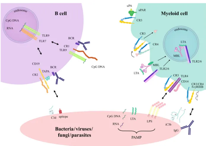
Crosstalk of complement receptors with other cell-membrane proteins Pathogens invading the body may get opsonized by plasma proteins other than complement – including antibodies, pentraxins [129] and fibronectin – further- more, microbes contain a wide variety of PAMPS. All of these factors allow the interaction of the pathogens with more than one receptor on the interacting immune cell, providing an additional level of regula- tion. Still, in most studies only the effect of single receptor–ligand interactions is dissected (Fig.2).
In addition to complement proteins, pathogen pat- terns can be recognized by several innate receptor fam- ilies, such as C-type lectin receptors (CLR), NOD-like receptors (NLR) or Toll-like receptors (TLR). The complex microbial surfaces offer multiple binding sites for these receptors, which modulate host-cell response.
Both synergistic and antagonistic interactions have been described for complement receptors and TLRs [142].
In the next paragraph, some examples of the cooper- ation between complement receptors and other sensors of nonopsonized and Ig-opsonized microbes are described briefly.
CR1-BCR
Simultaneous engagement of CR1 and BCR via com- plement opsonized antigens can significantly alter B- cell responses. Previously, our group was the first to show that in human system CR1 is a potent inhibitor of BCR-dependent B-cell activation– such as prolifer- ation, cytokine and antibody production of cells – in both physiological and pathological conditions [9–12].
Since then the inhibitory function of CR1 was con- firmed by others, using in vivo model systems as well [143–145].
CR1-TLR7, TLR9-BCR
In physiological and pathological conditions, when both complement and TLR activating microbial
products are present in the B-cell environment, the interaction between the two innate sensory systems has the potential to fundamentally alter or fine-tune B-cell responses. It has been shown recently that CR1 clus- tering has no effect on the TLR7-induced activation of tonsillar B cells. However, the TLR9-dependent responses of these cells were significantly and dose- dependently reduced by CR1 ligation [12]. These observations highlight diverse mechanisms of the inter- play between CR1 and TLR7 or CR1 and TLR9, in regulating humoral immune responses.
CR2-BCR
In mouse system, it is well accepted that C3d-op- sonized antigens, which crosslink CR2 with the BCR, augment antibody production by several order of mag- nitudes. Therefore, C3d is known as a molecular adju- vant with a capacity to bridge innate and adaptive immune responses [146]. However, in the case of human B cells the enhancement of antibody produc- tion by the C3-derived ligand has not been proven so far.
CR3-FccRII, FccRIII
Zhou and Brown reported that human neutrophils pla- ted on a surface coated with both anti-CR3 and anti- FccRIII antibodies, exert a strong respiratory burst.
They showed that the lectin-like site of CR3, which is sensitive to saccharides, binds to the extensively glyco- sylated FccRIIIB molecule. Furthermore, it has also been demonstrated that coligation of FccRIII is required for the tyrosine phosphorylation of FccRII.
Thus, the cooperation of these three receptors leads to the FccRII-dependent assembly of the NADPH oxi- dase[147].
CD14-TLR4-CR3
Hawley et al. [59] demonstrated that CD14, the GPI- linked LPS receptor expressed by phagocytes and CR3 are not associated under resting conditions; however, their interaction is rapidly induced in the presence of LPS, when they colocalize within lipid rafts.
Pereraet al.[148]observed a combined participation of CR3, CD14 and TLR4 in the response to LPS and taxol, suggesting the formation of a receptor complex of these molecules on the surface of mouse macro- phages. Later Hanet al.[149] showed that TLR4 sig- nalling activates CR3, which in turn provides negative feedback by enhancing the phosphorylation and subse- quent degradation of TLR signalling molecules,
MyD88 and TRIF. This inhibitory effect of CR3 was also proved for the TLR7/8-induced inflammatory response on human macrophages[150].
uPAR-CR3
The most studied interaction between integrins and GPI-linked proteins involves the urokinase-type plas- minogen activator receptor (uPAR; CD87) [151]. This receptor is expressed on a wide variety of cell types, including neutrophils and activated monocytes. uPAR- mediated calcium signalling was observed in the pres- ence of CR3, while it did not occur in cells of leuco- cyte adhesion deficiency (LAD) patients or in normal neutrophils treated with anti-CR3 mAb. It has also been shown that complex formation with uPAR facili- tates the adhesive functions of CR3[152].
CR3-CR4
Our group found a division of work between CR3 and CR4 in the process of the phagocytosis of iC3b-op- sonized S. aureus. In the case of monocyte-derived macrophages, we observed that blocking CR4 only decreased the amount of surface-bound particles, whereas internalization and digestion of the particles were dependent on CR3. While CR4 participates in the binding of iC3b-opsonized S. aureus, further steps leading to the digestion of the coccus are mediated by CR3[56].
MBL-TLR2
MBL binds to the surface of S. aureusthrough lipotei- choic acid (LTA) that is also the ligand of TLR2/6. Ip et al. [137] showed that the presence of S. aureus
Fig. 2.Crosstalk between complement receptors and other host-cell membrane proteins induced by encountering pathogens. Crosstalk through direct association orviasignalling cascade intersection has been described for complement receptors. The presence of pathogen-associated molecular patterns (PAMP) and opsonins on microbial surfaces promotes the simultaneous engagement of pattern recognition, complement, Fc- receptors and the BCR. CR3 may associate with FccRIIIB and uPAR, and in the presence of LPS, it forms a complex with TLR4 and CD14. In macrophages, there is a division of labour between CR3 and CR4 in the binding and internalization of iC3b-opsonized particles. MBL and TLR2/6 both bind the bacterial lipoteichoic acid (LTA) and become associated in the phagosomes after internalization. The cooperation between CR1 and TLR7 or TLR9 inhibits human B-cell functions. In mouse, C3d-opsonized antigens crosslink CR2 with the BCR, which augments B-cell activation.
induced the association of MBL and TLR2 in phago- somes, and this interaction resulted in an enhanced inflammatory response.
Conclusion
The complement system is a major component of innate immunity, which contributes to the maintenance of host homeostasis. The activation of the complement cascade generates various biologically active comple- ment protein-derived polypeptides that opsonize pathogens. Complement receptors interacting with these polypeptides are expressed by several cell types– including monocytes, macrophages, dendritic cells, neutrophil granulocytes, and T and B lymphocytes.
Engagement of the receptors leads to a wide array of responses, in many cases leading to the elimination of viruses, bacteria, fungi and parasites. On the other hand, pathogens utilize the same receptors to evade recognition and elimination. Learning more about the interaction and crosstalk between cells and microbes will help develop vaccines and treatment of infections.
Acknowledgements
This work was supported by the Hungarian National Science Fund (OTKA) grant K112011 and by the Hungarian Academy of Sciences.
References
1 Colten HR, Strunk RC, Perlmutter DH and Cole FS (1986) Regulation of complement protein-biosynthesis in mononuclear phagocytes.Ciba Found Symp118, 141–154.
2 Morgan BP and Gasque P (1997) Extrahepatic complement biosynthesis: where, when and why?Clin Exp Immunol107, 1–7.
3 Lubbers R, van Essen MF, van Kooten C and Trouw LA (2017) Production of complement components by cells of the immune system.Clin Exp Immunol188, 183–194.
4 Lambris JD, Ricklin D and Geisbrecht BV (2008) Complement evasion by human pathogens.Nat Rev Microbiol6, 132–142.
5 Geertsma MF, Nibbering PH, Haagsman HP, Daha MR and van Furth R (1994) Binding of surfactant protein A to C1q receptors mediates phagocytosis of Staphylococcus aureusby monocytes.Am J Physiol 267, L578–L584.
6 Klickstein LB, Bartow TJ, Miletic V, Rabson LD, Smith JA and Fearon DT (1988) Identification of
distinct C3b and C4b recognition sites in the human C3b/C4b receptor (CR1, CD35) by deletion mutagenesis.J Exp Med168, 1699–1717.
7 Erdei A, Isaak A, Torok K, Sandor N, Kremlitzka M, Prechl J and Bajtay Z (2009) Expression and role of CR1 and CR2 on B and T lymphocytes under physiological and autoimmune conditions.Mol Immunol46, 2767–2773.
8 Cornacoff JB, Hebert LA, Smead WL, VanAman ME, Birmingham DJ and Waxman FJ (1983) Primate erythrocyte-immune complex-clearing mechanism.
J Clin Invest71, 236–247.
9 Jozsi M, Prechl J, Bajtay Z and Erdei A (2002) Complement receptor type 1 (CD35) mediates inhibitory signals in human B lymphocytes.J Immunol 168, 2782–2788.
10 Kremlitzka M, Polgar A, Fulop L, Kiss E, Poor G and Erdei A (2013) Complement receptor type 1 (CR1, CD35) is a potent inhibitor of B-cell functions in rheumatoid arthritis patients.Int Immunol25, 25–33.
11 Kremlitzka M, Macsik-Valent B, Polgar A, Kiss E, Poor G and Erdei A (2016) Complement receptor type 1 suppresses human B cell functions in SLE patients.
J Immunol Res2016, 5758192.
12 Macsik-Valent B, Nagy K, Fazekas L and Erdei A (2019) Complement receptor type 1 (CR1, CD35), the inhibitor of BCR-mediated human B cell activation, differentially regulates TLR7, and TLR9 induced responses.Front Immunol10, 1493.
13 Wilson JG, Murphy EE, Wong WW, Klickstein LB, Weis JH and Fearon DT (1986) Identification of a restriction fragment length polymorphism by a CR1 cDNA that correlates with the number of CR1 on erythrocytes.J Exp Med164, 50–59.
14 Montefiori DC, Graham BS, Zhou JY, Zhou JT and Ahearn JM (1994) Binding of human
immunodeficiency virus type 1 to the C3b/C4b receptor CR1 (CD35) and red blood cells in the presence of envelope-specific antibodies and complement. National Institutes of Health AIDS Vaccine Clinical Trials Networks.J Infect Dis170, 429–432.
15 Horakova E, Gasser O, Sadallah S, Inal JM, Bourgeois G, Ziekau I, Klimkait T and Schifferli JA (2004) Complement mediates the binding
of HIV to erythrocytes. J Immunol173, 4236–4241.
16 Thieblemont N, Haeffner-Cavaillon N, Ledur A, L’Age-Stehr J, Ziegler-Heitbrock HW and Kazatchkine MD (1993) CR1 (CD35) and CR3 (CD11b/CD18) mediate infection of human monocytes and monocytic cell lines with complement-opsonized HIV independently of CD4.Clin Exp Immunol92, 106–113.
17 Fischer E, Delibrias C and Kazatchkine MD (1991) Expression of CR2 (the C3dg/EBV receptor, CD21) on normal human peripheral blood T lymphocytes.J Immunol146, 865–869.
18 Wilson JG, Tedder TF and Fearon DT (1983) Characterization of human T lymphocytes that express the C3b receptor.J Immunol131, 684–689.
19 Delibrias CC, Kazatchkine MD and Fischer E (1993) Evidence for the role of CR1 (CD35), in addition to CR2 (CD21), in facilitating infection of human T cells with opsonized HIV.Scand J Immunol38, 183–189.
20 Wang FS, Chu FL, Jin L, Li YG, Zhang Z, Xu D, Shi M, Wu H and Moulds JM (2005) Acquired but reversible loss of erythrocyte complement receptor 1 (CR1, CD35) and its longitudinal alteration in patients with severe acute respiratory syndrome.Clin Exp Immunol139, 112–119.
21 Fingeroth JD, Weis JJ, Tedder TF, Strominger JL, Biro PA and Fearon DT (1984) Epstein-Barr virus receptor of human B lymphocytes is the C3d receptor CR2.Proc Natl Acad Sci USA81, 4510–4514.
22 Ogembo JG, Kannan L, Ghiran I, Nicholson-Weller A, Finberg RW, Tsokos GC and Fingeroth JD (2013) Human complement receptor type 1/CD35 is an Epstein-Barr virus receptor.Cell Rep3, 371–385.
23 Newman SL, Becker S and Halme J (1985)
Phagocytosis by receptors for C3b (CR1), iC3b (CR3), and IgG (Fc) on human peritoneal macrophages.J Leukoc Biol38, 267–278.
24 Fallman M, Andersson R and Andersson T (1993) Signaling properties of CR3 (CD11b/CD18) and CR1 (CD35) in relation to phagocytosis of complement- opsonized particles.J Immunol151, 330–338.
25 Schwartz JT, Barker JH, Long ME, Kaufman J, McCracken J and Allen LA (2012) Natural IgM mediates complement-dependent uptake ofFrancisella tularensisby human neutrophils via complement receptors 1 and 3 in nonimmune serum.J Immunol 189, 3064–3077.
26 Amdahl H, Haapasalo K, Tan L, Meri T, Kuusela PI, van Strijp JA, Rooijakkers S and Jokiranta TS (2017) Staphylococcal protein Ecb impairs complement receptor-1 mediated recognition of opsonized bacteria.
PLoS ONE12, e0172675.
27 Ren B, McCrory MA, Pass C, Bullard DC, Ballantyne CM, Xu Y, Briles DE and Szalai AJ (2004) The virulence function ofStreptococcus pneumoniaesurface protein A involves inhibition of complement activation and impairment of complement receptor-mediated protection.J Immunol173, 7506–7512.
28 Ishibashi Y and Arai T (1996) A possible mechanism for host-specific pathogenesis ofSalmonellaserovars.
Microb Pathog21, 435–446.
29 Senbagavalli P, Geetha ST, Karunakaran K, Banu Rekha VV, Venkatesan P and Ramanathan VD (2008)
Reduced erythrocyte CR1 levels in patients with pulmonary tuberculosis is an acquired phenomenon.
Clin Immunol128, 109–115.
30 Schlesinger LS and Horwitz MA (1990) Phagocytosis of leprosy bacilli is mediated by complement receptors CR1 and CR3 on human monocytes and complement component C3 in serum.J Clin Invest85, 1304–1314.
31 Kretzschmar GC, Oliveira LC, Nisihara RM, Velavan TP, Stinghen ST, Stahlke ERS, Petzl-Erler ML, Messias-Reason IJT and Boldt ABW (2018)
Complement receptor 1 (CR1, CD35) association with susceptibility to leprosy.PLoS Negl Trop Dis12, e0006705.
32 Stoute JA (2011) Complement receptor 1 and malaria.
Cell Microbiol13, 1441–1450.
33 Rowe A, Obeiro J, Newbold CI and Marsh K (1995) Plasmodium falciparumrosetting is associated with malaria severity in Kenya.Infect Immun63, 2323– 2326.
34 Tham WH, Schmidt CQ, Hauhart RE, Guariento M, Tetteh-Quarcoo PB, Lopaticki S, Atkinson JP, Barlow PN and Cowman AF (2011)Plasmodium falciparum uses a key functional site in complement receptor type- 1 for invasion of human erythrocytes.Blood118, 1923–1933.
35 Tham WH, Wilson DW, Lopaticki S, Schmidt CQ, Tetteh-Quarcoo PB, Barlow PN, Richard D, Corbin JE, Beeson JG and Cowman AF (2010) Complement receptor 1 is the host erythrocyte receptor for Plasmodium falciparumPfRh4 invasion ligand.Proc Natl Acad Sci USA107, 17327–17332.
36 Sandri TL, Lidani KCF, Andrade FA, Meyer CG, Kremsner PG, de Messias-Reason IJ and Velavan TP (2018) Human complement receptor type 1 (CR1) protein levels and genetic variants in chronic Chagas disease.Sci Rep8, 526.
37 Da Silva RP, Hall BF, Joiner KA and Sacks DL (1989) CR1, the C3b receptor, mediates binding of infectiveLeishmania majormetacyclic promastigotes to human macrophages.J Immunol143, 617–622.
38 Rosenthal LA, Sutterwala FS, Kehrli ME and Mosser DM (1996)Leishmania major-human macrophage interactions: cooperation between Mac-1 (CD11b/
CD18) and complement receptor type 1 (CD35) in promastigote adhesion.Infect Immun64, 2206–2215.
39 Nemerow GR and Cooper NR (1984) Early events in the infection of human B lymphocytes by Epstein-Barr virus: the internalization process.Virology132, 186– 198.
40 Frade R, Myones BL, Barel M, Krikorian L, Charriaut C and Ross GD (1985) gp140, a C3b- binding membrane component of lymphocytes, is the B cell C3dg/C3d receptor (CR2) and is distinct from the neutrophil C3dg receptor (CR4).Eur J Immunol15, 1192–1197.
41 Tanner J, Weis J, Fearon D, Whang Y and Kieff E (1987) Epstein-Barr virus gp350/220 binding to the B lymphocyte C3d receptor mediates adsorption, capping, and endocytosis.Cell50, 203–213.
42 Tanner J, Whang Y, Sample J, Sears A and Kieff E (1988) Soluble gp350/220 and deletion mutant glycoproteins block Epstein-Barr virus adsorption to lymphocytes.J Virol62, 4452–4464.
43 Nemerow GR, McNaughton ME and Cooper NR (1985) Monoclonal antibody to the Epstein-Barr virus receptor induces human B lymphocyte activation and differentiation.Trans Assoc Am Physicians98, 290– 300.
44 Banki Z, Wilflingseder D, Ammann CG, Pruenster M, M€ullauer B, Holl€ander K, Meyer M, Sprinzl GM, van Lunzen J, Stellbrink H-Jet al. (2006) Factor I- mediated processing of complement fragments on HIV immune complexes targets HIV to CR2-expressing B cells and facilitates B cell-mediated transmission of opsonized HIV to T cells.J Immunol177, 3469–3476.
45 Erdei A, Lukacsi S, Macsik-Valent B, Nagy-Balo Z, Kurucz I and Bajtay Z (2019) Non-identical twins:
different faces of CR3 and CR4 in myeloid and lymphoid cells of mice and men.Semin Cell Dev Biol 85, 110–121.
46 Bajtay Z, Speth C, Erdei A and Dierich MP (2004) Cutting edge: productive HIV-1 infection of dendritic cells via complement receptor type 3 (CR3, CD11b/
CD18).J Immunol173, 4775–4778.
47 Bouhlal H, Galon J, Kazatchkine MD, Fridman WH, Sautes-Fridman C and Haeffner Cavaillon N (2001) Soluble CD16 inhibits CR3 (CD11b/CD18)-mediated infection of monocytes/macrophages by opsonized primary R5 HIV-1.J Immunol166, 3377–3383.
48 Ellegard R, Crisci E, Burgener A, Sj€owall C, Birse K, Westmacott G, Hinkula J, Lifson JD and Larsson M (2014) Complement opsonization of HIV-1 results in decreased antiviral and inflammatory responses in immature dendritic cells via CR3.J Immunol193, 4590–4601.
49 Cardosa MJ, Gordon S, Hirsch S, Springer TA and Porterfield JS (1986) Interaction of West Nile virus with primary murine macrophages: role of cell activation and receptors for antibody and complement.
J Virol57, 952–959.
50 Cardosa MJ, Porterfield JS and Gordon S (1983) Complement receptor mediates enhanced flavivirus replication in macrophages.J Exp Med158, 258–263.
51 Marinho CF, Azeredo EL, Torrentes-Carvalho A, Marins-Dos-Santos A, Kubelka CF, de Souza LJ, Cunha RV and de-Oliveira-Pinto LM (2014) Down- regulation of complement receptors on the surface of host monocyte even as in vitro complement pathway blocking interferes in dengue infection.PLoS ONE9, e102014.
52 Coulson BS, Londrigan SL and Lee DJ (1997) Rotavirus contains integrin ligand sequences and a disintegrin-like domain that are implicated in virus entry into cells.Proc Natl Acad Sci USA94, 5389– 5394.
53 Raftery MJ, Lalwani P, Krautkrӓmer E, Peters T, Scharffetter-Kochanek K, Kruger R, Hofmann J,€ Seeger K, Kr€uger DH and Sch€onrich G (2014) beta2 integrin mediates hantavirus-induced release of neutrophil extracellular traps.J Exp Med211, 1485– 1497.
54 Jawhara S, Pluskota E, Cao W, Plow EF and Soloviev DA (2017) Distinct effects of integrins alphaXbeta2 and alphaMbeta2 on leukocyte subpopulations during inflammation and antimicrobial responses.Infect Immun85, e00644-16.
55 Hirsch CS, Ellner JJ, Russell DG and Rich EA (1994) Complement receptor-mediated uptake and tumor necrosis factor-alpha-mediated growth inhibition of Mycobacterium tuberculosisby human alveolar macrophages.J Immunol152, 743–753.
56 Lukacsi S, Nagy-Balo Z, Erdei A, Sandor N and Bajtay Z (2017) The role of CR3 (CD11b/CD18) and CR4 (CD11c/CD18) in complement-mediated phagocytosis and podosome formation by human phagocytes.Immunol Lett189, 64–72.
57 Sandor N, Kristof K, Parej K, Pap D, Erdei A and Bajtay Z (2013) CR3 is the dominant phagocytotic complement receptor on human dendritic cells.
Immunobiology218, 652–663.
58 van Bruggen R, Zweers D, van Diepen A, van Dissel JT, Roos D, Verhoeven AJ and Kuijpers TW (2007) Complement receptor 3 and Toll-like receptor 4 act sequentially in uptake and intracellular killing of unopsonizedSalmonella entericaserovarTyphimurium by human neutrophils.Infect Immun75, 2655–2660.
59 Hawley Kl, Olson CM, Iglesias-Pedraz JM, Navasa N, Cervantes Jl, Caimano MJ, Izadi H, Ingalls RR, Pal U, Salazar JCet al. (2012) CD14 cooperates with complement receptor 3 to mediate MyD88-independent phagocytosis ofBorrelia burgdorferi.Proc Natl Acad Sci USA109, 1228–1232.
60 Peyron P, Bordier C, N’Diaye EN and Maridonneau- Parini I (2000) Nonopsonic phagocytosis of
Mycobacterium kansasiiby human neutrophils depends on cholesterol and is mediated by CR3 associated with glycosylphosphatidylinositol-anchored proteins.J Immunol165, 5186–5191.
61 Gluschko A, Herb M, Wiegmann K, Krut O, Neiss WF, Utermohlen O, Kronke M and Schramm M (2018) The beta2 integrin Mac-1 induces protective LC3-associated phagocytosis ofListeria
monocytogenes.Cell Host Microbe23, 324–337, e5.
62 Lei B, DeLeo FR, Hoe NP, Graham MR, Mackie SM, Cole RL, Liu M, Hill HR, Low DE, Federle MJ
et al. (2001) Evasion of human innate and acquired immunity by a bacterial homolog of CD11b that inhibits opsonophagocytosis.Nat Med7, 1298–1305.
63 Ben Nasr A, Haithcoat J, Masterson JE, Gunn JS, Eaves-Pyles T and Klimpel GR (2006) Critical role for serum opsonins and complement receptors CR3 (CD11b/CD18) and CR4 (CD11c/CD18) in phagocytosis ofFrancisella tularensisby human dendritic cells (DC): uptake ofFrancisellaleads to activation of immature DC and intracellular survival of the bacteria.J Leukoc Biol80, 774–786.
64 Orrskog S, Rounioja S, Spadafina T, Gallotta M, Norman M, Hentrich K, F€alker S, Ygberg-Eriksson S, Hasenberg M, Johansson Bet al. (2012) Pilus adhesin RrgA interacts with complement receptor 3, thereby affecting macrophage function and systemic pneumococcal disease.MBio4, e00535-12.
65 Payne NR and Horwitz MA (1987) Phagocytosis of Legionella pneumophilais mediated by human monocyte complement receptors.J Exp Med166, 1377–1389.
66 Hajishengallis G (2011) Immune evasion strategies of Porphyromonas gingivalis.J Oral Biosci53, 233–240.
67 Hajishengallis G, Wang M, Harokopakis E, Triantafilou M and Triantafilou K (2006) Porphyromonas gingivalisfimbriae proactively
modulate beta2 integrin adhesive activity and promote binding to and internalization by macrophages.Infect Immun74, 5658–5666.
68 Wang M, Shakhatreh MA, James D, Liang S, Nishiyama S, Yoshimura F, Demuth DR and Hajishengallis G (2007) Fimbrial proteins of porphyromonas gingivalis mediate in vivo virulence and exploit TLR2 and complement receptor 3 to persist in macrophages.J Immunol179, 2349–2358.
69 Hajishengallis G, Shakhatreh MA, Wang M and Liang S (2007) Complement receptor 3 blockade promotes IL-12-mediated clearance ofPorphyromonas gingivalis and negates its virulence in vivo.J Immunol179, 2359– 2367.
70 Levitz SM and Tabuni A (1991) Binding of Cryptococcus neoformansby human cultured
macrophages. Requirements for multiple complement receptors and actin.J Clin Invest87, 528–535.
71 Bullock WE and Wright SD (1987) Role of the adherence-promoting receptors, CR3, LFA-1, and p150,95, in binding ofHistoplasma capsulatumby human macrophages.J Exp Med165, 195–210.
72 Long KH, Gomez FJ, Morris RE and Newman SL (2003) Identification of heat shock protein 60 as the ligand onHistoplasma capsulatumthat mediates binding to CD18 receptors on human macrophages.J Immunol170, 487–494.
73 Brandhorst TT, Wuthrich M, Finkel-Jimenez B, Warner T and Klein BS (2004) Exploiting type 3
complement receptor for TNF-alpha suppression, immune evasion, and progressive pulmonary fungal infection.J Immunol173, 7444–7453.
74 Rambach G, Dum D, Mohsenipour I, Hagleitner M, Wurzner R, Lass-Florl C and Speth C (2010) Secretion of a fungal protease represents a complement evasion mechanism in cerebral aspergillosis.Mol Immunol47, 1438–1449.
75 Svoboda E, Schneider AE, Sandor N, Lermann U, Staib P, Kremlitzka M, Bajtay Z, Barz D, Erdei A and Jozsi M (2015) Secreted aspartic protease 2 ofCandida albicansinactivates factor H and the macrophage factor H-receptors CR3 (CD11b/CD18) and CR4 (CD11c/CD18).Immunol Lett168, 13–21.
76 Mayer CL, Diamond RD and Edwards JE Jr (1990) Recognition of binding sites onCandida albicansby monoclonal antibodies to human leukocyte antigens.
Infect Immun58, 3765–3769.
77 Gustafson KS, Vercellotti GM, Bendel CM and Hostetter MK (1991) Molecular mimicry inCandida albicans. Role of an integrin analogue in adhesion of the yeast to human endothelium.J Clin Invest87, 1896–1902.
78 Fukazawa Y and Kagaya K (1997) Molecular bases of adhesion ofCandida albicans.J Med Vet Mycol35, 87–99.
79 Zhou J, Feng G, Beeson J, Hogarth PM, Rogerson SJ, Yan Y and Jaworowski A (2015) CD14(hi)CD16+
monocytes phagocytose antibody-opsonised Plasmodium falciparuminfected erythrocytes more efficiently than other monocyte subsets, and require CD16 and complement to do so.BMC Med13, 154.
80 Wilson ME and Pearson RD (1988) Roles of CR3 and mannose receptors in the attachment and ingestion of Leishmania donovaniby human mononuclear phagocytes.Infect Immun56, 363–369.
81 Mosser DM, Springer TA and Diamond MS (1992) Leishmaniapromastigotes require opsonic complement to bind to the human leukocyte integrin Mac-1 (CD11b/CD18).J Cell Biol116, 511–520.
82 Carrera L, Gazzinelli RT, Badolato R, Hieny S, Muller W, Kuhn R and Sacks DL (1996)Leishmania promastigotes selectively inhibit interleukin 12 induction in bone marrow-derived macrophages from susceptible and resistant mice.J Exp Med183, 515– 526.
83 Ricardo-Carter C, Favila M, Polando RE, Cotton RN, Bogard Horner K, Condon D, Ballhorn W, Whitcomb JP, Yadav M, Geister RLet al. (2013) Leishmania majorinhibits IL-12 in macrophages by signalling through CR3 (CD11b/CD18) and down- regulation of ETS-mediated transcription.Parasite Immunol35, 409–420.
84 Woelbing F, Kostka SL, Moelle K, Belkaid Y, Sunderkoetter C, Verbeek S, Waisman A, Nigg AP,