International Journal of Dental Sciences
www.dentaljournal.in
E-ISSN: 2663-4708, P-ISSN: 2663-4694
Received Date: 01-03-2020 Accepted Date: 15-03-2020; Published: 02-04-2020 Volume 2; Issue 2; 2020; Page No. 01-05
Prosthetic treatment of a patient with Hallermann-Streiff syndrome
Anette Stájer1*, Edith László2, Márta Radnai3, Márió Gajdács4, Emese Horváth5, Zoltán Baráth6
1, 3, 6 Department of Prosthodontics, Faculty of Dentistry, University of Szeged, Szeged, Tiszta Lajos körút, Hungary
2 Folktandvården Skåne; Kristianstad, Östra Boulevarden 34, Sweden, Hungary
4 Department of Pharmacodynamics and Biopharmacy, Faculty of Pharmacy, University of Szeged; Szeged, Eötvös utca 6, Hungary
5 Department of Medical Genetics, Faculty of Medicine, University of Szeged; Szeged, Szőkefalvi-Nagy Béla utca 4/B, Hungary
Abstract
Hallermann–Streiff Syndrome or oculo-mandibulo-dyscephaly syndrome is a rare genetic disorder that is primarily characterized by head, face and dental abnormalities, with a penetrance of 50-80 %. A young female patient presented herself in the Prosthodontic Department of the Dental Faculty, University of Szeged for treatment. Her primary care dentist considered the case to be too complicated.
The patient’s principal symptoms were: microcephalia, hypodontia, hypoplasia mandibulae and bird face. The patient was not satisfied with the shape and esthetic of her teeth. In order to improve function and esthetics of the chewing apparatus and general appearance, a fixed upper bridge was prepared. The treatment of patients with Hallermann–Streiff Syndrome may be complicated, but careful planning and adequate bridge construction may fulfil the patient’s requirements. General dentists may also achieve the same result, instead of referring such patients to specialists.
Keywords: Hallermann-Streiff syndrome; oculo-mandibulo-dyscephaly; genetic disorders; rare disease; dentistry; prosthodontic treatment
Introduction
Hallermann-Streiff syndrome (HSS) is a rare genetic disorder, primarily characterized by abnormalities in cranial development
[1]. Manifestations may affect the growth of the organs on the head and face (dyscephaly), sparse hair growth, cataracts and dental abnormalities and short stature [2]. HSS has been included in the Orphanet Rare Disease Database (ordinal number 2108) [3]. Synonyms of the HSS disease include: Hallermann-Streiff- Francois syndrome, Francois dyscephaly syndrome, Ullrich–
Fremery-Dohna syndrome, oculo-mandibulo-dyscephaly syndrome with hypotrichosis and dysmorphia mandibulo-oculo- facialis syndrome [4]. The first mention of the disorder was by Aubry in 1893 [5]; however, the complete characterization of the illness was performed by the German opthtalmologist Wilhelm Hallermann (1948) and Swiss opthtalmologist Enrico Bernardo Streiff (1950) [6,7].
Most of cases published in the literature are sporadic cases (there are around 200-250 published cases worldwide), corresponding to sporadic mutations, where familiar accumulation was not identified [8]. The disorder is characterized by autosomal recessive inheritance, although occasionally autosomal dominant inheritance also occurs; only around 50-80% of cases are symptomatic (i.e. the penetrance is 50-80%) [6-8]. According to the hypotheses of Fraser and Friedmann [9], the pathography is a result of new mutation. The prevalence of this syndrome is random, and the mutation may already be detected at birth [6-8]. Because of the similar clinical presentation of oculodentodigital dysplasia (ODDD) and Hallerrman-Streiff syndrome, Pizutti et al. studied the causal role of the dominant gene mutation in the connexin 43 gene GJA1 (codon 76 c227, G>A, p.R76H), which
is the causative mutation in the development of ODDD; however, the results of their study did not verify the causative role of this gene mutation in the development of Hallerman-Streiff syndrome
[10]. On account of the observations of Bueno-Sanchez et al.
(based on the occurrence of the disease in two out of three children born out of consanguinity [11]) and Guyard et al. (the disease occurred in a child born out of a marriage of distant relatives [12]), the pseudodominant inheritance of Hallerman- Streiff syndrome may be presumed. During the chromosome analysis of a child with Hallermann-Streiff syndrome, Schanzlin et al. described a structural chromosomal abnormality (i.e.
chromosome end elongation) on chromosome 10 [13].
In case of most relevant genetic syndromes, the utilization of next-generation sequencing technology (NGS) has aided the understanding of disease development; there are only a handful of clinical syndromes left (e.g., like Hallerman-Streiff syndrome), where the causative mutations could not be identified
[14,15]. In case of such syndromes, the establishment of diagnosis is still based on the clinical presentation. The most commonly occurring abnormalities affecting the musculoskeletal system are scaphocephalia, unfused fontanelles, platybasia, microcephalia, hypoplasia of the cheekbone and/or mandible (the retrograde presentation of the mandible may also occur) [16]. In addition to these, osteoporosis, lordosis or scoliosis, wing-shaped scapula, spina bifida, hypoplasia of the ribs and collar bone and syndactyly has also been described [17]. Abnormalities may also affect the patients’ face, presenting as birds face, small and narrow lips, with a thin, beak-like and sharp nasofrontal angle
[16,17].
2. Case Report
A 24-year-old female patient visited our clinic with a dental problem, referred by a primary care dentist. As the pathography was unknown, the primary care dentist considered the case to be too complicated to treat. After the collection of the patient’s medical history, subsequent clinical examination and radiography, a cytogenetic analysis was performed from peripheral blood lymphocytes after 72-hour phytohaemagglutinin-induced cultivation to establish the etiology of the patient’s dental abnormalities. G-striped metaphasic chromosomes were examined using the CytoVision automated karyotyping instrument (Buffalo Grove, IL, USA). G- striping (550 stripes) was developed by acidic-salted Giemsa- staining, which allowed for the identification of the chromosomes and any chromosomal abnormalities or realignments in the prophasic and metaphasic chromosomes and for indicating the part of the genome with altered characteristics or function. The result of the patient’s karyotyping test was 46, XX, without any numerical or significant structural differences. The cytogenetic analysis did not confirm elongation in any of the arms of Chromosome 10 [14].
During pedigree analysis, it was revealed that the mother of the patient exhibited one of the dental symptoms of the disease, namely delayed tooth eruption; however, she showed no ophthalmological symptoms. Due to this, the possibility of maternal carriage with mild presentation was presumed.
Symptoms of the syndrome were not present in any other family members. Based on the assumed autosomal dominant inheritance pattern, the chance of inheritance and the presentation of clinical symptoms is 50%, independently of gender. To the best of our knowledge, a method for the genetic testing of this disease is not available (as the causal gene has not yet reliably identified) [14], there is no opportunity for a prenatal molecular genetic test to identify an affected fetus in utero.
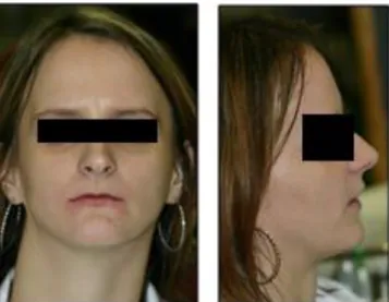
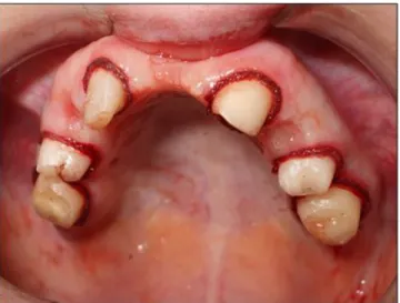
The patient’s symptoms and already diagnosed illnesses were the following (which were in line with the available literature): being born prematurely on the 33th gestational week, with a body weight of 2,400 grams and a length of 40 centimeters, hyperflexibility in the hand joints, hypotrichosis, cutis laxa, umbilical hernia, inguinal hernia, clinodactyly, dyscephalia and telesystolic mitral valve prolapse [18-22]. On physical examination, craniofacial abnormalities, such as projecting forehead, bird face, mandibular hypoplasia, small, thin lips, beak-like nose, bilateral microphthalmia and glaucoma juvenile were also observed (Figure 1.) [12-14]. During the examination of the oral cavity and teeth, hypodontia and a gothic palate were identified. Aplasia of the upper right central incisor was seen, together with the persistence of the upper right milky canine teeth (53). Some decayed teeth were also observed (25 and 36) (Figure 2.). The absence of additional permanent teeth was noted on the panoramic X-ray (Figure 3.). As the esthetic aspects were the most important to the patient, a preliminary treatment plan was made and discussed with her after the intraoral examinations.
Fig 1: The patient's face and profile, characterized by the distinctive features of the disease (before dental treatment)
Fig 2: Initial conditions in the oral cavity of the patient
Fig 3: Panoramic X-ray and X-ray of the upper right quadrant, showing the upper right canine to be removed
Preliminary treatment occurred in a stepwise fashion: following supra- and subgingival scaling and cleaning of the tooth surfaces, the extraction of the upper right deciduous canine was performed.
The filling of the carious teeth was also performed, there was no indication for root canal treatment. Six teeth remained in the maxilla, namely the first molar, the second premolar and the lateral incisor on both the right and the left side, which were suitable to serve as abutments for the placement of the upper bridge. After reassessment, a final prosthetic treatment plan was laid out; the patient was satisfied with the plan and consented to the treatment. The patient did not want any prosthetic restoration for the lower teeth. In the upper arch, a fixed prosthesis could be made, despite the fact that the number and position of the teeth
calculation, where in terms of materials different options were taken into consideration, the patient opted for a metal-ceramic material for the bridge, which was relatively cheap. The creation of the bridge in this patient, affected by Hallermann-Streiff syndrome will be discussed hereinafter.
Treatment had to be interrupted several times due to nausea and malaise of the patient. Antibiotic prophylaxis was not deemed necessary as the mitral prolapse of the patient was without regurgitation. The preparation of the abutment teeth was performed under local anesthesia. The vertical preparation design was used instead of chamfer finish line, as the size of the teeth was small, while the pulp was voluminous, therefore the chamfer preparation would cause too much structural tooth loss and would endanger the vitality of the pulp (Figure 4.). A precision impression was performed 15 minutes after inserting the gingival retraction cord. The impressions were taken with the two-step technique (two steps, two phases), with a small (children-size) impression tray. An acrylic temporary bridge (created by the technical laboratory) was applied on the prepared teeth for the time of the technical work to protect them against harmful physical and chemical effects. The temporary bridge was also useful as it showed a preliminary result of the intervention; what the patient could expect from the permanent bridge (Figure 5.).
Due to the positioning of the abutment, the midline was slightly shifted to the left side; in order to keep the ideal size and shape of the incisors, the position of the midline required to be slightly modified. This was a reasonable compromise made to provide the highest esthetic result. Five and four units were made inside the right and left side, respectively; however, it was only possible to place three crowns on each side and to replace one and two teeth on the left and right side, respectively (Figure 6.). Following the final cementation, the patient was satisfied with the result;
however, before the final delivery some additional, minor modifications had to be performed to achieve a general esthetic appearance (Figure 7.).
Fig 4: Abutement teeth after preparation and before impressions
Fig 5: The acrylic temporary bridge created by the laboratory
Fig 6: Finished dentures in front of the mouth and on the palate
Fig 7: The satisfied patient after the finished procedures.
3. Conclusions
Hallermann-Streiff syndrome is a rare genetic disorder primarily characterized by head, face and dental abnormalities. The identification of this disease requires the suspicion of various clinicians, including general
practitioners, pediatricians, geneticists, dentists, oral- and maxofacillary surgeons and opthtalmologists. The dental treatment of patients with HSS may be complicated, but careful planning and adequate bridge construction can aid complete restoration. The aim of our case presentation was to show such a case, where the oral health and esthetic aspects of a female patient with HSS both needed to be taken into consideration during the development of a treatment plane. In the end, our patient was satisfied with the progress of the interventions, resulting in significant improvements in her quality of life. Additionally, our case aims to serve as a blueprint for general dentists, who may also achieve the same result during the treatment of similar patients (if the correct treatment plan is selected), instead of referring such patients to the specialists in tertiary-care institutions.
4. Author contributions
The study was deemed exempt from ethics review by the Institutional Review Board of the University of Szeged, as the patient provided an informed written consent and approval for publishing the case. The patient signed an informed consent and approval regarding the publication of the photos taken. No differential surgical neither antibiotic protocol was implemented during the treatment of this patient.
5. Author contributions
A.S., M.R. and E.L. managed the patient, edited the case report and participated in the drafting of the manuscript.
E.H. performed microbiology diagnostics, analyzed and interpreted data, and formulated the manuscript; M.G. and Z.B. provided financial support, and supervised the manuscript writing, editing, and review. All authors have read and agreed to the published version of the manuscript.
6. Acknowledgments None.
7. Funding
This research received no external funding.
8. Conflict of interest
The authors declare no conflict of interest.
9. References
1. Thomas J, Ragavi BS, Ranessha PK, Ahmed NA, Cyntia S, Manoharan D, Manoharan R. Hallerman- Streiff Syndrome. Indian J Dermatol. 2013; 58:383- 384.
Available from: https://www.orpha.net/consor/cgi- bin/OC_Exp.php?lng=EN&Expert=2108
4. Dennis NR, Fairhurst J, Moore I.E. Lethal syndrome of slender bones, intrauterine fractures, characteristic facial appearance, and cataracts, resembling Hallermann-Streiff syndrome in two sibs. Am J Med Genet. 1995; 59:517-520.
5. Cohen MM. Hallermann-Streiff syndrome: a review.
Am J Med Gener 1991; 41:488-499.
6. Mirshekari A, Safar F. Hallermann-Streiff syndrome: a case review. Clin Exp Dermatol 2004; 29: 477-479.
7. Shen MQ, Li-Yang W. Hallermann-Streiff syndrome:
a case report. Chin Med J 2010; 123:3356-3357.
8. Kortüm F, Kortüm F, Chyrek M, Fuchs S, Albrecht B, Gillessen-Kaesbach G, Mütze U, Seemanova E, Tinschert S, Wieczorek D, Rosenberger G, Kutsche K.
Hallermann-Streiff Syndrome: No Evidence for a Link to Laminopathies. Mol Syndromol. 2011; 2:27-34.
9. Fraser GR, Friedmann AI. The Causes of Blindness in Childhood. A Study of 776 Children with Severe Visual Handicaps. John’s Hopkins Press (Baltimore), 1967; 16:89.
10. Pizutti A, Flex E, Mingarelli L, Salpietro C, Zelante L, Dallapiccola B. A homozygous GJA1 gene mutation causes a Hallermann-Streiff/ODDD spectrum phenotype. Hum Mutat. 2004; 23:286.
11. Bueno-Sanchez M. Sindrome de Hallermann-Streiff- Francois. A proposito de una presentacion familiar.
Boll Soc. Vasco-Navarra 1966; 1:21-35.
12. Guyard M, Perdriel G, Ceruti F. Sur deux cas de syndrome dyscephalique a tete d'oiseau. Bull Soc Ophtal Franc. 1962; 62:443-447.
13. Schanzlin DJ, Goldberg DB, Brown SI. Hallermann- Streiff syndrome associated with sclerocornea, aniridia and chromosomal abnormality. Am J Ophtalmol. 1980;
90:411-415.
14. Schmidt J, Wollnik B. Hallermann–Streiff syndrome:
A missing molecular link for a highly recognizable syndrome. Am J Med Genet Part C. 2018; 178:398–
406.
15. Fernandez-Marmiesse A, Gouveia S, Couce ML. NGS Technologies as a Turning Point in Rare Disease Research, Diagnosis and Treatment. Curr Med Chem.
2018; 25:404-432.
16. Carones AV. Francois's dyscephalic syndrome.
Ophthalmologica 1961; 142:510-518.
17. Caspersen I, Warburg M. Hallermann-Streiff syndrome. Acta Ophthal. 1968; 46:385-390.
18. Imaizumi K, Makita Y, Masuno M, Koruki Y.
Congenital heart defect in a patient with the
Ophthalmologica. 1961; 141:53-63.
20. Spaepen A, Schrander-Stumpel C, Fryns JP, Die- Smulders C, Borghgraef M, Van den Berghe H.
Hallermann-Streiff syndrome: clinical and psychological findings in children: nosologic overlap with oculodentodigital dysplasia? Am J Med Genet.
1991; 41: 517-520.
21. Dulong A, Bornert F, Gros CI, Garnier JF, Van Bellinghen X, Fioretti F, Lutz JC. Diagnosis and Innovative Multidisciplinary Management of Hallermann-Streiff Syndrome: 20-Year Follow-Up of a Patient. Cleft Palate Craniofac J. 2018; 55:1458- 1466.
22. Stájer A, Kajári S, Gajdács M, Musah-Eroje A, Baráth Z. Utility of Photodynamic Therapy in Dentistry:
Current Concepts. Dent. J. 2020; 8: 43.