Journal of International Dental and Medical Research ISSN 1309-100X A New Tooth Notation For Primary Teeth http://www.ektodermaldisplazi.com/journal.htm Ashfaq Akram and et al
AN ASSESSMENT OF CLINICAL APPLICATION OF A NEW TOOTH NOTATION FOR PRIMARY TEETH
Ashfaq Akram1,2*, Maher D Fuad Fuad2, Ulfat Bashir3, Nagendran Jayavel Pandiyan4, Kalyan Chakravathy5, Thirupathi rao Vishnumukkala6, Melinda Madlena1
1. Faculty of Dentistry, Department of Pediatric Dentistry, Semmelweis University, Budapest, Hungary.
2. International Medical School, Management & Science University – Shah Alam, Malaysia.
3. Department of Orthodontics, Islamic International Dental College, Islamabad, Pakistan.
4. Department of Pediatric Dentistry Penang International Dental College, Penang Malaysia . 5. Alliance Global Services Pvt. Ltd. Hyderabad India.
6. Perdana University- Serdang, Malaysia.
Abstract
A new tooth notation system records primary tooth classes and their types by letters ‘dI, dC, dM’ termed as ANAASEA letters and numbers (1,2) called as TOT digits respectively. The TOT digits are printed as superscript and subscript at both right and left sides of ANAASEA letters to indicate upper and lower teeth respectively. The new method is called MICAP (M-molar, I-incisor, C-canine, P- premolar) system.
To assess the clinical application of new tooth notation by dental health professionals.
Study design was cross sectional and tool was mock e dental chart based on MICAP format. The study participants were dental health professionals (N=225) who were divided into group A [dental specialists (n=44), dentists (n=60)] and group B [dental assistants (n=58), dental hygienists (n=38), dental technicians (n=25)]. They were demonstrated by video on MICAP format before they identified five primary teeth; three teeth to be written from word form to MICAP format and two teeth vice versa. Simple logistic regression and Pearson chi-square tests were performed to analyse the data.
From group A, specialists performed significantly better (p value = 0.031) as compared to dentists in correct writing of ‘deciduous maxillary right canine’ into MICAP format [#1dC]. From group B, dental hygienists had significantly higher association in correct translation of [#2dM] as ‘deciduous mandibular right 2nd molar’
compared to dental assistants (P value =0.043). Furthermore, dental technicians compared to dental assistants significantly performed better (p value =0.047) in writing ‘deciduous mandibular left central incisor’
into MICAP format [#dI1].
Majority of dental health professionals were able to translate and write MICAP format. Doctors were better than paramedics. However, reliability of the new system requires additional data. Further research is recommended to compare MICAP notation with currently used notations such as FDI & Universal systems.
Clinical article ( J Int Dent Med Res 2015; 8: (1), pp. 7-14 )
Keywords: Dental charting, tooth notation, primary teeth, incisor, canine, molar.
Received date: 10 April 2015 Accept date: 17 April 2015
Introduction
Primary teeth are identified by different notations. One method is Universal numbering system which uses letters ‘A-T’ to indicate primary teeth starting from upper right second
molar to lower right 2nd molar. However, the codes ‘d1- d20’ have been recommended for primary teeth in its revised method. 1 Another method is FDI two digit system which uses digits
‘51-55’ and ‘61-65’ for upper right and upper left teeth respectively. The primary lower left and lower right teeth are marked by ‘71-75’ and ‘81- 85’ respectively. 2 The Palmer notation which is the 3rd commonly used method, recognizes the primary teeth by letters A-E with a special right angle sign.3
The usage of multiple tooth notation systems or in other words, the communication gap in dental practices especially in referral cases is one of the major reasons of dental malpractice. 4-6 The current notation methods
*Corresponding author:
Dr. Ashfaq Akram
Faculty of Dentistry, Department of Pediatric Dentistry Semmelweis University, Szentkirályi u 47. 1088 Budapest, Hungary
Phone: +(36) 1266 2343 E-mail: ashfaqakram@hotmail.com E-mail:
Journal of International Dental and Medical Research ISSN 1309-100X A New Tooth Notation For Primary Teeth http://www.ektodermaldisplazi.com/journal.htm Ashfaq Akram and et al
which are based on Arabic numbers or English letters designate different deciduous teeth using the same letter or number. For example, letter ‘B’
is deciduous lateral incisor in Palmer notation whereas same letter (B) represents the deciduous first molar when dental charting is done by Universal system. Scenario is more complicated in mixed dentition which contains both permanent and deciduous teeth. For example, according to FDI system, permanent upper right canine is #13 and Universal system shows the same digit (#13) for upper left 2nd premolar. Such situation creates confusion and it is quite possible to make an error in the patients’
dental problem.
Hence a new method ‘MICAP’ 7 was suggested to record the teeth in dental charting.
MICAP is the abbreviation of ‘M-molar, I-incisor, C-canine, A-Akram, P-premolar’. According to its section associated with deciduous teeth, the primary tooth classes are identified by letters ‘dI- (deciduous incisor) dC- (deciduous canine) and dM- (deciduous molar)’. The letters dI, dC, dM are called ANAASEA letters 7. In each quadrant, the tooth classes are further classified: Two incisors (central and lateral incisor) One canine and Two molars (1st and 2nd molar) which are indicated by digits: central (1), lateral incisor (2), canine (1) first molar (1) and second molar (2).
The digits (1,2) are written as superscript and subscript on right and left side of the relevant tooth class ( dI, dC, dM) and indicate the relevant upper and lower teeth. A conceptual framework of MICAP format for primary teeth is shown ( Figure 1 & 2 ). Previously the conceptual frame work of this system was described. The present study was a step further towards its clinical application (Figure 3 & 4).
The improved performance of learners (dental students) by video methods has been observed. Many pre clinical lecture base courses have been incorporated with videos to give understanding of conceptual courses to dental students 8-11. The video base teaching is one of the preferred teaching methods for dental learners. 12, 13 MICAP is a novice method which is neither taught nor practised in any dental school or institution. The objective of this study was to give understanding of new system via video and to assess clinical application of MICAP system in terms of its format to write primary teeth. The study was approved by ethics committee of the Faculty of Dentistry – Semmelweis University.
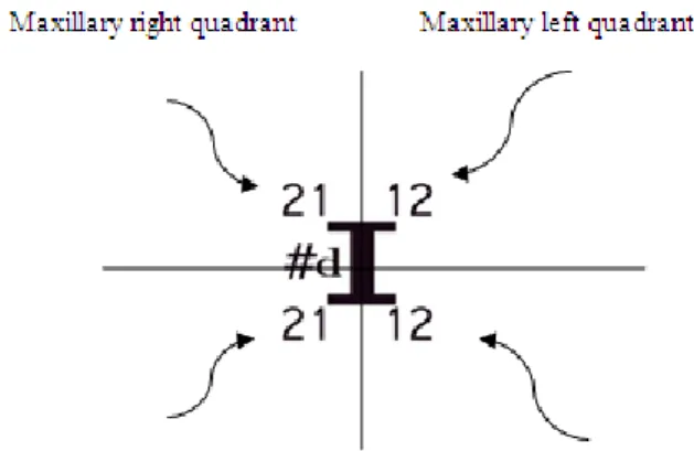
Figure 1. Deciduous incisor class is indicated by dI. Imaginary horizontal and vertical lines divide the letter dI into four quadrants. The digits (1,2) show central and lateral incisor respectively which are written as superscript and subscript on right and left side of ‘dI’ to represent all incisors present in four quadrants. The sign hash (#) represents the tooth number and helps write multiple tooth classes and differentiates MICAP format from word form.7 The digits written as 12 or 21 are read separately instead of twelve or twenty one.
Figure 2. MICAP notation for primary teeth uses the letters dI, dC, dM to identify deciduous Incisor, deciduous Canine and deciduous Molar respectively7,20. The letters dI, dC, dM are called ANAASEA letters. Each tooth class22 has its tooth types 22 which are indicated in digits such as central incisor (1) & lateral incisor (2), canine (1), first molar (1) & second molar (2). The digits (1, 2) are printed as superscript and subscript on right and left side of each relevant ANAASEA letter 7, 20.The term ANAASEA is the abbreviation of almost all continents; Asia, North America, South America, Europe & Africa because humans of all continents have the same tooth classes.20
Journal of International Dental and Medical Research ISSN 1309-100X A New Tooth Notation For Primary Teeth http://www.ektodermaldisplazi.com/journal.htm Ashfaq Akram and et al
Figure 3. MICAP format was described by video.
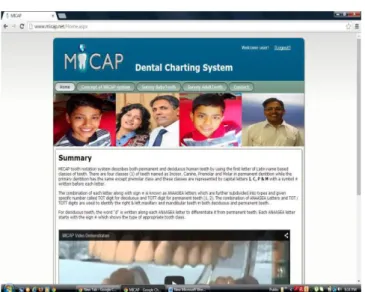
www.micap.net
Figure 4. Mock dental charting for primary teeth was developed by HTML and C+ programme.
This was the format to write the given primary teeth in MICAP format.
Methods
Dental specialists, general dentists, and dental paramedics (N=225) from Penang (Malaysia) and Islamabad (Pakistan) participated in a cross sectional study.
They were divided into two groups. The group A included dental specialists (n= 44) and general dentists (n=60). Dental specialists comprised of pedodontists (n=9), endodontists (n= 12), restorative dentists (n=15) and orthodontists (n=8).
For analysis purpose, they were combined as dental specialists due to small number from each respective speciality.
Group B had dental assistants (n=58), dental hygienist (n=38), dental technician (n=25).
An inclusion criteria was to be involved in dental practice for at least one year as clinician / academician / or supporting worker.
The new system (MICAP) was demonstrated by video to both groups before they participated in the study.
The written consents were obtained and data were collected from September 2014 to December 2014.
Study Instrument
Mock e dental charting based on MICAP notation was prepared.
Five primary teeth were randomly selected as independent variables. Among five teeth, two teeth were to be translated from MICAP format [#2dM #dC1] to word form and three teeth ‘deciduous maxillary left 2nd molar, deciduous mandibular left central incisor, deciduous maxillary right canine’ were to be converted (written) to MICAP format.
The focus was learning of MICAP notation rather than other common features of a dental charting. In addition, a closed end questionnaire based on five point likert scale (1= strongly disagree, 2=disagree, 3= not sure, 4 =agree, 5
=strongly agree) was added to obtain the perception on the prospective suitability of the new notation in dental charting and its usage as source of dental communication.
Data Analysis
SPSS version 20 was used to analyse the data. Pearson chi-square tests and Simple logistic regression were performed to analyse the data for group A and group B respectively.
Results
From group A, nearly ≥ 80 percent participants translated and wrote correctly the primary teeth in MICAP format e.g., #2dM was translated as deciduous mandibular right 2nd molar and ‘deciduous maxillary right canine’ was mirrored back as #1dC (MICAP format).
Dental specialists were statistically better than dentists associated with correct write up
‘deciduous maxillary right canine’ as #1dC (p value =0.031) (Table 1).
Journal of International Dental and Medical Research ISSN 1309-100X A New Tooth Notation For Primary Teeth http://www.ektodermaldisplazi.com/journal.htm Ashfaq Akram and et al
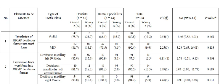
*Pearson chi-square analysis shows no significant difference (P>0.05) between dentists and specialists except deciduous maxillary right Canine to be written as [#1dC].
Table 1. Learning of MICAP method by dentists and specialists (n=104). Majority≥80 % translated and wrote MICAP format correctly.
*#2dM #dC1 [MICAP formats for deciduous lower right 2nd molar and deciduous upper left canine respectively].
** The word format [deciduous maxillary left 2nd molar, deciduous mandibular left central incisor, deciduous maxillary right canine] were to be written into MICAP format (#dM2 #dI1 #1dC) respectively.
Table 2a. Descriptive statistics for dental paramedics. Mixed results were found to translate and write the MICAP format.
From group B, more than fifty percent dental technicians wrote correctly ‘deciduous maxillary left 2nd Molar’ as [#dM2].
In contrast, dental assistants were much lower in percentage to learn the MICAP format.
For example almost 80 % dental assistants could not write ‘deciduous mandibular left central incisor’ as [#dI1] (Table 2a).
Simple logistic regression test showed that there was no significant association of correct translation of [#dC1] between dental assistants and dental hygienists (p value =0.097).
Dental technicians were significantly better (p value <0.05) than dental assistants in both translation of MICAP format as well as conversion into MICAP format except translation of [#dC1] such as ‘deciduous maxillary left canine’ (p value = 0.097) (Table 2b).
Journal of International Dental and Medical Research ISSN 1309-100X A New Tooth Notation For Primary Teeth http://www.ektodermaldisplazi.com/journal.htm Ashfaq Akram and et al
Dental technicians were the best among the three who translated MICAP format and vice versa. *Dental Assistant group was the reference in Simple logistic regression test.
Table 2b.Simple Logistic Regression Analysis.
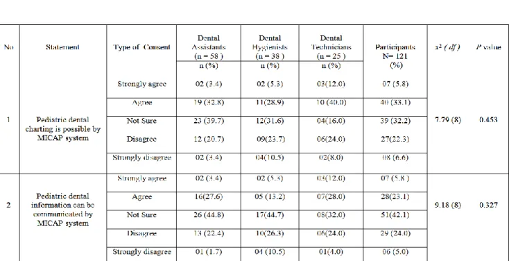
From descriptive statistics, approximately forty percent group A responded positively on the prospective use of MICAP notation for pediatric practice. Group B responded positively but a little less than group A. Small numbers of participants from both groups rejected the role of MICAP in dental charting. However, a quite large number of
specialists, doctors and paramedics were uncertain about its prospective use in dental charting as well as communication source of dental information (Table 3a &3b). Neither gender nor location based significant differences in identification of MICAP format and vice versa in group A & B were observed.
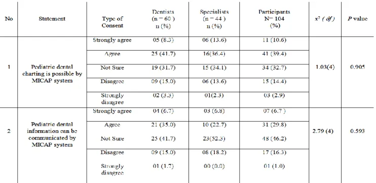
The values of Pearson chi-square (X2 1.03, p = 0.905) shows neither study group supported nor rejected the MICAP system for dental charting. The similar findings between two study groups were observed for dental communication.
Table 3a. Perception of dentists and dental specialists about MICAP notation as pediatric dental charting and communication source.
Journal of International Dental and Medical Research ISSN 1309-100X A New Tooth Notation For Primary Teeth http://www.ektodermaldisplazi.com/journal.htm Ashfaq Akram and et al
Approximate one third (33.1%) supported the prospective use of new system for pediatric dental charting.
Table 3b. Perception of dental paramedics about Pediatric dental charting and Communication source via MICAP system.
Discussion
MICAP system is a new concept. Dental team members were able to identify and write MICAP format with the help of video demonstration. Studies have shown the effectiveness of learning new technique by video teaching method. For an introductory dental public health course, affective learning outcomes in dental students were seen who were taught by video. 14 Similarly dental students found videos affective tool to learn prosthetic clinical procedures.15 Likewise, our participants learnt the skill to write MICAP format through video method. Though they were not students but they understood the format of MICAP. The results ≥ 80% show that new method is easy to understand. Over all, paramedics performed poorer than dental specialists and dentists. The reason could be their less educational level or more likely the lack of interest in new notation method because this is not a part of dental curriculum at the moment.
Considering the aspect of computer friendly, MICAP notation uses letters dI, dC,dM for primary teeth and digits (1,2) printed as superscript and subscript. All characters are available in Microsoft Word (window 7 & window 8). However, in writing both upper and lower
teeth of a particular tooth class, the digits may not appear up and down position in same alignment e.g., #dM121 [deciduous upper left 1st and 2nd and lower left 1st molar]. For this purpose, Microsoft Word ‘equation editor function’ is available.16 This is a little complex procedure but it solves the issue described earlier for upper and lower molar teeth. The use of MICAP system in power point and email to transmit dental information is a limitation of the system at the moment.
Universal numbering and FDI notation are computer friendly because they are based on numbers. If FDI and Universal system are used in e-dental charting and new notation (MICAP) is also computer friendly then question arises
‘why MICAP should be adopted?
The new system is based on recognized standard dental terminologies. For example, incisor is incisor in every dental curriculum no matter what language (English, French, Hindi etc) is used to pronounce incisor. Taking consideration of currently used notations, upper right canine could be marked by #13, #3 #6 in FDI, Palmer & Universal systems respectively. In MICAP system, it is simply #1C. The letter ‘C’
indicates canine. The digit 1 is superscript and printed on right side to C so it is maxillary (upper) right canine.
Journal of International Dental and Medical Research ISSN 1309-100X A New Tooth Notation For Primary Teeth http://www.ektodermaldisplazi.com/journal.htm Ashfaq Akram and et al
Dental hygienists undertake a number of activities such as dental charting, fissure sealing
& radiographs autonomously without dentists’
referral. They perform activities such as dental charting which is highly reviewed or validated in case of referral patients using a certain tooth notation.17,18 The new dental notation was recognised better (p=0.043) by dental hygienists than dental assistants. A higher score of translating and write up of MICAP format by dental specialists, dentists, dental hygienists and technicians showed that MICAP notation was easy to understand. Higher percentage of learning of a new teaching modality support the effectiveness of video method.14,15
The use of 3-D dental charting 19 enhances the charm of dental practice especially to children because the images appear in different angles and colours and attract the users.
The new notation system is blended with numbers and letters in superscript and subscript, it could be adopted in 3D charting with multiple colours of letters and digits. We suggest specific colours to be allocated especially deciduous teeth to give attraction and a specific sign as a standard to identify different or certain primary teeth and this would help understand not only staff but also to children. It would be an additional difference between deciduous and permanent teeth other than what has been proposed in earlier version of MICAP notation in terms of ANAASEA letters.20
In US, medico legal companies are concerned the legal issues raised by different tooth notation system. For example, orthodontists use Palmer notation whereas the oral surgeons use the Universal numbering system. In orthodontics, first premolar is usually recommended for extraction. Therefore tooth number #4 for orthodontist is actually the tooth number 5 for oral surgeon.21 If we consider primary teeth, the letter ‘A’ is deciduous upper right 2nd molar in Universal system. The same letter in Palmer notation is actually central incisor.
The problem is in dental charting and especially in dental communication; no one mentions what system s/he used. This is considered an
‘understood’ matter. The use of different tooth notation ‘why not one’ has been in debate from time to time. The new system gives a possible solution. It identifies the primary teeth by using tooth classes 22 ‘I- incisor, C- canine and M- molar’ which are constant all over the world. The
use of globally understood dental terminology suggests MICAP to be a beneficial addition in pediatric dentistry.
Conclusion
The findings of this study suggested that new tooth notation was mind cognitive because majority (≥ 80%), dental health professionals translated and wrote MICAP format correct.
There were discrepancy of understanding among doctors and dental paramedics. Doctor group understood the format of new tooth notation better than dental paramedics. Moreover, reliability of the new system does demand additional data. In addition, to get absolute error free identification of primary teeth, a proper training is suggested for dental staff members who are involved in patients’ data entry.
Limitation of the Study
This study had two limitations. First, there is lack of comparison of MICAP system with other notation systems (FDI tooth notation or Universal system). The comparison could give evidence of the effectiveness and reliability of the MICAP notation. Second the traditional method (lecture) could be introduced against video method.
Acknowledgement
Authors declare their sincere thanks for providing free video service by Mohd Faizan, Mohd Amin Zainol & Jamila Razak from Advance Medical & Dental Institute – USM- Penang.
References
1. American Dental Association. Council on Dental Practice.
www.ada.org/sections/professionals Resources / dental practice
2. FDI Two Digit Notation. http//www.fdiworldental.org/two-digit- notation
3. Ferguson JW. The Palmer notation system and its use with personal computer applications. Br Dent J 2005; 198 (9):551- 3.
4. Janice S L, Arthur W C, Richard A S. Prevention of wrong site tooth extraction: Clinical guidelines. J Oral Maxil Surg 2007;
65 (9):1793-9.
5. BBC News / Health. Dentists Warned on Tooth Extraction.
2004; October 4, 13:55 GMT.
http.//news.bbc.co.uk/1/hi/health/3714058.stm
6. Ricketts DNJ, Scott BJJ, Ali A, Chadwick RG, Murray CA, et al. Peer review amongst restorative specialists on the quality of their communication with referring dental practitioners. Br Dent J.2003; 195 (7):389-93.
Journal of International Dental and Medical Research ISSN 1309-100X A New Tooth Notation For Primary Teeth http://www.ektodermaldisplazi.com/journal.htm Ashfaq Akram and et al
7. Akram A, Abdel Hamid ZAH, Razak J, Hock TT. MICAP- a novel system for identification and communication of dental problems. Int Dent J. 2011;61(1): 31-6.
8. Smith W, Rafeek R, Marchan S, Paryag A. The use of video clips as a teaching aide. Eur J Dent Edu.2012; 16 (2) :91-96.
9. Kalwitzki M, Meller C, Beyer C. Does teaching method affects students’ perceptions regarding communication patterns in pediatric dentistry? A comparison of lecture and video methods. J Dent Edu.2011; 75(8): 1084-91.
10. Kalwitzki M, Beyer C, Meller C. Differences in the perceptions of seven behavior –modifying techniques in pediatric dentistry by undergraduate students using lecturing and video sequences for teaching. Euro J Dent Edu.2010; 14 (4):247-53.
11. Kamin C, O’Sullivan P, Deterding R, Younger M. A comparison of critical thinking in groups of third year medical students in text, video and virtual PBL case modalities. Acad Med 2003; 78 (2): 204-11.
12. Boynton JR, Johnson LA, Nainar SM, Hu JC. Portable digital video instruction in pre doctoral education of child behavior management. J Dent Edu.2007; 71(4): 545-9.
13. Aragon CE, Zibrowski EM. Does exposure to a procedural video enhance pre clinical dental student performance in fixed prosthodontics? J Dent Edu.2008; 72(1):67-71.
14. Chi DL, Pickrell JE, Riedy CA. Student learning outcomes associated with video vs paper cases in a public health dentistry course. J Dent Edu.2014; 78(1):24-30.
15. Kon H,Botelho MG, Bridges S, leung KC. The impact of complete denture making instructional videos on self –directed learning of clinical skills. J Prosth Res.2015.
doi:10.1016/j.jpor.2015.01.004.
16. Lewis DH. Dental notation (letter). Br Dent J.2000; 188L: 230- 31.
17. Turner S, Ross MK, Ibbetson RJ. Dental hygienists and therapists: how much professional autonomy do they have?
How much do they want? Results from a UK survey. Br Dent J.2011; 210: E16.
18. Demko CA,Victoroff KZ, Wotman S. Concordance of chart and billing data with direct observation in dental practice. Com Dent Oral Epid.2008;36 (5): 466-76.
19. Schieyer TK, Thyvalikakath TP, Malatack P, Marotta M, Shah TA, et al. The feasibility of a three dimensional charting interface for general dentistry. J Am Dent Assoc.2007;
138(8):1072-80.
20. Akram A, Abdus S, Ulfat B, Maroof N, Subhan M. Lesson plan on new method of teeth identification introduced at dental schools in Malaysia and Pakistan. J Dent Edu.2012; 76(12):
1691- 6.
21. Pogral MA. Tooth notation. Br Dent J.2003; 195:360.
22. Woelfel JB, Schied RC. Dental Anatomy. Its relevance to Dentistry (6th edn). Lippincott Williams & Wilkins, Maryland.2002; p. 2-3.