International Journal of
Molecular Sciences
Article
Synthesis and Spectrum of Biological Activities of Novel N -arylcinnamamides
Sarka Pospisilova1,2, Jiri Kos1,*, Hana Michnova1,2, Iva Kapustikova1, Tomas Strharsky1, Michal Oravec3, Agnes M. Moricz4, Jozsef Bakonyi4, Tereza Kauerova5, Peter Kollar5, Alois Cizek2and Josef Jampilek1 ID
1 Department of Pharmaceutical Chemistry, Faculty of Pharmacy, Comenius University, Odbojarov 10, 83232 Bratislava, Slovakia; sharka.pospisilova@gmail.com (S.P.); michnova.hana@gmail.com (H.M.);
kapustikova@fpharm.uniba.sk (I.K.); strharsky2@uniba.sk (T.S.); josef.jampilek@gmail.com (J.J.)
2 Department of Infectious Diseases and Microbiology, Faculty of Veterinary Medicine,
University of Veterinary and Pharmaceutical Sciences, Palackeho 1, 61242 Brno, Czech Republic;
cizeka@vfu.cz
3 Global Change Research Institute CAS, Belidla 986/4a, 60300 Brno, Czech Republic; oravec.m@czechglobe.cz
4 Plant Protection Institute, Centre for Agricultural Research, Hungarian Academy of Sciences, Herman Otto Str. 15, 1022 Budapest, Hungary; moricz.agnes@agrar.mta.hu (A.M.M.);
bakonyi.jozsef@agrar.mta.hu (J.B.)
5 Department of Human Pharmacology and Toxicology, Faculty of Pharmacy,
University of Veterinary and Pharmaceutical Sciences, Palackeho 1, 61242 Brno, Czech Republic;
tereza.kauerova@gmail.com (T.K.); kollarp@vfu.cz (P.K.)
* Correspondence: jirikos85@gmail.com; Tel.: +421-2-5011-7224
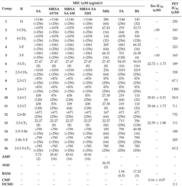
Received: 9 July 2018; Accepted: 3 August 2018; Published: 7 August 2018 Abstract: A series of sixteen ring-substituted N-arylcinnamamides was prepared and characterized. Primary in vitro screening of all the synthesized compounds was performed againstStaphylococcus aureus, three methicillin-resistantS. aureusstrains,Mycobacterium tuberculosis H37Ra,Fusarium avenaceum, andBipolaris sorokiniana. Several of the tested compounds showed antistaphylococcal, antitubercular, and antifungal activities comparable with or higher than those of ampicillin, isoniazid, and benomyl. (2E)-N-[3,5-bis(trifluoromethyl)phenyl]-3-phenylprop-2-enamide and (2E)-3-phenyl-N-[3-(trifluoromethyl)phenyl]prop-2-enamide showed the highest activities (MICs = 22.27 and 27.47 µM, respectively) against all four staphylococcal strains and against M. tuberculosis. These compounds showed an activity against biofilm formation of S. aureus ATCC 29213 in concentrations close to MICs and an ability to increase the activity of clinically used antibiotics with different mechanisms of action (vancomycin, ciprofloxacin, and tetracycline).
In time-kill studies, a decrease of CFU/mL of >99% after 8 h from the beginning of incubation was observed. (2E)-N-(3,5-Dichlorophenyl)- and (2E)-N-(3,4-dichlorophenyl)-3-phenylprop-2-enamide had a MIC = 27.38µM againstM. tuberculosis, while a significant decrease (22.65%) of mycobacterial cell metabolism determined by the MTT assay was observed for the 3,5-dichlorophenyl derivative. (2E)-N-(3-Fluorophenyl)- and (2E)-N-(3-methylphenyl)-3-phenylprop-2-enamide exhibited MICs = 16.58 and 33.71 µM, respectively, againstB.sorokiniana. The screening of the cytotoxicity of the most effective antimicrobial compounds was performed using THP-1 cells, and these chosen compounds did not shown any significant lethal effect. The compounds were also evaluated for their activity related to the inhibition of photosynthetic electron transport (PET) in spinach (Spinacia oleraceaL.) chloroplasts. (2E)-N-(3,5-dichlorophenyl)-3-phenylprop-2-enamide (IC50= 5.1µM) was the most active PET inhibitor. Compounds with fungicide potency did not show any in vivo toxicity against Nicotiana tabacumvar. Samsun. The structure–activity relationships are discussed.
Int. J. Mol. Sci.2018,19, 2318; doi:10.3390/ijms19082318 www.mdpi.com/journal/ijms
Int. J. Mol. Sci.2018,19, 2318 2 of 25
Keywords:cinnamamides; antistaphylococcal activity; time-kill assay; biofilm; antitubercular activity;
MTT assay; antifungal activity; PET inhibition; toxicity; structure–activity relationship
1. Introduction
Cinnamic acids and other hydroxy- or phenyl-substituted derivatives of cinnamic acids have been widely investigated by scientists due to their significant and varied biological effects. Cinnamic acids occur naturally in all plants [1]. They are formed in the biochemical pathway providing phenyl-propanoids, coumarins, lignans, isoflavonoids, flavonoids, stilbenes, aurones, anthocyanins, spermidines, and tannins [2]. The spectrum of their biological activities include anti-inflammatory, antioxidant, hepatoprotective, antidiabetic, antidepressant/anxiolytic, antifungal, antibacterial, antiviral, and anticancer effects [3–16]. Derivatives of cinnamic acids are used as agriculture fungicides as well [17].
The adaptation of microorganisms to external influences and, thus, the development of their resistance against antimicrobial agents is not a surprise; unfortunately, this process is faster and faster.
Thus, the emerging resistance of microbial pathogens to clinically used drugs, including second- and third-choice drugs, and the development of cross-resistant or multidrug-resistant strains are alarming. Microbial pathogens have developed a number of mechanisms to adapt to the effects of the environment. In addition, the increase in the number of infections and the occurrence of new, especially opportunistic species are also caused by the general immunosuppression of patients, and this fact makes these diseases extremely serious. Since the 1990s, only an inconsiderable number of really new drugs for systemic administration have been marketed for the treatment of infections, although the discovery of new molecules has been a priority [18].
Thus, in the light of the above mentioned facts, new simple anilides of cinnamic acid were designed as antimicrobial multitarget agents, synthesized using a modern microwave-assisted method and screened against a battery of bacterial/mycobacterial and fungal pathogens. These compounds were designed based on the experience with naphthalenecarboxamides—simple molecules with a number of biological activities, and in fact, the new ring-substituted (2E)-N-phenyl-3-phenylprop-2-enamides can be considered as open analogues of recently described naphthalene-2-carboxanilides [19,20].
As an amide moiety is able to affect photosystem II (PS II) by reversible binding [21], resulting in the interruption of the photosynthetic electron transport (PET) [22–24], and can be found in many herbicides acting as photosynthesis inhibitors [25–30], theseN-arylcinnamamides were additionally tested on the inhibition of PET in spinach (Spinacia oleraceaL.) chloroplasts using the Hill reaction.
The idea of this screening is based on the fact that both drugs and pesticides are designed to target particular biological functions. These functions/effects may overlap at the molecular level, which causes a considerable structural similarity between drugs and pesticides. Since different classes of herbicides are able to bind to different mammalian cellular receptors, the majority of pharmaceutical companies have pesticide divisions, and developed biologically active agents are investigated as both pesticides and drugs. Previously, several successful pesticides became pharmaceuticals and vice versa [31–35].
2. Results and Discussion
2.1. Chemistry and Physicochemical Properties
All the studied compounds1–16were prepared according to Scheme1. The carboxyl group of starting cinnamic acid was activated with phosphorus trichloride. In the reaction with an appropriate ring-substituted aniline, the generated acyl chloride subsequently gave the final amide in dry chlorobenzene via microwave-assisted synthesis. All the compounds were recrystallized from ethanol.
Int. J. Mol. Sci.2018,19, 2318 3 of 25
Many different molecular parameters/descriptors are used to determine structure-activity relationships (SAR). Lipophilicity and electronic properties are among the most frequent ones.
Hammett’sσparameters were used for the description of electronic properties. They were calculated for the whole substituted anilide ring using ACD/Percepta ver. 2012 (Advanced Chemistry Development Inc., Toronto, ON, Canada, 2012), see Table1. The lipophilicity of the studied compounds was predicted as logPusing ACD/Percepta software and ClogPusing ChemBioDraw Ultra 13.0 (CambridgeSoft, PerkinElmer Inc., Cambridge, MA, USA). LogPis the logarithm of the partition coefficient forn-octanol/water. ClogPis the logarithm ofn-octanol/water partition coefficient based on the established chemical interactions. In addition, the lipophilicity of studied compounds1–16 was investigated by means of reversed-phase high-performance liquid chromatography (RP-HPLC) determination of capacity factorskwith the subsequent calculation of logk[36]. The analysis was made under isocratic conditions with methanol as an organic modifier in the mobile phase using an end-capped nonpolar C18 stationary RP column. The results are shown in Table1.
Int. J. Mol. Sci. 2018, 19, x 3 of 24
partition coefficient for n-octanol/water. Clog P is the logarithm of n-octanol/water partition coefficient based on the established chemical interactions. In addition, the lipophilicity of studied compounds 1–16 was investigated by means of reversed-phase high-performance liquid chromatography (RP-HPLC) determination of capacity factors k with the subsequent calculation of log k [36]. The analysis was made under isocratic conditions with methanol as an organic modifier in the mobile phase using an end-capped nonpolar C18 stationary RP column. The results are shown in Table 1.
Scheme 1. Synthesis of (2E)-N-aryl-3-phenylprop-2-enamides 1–16. Reagents and conditions: (a) PCl3, chlorobenzene, and MW.
Table 1. Structure of ring-substituted (2E)-N-aryl-3-phenylprop-2-enamides 1–16, experimentally determined values of lipophilicity log k, calculated values of log P/Clog P, and electronic Hammett’s σ parameters.
Comp. R log k Clog P a log P b σAr b
1 H 0.1146 3.6640 3.18 0.60
2 3-CH3 0.2729 4.1630 3.40 0.48 3 4-CH3 0.2640 4.1630 3.40 0.46 4 2-F 0.1330 3.4646 3.17 1.02 5 3-F 0.2327 4.0646 3.32 0.82 6 3-CF3 0.4859 4.9978 4.26 0.89 7 2,5-CH3 0.2691 4.0120 3.57 0.59 8 2,5-Cl 0.5799 4.5878 4.65 1.22 9 2,6-Cl 0.0632 3.7378 4.56 1.33 10 3,4-Cl 0.6821 5.3178 4.70 1.19 11 3,5-Cl 0.8155 5.4378 4.79 1.11 12 2,6-Br 0.0992 3.9778 4.80 1.33 13 3,5-CF3 0.9814 6.0386 5.68 1.05 14 2-F-5-Br 0.4875 4.4178 4.07 1.28 15 2-Br-5-F 0.4588 4.1378 4.12 1.19 16 2-Cl-5-CF3 0.6178 4.9509 4.88 1.19
a calculated using ChemBioDraw Ultra 13.0; b calculated using ACD/Percepta ver. 2012
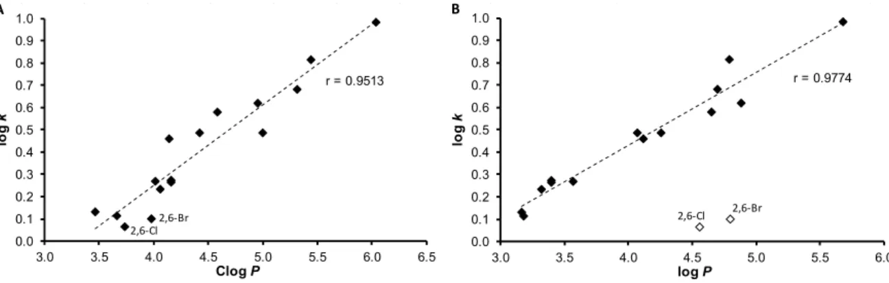
The results obtained with the discussed compounds show that the experimentally-determined lipophilicities (log k) are in accordance with the calculated Clog P values as illustrated in Figure 1A;
correlation coefficient r = 0.9513, n = 16. On the other hand, log P values calculated by ACD/Percepta show differences for compounds 9 (2,6-Cl) and 12 (2,6-Br), see Figure 1B. When these two compounds are excluded, r = 0.9774 (n = 14) is observed. This poor match for 2,6-disubstituted anilides 9 and 12 may be caused by intramolecular interactions that are probably caused by the steric effect of spatially- close moieties, which was not included in prediction by ACD/Percepta. The proximity of the di-ortho- substituents to the carboxamide group on the aniline ring leads to the twist of the aniline ring plane towards the carboxamide group, i.e., to the plane of the benzene ring of cinnamic acid. The described process resulted in the planarity violation of the molecule. Otherwise, (2E)-N-[3,5-
Scheme 1.Synthesis of (2E)-N-aryl-3-phenylprop-2-enamides1–16. Reagents and conditions: (a) PCl3, chlorobenzene, and MW.
Table 1. Structure of ring-substituted (2E)-N-aryl-3-phenylprop-2-enamides1–16, experimentally determined values of lipophilicity logk, calculated values of logP/ClogP, and electronic Hammett’s σparameters.
Int. J. Mol. Sci. 2018, 19, x 3 of 24
partition coefficient for n-octanol/water. Clog P is the logarithm of n-octanol/water partition coefficient based on the established chemical interactions. In addition, the lipophilicity of studied compounds 1–16 was investigated by means of reversed-phase high-performance liquid chromatography (RP-HPLC) determination of capacity factors k with the subsequent calculation of log k [36]. The analysis was made under isocratic conditions with methanol as an organic modifier in the mobile phase using an end-capped nonpolar C18 stationary RP column. The results are shown in Table 1.
Scheme 1. Synthesis of (2E)-N-aryl-3-phenylprop-2-enamides 1–16. Reagents and conditions: (a) PCl3, chlorobenzene, and MW.
Table 1. Structure of ring-substituted (2E)-N-aryl-3-phenylprop-2-enamides 1–16, experimentally determined values of lipophilicity log k, calculated values of log P/Clog P, and electronic Hammett’s σ parameters.
Comp. R log k Clog P a log P b σAr b
1 H 0.1146 3.6640 3.18 0.60
2 3-CH3 0.2729 4.1630 3.40 0.48 3 4-CH3 0.2640 4.1630 3.40 0.46 4 2-F 0.1330 3.4646 3.17 1.02 5 3-F 0.2327 4.0646 3.32 0.82 6 3-CF3 0.4859 4.9978 4.26 0.89 7 2,5-CH3 0.2691 4.0120 3.57 0.59 8 2,5-Cl 0.5799 4.5878 4.65 1.22 9 2,6-Cl 0.0632 3.7378 4.56 1.33 10 3,4-Cl 0.6821 5.3178 4.70 1.19 11 3,5-Cl 0.8155 5.4378 4.79 1.11 12 2,6-Br 0.0992 3.9778 4.80 1.33 13 3,5-CF3 0.9814 6.0386 5.68 1.05 14 2-F-5-Br 0.4875 4.4178 4.07 1.28 15 2-Br-5-F 0.4588 4.1378 4.12 1.19 16 2-Cl-5-CF3 0.6178 4.9509 4.88 1.19
a calculated using ChemBioDraw Ultra 13.0; b calculated using ACD/Percepta ver. 2012
The results obtained with the discussed compounds show that the experimentally-determined lipophilicities (log k) are in accordance with the calculated Clog P values as illustrated in Figure 1A;
correlation coefficient r = 0.9513, n = 16. On the other hand, log P values calculated by ACD/Percepta show differences for compounds 9 (2,6-Cl) and 12 (2,6-Br), see Figure 1B. When these two compounds are excluded, r = 0.9774 (n = 14) is observed. This poor match for 2,6-disubstituted anilides 9 and 12 may be caused by intramolecular interactions that are probably caused by the steric effect of spatially- close moieties, which was not included in prediction by ACD/Percepta. The proximity of the di-ortho- substituents to the carboxamide group on the aniline ring leads to the twist of the aniline ring plane towards the carboxamide group, i.e., to the plane of the benzene ring of cinnamic acid. The described process resulted in the planarity violation of the molecule. Otherwise, (2E)-N-[3,5-
Comp. R logk ClogPa logPb σArb
1 H 0.1146 3.6640 3.18 0.60
2 3-CH3 0.2729 4.1630 3.40 0.48
3 4-CH3 0.2640 4.1630 3.40 0.46
4 2-F 0.1330 3.4646 3.17 1.02
5 3-F 0.2327 4.0646 3.32 0.82
6 3-CF3 0.4859 4.9978 4.26 0.89
7 2,5-CH3 0.2691 4.0120 3.57 0.59
8 2,5-Cl 0.5799 4.5878 4.65 1.22
9 2,6-Cl 0.0632 3.7378 4.56 1.33
10 3,4-Cl 0.6821 5.3178 4.70 1.19
11 3,5-Cl 0.8155 5.4378 4.79 1.11
12 2,6-Br 0.0992 3.9778 4.80 1.33
13 3,5-CF3 0.9814 6.0386 5.68 1.05
14 2-F-5-Br 0.4875 4.4178 4.07 1.28
15 2-Br-5-F 0.4588 4.1378 4.12 1.19
16 2-Cl-5-CF3 0.6178 4.9509 4.88 1.19
acalculated using ChemBioDraw Ultra 13.0;bcalculated using ACD/Percepta ver. 2012.
The results obtained with the discussed compounds show that the experimentally-determined lipophilicities (logk) are in accordance with the calculated ClogPvalues as illustrated in Figure1A;
correlation coefficientr= 0.9513,n= 16. On the other hand, logPvalues calculated by ACD/Percepta show differences for compounds9(2,6-Cl) and12(2,6-Br), see Figure1B. When these two compounds
Int. J. Mol. Sci.2018,19, 2318 4 of 25
are excluded,r= 0.9774 (n= 14) is observed. This poor match for 2,6-disubstituted anilides9and 12 may be caused by intramolecular interactions that are probably caused by the steric effect of spatially-close moieties, which was not included in prediction by ACD/Percepta. The proximity of the di-ortho-substituents to the carboxamide group on the aniline ring leads to the twist of the aniline ring plane towards the carboxamide group, i.e., to the plane of the benzene ring of cinnamic acid. The described process resulted in the planarity violation of the molecule. Otherwise, (2E)-N-[3,5-bis(trifluoromethyl)phenyl]-3-phenylprop-2-enamide (13) is the most lipophilic, while compounds9,12andN-phenylcinnamamide (1) are characterized by the lowest lipophilicity. It can be stated that logkvalues specify lipophilicity within the series of the studied compounds.
Int. J. Mol. Sci. 2018, 19, x 4 of 24
bis(trifluoromethyl)phenyl]-3-phenylprop-2-enamide (13) is the most lipophilic, while compounds 9, 12 and N-phenylcinnamamide (1) are characterized by the lowest lipophilicity. It can be stated that log k values specify lipophilicity within the series of the studied compounds.
Figure 1. Comparison of experimentally found log k values of ring-substituted N-arylcinnamamides 1–16 with Clog P calculated using ChemBioDraw Ultra (A) and log P calculated using ACD/Percepta (B).
2.2. In Vitro Antibacterial Susceptibility Testing
All the cinnamanilides were tested on their antistaphylococcal activity against three clinical isolates of methicillin-resistant Staphylococcus aureus (MRSA) [37,38] and S. aureus ATCC 29213 as the reference and quality control strain. Although various derivatives of cinnamic acid were described as promising antibacterial agents [4–6,8,9,14,15], the compounds showed only limited activity (MICs
> 256 µg/mL), except for (2E)-3-phenyl-N-[3-(trifluoromethyl)phenyl]prop-2-enamide (6) and 3,5- bis(trifluoromethyl)phenyl derivative 13, see Table 2. As minimum inhibitory concentrations (MICs) of these compounds are the same against the reference and the MRSA strains (27.47 and 22.27 µM, respectively), it can be speculated about the specific effectivity against Staphylococcus sp. These compounds were also tested against Enterococcus faecalis ATCC 29212 as the reference strain and three isolates from American crows of vanA-carrying vancomycin-resistant E. faecalis (VRE) [39] but without any effect in the tested concentrations, which may indicate a specific mechanism of action [37,40]. From Table 2 it is obvious that compounds 6 and 13 exhibited activities comparable with those of the standards. Due to the small number of active compounds, no SAR could be established.
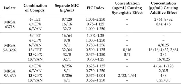
2.2.1. Synergy Effect with Clinically Used Drugs against MRSA
The most effective compounds 6 and 13 were tested for their ability of synergic activity with clinically used antibacterial drugs tetracycline, ciprofloxacin, and vancomycin. These antibiotics have different mechanisms of actions and different mechanisms of resistance to them, thus the prospective synergism could give an idea of the mechanism of action of the cinnamic derivatives. The investigation of synergistic activity was performed according to the methodology [41]. The method of fractional inhibitory concentration (FIC) was used [42]. For all the wells of the microtitration plates that corresponded to a MIC value, the sum of the FICs (ΣFIC) was calculated for each well, using the equation ΣFIC = FICA + FICB = (CA/MICA) + (CB/MICB), where MICA and MICB are the MICs of drugs A and B alone, respectively, and CA and CB are the concentrations of the drugs in the combination, respectively [42]. Synergy was defined as a ΣFIC ≤ 0.5; additivity was defined as 0.5 < ΣFIC < 1;
indifference was defined as 1 ≤ ΣFIC < 4; and antagonism was defined as ΣFIC ≥ 4 [41]. As the FIC index was evaluated for every single well corresponding to the MIC value, the results are presented as a range. The test was made with all 3 methicillin-resistant isolates, MRSA 63718, SA 3202, and SA 630. The isolates were also resistant to used antibiotics. Note that isolate MRSA SA 630 is susceptible to tetracycline. The results are mentioned in Table 3.
r = 0.9513
0.0 0.1 0.2 0.3 0.4 0.5 0.6 0.7 0.8 0.9 1.0
3.0 3.5 4.0 4.5 5.0 5.5 6.0 6.5
logk
Clog P A
2,6-Cl2,6-Br 2,6-Br
2,6-Cl
r = 0.9774
0.0 0.1 0.2 0.3 0.4 0.5 0.6 0.7 0.8 0.9 1.0
3.0 3.5 4.0 4.5 5.0 5.5 6.0
log k
log P B
Figure 1.Comparison of experimentally found logkvalues of ring-substitutedN-arylcinnamamides 1–16 with Clog P calculated using ChemBioDraw Ultra (A) and log P calculated using ACD/Percepta (B).
2.2. In Vitro Antibacterial Susceptibility Testing
All the cinnamanilides were tested on their antistaphylococcal activity against three clinical isolates of methicillin-resistant Staphylococcus aureus (MRSA) [37,38] and S. aureus ATCC 29213 as the reference and quality control strain. Although various derivatives of cinnamic acid were described as promising antibacterial agents [4–6,8,9,14,15], the compounds showed only limited activity (MICs > 256µg/mL), except for (2E)-3-phenyl-N-[3-(trifluoromethyl)phenyl]prop-2-enamide (6) and 3,5-bis(trifluoromethyl)phenyl derivative13, see Table2. As minimum inhibitory concentrations (MICs) of these compounds are the same against the reference and the MRSA strains (27.47 and 22.27µM, respectively), it can be speculated about the specific effectivity againstStaphylococcussp.
These compounds were also tested againstEnterococcus faecalisATCC 29212 as the reference strain and three isolates from American crows of vanA-carrying vancomycin-resistantE. faecalis(VRE) [39]
but without any effect in the tested concentrations, which may indicate a specific mechanism of action [37,40]. From Table2it is obvious that compounds6and13exhibited activities comparable with those of the standards. Due to the small number of active compounds, no SAR could be established.
2.2.1. Synergy Effect with Clinically Used Drugs against MRSA
The most effective compounds6 and13were tested for their ability of synergic activity with clinically used antibacterial drugs tetracycline, ciprofloxacin, and vancomycin. These antibiotics have different mechanisms of actions and different mechanisms of resistance to them, thus the prospective synergism could give an idea of the mechanism of action of the cinnamic derivatives.
The investigation of synergistic activity was performed according to the methodology [41]. The method of fractional inhibitory concentration (FIC) was used [42]. For all the wells of the microtitration plates that corresponded to a MIC value, the sum of the FICs (ΣFIC) was calculated for each well, using the equationΣFIC = FICA+ FICB= (CA/MICA) + (CB/MICB), where MICAand MICBare the MICs of drugs A and B alone, respectively, and CAand CBare the concentrations of the drugs in the combination,
Int. J. Mol. Sci.2018,19, 2318 5 of 25
respectively [42]. Synergy was defined as aΣFIC≤0.5; additivity was defined as 0.5 <ΣFIC < 1;
indifference was defined as 1≤ΣFIC < 4; and antagonism was defined asΣFIC≥4 [41]. As the FIC index was evaluated for every single well corresponding to the MIC value, the results are presented as a range. The test was made with all 3 methicillin-resistant isolates, MRSA 63718, SA 3202, and SA 630.
The isolates were also resistant to used antibiotics. Note that isolate MRSA SA 630 is susceptible to tetracycline. The results are mentioned in Table3.
Table 2. Structure of ring-substituted (2E)-N-aryl-3-phenylprop-2-enamides 1–16, IC50
(µM) values related to PET inhibition in spinach chloroplasts in comparison with 3-(3,4-dichlorophenyl)-1,1-dimethylurea (DCMU) standard, in vitro anti-Staphylococcus activities MIC (µM) in comparison with standard ampicillin (AMP), in vitro antitubercular activity MIC (µM (µg/mL)) in comparison with standard isoniazid (INH), in vitro antifungal activity MIC (µM (µg/mL)) of compounds1–16compared to standard benomyl (BNM), and in vitro antiproliferative (Tox) assay (IC50(µM)) of chosen compounds compared to standard camptothecin (CMP).
Comp. R
MIC (µM (µg/mL))
Tox IC50
(µM)
PET IC50
SA MRSA (µM) 63718
MRSA SA 630
MRSA SA
3202 Mtb FA BS
1 H >1146
(>256)
>1146 (>256)
>1146 (>256)
>1146 (>256)
286 (64)
1146 (256)
143
(32) – 250
2 3-CH3 >1078
(>256)
>1078 (>256)
>1078 (>256)
>1078 (>256)
67.43 (16)
270 (64)
33.71
(8) >30 343
3 4-CH3 >1078
(>256)
>1078 (>256)
>1078 (>256)
>1078 (>256)
134 (32)
1078 (256)
539
(128) – 320
4 2-F >1061
(>256)
>1061 (>256)
>1061 (>256)
>1061 (>256)
265 (64)
1061 (256)
66.32
(16) – 223
5 3-F >1061
(>256)
>1061 (>256)
>1061 (>256)
>1061 (>256)
66.31 (16)
531 (128)
16.58
(4) >30 165
6 3-CF3 27.47
(8)
27.47 (8)
27.47 (8)
27.47 (8)
27.47 (8)
54.93 (16)
54.93
(16) 22.72±1.73 189 7 2,5-CH3 >1018
(>256)
>1018 (>256)
>1018 (>256)
>1018 (>256)
254 (64)
1019 (256)
1019
(256) – 338
8 2,5-Cl >876
(>256)
>876 (>256)
>876 (>256)
>876 (>256)
876 (256)
876 (256)
876
(256) – 67.1
9 2,6-Cl >876
(>256)
>876 (>256)
>876 (>256)
>876 (>256)
876 (256)
876 (256)
876
(256) – 1380
10 3,4-Cl 438
(128) 876 (256)
438 (128)
876 (256)
27.38 (8)
219 (64)
110
(32) 29.81±0.31 54.9
11 3,5-Cl 438
(128) 876 (256)
109 (64)
438 (128)
27.38 (8)
219 (64)
110
(32) 29.44±1.73 5.1
12 2,6-Br >671
(256)
>671 (256)
>671 (256)
>671 (256)
167 (64)
671 (256)
671
(256) – 732
13 3,5-CF3 22.27
(8)
22.27 (8)
22.27 (8)
22.27 (8)
22.27 (8))
713 (256)
356
(128) 22.59±1.88 111 14 2-F-5-Br >799
(>256)
>799 (>256)
>799 (>256)
>799 (>256)
199 (64)
799 (256)
49.98
(16) – 188
15 2-Br-5-F >799 (>256)
>799 (>256)
>799 (>256)
>799 (>256)
199 (64)
799 (256)
799
(256) – 205
16 2-Cl-5-CF3 >785 (>256)
>785 (>256)
>785 (>256)
>785 (>256)
785 (256)
785 (256)
785
(256) – 63.2
AMP – 5.72
(2)
45.81 (16)
45.81 (16)
45.81
(16) – – – – –
INH – – – – – 36.55
(5) – – – –
BNM – – – – – – 1.94
(0.5)
17.22
(5) – –
CMP – – – – – – – – 0.16±0.07 –
DCMU – – – – – – – – – 2.1
SA= Staphylococcus aureusATCC 29213; MRSA=clinical isolates of methicillin-resistantS. aureus63718, SA 630, and SA 3202 (National Institute of Public Health, Prague, Czech Republic); Mtb =Mycobacterium tuberculosisH37Ra;
FA =Fusarium avenaceum(Fr.) Sacc. IMI 319947; BS =Bipolaris sorokiniana(Sacc.) Shoemaker H-299 (NCBI GenBank accession No. MH697869).
Int. J. Mol. Sci.2018,19, 2318 6 of 25
Table 3.Combined effect of most potentN-arylcinnamamides and tetracycline (TET), ciprofloxacin (CPX), and vancomycin (VAN).
Isolate Combination of Compds.
Separate MIC
(µg/mL) FIC Index
Concentration (µg/mL) Causing Synergistic Effect
Concentration (µg/mL) Causing
Additive Effect MRSA
63718
6/TET 8/128 1.004–2.250 – 2/64; 8/32
6/CPX 16/16 0.75–1.125 – 8/4; 4/8
6/VAN 32/2 1.000–1.250 – –
MRSA SA 3202
6/TET 16/64 1.002–1.25 – –
6/CPX 8/8 1.000–1.250 – –
6/VAN 8/1 0.750–1.256 – 4/0.25
13/TET 32/64 0.500–1.125 8/16 16/16; 4/32; 2/64
13/CPX 32/8 0.375–1.250 8/1 2/4
13/VAN 32/1 0.750–1.25 – 16/0.25
MRSA SA 630
6/CPX 8/256 0.625–1.125 – 4/64; 1/128
6/VAN 8/1 0.750–1.250 – 2/0.5
13/CPX 8/256 0.375–1.004 2/32; 1/64 4/8
13/VAN 4/1 0.562–1.250 – 0.25/0.5
Although the activity of cinnamic acid derivatives is known for a long time, the exact mechanism of action is still unknown. The most reported mechanism of action is interaction with plasmatic membrane. The compounds can cause disruption of the membrane, damage the membrane proteins, etc. [43–46]. There are also specific targets for cinnamic acid derivatives [46]. Nevertheless, it is possible that the wide spectrum of effects to cells is caused by the primary activity of the compounds, which is membrane destabilization [46].
Both compounds 6 and 13 tested for synergy showed additivity with vancomycin against MRSA SA 630 and SA 3202. A similar effect was reported by Hemaiswarya et al. [47]; compound 13 had synergistic effect with ciprofloxacin against both tested strains. The effect of derivative 13 was also synergistic with tetracycline against MRSA SA 3202. The rest combinations with compound 13 had additive effect. Whereas compound 13 had a potential to increase the activity of all tested antibiotics, which have different mechanisms of actions and to which bacteria develop different resistance mechanisms, it can be expected that compound 13 acts by its own mechanism of action or increases the availability of the antibiotics by interaction with the membrane.
2.2.2. Dynamics of Antibacterial Activity
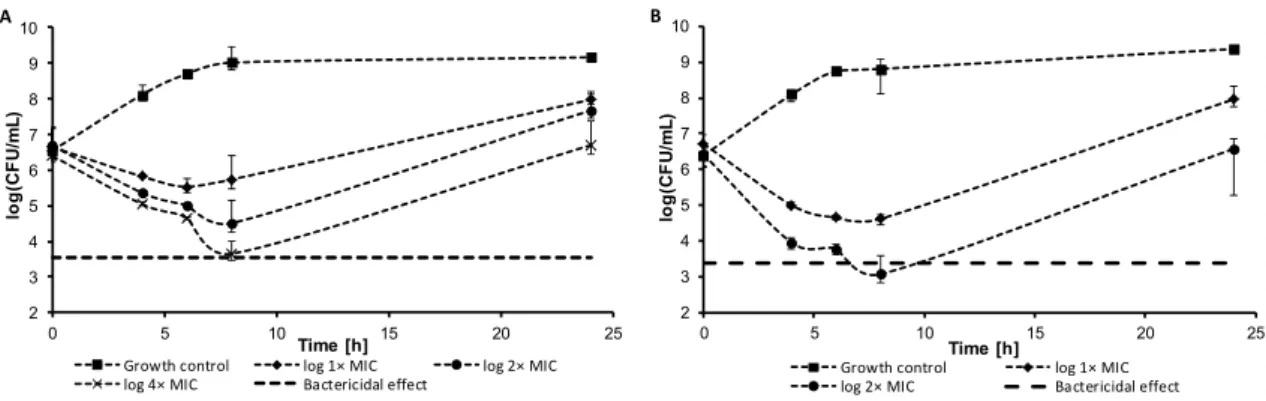
Within the pre-test subcultivation aliquots on agar, antistaphylococcal-effective compounds6 and13showed bactericidal activity, i.e., minimal bactericidal concentrations were≤4×MIC. These facts were verified using the time-kill curve assay for testing the bactericidal effect. The dynamics of antibacterial activity was tested againstS. aureusATCC 29213 for the most active compounds6 (Figure2A) and13(Figure2B). Both compounds showed concentration-dependent activity that was bactericidal in concentration 4×MIC in the case of compound13or very close to the bactericidal level for compound6after 8 h from the beginning of incubation. The increase of bacterial growth at 24 h could be caused by the selection of resistant mutants, as observed previously [37].
Int. J. Mol. Sci.Int. J. Mol. Sci. 2018, 19, x 2018,19, 2318 7 of 24 7 of 25
Figure 2. Time-kill curve of compound 6 (A) and compound 13 (B) against S. aureus ATCC 29213.
2.2.3. Inhibition of Bofilm Formation
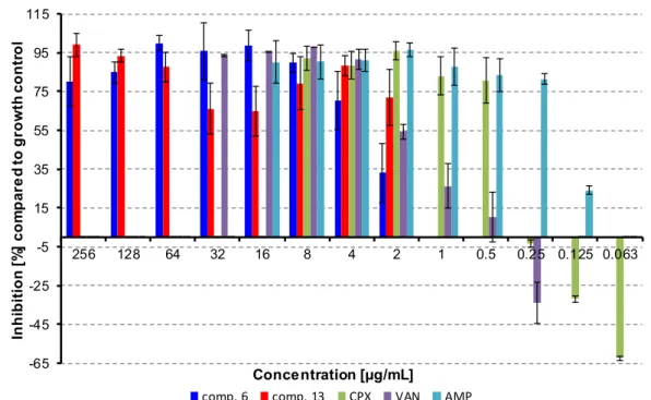
There are many evidences in literature that cinnamic acid derivatives are inhibitors of biofilm formation [47–51]. The most studied derivate is cinnamaldehyde that interacts with quorum sensing system in bacterial biofilms [46,52,53]. Thus, selected compounds were also tested for their ability to inhibit biofilm formation. Compounds 6 and 13 were tested as inhibitors of biofilm formation against S. aureus ATCC 29213. MRSA strains were not producers of biofilm.
The activity of compound 6 does not depend on concentration in concentrations above 8 µg/mL;
only the highest concentration showed lower inhibition effect. This could be caused the higher lipophilicity of the compound and potential formation of precipitates, which could decrease the antibacterial activity of the compound. The lowest concentration of the compound, which inhibited
≥ 80% of biofilm formation, was 8 µg/mL, then the activity sharply decreased. Interestingly, on the other hand, concentrations of compound 13 close to MIC against planktonic cells had the lowest inhibition activities against biofilm forming, and the activity increased for sub-MIC values. These conditions could be potentially toxic for planktonic cells, but they can induce biofilm formation [54].
In general, the inhibition activity against biofilm formation was comparable with the activity against planktonic cells. Despite many studies reported a higher resistance of biofilm, there were also studies that proved a similar or only little lower antibiofilm activity of tested compounds compared to planktonic cells. [13,55]. Budzynska et al. [55] described high antibiofilm activity of plant essential oils compared to MICs. De Vita et al. [13] studied the activity of cinnamic acid derivatives against candida biofilm. These compounds had good effect against biofilm formation, and the effective concentrations were lower than 10× MIC. Thus, the high activity of our compounds can be explained due to their structure, based on cinnamic acid.
Ampicillin (16–0.125 µg/mL), vancomycin (32–0.25 µg/mL), and ciprofloxacin (8–0.063 µg/mL) were used as positive controls. Ciprofloxacin and vancomycin caused the induction of biofilm formation in sub-MIC concentrations, which is in line with already published results [56,57]. All the results are shown in Figure 3.
2.3. In Vitro Antitubercular Activity
The evaluation of the in vitro antitubercular activity of the compounds was performed against Mycobacterium tuberculosis ATCC 25177/H37Ra, see Table 2. In order to reduce risks, a replacement of model pathogens is commonly used in basic laboratory screening. For M. tuberculosis, avirulent strain H37Ra is used that has a similar pathology as M. tuberculosis strains infecting humans and, thus, represents a good model for testing antitubercular agents [58]. The potency of the compounds was expressed as the MIC that is defined for mycobacteria as 90% or greater (IC90) reduction of growth in comparison with the control.
mr
2 3 4 5 6 7 8 9 10
0 5 10 15 20 25
log(CFU/mL)
Time [h]
Growth control log 1× MIC log 2× MIC
log 4× MIC Bactericidal effect A
2 3 4 5 6 7 8 9 10
0 5 10 15 20 25
log(CFU/mL)
Time [h]
Growth control log 1× MIC
log 2× MIC Bactericidal effect
B
Figure 2.Time-kill curve of compound6(A) and compound13(B) againstS. aureusATCC 29213.
2.2.3. Inhibition of Bofilm Formation
There are many evidences in literature that cinnamic acid derivatives are inhibitors of biofilm formation [47–51]. The most studied derivate is cinnamaldehyde that interacts with quorum sensing system in bacterial biofilms [46,52,53]. Thus, selected compounds were also tested for their ability to inhibit biofilm formation. Compounds6and13were tested as inhibitors of biofilm formation against S. aureusATCC 29213. MRSA strains were not producers of biofilm.
The activity of compound6does not depend on concentration in concentrations above 8µg/mL;
only the highest concentration showed lower inhibition effect. This could be caused the higher lipophilicity of the compound and potential formation of precipitates, which could decrease the antibacterial activity of the compound. The lowest concentration of the compound, which inhibited
≥80% of biofilm formation, was 8µg/mL, then the activity sharply decreased. Interestingly, on the other hand, concentrations of compound13close to MIC against planktonic cells had the lowest inhibition activities against biofilm forming, and the activity increased for sub-MIC values. These conditions could be potentially toxic for planktonic cells, but they can induce biofilm formation [54].
In general, the inhibition activity against biofilm formation was comparable with the activity against planktonic cells. Despite many studies reported a higher resistance of biofilm, there were also studies that proved a similar or only little lower antibiofilm activity of tested compounds compared to planktonic cells. [13,55]. Budzynska et al. [55] described high antibiofilm activity of plant essential oils compared to MICs. De Vita et al. [13] studied the activity of cinnamic acid derivatives against candida biofilm. These compounds had good effect against biofilm formation, and the effective concentrations were lower than 10×MIC. Thus, the high activity of our compounds can be explained due to their structure, based on cinnamic acid.
Ampicillin (16–0.125µg/mL), vancomycin (32–0.25µg/mL), and ciprofloxacin (8–0.063µg/mL) were used as positive controls. Ciprofloxacin and vancomycin caused the induction of biofilm formation in sub-MIC concentrations, which is in line with already published results [56,57]. All the results are shown in Figure3.
2.3. In Vitro Antitubercular Activity
The evaluation of the in vitro antitubercular activity of the compounds was performed against Mycobacterium tuberculosisATCC 25177/H37Ra, see Table2. In order to reduce risks, a replacement of model pathogens is commonly used in basic laboratory screening. ForM. tuberculosis, avirulent strain H37Ra is used that has a similar pathology asM. tuberculosisstrains infecting humans and, thus, represents a good model for testing antitubercular agents [58]. The potency of the compounds was expressed as the MIC that is defined for mycobacteria as 90% or greater (IC90) reduction of growth in comparison with the control.
Int. J. Mol. Sci.2018,19, 2318 8 of 25
Int. J. Mol. Sci. 2018, 19, x 8 of 24
Figure 3. Inhibition of bacterial film formation. (CPX = ciprofloxacin, AMP = ampicillin, VAN = vancomycin).
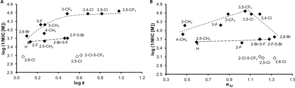
In comparison with antibacterial activities, the investigated compounds exhibited much higher effect against M. tuberculosis. Compounds 13 (R = 3,5-CF3), 10 (R = 3,4-Cl), 11 (R = 3,5-Cl), and 6 (R = 3-CF3) were the most effective; their activity ranged from 22.27 to 27.47 µM. The dependences of the antitubercular activity of the compounds against M. tuberculosis expressed as log (1/MIC (M)) on lipophilicity expressed as log k are illustrated in Figure 4A. When inactive compounds 8 (R = 2,5-Cl), 9 (R = 2,6-Cl), and 16 (R = 2-Cl-5-CF3) are eliminated from the SAR study (illustrated by empty symbols), two different dependences in relation to the position and the type of substituents can be observed. Based on Figure 4A, it can be stated that compounds substituted in positions C(3)’, C(3,4)’,
C(3,5)’, or C(2,6)’ showed an increasing trend of activity with the lipophilicity increase up to compound
6 (R = 3-CF3), at which the activity achieved plateau and from approximately log k ≈ 0.5 had an insignificant increase. The second, in fact, a linear, insignificantly increasing dependence can be found for the compounds substituted in positions C(2)’ and C(2,5)’. It is important to note that the antitubercular activity of the discussed cinnamanilides is also dependent on electronic σ parameters, see Figure 4B. As mentioned above, the linear insignificantly increasing dependence can be found for the derivatives substituted on the anilide in positions C(2)’ and C(2,5)’, while a bilinear dependence of activity on σ (for derivatives substituted in positions C(3)’, C(3,4)’, C(3,5)’, and C(2,6)’) can be observed. The activity increases with the increasing electron-withdrawing effect with r = 0.8803 (n = 5) to optimum σAr ca. 1 (compound 13, R = 3,5-CF3) and then decreases (r = 0.9162, n = 4) with increasing values of the electron-withdrawing parameter.
Additionally, a standard MTT (3-(4,5-dimethylthiazol-2-yl)-2,5-diphenyltetrazolium bromide) assay was performed on selected compounds that were the most effective against M. tuberculosis H37Ra and the MICs of which were previously determined, see Table 2. The MTT test can be used to assess cell growth by measuring respiration. The MTT measured viability of M. tuberculosis H37Ra less than 70% after exposure to the MIC values for each test agent is considered as a positive result of this assay. This low level of cell viability indicates inhibition of cell growth by inhibition of respiration [59]. All the selected compounds, i.e., 6 (R = 3-CF3, 40.99%), 10 (R = 3,4-Cl, 59.65%), 11 (R = 3,5-Cl, 22.65%), and 13 (R = 3,5-CF3, 66.09%) showed less than 70% viability of M. tuberculosis H37Ra at the tested concentration equal to MICs (i.e., 8 µg/mL or 22 and 27 µM). At MIC = 16 µg/mL (33 µM) compound 2 (R = 3-CH3) showed inhibition of viability 13.23%, and compound 5 (R = 3-F) showed inhibition of viability 11.04% at MIC = 32 µg/mL (33 µM).
-65 -45 -25 -5 15 35 55 75 95 115
256 128 64 32 16 8 4 2 1 0.5 0.25 0.125 0.063
Inhibition [%] compared to growth control
Concentration [µg/mL]
comp. 6 comp. 13 CPX VAN AMP
Figure 3. Inhibition of bacterial film formation. (CPX = ciprofloxacin, AMP = ampicillin, VAN = vancomycin).
In comparison with antibacterial activities, the investigated compounds exhibited much higher effect againstM. tuberculosis. Compounds13 (R = 3,5-CF3), 10(R = 3,4-Cl), 11 (R = 3,5-Cl), and 6(R = 3-CF3) were the most effective; their activity ranged from 22.27 to 27.47µM. The dependences of the antitubercular activity of the compounds againstM. tuberculosisexpressed as log (1/MIC (M)) on lipophilicity expressed as logkare illustrated in Figure4A. When inactive compounds8(R = 2,5-Cl), 9 (R = 2,6-Cl), and16 (R = 2-Cl-5-CF3) are eliminated from the SAR study (illustrated by empty symbols), two different dependences in relation to the position and the type of substituents can be observed. Based on Figure4A, it can be stated that compounds substituted in positions C(3)’, C(3,4)’, C(3,5)’, or C(2,6)’ showed an increasing trend of activity with the lipophilicity increase up to compound6(R = 3-CF3), at which the activity achieved plateau and from approximately logk≈0.5 had an insignificant increase. The second, in fact, a linear, insignificantly increasing dependence can be found for the compounds substituted in positions C(2)’ and C(2,5)’. It is important to note that the antitubercular activity of the discussed cinnamanilides is also dependent on electronicσ parameters, see Figure4B. As mentioned above, the linear insignificantly increasing dependence can be found for the derivatives substituted on the anilide in positions C(2)’ and C(2,5)’, while a bilinear dependence of activity onσ(for derivatives substituted in positions C(3)’, C(3,4)’, C(3,5)’, and C(2,6)’) can be observed. The activity increases with the increasing electron-withdrawing effect withr= 0.8803 (n= 5) to optimumσArca. 1 (compound13, R = 3,5-CF3) and then decreases (r= 0.9162,n= 4) with increasing values of the electron-withdrawing parameter.
Additionally, a standard MTT (3-(4,5-dimethylthiazol-2-yl)-2,5-diphenyltetrazolium bromide) assay was performed on selected compounds that were the most effective againstM. tuberculosis H37Ra and the MICs of which were previously determined, see Table2. The MTT test can be used to assess cell growth by measuring respiration. The MTT measured viability ofM. tuberculosisH37Ra less than 70% after exposure to the MIC values for each test agent is considered as a positive result of this assay. This low level of cell viability indicates inhibition of cell growth by inhibition of respiration [59].
All the selected compounds, i.e.,6(R = 3-CF3, 40.99%),10(R = 3,4-Cl, 59.65%),11(R = 3,5-Cl, 22.65%), and13(R = 3,5-CF3, 66.09%) showed less than 70% viability ofM. tuberculosisH37Ra at the tested concentration equal to MICs (i.e., 8µg/mL or 22 and 27µM). At MIC = 16µg/mL (33µM) compound
Int. J. Mol. Sci.2018,19, 2318 9 of 25
2(R = 3-CH3) showed inhibition of viability 13.23%, and compound5(R = 3-F) showed inhibition of viability 11.04% at MIC = 32Int. J. Mol. Sci. 2018, 19, x µg/mL (33µM). 9 of 24
Figure 4. Relationships between in vitro antitubercular activity against M. tuberculosis log (1/MIC (M)) and lipophilicity expressed as log k (A) and electronic Hammett’s σ parameters of ring-substituted anilide ring (B) of studied compounds. (Derivatives excluded from SAR are illustrated by empty symbols.).
Similar effects were observed previously, for example, with ring-substituted 6- hydroxynaphthalene-2-carboxanilides, where 3-Cl, 4-Cl, 3-Br, and 3-CF3 substituted derivatives decreased the viability of M. tuberculosis H37Ra in the range from 41.2% to 46.5% at the lowest tested concentration (MICs = 8 µg/mL) [20], with 8-hydroxy-N-(3-trifluoromethylphenyl)quinoline-2-carbox- amide, where the decrease of the viability of M. tuberculosis H37Ra was to 18.8% at MIC = 8 µg/mL [60], and with N-alkoxyphenylhydroxynaphthalenecarboxanilides substituted in C(3)’ position of the anilide core by a longer alkoxy tail [61,62]. Since the MTT assay was positive, it can be stated that the tested compounds caused a decrease of mycobacterial cell metabolism. Thus, based on the structure analogy of (2E)-N-aryl-3-phenylprop-2-enamides with naphthalene-2-carboxanilides, it may be hypothesized that the mechanism of action of these ring-substituted anilides of cinnamic acid could be connected with the affection of mycobacterial energy metabolism [59,63–66]; nevertheless, another possible site of action of the studied compounds in the mycobacteria cannot be excluded [67–70].
2.4. In Vitro Activity against Plant Pathogenic Fungi
Fungal infections are not only a problem in human and veterinary medicine, but also an important problem in agriculture. Plants diseases in general are a major factor limiting the crop quality. Fungal pathogens cause production losses and also can product mycotoxins, which are dangerous for consumers. The widespread use of fungicides increases food availability and safety, but it can leads to the selection of resistant pathogens and an increase of the production of mycotoxins [71]. As cinnamic acid and its derivatives do not have only antibacterial and antimycobacterial activity, but also activity against plant pathogens [7,72], all the prepared compounds were tested for their potency against Fusarium avenaceum (Fr.) Sacc. IMI 319947 and Bipolaris sorokiniana (Sacc.) Shoemaker H-299. B. sorokiniana is a wide-spread wheat and barley pathogen. It causes many diseases, such as head blight, seedling blight, common root rot, spot blotch, etc. [73]. The last one is a big problem, especially in Southern Asia, where 20% of crop yield is lost because of leaf blight disease [74]. F. avenaceum is one of the most common Fusarium species causing head blight disease of cereals. It can be isolated from cereal seeds and feed products [75]. Fusarium spp. produces a wide spectrum of mycotoxins. The most important are the trichothecenes, zearalenone, moniifromin, and the fumonisins. These compounds have toxic effect on humans and animals [76].
Only compound 6 (R = 3-CF3) showed moderate activity (MIC = 54.93 µM) against F. avenaceum within the series of compounds, see Table 2. On the other hand, the investigated compounds demonstrated higher effect against B. sorokiniana. (2E)-N-(3-Fluorophenyl)- (5) and (2E)-N-(3- methylphenyl)-3-phenylprop-2-enamide (2) had MICs = 16.58 and 33.71 µM, respectively, which is comparable with the benomyl standard. Also compounds 6, 4 (R = 2-F), and 14 (2-F-5-Br) demonstrated moderate activity (MIC range 49.98–66.32 µM) against B. sorokiniana. Surprising was the inactivity of compound 13 (R = 3,5-CF3) against both fungal pathogens.
H
3-CH3
4-CH3
2-F 3-F
3-CF3
2,5-CH3
2,5-Cl 2,6-Cl
3,4-Cl 3,5-Cl
2,6-Br
3,5-CF3
2-F-5-Br 2-Br-5-F
2-Cl-5-CF3
2.5 2.8 3.1 3.4 3.7 4.0 4.3 4.6 4.9
0.0 0.2 0.4 0.6 0.8 1.0 1.2
log (1/MIC[M])
log k A
H 3-CH3
4-CH3
2-F 3-F
3-CF3
2,5-CH3
2,5-Cl 2,6-Cl 3,4-Cl 3,5-Cl
2,6-Br 3,5-CF3
2-F-5-Br 2-Br-5-F 2-Cl-5-CF3
2.5 2.8 3.1 3.4 3.7 4.0 4.3 4.6 4.9
0.3 0.5 0.7 0.9 1.1 1.3 1.5
log (1/MIC[M])
σAr B
Figure 4.Relationships between in vitro antitubercular activity againstM. tuberculosislog (1/MIC (M)) and lipophilicity expressed as logk(A) and electronic Hammett’sσparameters of ring-substituted anilide ring (B) of studied compounds. (Derivatives excluded from SAR are illustrated by empty symbols.).
Similar effects were observed previously, for example, with ring-substituted 6-hydroxynaphthalene- 2-carboxanilides, where 3-Cl, 4-Cl, 3-Br, and 3-CF3 substituted derivatives decreased the viability of M. tuberculosis H37Ra in the range from 41.2% to 46.5% at the lowest tested concentration (MICs = 8µg/mL) [20], with 8-hydroxy-N-(3-trifluoromethylphenyl)quinoline-2-carbox- amide, where the decrease of the viability ofM. tuberculosisH37Ra was to 18.8% at MIC = 8µg/mL [60], and withN-alkoxyphenylhydroxynaphthalenecarboxanilides substituted in C(3)’ position of the anilide core by a longer alkoxy tail [61,62]. Since the MTT assay was positive, it can be stated that the tested compounds caused a decrease of mycobacterial cell metabolism. Thus, based on the structure analogy of (2E)-N-aryl-3-phenylprop-2-enamides with naphthalene-2-carboxanilides, it may be hypothesized that the mechanism of action of these ring-substituted anilides of cinnamic acid could be connected with the affection of mycobacterial energy metabolism [59,63–66]; nevertheless, another possible site of action of the studied compounds in the mycobacteria cannot be excluded [67–70].
2.4. In Vitro Activity against Plant Pathogenic Fungi
Fungal infections are not only a problem in human and veterinary medicine, but also an important problem in agriculture. Plants diseases in general are a major factor limiting the crop quality. Fungal pathogens cause production losses and also can product mycotoxins, which are dangerous for consumers. The widespread use of fungicides increases food availability and safety, but it can leads to the selection of resistant pathogens and an increase of the production of mycotoxins [71]. As cinnamic acid and its derivatives do not have only antibacterial and antimycobacterial activity, but also activity against plant pathogens [7,72], all the prepared compounds were tested for their potency againstFusarium avenaceum(Fr.) Sacc. IMI 319947 andBipolaris sorokiniana(Sacc.) Shoemaker H-299.
B. sorokinianais a wide-spread wheat and barley pathogen. It causes many diseases, such as head blight, seedling blight, common root rot, spot blotch, etc. [73]. The last one is a big problem, especially in Southern Asia, where 20% of crop yield is lost because of leaf blight disease [74].F. avenaceumis one of the most commonFusariumspecies causing head blight disease of cereals. It can be isolated from cereal seeds and feed products [75].Fusariumspp. produces a wide spectrum of mycotoxins. The most important are the trichothecenes, zearalenone, moniifromin, and the fumonisins. These compounds have toxic effect on humans and animals [76].
Only compound 6 (R = 3-CF3) showed moderate activity (MIC = 54.93 µM) against F. avenaceum within the series of compounds, see Table 2. On the other hand, the investigated compounds demonstrated higher effect against B. sorokiniana. (2E)-N-(3-Fluorophenyl)- (5) and (2E)-N-(3-methylphenyl)-3-phenylprop-2-enamide (2) had MICs = 16.58 and 33.71µM, respectively,
Int. J. Mol. Sci.2018,19, 2318 10 of 25
which is comparable with the benomyl standard. Also compounds6,4(R = 2-F), and14(2-F-5-Br) demonstrated moderate activity (MIC range 49.98–66.32µM) againstB. sorokiniana.Surprising was the inactivity of compound13(R = 3,5-CF3) against both fungal pathogens.
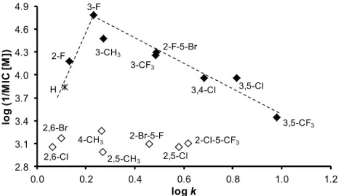
The dependences of the antifungal activity of the compounds againstB. sorokinianaexpressed as log (1/MIC (M)) on lipophilicity expressed as logkare illustrated in Figure5. In general, effective compounds are preferentially substituted in positions C(3)’, C(3,5)’, or C(3,4)’. When inactive compounds (illustrated by empty symbols) substituted in C(4)’, C(2,6)’, or C(2,5)’ are eliminated from the SAR study, a bilinear dependence can be found. The activity increases with increasing lipophilicity from unsubstituted derivative1to compound5(R = 3-F) with the supposed lipophilicity optimum log k= 0.23 and then decreases to derivative13(R = 3,5-CF3);r= 0.9678,n= 7. It can be stated that it is an opposite trend in comparison with antitubercular findings, which can be caused by differences in the composition and structure of mycobacterial and fungal cell walls [77]. A similar trend can be found for electronic properties of substituents in individual derivatives. The activity increases with an increase of electron-withdrawing effect from compound2(R = 3-CH3,σAr= 0.48) to an optimumσAr= 0.82 (compound5, R = 3-F) and then decreases with increasing electron-withdrawing effect as follows:
σAr= 0.89 (compound6, R = 3-CF3), 1.02 (compound4, R = 2-F), 1.11 (compound11, R = 3,5-Cl), and 1.19 (compound10, R = 3,4-Cl).
Int. J. Mol. Sci. 2018, 19, x 10 of 24
The dependences of the antifungal activity of the compounds against B. sorokiniana expressed as log (1/MIC (M)) on lipophilicity expressed as log k are illustrated in Figure 5. In general, effective compounds are preferentially substituted in positions C(3)’, C(3,5)’, or C(3,4)’. When inactive compounds (illustrated by empty symbols) substituted in C(4)’, C(2,6)’, or C(2,5)’ are eliminated from the SAR study, a bilinear dependence can be found. The activity increases with increasing lipophilicity from unsubstituted derivative 1 to compound 5 (R = 3-F) with the supposed lipophilicity optimum log k = 0.23 and then decreases to derivative 13 (R = 3,5-CF3); r = 0.9678, n = 7. It can be stated that it is an opposite trend in comparison with antitubercular findings, which can be caused by differences in the composition and structure of mycobacterial and fungal cell walls [77]. A similar trend can be found for electronic properties of substituents in individual derivatives. The activity increases with an increase of electron-withdrawing effect from compound 2 (R = 3-CH3, σAr = 0.48) to an optimum σAr = 0.82 (compound 5, R = 3-F) and then decreases with increasing electron-withdrawing effect as follows:
σAr = 0.89 (compound 6, R = 3-CF3), 1.02 (compound 4, R = 2-F), 1.11 (compound 11, R = 3,5-Cl), and 1.19 (compound 10, R = 3,4-Cl).
Figure 5. Relationships between in vitro antifungal activity against B. sorokiniana log (1/MIC (M)) and lipophilicity expressed as log k of studied compounds. (Derivatives excluded from SAR are illustrated by empty symbols.).
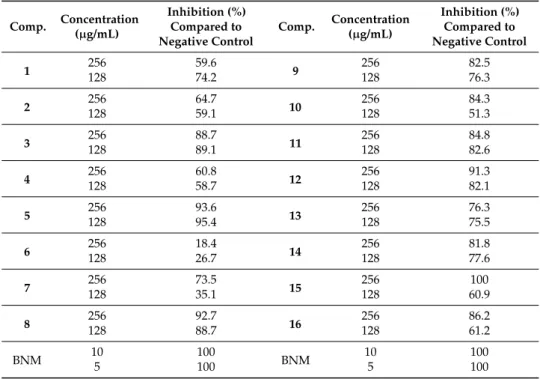
Inhibition of B. sorokiniana Germination
All the compounds were additionally evaluated for the inhibition of B. sorokiniana conidium germination at two concentrations (128 and 256 µg/mL), see results in Table 4. It can be stated that at both concentrations, the compounds showed the inhibition of germination. The effect was concentration-dependent for compounds 7, 10, 15, and 16, and the rest of compounds had concentration-independent effect on the germination. Interestingly, compound 6 that was one of the most active against all bacterial cells including M. tuberculosis and displayed strong inhibition on the mycelial growth of both investigated fungi (F. avenaceum and B. sorokiniana) (Table 2), showed the weakest effect in the germination test. Apart from compound 6, compounds 2, 4, 5, and 14 exerted the highest B. sorokiniana mycelial growth inhibitory effect and had characteristic anti-germination activity. Moreover, compound 5 was the most effective in both assays (IC50 = 16.58 µM in mycelial growth test and 95.4% germination inhibition at 128 µg/mL).
2.5. In Vitro Antiproliferative Assay
The preliminary in vitro screening of the antiproliferative activity of the most effective antimicrobial compounds was performed using a Water Soluble Tetrazolium salts-1 (WST-1) assay kit [78] and the human monocytic leukemia THP-1 cell line by means of the method described recently [20,79]. The principle of the WST-1 assay kit is that antiproliferative compounds inhibit mitochondrial dehydrogenases. The activity of this enzyme directly correlates with the number of metabolically active cells in the culture. Antiproliferative effect was evaluated as IC50 value (concentration of compound causing 50% inhibition of cell proliferation). It can be stated that a compound is
H
3-CH3
4-CH3
2-F 3-F
3-CF3
2,5-CH3 2,5-Cl 2,6-Cl
3,4-Cl 3,5-Cl
2,6-Br 3,5-CF3
2-F-5-Br
2-Br-5-F 2-Cl-5-CF3
2.8 3.1 3.4 3.7 4.0 4.3 4.6 4.9
0.0 0.2 0.4 0.6 0.8 1.0 1.2
log (1/MIC[M])
log k
Figure 5.Relationships between in vitro antifungal activity againstB. sorokinianalog (1/MIC (M)) and lipophilicity expressed as logkof studied compounds. (Derivatives excluded from SAR are illustrated by empty symbols.).
Inhibition ofB. sorokinianaGermination
All the compounds were additionally evaluated for the inhibition ofB. sorokinianaconidium germination at two concentrations (128 and 256µg/mL), see results in Table 4. It can be stated that at both concentrations, the compounds showed the inhibition of germination. The effect was concentration-dependent for compounds 7, 10, 15, and 16, and the rest of compounds had concentration-independent effect on the germination. Interestingly, compound6that was one of the most active against all bacterial cells includingM. tuberculosisand displayed strong inhibition on the mycelial growth of both investigated fungi (F. avenaceumandB. sorokiniana)(Table2), showed the weakest effect in the germination test. Apart from compound6, compounds2,4,5, and14exerted the highestB. sorokinianamycelial growth inhibitory effect and had characteristic anti-germination activity.
Moreover, compound5was the most effective in both assays (IC50= 16.58µM in mycelial growth test and 95.4% germination inhibition at 128µg/mL).
2.5. In Vitro Antiproliferative Assay
The preliminary in vitro screening of the antiproliferative activity of the most effective antimicrobial compounds was performed using a Water Soluble Tetrazolium salts-1 (WST-1) assay kit [78] and the human monocytic leukemia THP-1 cell line by means of the method described