REVIEW ARTICLE published: 20 August 2014 doi: 10.3389/fimmu.2014.00348
Placental Protein 13 (PP13) – a placental immunoregulatory galectin protecting pregnancy
Nándor Gábor Than1,2,3,4*, Andrea Balogh5, Roberto Romero1, Éva Kárpáti5, Offer Erez6, András Szilágyi4, Ilona Kovalszky7, Marei Sammar8, Sveinbjorn Gizurarson9, János Matkó5, Péter Závodszky4, Zoltán Papp3 and Hamutal Meiri10,11*
1Perinatology Research Branch, Eunice Kennedy Shriver National Institute of Child Health and Human Development, National Institutes of Health, U.S. Department of Health and Human Services, Bethesda, MD, and Detroit, MI, USA
2Department of Obstetrics and Gynecology, Wayne State University School of Medicine, Detroit, MI, USA
3Maternity Private Department, Kútvölgyi Clinical Block, Semmelweis University, Budapest, Hungary
4Institute of Enzymology, Research Centre for Natural Sciences, Hungarian Academy of Sciences, Budapest, Hungary
5Department of Immunology, Eötvös Loránd University, Budapest, Hungary
6Department of Obstetrics and Gynecology, Soroka University Medical Center, Ben-Gurion University of the Negev, Beer-Sheva, Israel
7First Department of Pathology and Experimental Cancer Research, Semmelweis University, Budapest, Hungary
8Prof. Ephraim Katzir Department of Biotechnology Engineering, ORT Braude College, Karmiel, Israel
9Faculty of Pharmaceutical Sciences, School of Health Science, University of Iceland, Reykjavik, Iceland
10TeleMarpe Ltd., Tel Aviv, Israel
11Hylabs Ltd., Rehovot, Israel
Edited by:
Sinuhe Hahn, University Clinics Basel, Switzerland
Reviewed by:
Stefan Gebhardt, Stellenbosch University, South Africa
Kai Fang, University of California Los Angeles, USA
*Correspondence:
Nándor Gábor Than, Institute of Enzymology, Research Centre for Natural Sciences, Hungarian Academy of Sciences, Magyar tudósok körútja 2, Budapest H-1117, Hungary e-mail: than.gabor@ttk.mta.hu;
Hamutal Meiri , TeleMarpe Ltd., 12 Barasani Street, Tel Aviv 69127, Israel e-mail: hamutal62@hotmail.com
Galectins are glycan-binding proteins that regulate innate and adaptive immune responses, and some confer maternal-fetal immune tolerance in eutherian mammals. A chromosome 19 cluster of galectins has emerged in anthropoid primates, species with deep placentation and long gestation. Three of the five human cluster galectins are solely expressed in the placenta, where they may confer additional immunoregulatory functions to enable deep placentation. One of these is galectin-13, also known as Placental Protein 13 (PP13). It has a
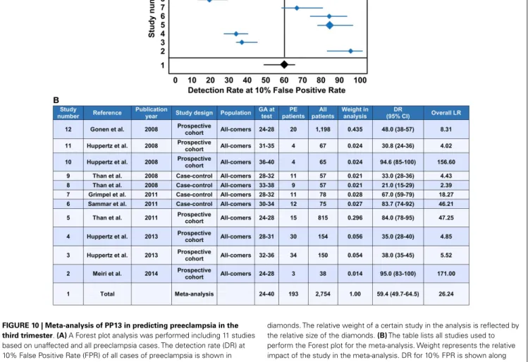
“jelly-roll” fold, carbohydrate-recognition domain and sugar-binding preference resembling other mammalian galectins. PP13 is predominantly expressed by the syncytiotrophoblast and released from the placenta into the maternal circulation. Its ability to induce apoptosis of activated T cells in vitro, and to divert and kill T cells as well as macrophages in the maternal deciduain situ,suggests important immune functions. Indeed, mutations in the promoter and an exon ofLGALS13presumably leading to altered or non-functional protein expression are associated with a higher frequency of preeclampsia and other obstetrical syndromes, which involve immune dysregulation. Moreover, decreased placental expres- sion of PP13 and its low concentrations in first trimester maternal sera are associated with elevated risk of preeclampsia. Indeed, PP13 turned to be a good early biomarker to assess maternal risk for the subsequent development of pregnancy complications caused by impaired placentation. Due to the ischemic placental stress in preterm preeclampsia, there is increased trophoblastic shedding of PP13 immunopositive microvesicles starting in the second trimester, which leads to high maternal blood PP13 concentrations. Our meta-analysis suggests that this phenomenon may enable the potential use of PP13 in directing patient management near to or at the time of delivery. Recent findings on the beneficial effects of PP13 on decreasing blood pressure due to vasodilatation in pregnant animals suggest its therapeutic potential in preeclampsia.
Keywords: actin cytoskeleton, biomarker, danger signal, evolution, extracellular vesicles, glycans, lectins, maternal-fetal interface
PREFACE
Many authors of this review have collaborated with Dr. Hans Bohn, the discoverer of Placental Protein 13 (PP13), who passed away on January 25, 2014. We dedicate this manuscript to his memory. His scientific legacy and enormous contribution to placental protein research have strongly influenced placentology and inspired our studies on PP13 (1).
Hans Bohn was born in Munich on October 18, 1928. He gradu- ated in 1954 and completed his doctoral thesis in 1956 in chemistry
at the University of Würzburg. A research fellowship starting in 1963 in the Protein Research Laboratory at the University of Pitts- burgh was critical in directing his interest in protein research.
After two years, Dr. Bohn returned to Germany to work on pro- teins in Behringwerke in Marburg/Lahn (Figure 1A). He was the first to isolate factor XIII from human placenta for the treatment of patients with factor XIII deficiency and wounds after injury or surgery, and a side-fraction of this experiment yielded human placental lactogen. This experience strongly influenced him to
focus his research on the systematic isolation and characterization of placental, endometrial and pregnancy serum proteins. These studies have greatly supported our knowledge on pregnancy- related proteins and their application in the diagnosis of pregnancy complications (1).
Dr. Hans Bohn processed large amounts of human placental, amniotic fluid and serum specimens and utilized combinations of classical fractionation techniques to isolate more than 50 proteins, which he named sequentially. He characterized these proteins for their physico-chemical characteristics, and then developed specific rabbit antisera against them for further protein purification and for the development of immunoassays to determine these proteins’
diagnostic significance. In collaboration with scientists around the world, Dr. Bohn also determined the amino acid sequence as well as biological functions of many of these. Among the proteins he isolated were Placental Protein (PP) 4 (annexin-V), PP5 (tissue factor pathway inhibitor-2, TFPI-2), PP10 (plasminogen activa- tor inhibitor-2, PAI-2), PP12 (insulin-like growth factor binding protein-1, IGFBP-1) and PP13 (galectin-13), which were subse- quently identified to be important regulators of the fundamental processes in pregnancy (1).
Dr. Bohn’s collaboration with Professor Gábor N. Than (Uni- versity of Pécs, Pécs, Hungary) had a fundamental impact on the cloning, sequencing, structural, and molecular biological charac- terization of a large number of PPs including PP13, and their pio- neering collaborative research significantly improved our under- standing on the biological role and diagnostic significance of these proteins in pregnancy complications and malignancies. Beyond
these scientific discoveries, their friendship and close collabora- tion strongly inspired a new generation of scientists. The existing knowledge and advancements in the field were summarized in their book entitledAdvances in Pregnancy-Related Protein Research, co-written with Dr. Dénes G. Szabó in 1993 (2) (Figure 1B).
For Dr. Bohn’s founding research and discovery of PP14 (gly- codelin), he shared the prestigious Abbott Award in 1997. His remarkable contributions to placental and pregnancy-related pro- tein research were published in 198 research articles. Dr. Bohn continued to contribute to placental protein research after his retirement, and closely followed the studies implementing novel molecular and cellular biological techniques on the proteins he iso- lated, leading to further discoveries and improvements in clinical diagnostics and patient care (1).
Dr. Hans Bohn was an exceptional scientist, an enthusiastic cat- alyzer of collaborations and friendships who inspired many peers and followers. He was a wonderful, kind and charismatic person, a silent giant, who will be greatly missed.
THE DISCOVERY AND MOLECULAR CHARACTERIZATION OF PP13
ISOLATION, PURIFICATION AND PHYSICO-CHEMICAL CHARACTERIZATION OF PP13
Dr. Bohn’s scientific vision combined with his thorough work using state-of-the-art methods of the 70’s and 80’s yielded the dis- covery of 26 soluble placental tissue proteins, among which PP13 was purified, physico-chemically characterized and described in 1983 (3). Dr. Bohn homogenized term placental tissues and
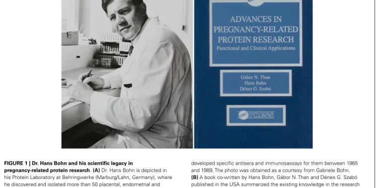
FIGURE 1 | Dr. Hans Bohn and his scientific legacy in
pregnancy-related protein research.(A)Dr. Hans Bohn is depicted in his Protein Laboratory at Behringwerke (Marburg/Lahn, Germany), where he discovered and isolated more than 50 placental, endometrial and pregnancy serum proteins including Placental Protein 13 (PP13), and
developed specific antisera and immunosassays for them between 1965 and 1989. The photo was obtained as a courtesy from Gabriele Bohn.
(B)A book co-written by Hans Bohn, Gábor N. Than and Dénes G. Szabó published in the USA summarized the existing knowledge in the research field in 1993.
Than et al. PP13 in health and disease
fractionated the protein extracts by a step-wise process that included Rivanol and ammonium sulphate fractionation, gel- filtration, ethanol precipitation, and immunoabsorption tech- niques. The resulting PP13 protein was>99% pure, and SDS- polyacrylamide gel electrophoresis found it to be composed of two identical ~16 kDa subunits held together by disulphide bonds.
The carbohydrate content of PP13 was found negligible, a feature that later became important in understanding its functions (2, 3). Utilizing the purified PP13-specific rabbit antiserum, an elec- troimmunoassay and an Ouchterlony’s gel-diffusion test found an average amount of 3.7 mg PP13 in human term placentas and detected PP13 solely in the placenta among fetal and adult tissues.
The sensitivity of a radioimmunoassay (0.8 ng/ml) that utilized this rabbit antiserum was insufficient to detect PP13 in maternal and fetal serum or in amniotic fluid (2–4). Indeed, this is in accord with the 0.1–0.4 ng/ml concentration range of PP13 in maternal blood as was discovered with sandwich ELISA techniques using mouse monoclonal antibodies a decade later (5).
CLONING, SEQUENCING AND INITIAL MOLECULAR BIOLOGICAL ANALYSIS OF PP13
Professor Gábor N. Than’s team in Hungary isolated the full- length cDNA (GenBank Acc. No.: AF117383) encoding PP13 from a human placental cDNA expression library using Dr. Bohn’s rab- bit anti-PP13 antiserum. Sequence analysis revealed a 578 bp insert with a 417 bp open reading frame encoding a 139 amino-acid pro- tein (6). The predicted molecular mass and amino-acid composi- tion of the cloned protein corresponded with Dr. Bohn’s estimate of the purified PP13 protein. A BLAST search of nucleotide and protein sequences showed PP13 to be homologous to members of the beta-galactoside binding galectin family, and computer analy- sis detected 8 out of 16 invariant residues in galectins conserved in PP13, suggesting its place in the galectin family (6). The high- est sequence similarity of PP13 was found with the eosinophil Charcot–Leyden Crystal (CLC) protein, which forms crystals at sites of eosinophil-associated inflammation (7), a phenome- non similar to that found with PP13 immunostainings on first trimester decidual tissue sections (8). Interestingly, using a func- tional assay and highly sensitive NMR measurements, the native and recombinant PP13 was observed to have weak lysophopholi- pase activity (6,9) similar to CLC protein. This lysophopholipase activity was also inferred from the release of free fatty acids from cultured primary trophoblasts exposed to PP13 (5). However, it was later revealed that this enzymatic activity of CLC protein is caused by an associated lysophospholipase (10), and further research is required to understand whether the lysophospholipase activity of PP13 is intrinsic or indeed related to an associated protein.
DETAILED STRUCTURAL AND FUNCTIONAL CHARACTERIZATION OF PP13 AS A GALECTIN
The homology of PP13 to members of the galectin family inspired further structural and functional investigations. Homology mod- elling based on the “jelly-roll” fold of galectins observed by X-ray crystallography revealed the 3D model of PP13, which was deposited into the Protein Data Bank (Acc. No.: 1F87) (11). This fold consists of five- and six-stranded β-sheets
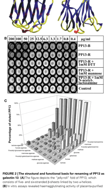
FIGURE 2 | The structural and functional basis for renaming of PP13 as galectin-13.(A)The figure depicts the “jelly-roll” fold of PP13, which consists of five- and six-strandedβ-sheets linked by twoα-helices.
(B)In vitroassays revealed haemagglutinating activity of placenta-purified (PP13-B) and recombinant (PP13-R) PP13. In non-reducing conditions, both PP13-B and PP13-R agglutinated human erythrocytes at≥50µg/ml protein concentrations. The haemagglutinating ability of PP13-R was inhibited by dithiothreitol or sugars at≥1 mM concentrations.(C)The strength of PP13-R binding to sugar-coupled agarose beads increased from lactose-agarose to glucose-agarose (left to right). PP13 bound to sugar-coupled agarose beads was competitively eluted by sugars (1 M) listed back to front. PP13 had the best eluting capacity (sugar affinity) for N-acetyl-lactosamine, mannose andN-acetyl-galactosamine.
Figure(A)was published in Ref. (11), and Figures(B,C)in Ref. (9). Kind permission for the reuse of figures was obtained from Oxford University Press(A)and John Wiley and Sons(B,C).
linked by two α-helices characteristic for “prototype” galectins (Figure 2A). Out of the eight consensus residues in the galectin carbohydrate-recognition domains (CRDs), four identical and
three conservatively substituted residues were found in PP13.
Computational docking simulations showed that the PP13 CRD may bind sugars, e.g. N-acetyl-lactosamine and lactose, simi- lar to most galectins (Figure 2A). Since these lines of evidence demonstrated that PP13 is a novel galectin, it was designated as galectin-13 (11).
Galectins constitute a subgroup among the superfamily of lectins, carbohydrate-binding proteins that are important in the regulation of cellular interactions with cells, the extracellular matrix and pathogens. They bind to glycans residing on glyco- proteins, glycolipids and other glycoconjugates that constitute a complex array coined the “glycome”, which stores orders of magni- tude larger biological information than nucleic acids and proteins store. For example, the numbers of 4,096 hexanucleotides and 64 million hexapeptides are far surpassed by the 1.44×10¹⁵iso- mer quantity of hexasaccharides (12). Galectins can bind to a diverse set of glycoconjugates, and therefore, they have pleiotropic functions in a variety of key biological processes including sig- nal transduction, cell differentiation, apoptosis, or cell adhesion.
Moreover, galectins are positioned at the cross-roads of adaptive and innate immune functions as they are key determinants of acute and chronic inflammation, immune tolerance and host-pathogen interactions (12–18).
Triggered by the recognition that these galectin functions are important determinants of healthy pregnancies (19), the detailed molecular characterization of PP13 allowed greater insight into its function in the placenta during pregnancy. Similar to other galectins, PP13 also hemagglutinated human erythrocytesin vitro (Figure 2B). Furthermore, sugar-binding assays showed the affin- ity of PP13 for carbohydrates widely expressed in the human placenta (Figure 2C), particularly forN-acetyl-lactosamine, man- nose andN-acetyl-galactosamine (9) as already predicted by mol- ecular modelling (11). Assay performance in reducing conditions decreased the hemagglutinating (Figure 2B) and sugar binding activity of PP13, suggesting that homodimerization of PP13 sub- units by disulphide bridges are important for these functions (9).
Through placental immunostaining, PP13 was found in the cytoplasm and brush border membrane of the syncytiotro- phoblast. Using affinity chromatography and mass spectrometry, annexin II and beta/gamma actin were identified as ligands of PP13, a finding that was also supported by high colocalization of PP13 with annexin II in the syncytiotrophoblast brush border membrane. These results suggested the galectin-like externaliza- tion of PP13 to the cell surface by extracellular vesicles containing actin and annexin II (9). It has to be elucidated whether, similar to other galectins (20), PP13 may bind to glycoconjugates on cell surfaces and form “galectin-glycan lattices” that are important in cellular interactions and signaling.
THE INTERACTIONS OF PP13 WITH ABO BLOOD GROUP ANTIGENS As an indirect sign of PP13 binding to glycoconjugates on cell surfaces, placental immunostainings showed PP13 positiv- ity of maternal and fetal erythrocytes, confirming the in vivo erythrocyte-binding of PP13 (21). These results were consistent with the tendency of PP13, similar to other galectins, to bind beta- galactosides that are present at terminal positions on ABO blood- group antigens (9, 11, 22, 23). Flow cytometry measurements
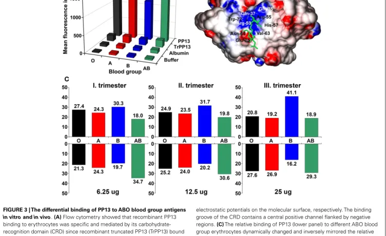
further demonstrated the binding of PP13 but not its CRD- truncated variant to erythrocytes, proving that PP13 binding is mediated by its CRD (Figure 3A). PP13 binding was similar in intensity to blood group A and O erythrocytes, while PP13 had the weakest binding intensity to blood group B and the strongest binding intensity to blood group AB erythrocytes (Figure 3A).
Similar to other galectins (24,25), PP13 binding to various ABO blood group erythrocytes changed dynamically with increasing PP13 concentrations (Figure 3C).
Computational studies have indicated that the structural basis for ABO blood group antigen binding includes the following: (1) three out of four residues in the core galectin CRD involved in disaccharide-binding are conserved in PP13 (11, 23); (2) PP13 has a similar “B-site” involved in ABO antigen binding as human galectins that exhibit ABO blood group antigen binding (21,25);
and (3) PP13 accommodates blood group H trisaccharide in its CRD similar to a fungal galectin’s CRD (21) (Figure 3B).
It is interesting to note that ABO blood group antigens are oligosaccharides attached to cell-surface glycoconjugates on epithelia, endothelia and erythrocytes, which might have been evolutionarily advantageous in conferring resistance against cer- tain pathogens (26). The gene encoding for the enzymes that catalyze the transfer of these oligosaccharides to cell-surface gly- coconjugates emerged in primates (22, 27). If the evolution of the ABO blood group system and genes encoding for PP13 and closely related galectins was somehow associated, that would sug- gest a potential functional relevance of PP13 binding to ABO blood group antigens.
THE EVOLUTION AND HUMAN DISEASE-RELATED POLYMORPHISMS OFLGALS13
EVOLUTIONARY ANALYSES OF GENES ENCODING FOR PP13 AND CLOSELY RELATED GALECTINS
An evolutionary study presented compelling evidence that a cluster of galectin genes, includingLGALS13that encodes PP13, emerged on chromosome 19 in anthropoid primates, which differ from other primates by having larger brains and longer gestations (23).
The analysis of this galectin cluster in the available genome assem- blies revealed frequent gene duplication, inversion and deletion events characteristic of repeat-mediated “birth-and-death” evolu- tion, a process that leads to novel phenotypes in species adapting to their changing environment (28). Detailed analysis showed that transposable long interspersed nuclear elements (LINEs) were positioned at the majority of boundaries of large inversions and gene duplication units, suggesting that LINEs had mediated the rearrangements within this cluster. Genes in this cluster have four- exon structures as other “prototype” galectin genes. Of the two major clades in the cluster, one contains genes with predominant placental expression includingLGALS13and related pseudogenes.
Of note,LGALS13 was only found in Old World monkeys and apes. Sequence analyses of 24 newly determined sequences and 69 annotated sequences in 10 anthropoid species indicated func- tional diversification among PP13 and related galectins during evolution as can be inferred from the amino acid replacements in their CRDs (23).
Sequence comparison showed a strong conservation of more than half of the residues of PP13 and cluster galectins
Than et al. PP13 in health and disease
FIGURE 3 | The differential binding of PP13 to ABO blood group antigens in vitroandin vivo.(A)Flow cytometry showed that recombinant PP13 binding to erythrocytes was specific and mediated by its carbohydrate- recognition domain (CRD) since recombinant truncated PP13 (TrPP13) bound negligibly similar to bovine serum albumin (BSA). PP13 had the strongest affinity to blood group AB erythrocytes and weakest affinity to blood group B erythrocytes.(B)Surface representation of PP13 complexed with blood group H trisaccharide (green). Blue and red colors indicate positive and negative
electrostatic potentials on the molecular surface, respectively. The binding groove of the CRD contains a central positive channel flanked by negative regions.(C)The relative binding of PP13 (lower panel) to different ABO blood group erythrocytes dynamically changed and inversely mirrored the relative serum PP13 concentrations (upper panel) in women with different ABO blood groups with advancing gestation from the first to third trimesters. All figures were published in Ref. (21). Kind permission for the reuse of figures was obtained from the Public Library of Science.
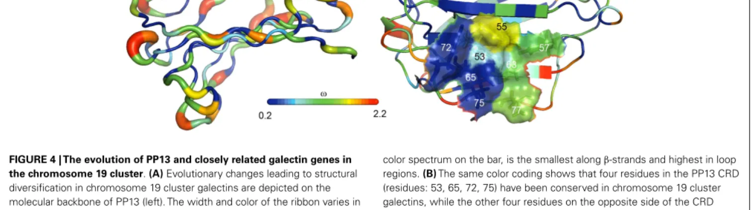
predominantly located in the protein cores that determine their overall structure, whereas residues on their surface, espe- cially in the loop regions, have undergone rapid evolution (23) (Figure 4A). From the eight conserved residues in the galectin CRD, four (positions: 53, 65, 72, and 75) that are key in the overall sugar binding form a pocket in one side of the CRD and were under purifying selection in PP13 and cluster galectins. The other four residues (positions: 55, 57, 63, and 77) on the opposite side of the CRD had more variability among cluster galectins, with frequent replacements in several lineages following gene duplica- tions including K>T77 in PP13 (Figure 4B). As these latter four residues are crucial for galactose or glucose binding, the structural differences might have resulted in differing functions between PP13 and other cluster galectins. Indeed, functional experiments with human recombinant PP13 and five other galectins proved the different sugar-binding profiles of these investigated proteins (23).
A large number of pseudogenes in the studied species were found in the cluster (23). These emerged by the deletion of exons, mutations of the exon–intron boundaries, and by the introduction of one or more in-frame premature stop codons. As a strik- ing observation, 18 out of the identified 38 pseudogene variants
contained the “163C>T” DNA variant leading to the introduc- tion of a premature stop codon at the site encoding residue 55, which may result in truncated proteins of 54 amino acids that lack the entire CRD. In fact, functional experiments proved that this “163C>T” DNA variant results in the expression of a trun- cated PP13 that cannot bind carbohydrates (23). The question why nature utilized the same process to silence so many galectin genes in certain lineages, includingLGALS13in baboon, Bornean orangutan and Sumatran orangutan, remains unanswered.
DNA VARIANTS IN HUMANLGALS13
Somewhat related to evolutionary selection, a total of 933 LGALS13 DNA variants have been already identified in the genomes of 1,092 individuals from 14 populations by the “1000 Genomes Project”1. These included mostly upstream (n=277), downstream (n=261) or intron (n=240) variants. Besides these, non-coding transcript (n=98) or exon (n=56) variants and mis- sense variants (n=56) were frequently detected. Nevertheless, the
1www.1000genomes.org
FIGURE 4 | The evolution of PP13 and closely related galectin genes in the chromosome 19 cluster.(A)Evolutionary changes leading to structural diversification in chromosome 19 cluster galectins are depicted on the molecular backbone of PP13 (left). The width and color of the ribbon varies in proportion with site-specificωvalues (dN/dS;ω<1, purifying selection;ω>1, positive selection) for chromosome 19 cluster galectins.ω, indicated by the
color spectrum on the bar, is the smallest alongβ-strands and highest in loop regions.(B)The same color coding shows that four residues in the PP13 CRD (residues: 53, 65, 72, 75) have been conserved in chromosome 19 cluster galectins, while the other four residues on the opposite side of the CRD (residues: 55, 57, 63, 77) show more evolutionary changes among these galectins.
“1000 Genomes Project” has not provided information regarding the association ofLGALS13 DNA variants with disease suscep- tibility. In search ofLGALS13DNA polymorphisms by targeted genotyping studies, the association of certainLGALS13DNA vari- ants with severe complications of pregnancy has been identified.
These studies utilized whole blood DNA samples obtained from pregnant women and their neonates in a South African cohort of the Black and Coloured population, and the following DNA vari- ants were revealed forLGALS13: (1) variants in Exon 3 including single nucleotide polymorphisms (SNPs) and a single nucleotide deletion, which latter causes a frame-shift in the open reading frame, leading to the formation of a premature stop codon and a truncated protein (221delT); (2) SNPs in Introns 2 and 3 including an intron boundary polymorphism that is associated with alter- native splicing and the deletion of Exon 2; and (3) an SNP in the promoter region (29–34).
Of interest, in a prospective cohort of 450 low-risk primigravid women of Black and Coloured origin, carrying the naturally occur- ring “221delT” mutation conferred a 2.27-fold relative risk for preterm labor (34). The frequency of heterozygous carriers of this mutation was higher in the group of women with preterm preeclampsia (5.7%) than in controls (2.4%), and no individuals were found to be homozygous. In another study conducted on the same population, there was a significant association for this muta- tion and preeclampsia, particularly among Coloured women (33).
These results suggest that the placental expression of a function- ally impaired, truncated PP13 may put women at increased risk for severe pregnancy complications. However, so far no polypep- tide derived from the “221delT” DNA polymorphism could be identified in placental or body fluid samples, most likely due to the rapid degradation or insufficient immunodetection of such a protein because of the anticipated major misfolding (35).
The “-98A/C” promoter polymorphism was also associated with the risk of preeclampsia (31,33,34). In a prospective cohort of low-risk pregnant women, controls were in the Hardy-Weinberg equilibrium, while cases deviated from that, and the heterozy- gous A/C genotype appeared to be protective against preeclampsia
(31). Another study comprising the same population found a sig- nificant difference between “-98A/C” genotype distributions in patients with placental abruption and controls among Coloured women (33). These results suggest that the “-98A/C” promoter polymorphism may negatively affect LGALS13 expression and PP13 functions.
THE EXPRESSION PATTERN OF PP13 IN HUMANS
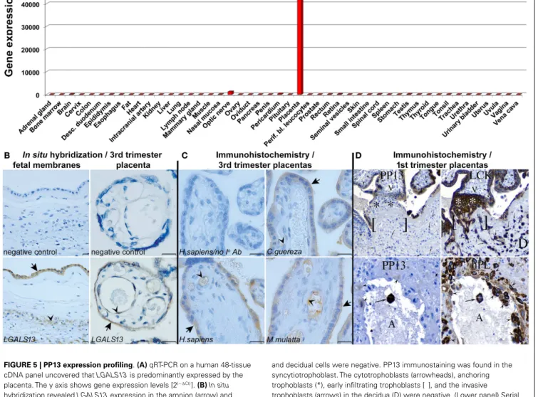
WIDE-SCALE EXPRESSION PROFILING OF PP13 IN HUMAN TISSUES Besides the studies on LGALS13 DNA variants, the investiga- tions on the expression patterns of PP13 have revealed interesting insights. The study describing the cloning of PP13 also presented compelling evidence for the predominant placental expression of PP13 in the human body (6). In fact, the expression profiling of human adult and fetal, normal and tumorous tissues by West- ern blot (26 tissues) and Northern blot (16 tissues) detected a 16kDa PP13 immunopositive band in extracts of human term placentas, and unique placental PP13 mRNA expression, respec- tively. These findings were supported by GenBank evidence of only placental expressed sequence tags forLGALS13. Later, the wide- scale expression profiling ofLGALS13and related chromosome 19 cluster galectin genes using qRT-PCR on a human 48-tissue cDNA panel confirmed the predominant placental expression of LGALS13(Figure 5A) (23).
PLACENTAL EXPRESSION PROFILING OF PP13 IN NORMAL PREGNANCIES
In human villous placental tissues at term, immunohistochemistry and immunofluorescence consistently found predominant PP13 positivity of the syncytiotrophoblast and villous capillary endothe- lium but not the cytotrophoblasts (8, 9, 21,23,36,37).In situ hybridization (23) detected PP13 mRNA expression in the same placental cells, further confirming the specificity of the immunos- tainings (Figure 5B). The same PP13 expression pattern was found in the placentas of Old World monkeys, suggesting the conserva- tion of PP13 expression during evolution (Figure 5C). Moreover, in situhybridization revealedLGALS13expression in the amnion
Than et al. PP13 in health and disease
FIGURE 5 | PP13 expression profiling.(A)qRT-PCR on a human 48-tissue cDNA panel uncovered thatLGALS13is predominantly expressed by the placenta. The y axis shows gene expression levels [2(−∆Ct)].(B)In situ hybridization revealedLGALS13expression in the amnion (arrow) and chorionic trophoblasts (arrowhead) in the fetal membranes in normal term pregnancy (left). In normal term placenta,LGAL13is predominantly expressed by the syncytiotrophoblast (arrow) and endothelium (arrowhead) (right). Scale bars: 20µm.(C)PP13 immunostaining is conserved in the syncytiotrophoblast, its apical membrane (arrows), and the endothelia (arrowheads) of human and anthropoid primate placentas. (Scale bars:
20µm.)(D)(Upper panel) Serial sections of a 15 week junctional complex stained for PP13 and low molecular weight cytokeratin (LCK). LCK immunostained epithelial cells, including cytotrophoblasts (arrowheads), anchoring trophoblasts (*), early infiltrating trophoblasts [ ], and invasive trophoblasts (arrows) in the decidua (D). Mesenchymal villous core cells (V)
and decidual cells were negative. PP13 immunostaining was found in the syncytiotrophoblast. The cytotrophoblasts (arrowheads), anchoring trophoblasts (*), early infiltrating trophoblasts [ ], and the invasive trophoblasts (arrows) in the decidua (D) were negative. (Lower panel) Serial sections of 8 week maternal spiral arterioles immunostained for PP13 and human placental lactogen (hPL). All the decidual invasive, intravascular and endovascular trophoblasts (arrowheads), and a single luminal (A)
syncytiotrophoblast (arrow) were stained for hPL. The monoclonal anti-PP13 antibody did not stain decidual invasive trophoblasts, lightly stained endovascular trophoblasts (arrowheads), and it intensely stained luminal syncytiotrophoblasts (arrow). Figure(A)represents data published in Ref.
(23). Figures(B,C)were published in Ref. (23). Figure(D)was published in Ref. (8). Kind permission for the reuse of the figures was obtained from the National Academy of Sciences of the United States of America(A-C)and SAGE US(D).
and chorionic trophoblasts in the fetal membranes. These find- ings showed PP13 expression predominantly in locations where maternal-fetal immune interactions occur.
In the first trimester, PP13 was immunolocalized to the syn- cytiotrophoblast and multinucleated luminal trophoblasts within converted decidual spiral arterioles (8). Villous cytotrophoblasts and invasive extravillous trophoblasts in the anchoring tro- phoblastic columns were immunonegative (Figure 5D). The syn- cytiotrophoblastic staining intensity declined with gestational age,
being the strongest between 6 to 8 weeks of gestation. This study also confirmed previous findings in term placental tissues on pre- dominant, diffuse cytoplasmic and also nuclear immunopositivity of the syncytiotrophoblast.
EXPRESSION PROFILING OF PP13 DURING VILLOUS TROPHOBLAST DIFFERENTIATION AND FUSION
Based on this immunohistochemical evidence, it was hypoth- esized that PP13 expression is related to the biochemical and
morphological differentiation and syncytialization of the villous trophoblast (36). These processes are primarily governed by cyclic adenosine monophosphate (cAMP) and protein kinase A (PKA), which regulate the resetting of the transcriptional program dur- ing the shift from cytotrophoblast into the syncytiotrophoblast (38–40). The resulting unique transcriptome of the syncytiotro- phoblast (41) controls the production of placental hormones, immune proteins and other proteins predominantly expressed by the placenta, which support pregnancy. Besides the exchange of feto-maternal gas, nutrients and waste, and the hormonal regula- tion of fetal development, the syncytiotrophoblast is also active in generating immune tolerance between the mother and her semi-allogeneic fetus (2,42,43).
In vitro assays with trophoblast-like BeWo cells demon- strated that indeedLGALS13expression is related to trophoblast fusion and syncytium formation induced by the cAMP-analogue Forskolin, and that a PKA inhibitor could block BeWo cell syncy- tialization andLGALS13expression (44). A recent study confirmed these findings in BeWo cells, and demonstrated the syncytializa- tion and differentiation-relatedLGALS13expression in primary villous trophoblasts (45). The evolutionary and functional inves- tigations of the trophoblastic regulatory mechanisms ofLGALS13 expression showed that promoter evolution and the insertion of an anthropoid-specific LINE element into the 50untranslated region (UTR) of an ancestral gene introduced binding sites for several transcription factors (e.g. ESRRG) key in villous trophoblastic gene expression, leading to the gain of placental expression of LGALS13and related chromosome 19 cluster galectin genes (43, 45). Glial cell missing-1 (GCM1), the transcription factor that governs villous trophoblast fusion and syncytialization (46), was shown to facilitate the expression of ESRRG and other key villous trophoblastic transcription factors, and thus, to indirectly pro- mote the placental expression of LGALS13 and cluster galectin genes. In addition, DNA methylation was also observed to reg- ulate developmental expression ofLGALS13and cluster galectin genes (45).
PLACENTAL ASPECTS OF PREECLAMPSIA
It is important that the impairment of villous trophoblast syncy- tialization characterized by the decreased trophoblastic expression of GCM1 and syncytin-1, a fusogenic protein regulated by GCM1, has been observed in preeclampsia (47,48), an obstetrical syn- drome originating from impaired early placentation (49, 50).
Preeclampsia is diagnosed by new-onset hypertension and pro- teinuria after 20 weeks of gestation, and it is a major cause of maternal, fetal and neonatal morbidity and mortality (51). More- over, this syndrome consists of various subtypes defined by gesta- tional age (e.g.: early-onset:<34th weeks; preterm:<37 weeks; and term:≥37 weeks) (52,53). Early-onset and preterm preeclampsia are severe subtypes of the disease that require premature delivery and are more often associated with intrauterine growth restriction (IUGR), hemolysis, elevated liver enzymes, and low platelet count (HELLP) syndrome, and fetal death, while term preeclampsia may be severe or mild in its clinical presentation (51–55). Although the molecular pathways of preeclampsia are incompletely understood, it appears to be associated with impaired placentation as the only definite therapy of preeclampsia is still the delivery of the fetus
and the removal of the placenta (50,51,53). It is also evident that heterogeneous causes can trigger early placental pathologic events, and that these are followed by the onset of the terminal pathway of preeclampsia in a later stage, leading to the subsequent clinical onset of the symptoms (50,51,56).
Several studies providing histopathologic or transcriptomic evidence have suggested that the placental pathogenesis of preeclampsia may differ in its subtypes as more pronounced differ- ences could be observed in early-onset than late-onset preeclamp- sia when compared to gestational age-matched controls (57–61).
In line with these findings, the extent of histopathologic changes in the placental bed was most extensive in early-onset preeclampsia, especially in cases associated with IUGR. These abnormal find- ings were consistent with impaired trophoblast invasion into the uterine tissues and the consequent abnormal remodelling of the maternal spiral arterioles, placental pathologic events that occur in the first trimester (62,63).
Previously it was thought that impaired early placentation is associated with placental hypoxia (64); however, it has recently become evident that the resulting fluctuation in uterine blood supply leads to placental ischemic injury, causing oxidative stress, pro-inflammatory conditions, and apoptosis (65–67). In response, the placenta expresses and releases increased amounts of anti- angiogenic factors, pro-inflammatory cytokines and aponecrotic syncytiotrophoblast microvesicles. The latter might induce mater- nal anti-angiogenic and exaggerated systemic pro-inflammatory states, hypertension and proteinuria (50,51,53,66,68–70).
DECREASED PLACENTAL PP13 EXPRESSION IN PREECLAMPSIA In this context, LGALS13 expression was found to be down- regulated in villous placental tissues in preeclampsia. This was first described for preterm preeclampsia compared to gestational age- matched controls at the time of disease, and this phenomenon was suggested to be associated with problems in trophoblast syncytial- ization (36) (Figures 6A,B). Since then, other studies confirmed these findings in the third trimester, including one that investi- gated placentalLGALS13expression at the time of disease (71) and another that looked for syncytiotrophoblasticLGALS13 expres- sion in laser captured specimens in the first trimester (72). The latter study detected decreasedLGALS13expression in the syncy- tiotrophoblast from chorionic villus samples obtained at 11 weeks of gestation in women who subsequently developed preeclampsia compared to controls. Such first trimester down-regulation of pla- centalLGALS13expression may be one of the earliest pathological indications for the subsequent development of preeclampsia.
A recent study has revealed the possible molecular mechanisms leading to decreased placentalLGALS13expression in women with severe preterm preeclampsia. It was found that in this subform of preeclampsia there is a decreased placental expression ofGCM1 andESRRG, genes encoding transcription factors that regulate tro- phoblasticLGALS13expression (45). Moreover, functional exper- iments showed that the knock-down ofGCM1in BeWo cells led to the down-regulation of ESRRG and other transcription factors that regulateLGALS13expression. Accordingly, it was concluded that there is a decreased GCM1-mediated trophoblast fusion and trophoblastic gene expression in severe preterm preeclamp- sia that leads to the down-regulation ofLGALS13. Furthermore,
Than et al. PP13 in health and disease
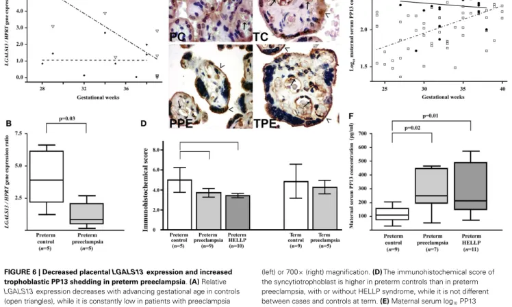
FIGURE 6 | Decreased placentalLGALS13expression and increased trophoblastic PP13 shedding in preterm preeclampsia.(A)Relative LGALS13expression decreases with advancing gestational age in controls (open triangles), while it is constantly low in patients with preeclampsia (diamonds).(B)RelativeLGALS13expression is lower in women with preterm preeclampsia than in controls.(C)Syncytiotrophoblastic PP13 immunostaining is decreased in preterm preeclampsia compared to controls. PC: preterm control, 35 GW (weeks of gestation); TC: term control, GW38; PPE: preterm preeclampsia, GW29; TPE: term preeclampsia, GW37.
The endothelium (arrows) is also PP13 immunopositive in all sections. The microvillous membrane (open arrowheads) stains moderately for PP13 in controls, while it is strongly PP13 immunopositive in preeclampsia. 500×
(left) or 700×(right) magnification.(D)The immunohistochemical score of the syncytiotrophoblast is higher in preterm controls than in preterm preeclampsia, with or without HELLP syndrome, while it is not different between cases and controls at term.(E)Maternal serum log10PP13 concentrations increase as a function of gestational age in control women (open rectangle), while these do not correlate with gestational age in patients with preeclampsia (filled circle). The regression line for log10PP13 concentrations is significantly different in the two groups.(F)Median maternal serum PP13 concentrations are higher in women with preterm preeclampsia, with or without HELLP syndrome, than in controls. All the figures were published in Ref. (36). Kind permission for the reuse of figures was obtained from Springer Science+Business Media.
the differential methylation ofLGALS13 was also found in the villous trophoblast in preterm preeclampsia, which may inter- fere withLGALS13expression, suggesting that potential additional disease-mechanisms may account for the trophoblastic pathology in preterm preeclampsia (45).
ALTERED PLACENTAL LOCALIZATION AND INCREASED SHEDDING OF PP13 IN PREECLAMPSIA
In accord with gene expression data, immunostainings revealed that cytoplasmic PP13 positivity of the syncytiotrophoblast was weaker in preeclampsia compared to controls, particularly in preterm cases. Similar changes were also observed at the time of disease in preterm HELLP syndrome (36) (Figures 6C,D).
Paradoxically, PP13 immunostaining of the syncytiotrophoblast microvillous membrane was stronger in preeclampsia and HELLP syndrome compared to controls (Figures 6C,7A). Syncytial cyto- plasmic protrusions and membrane microvesicles shed from the syncytiotrophoblast stained strongly for PP13 in preeclampsia
(Figure 7A). It was suggested that the increased release of PP13 positive microvesicles from the syncytiotrophoblast may lead to elevated maternal serum PP13 concentrations in preterm preeclampsia and HELLP syndrome before or at the time when the clinical symptoms of preeclampsia appear (Figures 6E,F) (36,37).
The subcellular redistribution of PP13 in the syncytiotro- phoblast was further observed by confocal imaging of placental samples from patients with preeclampsia and HELLP syndrome compared to gestational age-matched controls (73). In all study groups, PP13 highly colocalized with placental alkaline phos- phatase, a glycophosphatidylinositol-anchored lipid raft-resident protein. However, there was also a high degree of colocalization of PP13 with CD71, a non-raft plasma membrane protein, which decreased in preterm preeclampsia and HELLP syndrome. In con- trast, the colocalization of PP13 with cytoskeletal actin, a protein earlier found to bind to PP13 with high affinity (9), was increased in all patient groups compared to controls. These results indicated that the translocation of PP13 to the juxta-membrane region of
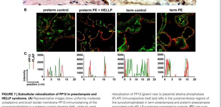
FIGURE 7 | Subcellular relocalization of PP13 in preeclampsia and HELLP syndrome.(A)Representative images show uniformly moderate cytoplasmic and brush border membrane PP13 immunostaining of the syncytiotrophoblast in a preterm control placenta (left), while its weak cytoplasmic and strong membrane immunostaining in a placenta from a woman with preterm preeclampsia (PE) associated with HELLP syndrome (right). Cytoplasm protrusions, membrane blebs and shed membrane microvesicles immunostained intensely for PP13 (right). 800 x magnification.(B)Representative confocal images show the subcellular
relocalization of PP13 (green) near to placental alkaline phosphatase (PLAP) immunopositive (red) lipid rafts in the juxtamembrane regions of the syncytiotrophoblast in term preeclampsia and preterm preeclampsia associated with HELLP syndrome compared to controls.(C)Line scan intensity distributions of PP13 and PLAP in representative confocal images shown in subfigure(B). Figure(A)was published in Ref. (36).
Figures(B,C)were published in Ref. (73). Kind permission for the reuse of the figures was obtained from Springer Science+Business Media(A)and Elsevier(B,C).
the syncytiotrophoblast in preeclampsia and HELLP syndrome is associated with actin (Figures 7B,C) (73). Supporting these obser- vations in the placenta, subsequentin vitroexperiments revealed that Latrunculin B, a selective blocker of actin polymerization, decreased PP13 release from BeWo cells and led to its intracellular accumulation (Figure 8A) (73).
This result may also explain how PP13 is released from the syncytiotrophoblast since the actin cytoskeleton and associated motor proteins drive intracellular and plasma membrane traf- ficking amongst a wide variety of cellular processes (74,75). In this regard, galectins predominantly utilize unconventional traf- ficking routes, either vesicular or direct translocational, avoiding the endoplasmic reticulum (ER) and Golgi apparatus, since they are synthetized on free ribosomes and lack anN-terminal signal sequence for the translocation to the ER/Golgi system (76–78).
However, other vesicular transport mechanisms for PP13 can- not be excluded, such as the “kiss and run” exocytosis, which was described for many hormones and neurotransmitters and was proved to be an actin- and calcium-dependent process (79,80).
The role of actin cytoskeleton in the release of extracellular vesi- cles (EV; e.g. microvesicles/microparticles and exosomes), which also carry various galectins, has also been demonstrated (81–83).
In addition, annexin II, another protein that specifically bound to PP13 (9), has also been found in various types of EVs along with actin (84,85). Similar to galectin-9, which was shown to be
associated with many different types of EV fractions (86), PP13 may also translocate with different EVs through the syncytiotro- phoblast membrane. This type of release is supported by evidence on the PP13 release from BeWo cells mediated by exosomes (73) and the observed PP13 immunopositivity of microvillous membrane microvesicles shed from the syncytiotrophoblast (36).
THE ROLE OF CALCIUM AND ISCHEMIA IN TROPHOBLASTIC PP13 RELEASE
Recent in vitro experiments with BeWo cells transfected with LGALS13 to enable high PP13 expression (73) also showed an increased PP13 release from calcium ionophore-treated cells, evi- denced by decreased cellular PP13 content and elevated amounts of PP13 in cell culture supernatants (Figure 8B). This finding is in accord with a previous report showing that galectin-3 is secreted by exosomes from monocytes upon calcium ionophore treatment (87). Calcium serves as a ubiquitous second messenger responsi- ble for controlling numerous cellular processes including exosome secretion (88). Since calcium regulates the actin cytoskeleton at multiple levels including the organization of actin monomers into actin polymers and the super-organization of actin polymers into a filamentous network (89), it is not surprising that stimuli result- ing in the elevation of intracellular calcium concentration can induce microvesiculation and membrane shedding of exposed cells (90,91). As a mechanism, the dynamics of actin assembly and
Than et al. PP13 in health and disease
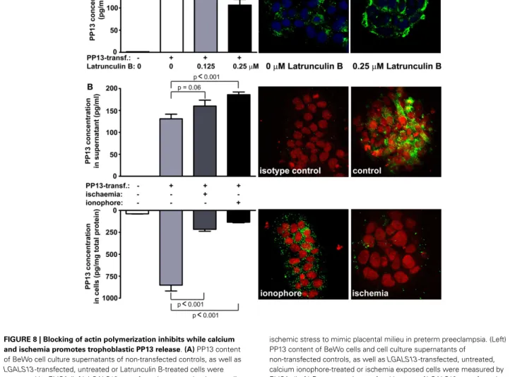
FIGURE 8 | Blocking of actin polymerization inhibits while calcium and ischemia promotes trophoblastic PP13 release.(A)PP13 content of BeWo cell culture supernatants of non-transfected controls, as well as LGALS13-transfected, untreated or Latrunculin B-treated cells were measured by ELISA (left).LGALS13-transfected, untreated or Latrunculin B-treated cells were stained with anti-PP13 (green) and nuclei were counterstained with DRAQ5 (blue) followed by confocal microscopic analysis (right; 40x magnifications). The disruption of the actin cytoskeleton with Latrunculin B treatment decreased PP13 release from BeWo cells.(B)LGALS13-transfected BeWo cells were treated with calcium ionophore to increase intracellular calcium level, or kept under
ischemic stress to mimic placental milieu in preterm preeclampsia. (Left) PP13 content of BeWo cells and cell culture supernatants of
non-transfected controls, as well asLGALS13-transfected, untreated, calcium ionophore-treated or ischemia exposed cells were measured by ELISA. (Left) Representative confocal images ofLGALS13-transfected control, calcium ionophore-treated or ischemia exposed BeWo cells immunostained with monoclonal anti-PP13 antibody (green) and counterstained with DRAQ5 (red) nuclei dye. Either ionophore treatment or ischemia induced the release of PP13 from BeWo cells. Figures were published in Ref. (73). Kind permission for the reuse and modification of the figures was obtained from Elsevier.
disassembly is regulated by certain actin-binding proteins such as annexin II in a calcium-dependent manner (92–96).
The release of PP13 from BeWo cells appears to be similar to the in vivorelease when comparing the effect of calcium ionophores and ischemic stress (Figure 8B) (73). Ischemic stress of the pla- centa is a major component of the pathophysiology of preterm preeclampsia (97). In accord, higher PP13 release was observed in placental villous tissue explants obtained from women with preeclampsia compared to gestational age-matched controls in the third trimester (98). A possible explanation is that ischemic stress causes elevation in intracellular calcium levels, which leads to actin depolymerization supported by findings of separate stud- ies (99,100). All of these results indicate that different kind of actin- and calcium-dependent release mechanisms exist side by
side for PP13, and most probably the dominant sort depends on the cell type and also on the nature of the received stimuli.
As a functional aspect of the increased PP13 release from the placenta in preeclampsia, ischemic and other stress conditions pose danger to the organism, which is signaled to the immune system by endogenous danger signals called “alarmins” (101,102).
Indeed,“danger signals” in the placenta have also been proposed to create an abnormal placental cytokine milieu and link the activa- tion of the innate immune system and preeclampsia (8,103–105).
In this context, some galectins with cytokine-like properties (106, 107) may act as alarmins, since they are increasingly secreted from inflamed or damaged tissues, and they may elicit effector responses from innate and adaptive immune cells (19,102,108). Although direct evidence for the role of PP13 as an alarmin has not yet been
established, these findings suggest that PP13 may function in such way in the placenta in preeclampsia (19,73).
IN VITROANDIN VIVOFUNCTIONAL STUDIES ON PP13 IN VITROPARACRINE EFFECTS OF PP13 ON HUMAN IMMUNE CELLS PP13 released from the trophoblast into the extracellular space may have various functions similar to other galectins, which may exert their pleiotropic extracellular functions in an autocrine and paracrine manner. Since PP13 is secreted by the trophoblast to the maternal circulation from where it gets into the decidual extra- cellular matrix (8), PP13 may affect various types of circulating and tissue-resident maternal leukocytes throughout pregnancy.
Thus far only a couple of functional experiments were carried out, focusing on the examination of potential extracellular effects of PP13. As several members of the galectin family regulate adap- tive immune responses by the induction of apoptosis of activated T lymphocytes (19,109–111), the apoptosis-inducing effects of PP13 and other chromosome 19 cluster galectins were investi- gated on activated T cells freshly isolated from healthy donors (23).
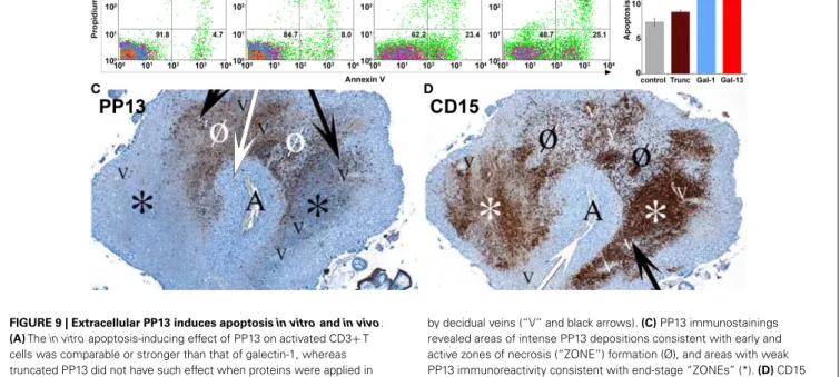
Among the studied recombinant galectins, PP13 had the strongest apoptosis-inducing effect (Figures 9A,B), stronger than galectin- 1, a protein that has central role in maintaining maternal-fetal immune tolerance in eutherian mammals (16,19,112). A subse- quent study also investigated the effect of PP13 on the secretion of cytokines and chemokines from mononuclear cells isolated from
peripheral blood of pregnant women (8). The treatment with placenta-purified PP13 slightly increased the secretion of inter- leukin (IL)-1αand IL-6 into the culture medium. Thesein vitro experimental evidences suggest various effects of PP13 on immune cells, which may also be largely dependent on the type, activation and differentiation status of the affected cells, the microvesicle- bound or free nature and concentration of PP13, and the redox status of the environment, similarly to other galectins (19).
As with galectins that bind ABO blood group antigens, the paracrine effects of PP13 may also be affected by this phenomenon (21). In this context, large cohort studies showed that preeclampsia is more frequent among patients with AB blood group compared to those with non-AB blood groups (113,114). Thus, recently it has been hypothesized that the higher susceptibility to preeclamp- sia among AB blood group women may be related to the decreased bioavailability and paracrine effects of PP13 on maternal immune cells (21). As a similar phenomenon, the ABO blood group anti- gens linked to the protein backbone of coagulation factor VIII and von Willebrand factor significantly affect the bioavailability of these blood clotting factors and coagulation (115–118). These findings altogether further underline the important immunoreg- ulatory functions that PP13 may have in early pregnancy and warrant further investigation of the effect of ABO blood group system on PP13 bioavailability and functions. In summary of all of the above, PP13 may have a complex role in the regulation
FIGURE 9 | Extracellular PP13 induces apoptosisin vitroandin vivo.
(A)Thein vitroapoptosis-inducing effect of PP13 on activated CD3+T cells was comparable or stronger than that of galectin-1, whereas truncated PP13 did not have such effect when proteins were applied in 8µM concentration. Numbers in quadrants indicate the percentage of CD3+T cells.(B)Thein vitroapoptosis-inducing effect of PP13 on activated CD3+T cells was stronger than that of galectin-1. Apoptosis rate was calculated as the percentage of Annexin V and propidium iodide double-positive cells. Gal: galectin; Trunc: truncated galectin-13;
*P<0.05; **P<0.01.(C,D)Serial sections of decidua basalis samples in the first trimester. A spiral arteriole (“A” and white arrows) is surrounded
by decidual veins (“V” and black arrows).(C)PP13 immunostainings revealed areas of intense PP13 depositions consistent with early and active zones of necrosis (“ZONE”) formation (Ø), and areas with weak PP13 immunoreactivity consistent with end-stage “ZONEs” (*).(D)CD15 immunostainings revealed neutrophil accumulation showing an inverse pattern with PP13 depositions. The least intense staining was observed in early “ZONEs” (Ø), while the most intense staining in end-stage
“ZONEs” (*). Figures and data(A,B)were published in Ref. (23).
Figures(C,D)were published in Ref. (8). Kind permission for the reuse and modification of figures was obtained from the National Academy of Sciences of the United States of America(A,B)and SAGE US(C,D).
Than et al. PP13 in health and disease
of adaptive and innate immune functions at the maternal-fetal interface depending on the changing environment.
THEIN VIVOPARACRINE EFFECTS OF PP13 ON HUMAN IMMUNE CELLS This latter study also described an interesting finding in placental tissue specimens obtained from elective terminations of pregnan- cies between 6 to 15 weeks of gestation (8). In addition to the PP13 immunopositivity of the syncytiotrophoblast, crystal-like PP13 deposits in the decidual extracellular matrix and phagocytosed PP13 immunopositive material in immune cells were also doc- umented. These deposits were always adjacent to decidual veins but not to arteries, and they were coincident with unique zones of necrotic and apoptotic immune cells (“ZONEs”) (Figures 9C,D) (8), a phenomenon that may be consistent with previous find- ings on zones of decidual necrosis in the first trimester (119). In addition, immunostainings demonstrated the expression of IL- 1αand IL-6 within and around macrophages in these “ZONEs”, suggesting the potential pro-inflammatory effect of PP13 (8).
The highest number of “ZONEs” appeared to be between 7 to 8 weeks of gestation, and their occurrence declined by the end of the first trimester in parallel with the completion of the spiral artery remodelling and the establishment of the blood circulation to the placenta. This study also revealed that spiral artery trans- formation by invasive trophoblasts and “ZONEs” were rarely seen in specimens obtained from women with low maternal serum PP13 concentration compared to those with normal PP13 val- ues. In summary, these findings prompted the authors to sug- gest that syncytiotrophoblast-secreted PP13 reaches the decidual veins, crosses their wall, deposits into the extracellular matrix and forms perivenous crystal-like aggregates near the veins. The lysophospholipase activity associated with PP13 was implicated in this process, but no evidence yet exists to prove it. These PP13 deposits were suggested to serve as “diversion sites” to attract, activate and kill maternal immune cells, drawing these away from sites where the semi-allogeneic fetal trophoblasts invade and remodel maternal spiral arteries. It was also hypothesized that decreased PP13 expression may lead to deficient “ZONE” forma- tion, decreased trophoblast invasion, and the subsequent failure of spiral artery transformation (8). Functional and causal evi- dence for thesein situobservations needs to be provided in the future.
PP13 AND UNIQUE ASPECTS OF DEEP PLACENTATION IN ANTHROPOID PRIMATES
Thesein vivofindings are important from an evolutionary point of view since PP13 evolved in Old World monkeys and apes (23), species that have endovascular trophoblast invasion and spiral artery remodelling different from lower primates (120–123). In fact, a growing body of evidence suggests that PP13 may belong to primate-specific molecules (e.g. human leukocyte antigen C, killer-cell immunoglobulin-like receptors), which are involved in the regulation of immune mechanisms related to invasive placen- tation (124). The findings on PP13 and “ZONE” formation may be mostly related to the pro-apoptotic effect of PP13, similar to the effect of galectin-1 on activated decidual T cells, which is critical in the down-regulation of maternal adaptive immune responses at the maternal-fetal interface in early pregnancy (111). However, the
pro-inflammatory action of extracellular PP13 may also fit with early placentation events.
In fact, the early pregnancy decidua is infiltrated by a large number of leukocytes, mainly natural killer (NK) cells (70%), macrophages (20–25%), and T cells (10%) (125–127).
These leukocytes, especially decidual natural killer (dNK) cells, macrophages and T regulatory cells, are indispensable for the suc- cess of pregnancy since they produce a large variety of chemokines, cytokines, matrix metalloproteinases and angiogenic molecules that regulate maternal-fetal interactions, trophoblast invasion and spiral artery remodelling (127–130). On one hand, these immune cells are involved in the establishment of a delicate immune toler- ance between the mother and the fetus, and on the other hand they promote local pro-inflammatory responses that facilitate implan- tation, trophoblast invasion and placentation events (126,127).
These complex immune interactions between the mother and the fetus are conveyed by cell membrane- and vesicle-bound as well as soluble molecules. Among the best studied molecular mech- anisms are the effect of progesterone-induced blocking factor (PIBF) on the shift towards Th2 over Th1 cytokine production (131), the anti-inflammatory role of decidual macrophages (132), the immunosuppressive effects of decidual galectin-1 (16), tro- phoblastic indoleamine 2,3-dyoxignease (IDO), FAS/FAS ligand and galectin-1 (133,134), and the roles of human leukocyte anti- gen (HLA)-C and HLA-G in protecting fetal cells from NK- and cytotoxic lymphocyte (CTL)-mediated cytolysis (135,136). It is a question for future studies how the actions of PP13 are related to this complex, dynamically changing cellular and molecular network during placentation.
As the result of these complex interactions at the maternal-fetal interface, aggregates of extravillous endovascular trophoblasts plug the openings of uterine spiral arteries; therefore, they inhibit intervillous circulation at the beginning of gestation (49,120,128).
This is suggested to be a protective mechanism to keep the devel- oping embryo in a relatively low oxygen environment, minimizing oxidative stress that would lead to developmental defects during organogenesis (137). Of importance, the observed “ZONE” for- mation peaks when placental circulation is not yet established (8), and the low flow of endometrial gland secretions around the pla- centa allows the increased transport of PP13 from decidual veins into the decidua. This is also the period when extravillous tro- phoblast invasion starts into the decidua (128,137). Remarkably, after the start of the placental intervillous circulation at around 8–10 weeks of gestation (49,128,137, 138) PP13 deposits and
“ZONE” formation rapidly declines and diminishes by the time intervillous circulation is fully established at about 10–14 weeks of gestation (8). This suggests that PP13 transport is reduced to the decidual extracellular matrix due to the continuously increas- ing blood flow in spite of the increasing total production of PP13 by the placenta. Importantly, if trophoblastic plug formation is incomplete, placental circulation starts earlier, which leads to the oxidative stress of the placenta, the subsequent development of preeclampsia, and early pregnancy loss in more severe cases (49, 137, 138). In this context, the earlier start of placental blood flow would theoretically restrict PP13 transport into the decidua and “ZONE” formation, providing another mechanism to hamper normal placentation.