INTERACTION OF TWO FLAVONOIDS WITH CALF THYMUS DNA: A MULTI - SPECTROSCOPIC, ELECTROCHEMICAL
AND MOLECULAR MODELLING APPROACH
P. Venmathy
[a], J. Jeyasundari
[a], V. S. Vasantha
[b]*, P. Nandha Kumar
[b]and M. Sakthi
[c]Keywords: Flavonoid interaction, Ct-(ds) DNA, spectroscopy, cyclic voltammetry, docking.
Interaction of naturally occurring bioactive flavonoids 5,6,7-trihydroxyflavone (Baicalein) and 7,8-dihydroxyflavone (DHF) binding with calf thymus deoxyribose nucleic acid (dsDNA) was studied by employing UV absorption, fluorescence, circular dichroism, cyclic voltammetric and molecular modeling techniques. All studies were confirmed that the structural changes of DNA binding to the flavonoid.
From the CV results positive shift in peak potential and increased peak current of the flavonoid in the presence of DNA and then the fluorescence quenching of DNA-flavonoids system indicated the intercalative mode of binding between flavonoid and DNA. CD studies suggest the conformational changes in DNA upon interaction with the flavonoids. Molecular docking simulation methods are used as tools to delineate the binding mode and probable location of the flavonoids and their effects on the stability and conformation of Ct-(ds) DNA.
Furthermore, Baicalein can bind with more potential with Ct-(ds) DNA than DHF. This is helpful to understand the molecular aspects of binding mode and provides direction for the use and the design of new effective therapeutic agents. These results could provide useful information for insight into the pharmacological mechanism of flavonoids.
* Corresponding Authors
Mobile Number: +919442357392 Email: vasantham999@yahoo.co.in
[a] Nadar Mahajana Sangam S.Vellaichamy Nadar College, Nagamalai, Madurai 625019, India.
[b] Department of Natural Products Chemistry, School of Chemistry, Madurai Kamaraj University, Madurai 625021, India.
[c] Department of Inorganic Chemistry, School of Chemistry, Madurai Kamaraj University, Madurai 625021, India.
INTRODUCTION
Flavonoids are polyphenolic compounds and due to their tremendous biological importance and broader range of pharmacological activities including antioxidant, anticancer, antitubercular, antibacterial, antiallergic, antimicrobial, anti- inflammatory, antiviral, antitumor, antimutagenic, antidiabetic, hepatoprotective and cardiovascular activities, flavonoids gained much attention and become a topic of interest for researchers in the last decades.1-8 Over 5000 flavonoids have been isolated from plants, most of which are divided into subclasses, including anthocyanidins, flavanones, flavonols, flavones, and isoflavones. Among them, flavones are less common than flavonols, including the well-known flavones 5,6,7- trihydroxyflavone and 7, 8- dihydroxyflavone. 7,8-DHF (Figure 1A) flavone is isolated from Wagatea spicata, then baicalein (Figure 1B) flavone is originally isolated from Oroxylum indicum,9 a Chinese medicinal plant with various biological properties.
Flavonoids are everywhere in plants. They are rich in seeds, citrus fruits, olive oil, tea, and vegetables.10
Over the last few decades structure of DNA and its interaction with different bioactive molecular moieties have gained a great interest in the field of organic synthesis and pharmacology. DNA is a nucleic acid that contains all the information necessary for specifying the biological development of all living bodies. It is a molecule that controls hereditary information transferred to the offspring.
During reproduction, DNA is replicated and transmitted to the new trait. In this process, the sequence of DNA base pairs defines the characters of individuals ranging from physical traits to disease susceptibility. It is necessary to understand at molecular level gene expression and their mechanism of transfer to offspring.11-13 This could be helpful to understand the transfer of many diseases. It is also a key step towards the development of new chemotherapeutic strategies. The interaction of many naturally occurring compounds with DNA adducts is an active area of research in chemistry and biology which leads to the understanding of drug–DNA interaction and the consequent design of new efficient drugs targeted to DNA.
14 Due to the central role of DNA in replication and transcription, DNA has been a major role for the antibiotic, anticancer and antiviral, anti-inflammatory drugs.
Interaction of small molecules and DNA are mainly of two types. One is covalent interactions and another one is non-covalent interactions. Three major modes of non- covalent interactions are electrostatic interactions, groove binding, and intercalative binding. A small molecule can interact with DNA involving a single mode of binding or mixed binding modes. It is worth noting that the property of mixed binding mode can be linked to their mechanism of action and therapeutic efficiency.15,16 Intercalation and minor-groove binding are the predominant DNA-binding modes of small ligands17 while electrostatic interactions between the cationic species and negatively charged DNA phosphate backbone usually occur along the exterior of the helix. DNA is an antiparallel double helix held together by hydrogen bonding interactions between DNA base pairs.
The drugs could interact with DNA in different ways.18 The drugs could interact at DNA base pairs by the breakdown of hydrogen bonding (intercalators) while some moieties could interact at groove sites (groove binders). The first evidence of interaction was published in 1961 when Lerman demonstrated that acridine dye could intercalate
between DNA base pairs. It was concluded after this research that only molecules with flat, an aromatic structure can intercalate with DNA and are considered to be good anticancer drugs.19 There are certain cases where the cytotoxicity is parallel to anticancer activity. A number of compounds like vitamins, hormones, vitamin antagonists, antidepressants, and antihistamines are also good intercalators.20-22 The non-planer structures mostly interact with DNA through groove bindings, which do not disturb the base pairs but just interact through outside bindings.
Generally, those drugs are considered to be best anticancer which are organometallic in nature.23 These drugs are intercalators as well. In this paper DNA interaction of two compounds with DNA is reported.24-25 The interaction is carried out with Calf Thymus DNA and studied via spectroscopic and cyclic voltammetric analysis.26 Furthermore, molecular modeling methods can be applied to study of interaction drugs and biomacromolecules for saving time and money, especially since the reactivity of newly designed drugs with their targeted biomolecules can be predicted prior to chemical synthesis.27
The present investigation attempts to understand the mechanism of binding of two flavonoids with DNA by employing spectroscopic, electrochemical and molecular modeling techniques. For this, UV–vis spectroscopy, fluorescent spectrometry, voltammetry, circular dichroism and molecular modeling (using Autodock 4.228) are employed, and the results could provide useful pharmacological and toxicity information and insight into the redox reactions of these molecules in the living body.
EXPERIMENTAL
Materials
Calf thymus-(ds) DNA was purchased from Sisco Research Laboratories Private Limited (SRL), India and used without any purification. 5,6,7-Trihydroxyflavone (Baicalein) was extracted from Oroxylum indicum, 7,8- dihydroxyflavone (DHF) was extracted from Wagatea Spicata. Stock solutions of DNA and flavonoids were prepared by dissolving the appropriate amounts of DNA with flavonoids in Tris-HCl buffer pH 7.4 and double distilled water containing 10 % DMSO, respectively. Both DNA and flavonoid solutions were stored at 4 ˚C.
The concentration of DNA was determined spectrophotometrically using the extinction coefficient value of 6600 L mol−1 cm−1 at 260 nm. The solution of DNA was found to be free from protein as evident from its absorbance ratio value in the range of 1.7.29 All measurements were carried out at the physiological pH of 7.4 by using the Tris- HCl buffer.
UV-VIS absorption studies
All absorption spectra were recorded by using Agilent diode array spectrometer (Agilent 8453) at room temperature (25 °C). UV absorption spectra of flavonoids in the absence and presence of increasing concentrations of DNA was recorded in the wavelength range of 250-300nm.
Matched quartz cells of 1 cm path length were used in this study. Respective buffer solutions (Tris-HCl buffer, pH 7.4) were used as the reference. During optical titration of the flavonoids, an equal amount of DNA was added to both the sample and the reference cells. The temperature was maintained at 4˚C especially for Ct-(ds) DNA. In the spectrophotometric titrations to a fixed concentration of flavonoid, the concentration of DNA was varied and the change in the absorption at λ max of the flavonoid was noted at each P/D [DNA/flavonoid molar ratio]. The purity of DNA was verified by monitoring the ratio of absorbance at 260/280 nm (A260/A280). Appropriate blanks were run under the same conditions and subtracted from the sample spectra.
CD analysis
Circular dichroism (CD) is a powerful and reliable tool to understand the conformational changes in a biomacromolecule upon interaction. It is known that the intercalation of linear or flat aromatic molecules into double-stranded DNA induce large chirality changes and consequently significant affects on their CD spectra. The CD spectra of buffer were used and were automatically subtracted from the CD spectra of the samples as baselines.
CD band intensities were expressed in terms of mean residue ellipticity (MRE) in deg cm2 dmol-1. CD studies support the conformational changes in DNA upon interaction with the flavonoid.
Fluorescence spectral analysis
Fluorescence measurements were carried out on a carry eclipse fluorescence spectrophotometer. Measurements were made in a fluorescence free quartz cell of 1 cm path length.
The fluorescence characteristics of flavonoids (λex = 270 nm and λem = 350 nm) were used to investigate Ct-(ds) DNA–flavonoids interaction in Tris HCl buffer solution (pH
= 7.4) at room temperature. Fluorescence spectra were recorded in the range of 300–500 nm while maintaining the constant concentration of flavonoids and varying concentrations of DNA.
Voltammetric studies
The electrochemical behaviors of flavonoids were studied before and after adding DNA by cyclic voltammetry (CV) using Tris HCl buffer solution of pH 7.4 as supporting electrolyte. The mixed flavonoids–DNA solution was allowed to equilibrate for 5 min at room temperature. The voltammetric behaviours of both flavonoids and flavonoids–
DNA adduct were studied at different scan rates (25–250 mV s-1). During the determination, a nitrogen atmosphere was maintained over the solutions. All the measurements were carried out at room temperature.
Molecular docking
Docking operations were performed using version 4.2 of the AutoDock program package and the Lamarckian genetic algorithm (LGA) available in AutoDock 4.2, which was proven to be most reliable, successful and effective.19,20
Structures of the compounds, Baicalein and 7,8- dihydroxyflavones were modeled by using ChemDraw (version 11.0) and geometrically optimized using the LigPrep module (Schrodinger, LLC). These two compounds differed in the number of hydroxyl groups attached to the phenyl ring. Then it was converted into PDB format from
mol format by online OPENBABEL
(http://www.vcclab.org/lab/babel/). The LGA was used in this docking study of the compounds Baicalein and 7, 8- dihydroxyflavone with double-stranded DNA. The DNA duplex receptor structure from the Protein Data Bank (PDB ID: 2dyw) contained 12 base pairs. The base pair sequence was CGCGAATTCGCG: GCGCTTAAGCGC.
In all cases, we used grid maps with a grid box size of 120×60×120 points with a grid-point spacing of 0.371 Ǻ.
Then, we started the molecular docking via the LGA using default parameters. For ligands, ten independent docking runs were carried out. Visualization of the docked pose was done using Discovery digital studio molecular graphics programs.
RESULTS AND DISCUSSION
Binding modes of flavonoids with DNA
It is generally accepted that small molecules are bound to DNA double helix by three modes viz., electrostatic binding, groove binding and intercalative binding. In fact, each small molecule has a particular structure, which possibly has different binding modes with DNA and each mode results in a characteristic distortion of the DNA, in theory giving rise to a specific pharmacological effect.
Absorption spectroscopy
Interaction of Baicalein and DHF with ct-DNA
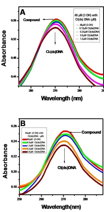
UV-Vis absorption spectra were obtained by titration of a 1.0 × 10⁻³mol L-1flavonoids with double strand DNA (ds- DNA) solution. The results, shown in Figures 1A and 1B, show a single absorption band of 270 nm for flavonoids in the absence of Ct-(ds) DNA. As the DNA concentration increases the intensity of the absorption band decreases. The phenomenon indicated an interaction between DNA and the flavonoid and it is typical of intercalate mode. 30
The hypochromic shift observed in the spectra of flavonoids indicates helical ordering of flavonoids in the DNA helix. Flavonoids binding to DNA through intercalation is characterized by change in the absorbance hypochromic and a red shift in wavelength, due to the intercalative binding mode involving a stacking interaction between the DNA base pairs.31
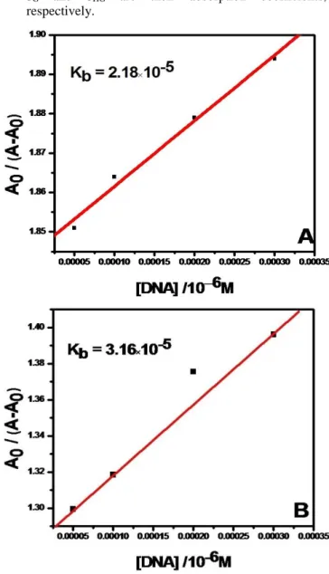
The absorption spectra of Baicalein and DHF when titrated with ct-DNA showed the isosbestic point at 250 nm and 300 nm respectively.32 The binding constants were calculated from the ratio of the intercept to the slope of the linear fitting of the curve obtained by plotting 1/(A-A0) versus 1/[DNA] for both the flavonoid. The binding constants of flavonoids with Ct-(ds) DNA were observed to be in the order of Baicalein ˃ DHF.
Figure 1. Absorption spectra of (A) 48 μM of Baicalein and (B) of 84 μM of DHF with varying concentration of Ct-(ds) DNA.
The values of the binding constants K were obtained according to the methods reported. To calculate the flavonoid–polynucleotide binding constant, the data are treated according to the following equations.
DNA + flavonoid ⇄ DNA flavonoid complex (1)
𝐾 = [𝐷𝑁𝐴 𝑓𝑎𝑙𝑣𝑜𝑛𝑜𝑖𝑑 𝑐𝑜𝑚𝑝𝑙𝑒𝑥]
[𝐹𝑟𝑒𝑒 𝐷𝑁𝐴][𝐹𝑟𝑒𝑒 𝑓𝑙𝑎𝑣𝑜𝑛𝑜𝑖𝑑] (2) The values of the binding constants K were obtained from the DNA absorption at 260 nm according to the published methods,33,34 where the bindings of various ligands to hemoglobin were described. For weak binding affinities the data were treated using linear reciprocal plots based on the following equation.
Based on the variations of absorbance in the spectral band, the binding constant, K of the complex-DNA can be obtained according to the following equation.
(3)
where
A0 and A are the absorbencies of the complex in the absence and presence of DNA, respectively, and
G and HG are their absorption coefficients, respectively.
Figure 2. Fitting of experimental data of Baicalein and DHF with Eqn. (4).
The association constant demonstrated that flavonoid binds to CT-DNA through outside binding such as an electrostatic interaction. Planarity, hydrophobicity, and electrostatic component of flavonoids play important roles in its binding to DNA through intercalative mode.35
The result of fitting the experimental data with Eqn. (4) is shown in Figure 2. It is suggested that the complex of flavonoids with DNA is to be a kind of 1:1 ratio. From a
plot of A0/(A-A0) vs. 1/[DNA], the ratio of the intercept to the slope gives the binding constant, K = 2.18 ×10⁻⁵ mol-1L and K = 3.16 ×10⁻⁵ mol-1L.
Circular dichroism
Circular dichroism (CD) is a reliable tool to understand the conformational changes in a biomacromolecule upon interaction. Conformational changes associated with the binding of flavonoids to DNA were investigated by CD studies. Small molecules bind to the DNA double helix by three dominant modes referred to as (i) intercalative binding where the probe intercalates within the nucleic acid base pairs, (ii) groove binding involving van der Waal’s interaction in the major groove or the minor groove of the DNA helix and (iii) electrostatic binding between the negatively charged DNA phosphate backbone and cationic end of the molecules.
Intercalated probes are comparatively more protected from the external agents compared to those bound through other interactions.36,37 Electrostatic, hydrogen bonding and hydrophobic interactions generally contribute to the stability of groove binding,38 whereas intercalative binding39 is mostly favoured by stacking interaction with the adjacent DNA bases.40
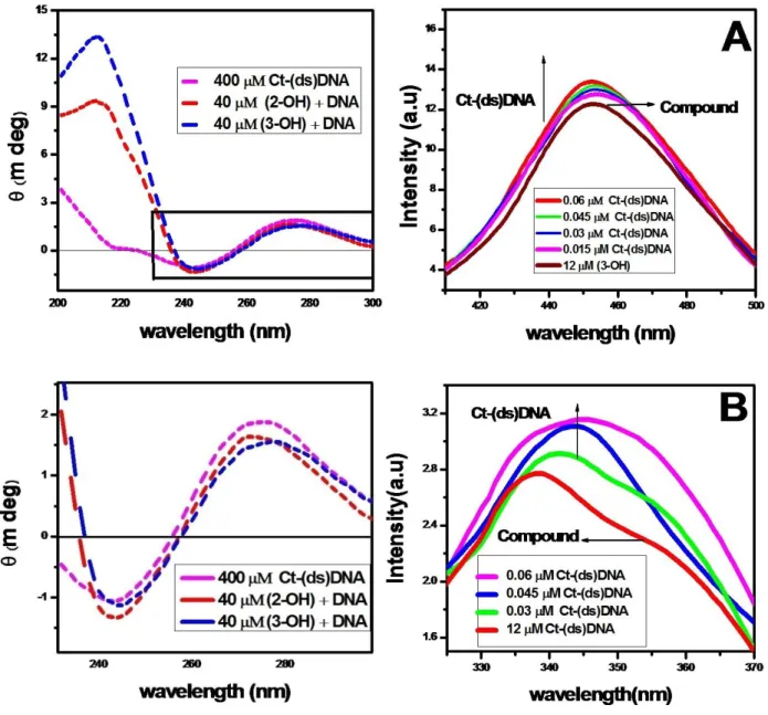
It is well known that the intercalation binding of linear or flat aromatic molecules into calf thymus (double-stranded) DNA induce large chirality changes and consequently significant effects on their CD spectra.41 The CD spectrum of free DNA shows a negative band at 245 nm due to helicity (Figure 3), and a positive band at 275 nm due to the base stacking, which is the characteristic of DNA in the right-hand B form.42 The CD band of DNA at 270-280 nm is assigned to base stacking interactions between the bases and the band at 245 nm is attributed to the polynucleotide helical structure.43
These bands are caused by the stacking interactions between the bases pairs and the helical suprastructure of the polynucleotide that provides an asymmetric environment for the bases. The secondary structure of DNA is perturbed markedly by the intercalation of small molecules leaving its signature through the conformational changes in the intrinsic CD spectra of ct-(ds) DNA. Groove binding, however, does not put so much impact on the CD signal in this report.44-47
Emission spectroscopy
The fluorescence signal of 452 nm was responsible for Baicalein and 338 nm for DHF. When the DNA solution was added to the flavonoids, Baicalein peak shifted from 452 nm to 450 nm and DHF is shifted from 338 nm to 346 nm. This information indicates that flavonoids were more binding (turn on) with Ct-(ds) DNA. Figures 6A and 6B shows the fluorescence spectra of flavonoids in the presence and absence of calf thymus DNA.
The stronger enhancement in fluorescence intensity of Baicalein with DNA may be largely due to the increase of the molecular planarity of the complex and the decrease of the collisional frequency of the solvent molecules with the complex which is caused by the planar aromatic group of the complex stacks between adjacent base pairs of the DNA.
0 G G
0 H G G H G G
1 DNA
A
A A K
Figure 3. Circular dichroism spectra of Ct-(ds) DNA in the absence and presence of flavonoids.
Flavonoids were binding to DNA leading to a marked increase in fluorescence emission intensity also agrees with observations for other intercalators.49,50
In the presence of DNA, emission quenching of flavonoids may be caused by the fact that, flavonoids being a small hydrophobic molecule and can be absorbed by hydrophobic groups on the surface of DNA.
Cyclic voltammetry
In a cyclic voltammetry experiment, scanning the potential in both directions provides an opportunity to explore the electrochemical behaviour of species generated at the electrode.
Electrochemical behavior of GCE was carefully investigated in Tris HCl buffer by cyclic voltammetry. The electrochemical response of flavonoids in DNA solution is a rich source of information about binding and reactivity.51
Figure 4. Emission spectrum of flavonoids in presence of increasing amount of Ct(ds)DNA, (A) Baicalein and (B) DHF.
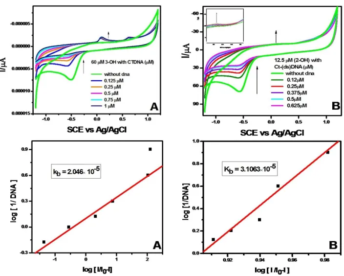
The cyclic voltammograms of flavonoids in the presence of different amounts of DNA were recorded in Tris HCl buffer solution pH 7.4 and are shown in Fig. 7A and 7B.
The voltammograms of Baicalein and DHF showed a prominent oxidation peak at 0.15 and 0.25 V, respectively.
These peaks were found to be shifted towards positive potential (from 0.12 to 0.26 V for Baicalein and from 0.03 to 0.24 V for DHF) in the presence of DNA. Such an observation might be attributed to one of the two factors i.e.
either the nonconducting DNA can block the electron transfer from the flavonoid or the DNA–flavonoid complex formed is electrochemically inactive. If the electron transfer was not blocked, then the current should increase but peak shift would be expected.52 The observed shift in the peak potential and increased peak current was attributed to the formation of the DNA-flavonoid complex through the intercalative mode of binding.53 Thus, the electrochemical studies supported the spectroscopic results indicating the intercalative mode of binding between the flavonoid and DNA. Typical cyclic voltammetric behaviours of flavonoids without and with Ct-(ds) DNA was studied in pH 7.4 Tris HCl buffer solution under the potential range of (-1.2 to
1.2 V) for Baicalein and (-1.0 to 1.0 V) for DHF with the rate of 50 mV s−1 (Fig.5A, B) and oxidation and reduction peaks (redox reaction) appeared.
Figure 5A. Cyclic voltammograms of interaction of Baicalein with various concentrations of Ct-(ds) DNA.
As can be seen, on the bare GCE, flavonoids had a small stable and quasi-reversible redox response, however, on the GCE, much more obviously redox peaks of flavonoids were found at 0.15 and 0.25 V, which were ascribed to the good conductivity for flavonoids.54 Binding constant for Baicalein is 2.046×10⁻⁵ and 3.1063×10⁻⁵ for DHF. A further study showed that the peak currents of flavonoids were linear with the scan rates (v), in the scan rates range from 25– 250 mV s−1.55 In the presence of 0.016 mg mL-1 dsDNA, the peak current of the flavonoids decreased apparently and the peak potentials show almost no changes, indicating that flavonoids combined with DNA forming an electroactive complex.
Molecular docking
Molecular docking techniques are an attractive scaffold to understand the drug–DNA interactions in rational drug design, as well as in the mechanistic study by placing a small molecule into the binding site of the target specific region of the DNA mainly in a non-covalent fashion.
Structure of drug is made flexible to attain different conformations in order to predict the best-fit orientation, and the best energy docked structure is analyzed.56
Figure 5B. Cyclic voltammograms of interaction of DHF with various concentrations of Ct-(ds) DNA.
Molecular docking is an exemplary platform to interpret ligand DNA interactions leads to drug discovery and design.
Here the ligands were individually docked with dodecamer duplex sequence of DNA d(CGCGAATTCGCG)2 (PDB ID:
1BNA) so as to find out the binding site besides position of the ligand. From the docking results, Minimum binding energy conformer is elected for calculations.57
The binding interactions of DHF and Baicalein with Ct- (ds) DNA (1BNA) seems to had intercalation mode of binding with DNA. Both ligands interacted on the same site of the DNA molecule but Baicalein seems to have more interaction with the nucleotide than the other. Maximum of 10 poses will be generated depending upon torsions in the ligands.58
The above studies confirm that both Baicalein and the 7,8- dihydroxyflavone interact with the double-stranded DNA by intercalation binding. Among these, the Baicalein has a better binding efficiency compared to 7,8-dihydroxyflavone.
Figure 6A. Docking of interaction between Baicalein to Ct-(ds) DNA
Figure 6B. Docking of interaction between 7, 8-DHF to Ct-(ds) DNA
Conclusion
In conclusion, the binding interactions of flavonoids with calf thymus DNA have been studied using UV-VIS absorption, fluorescence, CD spectroscopy and electrochemical studies. The results indicated that the binding mode of flavonoids to DNA is an intercalation binding, which was supported by the results from electrochemical studies [Guowen Zhang et al.]. It was found that both hydrophobic interactions and hydrogen bonds play a major role in the binding of flavonoids to DNA. Their studies indicated that these flavonoids intercalated into the dsDNA helix, which accounts for the high binding constant.
Their experimental results are similar to those of our present study, which might be attributed to the similar chemical structures of the flavonoids. Based on binding constants, it is apparent that Baicalein (2.046×10⁻⁵) can bind more strongly with than DHF (3.1063×10⁻⁵). These studies may provide useful information for further study of the pharmacological effect and insight into the redox reactions of these molecules in the living body.
References
1Arshad, N., Janjua, N. K., Skibsted, L. H., Anderson, M. L., UV- absorption studies of the interaction of karanjin and karanjachromene with ds. DNA: Evaluation of binding and antioxidant activity. Cent. Eur. J. Chem., 2013, 11(12), 2040- 2047 DOI: 10.2478/s11532-013-0327-z
2Tu, B., Chen, Z.-F., Liu, Z.-J., Cheng, L.-Y., Hu, Y.-J., Hbei, Collaborative Innovation Center for Rare Metal Chemistry, Hubei Key. RSC Adv., 2015, 5, 33058-33066.
DOI:https://doi.org/10.1039/C5RA04505C
3Middleton, E., Kandaswami C., and Theoharides, T. C., The effects of plant flavonoids on mammalian cells: Implication for inflammation, heart disease, and cancer. Pharmacol. Rev., 2000, 52, 673–751. DOI:0031-6997/00/5204-0673$03.00/0
4Hannon, M. J., Supramolecular DNA recognition. Chem. Soc.
Rev., 2007, 36, 280-295
DOI:https://doi.org/10.1039/B606046N
5MacMillan, A. M., Fifty years of "Watson-Crick". Pure Appl.
Chem., 2004, 76, 1521-1524.
DOI:https://doi.org/10.3390/molecules14051725
6Theodore, K., Christopoulos, S., Acid Analysis. Anal.
Chem., 1999, 71(18), 425–438.
DOI:https://doi.org/ 10.1021/a19900161
7Brenno, A., Neto, D., Alexandre, A., Lapis. M., Recent Developments in the Chemistry of Deoxyribonucleic Acid (DNA) Intercalators: Principles, Design, Synthesis, Applications, and Trends. Molecules, 2009, 14, 1725-1746.
DOI:https://doi.org/10.3390/molecules14051725
8Chen, A. Y , Liu, L. F., DNA Topoisomerases: Essential Enzymes and Lethal Targets. Annu Rev Pharmacol Toxicol, 1994, 34, 191–218.
DOI:https://doi.org/10.1146/annurev.pa.34.040194.001203
9Pilch, D. S., Kirolos, M. A., Liu X., Plum G. E., Breslauer K. J., Berenil [1,3-bis(4-aminophenyl)triazene] Binding to DNA Duplexes and to a RNA Duplex: Evidence for Both Intercalative and Minor Groove Binding Properties.
Biochemistry, 2017, 34, 9962–9976. DOI:https://doi.org/
10.1021/acs.jpclett.7b00472
10Qu, X., Wan, C., Becker, H. C., Zhong, D., Zewail, A. H., The anticancer drug–DNA complex: femtosecond primary dynamics for anthracycline antibiotics function. Proc. Natl.
Acad. Sci. The U.S.A., 2001, (98), 14212–14217.
DOI:https://doi.org/ 10.1073/pnas.241509698
11Swaran, F. J. S., Structural, chemical and biological aspects of antioxidants for strategies against metal and metalloid exposure. J. List Oxid. Med. Cell Longev., 2009, 2(4), 191–
206. DOI: https://doi.org/10.4161/oxim.2.4.9112
12Erlejman, A. G., Verstraeten, S. V., Fraga, C. G., Oteiza, P. I., The Interaction of Flavonoids with Membranes: Potential Determinant of Flavonoid Antioxidant Effects. Free Radical Res., 2004, 38(12),
DOI:https://doi.org/10.1080/10715760400016105
13Chen, H., Liu, X., Patel, D. J., DNA bending and unwinding associated with actinomycin D antibiotics bound to partially overlapping sites on DNA. J. Mol. Biol., 1996, 258, 457–479.
DOI:https://doi.org/ 10.1006/jmbi.1996.0262
14Tsuboi, T. M., Benevides, J. M., Thomas, G. J., The complex of ethidium bromide with genomic DNA: structure analysis by polarized Raman spectroscopy. Biophys. J, 2007, 92, 928–
934. DOI: https://doi.org/ 10.1529/biophysj.106.093633
15Bruijnincx, P. C. A. and Sadler, P. J., New trends for metal complexes with anticancer activity. Current Opin. Chem.
Biol., 2007, 12(2), 197-206. DOI: http://dx.doi.org/
10.1016/j.cbpa.2007.11.013
16Ashwini, H., Hegde, S. N., Prashanth, J., Seetharamappa, Interaction of antioxidsnt flavonoids with calf thymus DNA analyzed by spectroscopic and electrochemical methods. J.
Pharm. Biomed. Anal., 2012, 63, 40–46.
DOI:https://doi.org/10.1016/j.jpba.2012.01.034
17Pospisil, P., Wang, K., Al Aowad, A . F., Lyer, L. K., Adelstein, S. J., Kassis, A. I., Computational modeling and experimental evaluation of a novel prodrug for targeting the extracellular space of prostate tumors. Cancer Res. 2007, 67, 2197–2205.
DOI: https://doi.org/10.1158/0008-5472.CAN-06-3309
18Husain, M. A., Ishqi, H. M., Sarwar, T., Rehman S. U. and Tabish, M., Interaction of indomethacin with calf thymus DNA: a multi-spectroscopic, thermodynamic and molecular modelling approach. MedChemCom., 2017(6), c7md00094d.
DOI: https://doi.org/10.1039/C7MD00094D
19Ali, A., Ensafi, A., Hajiana, R. and Ebrahimi, S., Study on the Interaction between Morin-Bi(III) Complex and DNA with the use of Methylene Blue Dye as a Fluorophor Probe. J. Braz.
Chem. Soc., 2009, 20(2), 266-276,
DOI:http://dx.doi.org/10.1590/S0103-50532009000200011
20Iyyam Pillai, S., Vijaya Raghavan, K., and Subramanian, S., Evaluation of DNA-binding, cleavage, BSA interaction of Zn-hydroxy flavone complex. Der Pharma Chem., 2014, 6(1), 379-389. http://derpharmachemica.com/archive.html
21Stephanos, J. J., Drug-protein interactions: Two-site binding of heterocyclic ligands to a monomeric hemoglobin, J.
Photochem. Photobiol., 2017, 170, 256–262.
DOI: 10.1016/j.jphotobiol.2017.04.019
22Stephanos, J. J., Farina, S. A., and Addison, A. W., Iron ligand recognition by monomeric hemoglobins, Biochim. Biophys.
Acta, 1996, 1295, 209–221.
DOI:https://doi.org/10.1016/0167-4838(96)00041-6
23Saenger, W. Principles of Nucleic Acid Structure; Springer-Verlag:
New York, 1983, DOI:https://doi.org/10.1007/978-1-4612- 5190-3
24Sarkar, D., Das, P., Basak, S., Chattopadhyay, N., Binding Interaction of Cationic Phenazinium Dyes with Calf Thymus DNA: A Comparative Study J. Phys. Chem. B 2008, 112, 9243–9249. DOI:https://doi.org/ 10.1021/jp801659d
25Teng, M. K., Usman, N., Frederick, C. A., Wang, A. H. J., The molecular structure of the complex of Hoechst 33258 and the DNA dodecamer d(CGCGAATTCGCG). Nucleic Acids Res.
1988, 16, 2671–2690.
26Pyle, A. M., Rehmann, J. P., Meshoyrer, R., Kumar, C. V., Turro, N. J., Jacqueline Barton K., Mixed-ligand complexes of ruthenium(II): factors governing binding to DNA. J. Am.
Chem. Soc., 1989, 111(8), 3051–3058.
DOI: https://doi.org/10.1021/ja00190a046
27Fasman, G. D., Circular Dichroism and Conformational Analysis of Biomolecules, Plenum Press, New York, 1996, 413.
DOI: https://doi.org/10.1021/ja965689f
28Rich, A., Nordheim, A., Wang, A .H. J., The chemistry and biology of left-handed Z-DNA, Ann. Rev. Biochem. 1984, 53, 791–843.
DOI: https://doi.org/10.1146/annurev.bi.53.070184.004043
29Zhang, G., Fu, P., Wang, L. and Hu, M., Molecular Spectroscopic Studies of Farrerol Interaction with Calf Thymus DNA. J.
Agric. Food Chem., 2011, 59, 8944–8952.
DOI:https://doi.org/ 10.1021/jf2019006
30Sarkar, D., Das, P., Basak, S., Chattopadhyay, N., Binding Interaction of Cationic Phenazinium Dyes with Calf Thymus DNA: A Comparative Study. J. Phys. Chem. B, 2008, 112, 9243–9249. DOI: https://doi.org/ 10.1021/jp801659d
31Jain, S. S., Polak, M., Hud, N. V., Controlling nucleic acid secondary structure by intercalation: effects of DNA strand length on coralyne‐driven duplex disproportionation.
Nucleic Acids Res. 2003, 31, 4608–4615.
DOI:https://doi.org/10.1093/nar/gkg648
32Gago, F., Stacking Interactions and Intercalative DNA Binding. : A Companion to Methods in Enzymology, 1998, 14, 277–292.
DOI:https://doi.org/10.1006/meth.1998.0584
33Garab, Gy., van Amerongen, H., Linear dichroism and circular dichroism in photosynthesis research. Photosynth Res., 2009, 101(2-3), 135–146.
DOI:https://doi.org/10.1007/s11120-009-9424-4
34Long, Y. F., Liao, Q. G., Huang, C. Z., Ling, J., Li, Y. F., Conformational Change Detection of DNA with the Fluorogenic Reagent of o-Phthalaldehyde-β- Mercaptoethanol, J. Phys.Chem. B 2008, 112, 1783–178.
DOI:https://doi.org/ 10.1021/jp071601g
35Barton, J. K., Goldberg, J. M., Kumar, C.V., Turro, N. J., Binding
modes and base specificity of
tris(phenanthroline)ruthenium(II) enantiomers with nucleic acids: tuning the stereoselectivity. J. Am. Chem. Soc. 1986, 108, 2081. DOI: https://doi.org/ 10.1021/ja00268a057
36Ni, Y., Du, S., Kokot, S., Continuous flow microfluidic device for cell separation, cell lysis and DNA purification. Anal. Chim.
Acta, 2007, 584, 19.
DOI:https://doi.org/10.1016/j.aca.2006.11.006
37Ling, X., Zhong, W., Huang, Q., Ni, K., Spectroscopic studies on the interaction of pazufloxacin with calf thymus DNA. J.
Photochem. Photobiol. B: Biology 2008, 93, 172-176.
DOI:https://doi.org/10.1016/j.jphotobiol.2008.07.008
38Welch, T. W., Thorp, H., Distribution of Metal Complexes Bound to DNA Determined by Normal Pulse Voltammetry. J. Phys.
Chem. 1996, 100, 13829-13836.
DOI:https://doi.org/10.1021/jp960251n
39Wang, Y., Ni, Y., Kokot, S., Voltammetric behaviour of complexation of salbutamol with calf thymus DNA and its analytical application, Anal. Biochem., 2011, 419, 76–80.
DOI: https://doi.org/10.1039/C6RA03062A
40Carter, M. T., Rodriguez, M., Bard, A. J., Voltammetric studies of the interaction of metal chelates with DNA. 2. Tris-chelated complexes of cobalt (III) and iron (II) with 1,10- phenanthroline and 2,2-bipyridine. J. Am. Chem. Soc., 1989, 111, 8901–8911. DOI:https://doi.org/10.1021/ja00206a020
41Molina, A., Gonzalez, J., Laborda,E., Wang, Y., Compton, R. G., Catalytic mechanism in cyclic voltammetry at disc electrodes:
an analytical solution, Phys. Chem. Chem. Phys., 2011, 13, 14694-14704. DOI:https://doi.org/ 10.1039/C1CP21181A
42Zou, N., Wang, X., Li, G., Spectroscopic and electrochemical studies on the interaction between luteolin and DNA. J Solid State Electrochem, 2016, 20, 1775–1782.
DOI:https://doi.org/10_1007-S10008-016-3174
43Rehman, S. U., Yaseen, Z., Husain, M. sA., Sarwar, T., Ishqi, H.
M., Interaction of 6 Mercaptopurine with Calf Thymus DNA – Deciphering the Binding Mode and Photoinduced DNA Damage. PLoS ONE , 2014, 9(4), e93913.
DOI:https://doi.org/ 10.1371/journal.pone.0093913
44Trott, O., Olson, A. J., AutoDock Vina: improving the speed and accuracy of docking with a new scoring function, efficient optimization, and multithreading. J. Comput. Chem., 2010, 31, 455-461. DOI:https://doi.org/ 10.1002/jcc.21334
45Roy, S., Ganai, S., Nandi, R.K., Majumdar, K. C., Taipans, D. K., Report of Interaction Between Calf Thymus DNA and Pyrimidine-Annulated Spiro-Dihydrofuran. Biochem. Anal.
Biochem. , 2016, 5, 2.
DOI:https://doi.org/10.4172/21611009.1000278
46Garab, GY., van Amerongen, H., Linear dichroism and circular dichroism in photosynthesis research. Photosynth Res., 2009, 101(2-3), 135–146.
DOI:https://doi.org/10.1007/s11120-009-9424-4
47Long, Y. F., Liao, Q. G., Huang, C. Z., Ling, J., Li, Y. F., Conformational Change Detection of DNA with the Fluorogenic Reagent of o-Phthalaldehyde-β- Mercaptoethanol. J. Phys. Chem. B 2008, 112, 1783–178.
DOI:https://doi.org/ 10.1021/jp071601g
48Barton, J. K., Goldberg, J. M., Kumar, C. V., Turro, N. J., Binding modes and base specificity of tris(phenanthroline)ruthenium(II) enantiomers with nucleic acids: tuning the stereoselectivity. J. Am. Chem. Soc. 1986, 108, 2081. DOI: https://doi.org/ 10.1021/ja00268a057
49Ni, Y., Du, S., Kokot, S., Continuous flow microfluidic device for cell separation, cell lysis and DNA purification. Anal.
Chim. Acta, 2007, (584), 19.
DOI:https://doi.org/10.1016/j.aca.2006.11.006
50Ling, X., Zhong, W., Huang, Q., Ni, K., Spectroscopic studies on the interaction of pazufloxacin with calf thymus DNA. J.
Photochem. Photobiol. B: Biology, 2008, 93(3), 172-176.
DOI:https://doi.org/10.1016/j.jphotobiol.2008.07.008
51Welch, T. W., Thorp, H., Distribution of Metal Complexes Bound to DNA Determined by Normal Pulse Voltammetry.
J. Phys. Chem. 1996, 100, 13829-13836.
DOI:https://doi.org/10.1021/jp960251n
52Wang, Y., Ni, Y., Kokot, S., the Voltammetric behaviour of complexation of salbutamolwith calf thymus DNA and its analytical application, Anal. Biochem., 2011, 419, 76–80.
DOI: https://doi.org/10.1039/C6RA03062A
53Carter, M. T., Rodriguez, M., Bard, A. J., Voltammetric studies of the interaction of metal chelates with DNA. 2.
Tris-chelated complexes of cobalt (III) and iron (II) with 1,10-phenanthroline and 2,2-bipyridine. J. Am. Chem. Soc., 1989, 111, 8901–8911.
DOI:https://doi.org/10.1021/ja00206a020
54Molina, A., Gonzalez, J., Laborda, E., Wang, Y., and Compton, R.
G., Catalytic mechanism in cyclic voltammetry at disc electrodes: an analytical solution, Phys. Chem. Chem. Phys., 2011, 13, 14694-14704.
DOI:https://doi.org/ 10.1039/C1CP21181A
55Zou, N., Wang, X., Li, G., Spectroscopic and electrochemical studies on the interaction between luteolin and DNA. J.
Solid State Electrochem, 2016, 20, 1775–1782.
DOI:https://doi.org/10_1007-S10008-016-3174
56Rehman, S. U., Yaseen, Z., Husain, M. sA., Sarwar, T., Ishqi, H. M., Interaction of 6 Mercaptopurine with Calf Thymus DNA – Deciphering the Binding Mode and Photoinduced DNA Damage. PLoS ONE, 2014, 9(4), e93913.
DOI:https://doi.org/ 10.1371/journal.pone.0093913
57Trott, O., Olson, A. J., AutoDock Vina: improving the speed and accuracy of docking with a new scoring function, efficient optimization, and multithreading. J. Comput. Chem., 2010, 31, 455-461.
DOI:https://doi.org/ 10.1002/jcc.21334
58Swarup Roy, Sintu Ganai, Raj Kumar Nandi, Majumdar, K. C., and Taipans, Das, K., Report of Interaction Between Calf Thymus DNA and Pyrimidine-Annulated Spiro- Dihydrofuran. Biochem. Anal. Biochem., 2016, 5, 2.
DOI:https://doi.org/10.4172/21611009.1000278
Received: 16.01.2018.
Accepted: 03.03.2018.