THESIS FOR THE DEGREE OF DOCTOR OF PHILOSOPHY (PHD)
8-Oxoguanine DNA glycosylase-1 links DNA repair to cellular responses via the activation of the small GTPases, Ras and Rac1
by
György Hajas
Supervisor: Attila Bácsi, PhD
UNIVERSITY OF DEBRECEN
DOCTORAL SCHOOL OF MOLECULAR CELL AND IMMUNE BIOLOGY
DEBRECEN,2015
Table of contents
1. Introduction ... 7
1.1 Oxidative stress ... 7
1.1.1 Formation of 8-oxo-7,8-dihydroguanine (8-oxoG) ... 9
1.1.2 Defense mechanisms against ROS ... 11
1.2 DNA repair mechanisms ... 12
1.2.1 Mismatch Repair (MMR) ... 13
1.2.2 Nucleotide Excision Repair (NER) ... 15
1.2.3 Double-Strand Break Repair ... 16
1.2.4 Base Excision Repair (BER) ... 18
1.2.5 OGG1, a versatile DNA repair enzyme ... 20
1.3 Small GTPases... 22
1.3.1 Ras ... 23
1.3.2 Rac ... 25
1.4. DNA repair-independent functions of OGG1 ... 28
2. Objectives of the study ... 29
3. Materials and methods ... 30
3.1 Reagents ... 30
3.2 Cell cultures ... 31
3.3 Animals and treatments ... 31
3.4 Assessment of GTP-bound Ras and Rac levels ... 32
3.5 Assessment of 8-oxoG’s cellular uptake ... 32
3.6 Protein interaction assays ... 33
3.7 Western blot analysis ... 33
3.8 Preparation of 8-oxoG solution ... 34
3.9 Fluorescence spectroscopy ... 34
3.10 Guanine nucleotide exchange assay ... 34
3.11 Gene expression and molecular network analysis ... 35
3.12 Flow cytometry ... 35
3.13 Latex bead uptake ... 36
3.14 Lucifer Yellow uptake ... 36
3.15 FITC-dextran uptake ... 36
3.16 Chemotaxis assay ... 36
3.17 Down-regulation of gene expression ... 37
3.18 Quantitative real-time PCR ... 37
3.19 Oligonucleotide excision assay ... 37
3.20 Assessment of cellular ROS levels ... 38
3.21 Microscopic imaging ... 38
3.22 Statistical analysis ... 39
4. Results and Discussion ... 40
4.1 Signaling pathways induced at the transcriptomal level by 8-oxoG ... 40
4.2 Ras activation in cell culture and lungs ... 41
4.3 Ras is not activated when OGG1 silenced ... 42
4.4 Ras activation by excised 8-oxoG: the KG-1 model ... 43
4.5 OGG1 binds free 8-oxoG at an independent site and not in the DNA lesion-recognition site .... 48
4.6 OGG1/8-oxoG complex functions as a GEF on Ras ... 50
4.7 Ras activation induced by 8-oxoG treatment leads to phosphorylation of MAPKs ... 52
4.8 Treatment with 8-oxoG also increases cellular ROS levels ... 54
4.9 8-OxoG induces ROS via activating NADPH oxidase... 56
4.10 Rac1 activation in cultured cells ... 57
4.11 Rac1 activation in lung ... 59
4.12 Changes in ROS levels begin at perinuclear membrane ... 60
4.13 Rac1 and OGG1 colocalize with NOX4 in nuclear membrane ... 61
4.14 OGG1 physically interacts with small GTPase Rac1 ... 63
4.15 OGG1/8-oxoG complex acts as a GEF on Rac1 ... 64
4.16 The role of ROS-OGG1-Rac1/Ras triangle in cellular responses ... 65
5. New scientific results ... 68
6. Summary ... 69
Összefoglalás ... 70
7. References ... 71
7.1 References related to dissertation ... 71
7.2 Publication list prepared by the Kenézy Life Siences Library ... 87
8. Keywords ... 90
9. Acknowledgement ... 91
SUPPLEMENTARY 1
Boldogh I, Hajas G, Aguilera-Aguirre L, Hegde ML, Radak Z, Bacsi A, Sur S, Hazra TK, Mitra S. Activation of ras signaling pathway by 8-oxoguanine DNA glycosylase bound to its excision product, 8-oxoguanine.
J Biol Chem. 2012 Jun 15;287(25):20769-73.
SUPPLEMENTARY 2
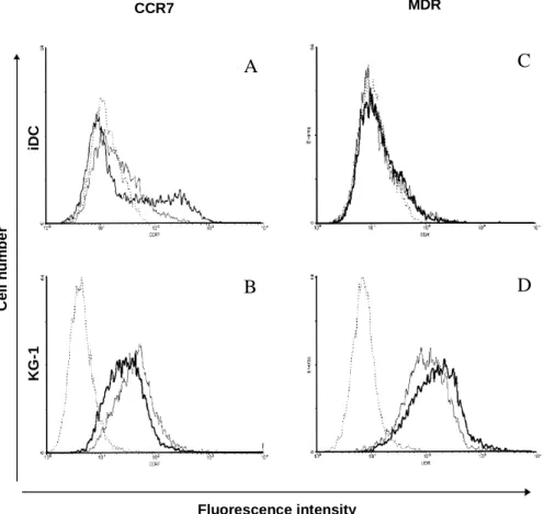
Hajas G, Zsiros E, László T, Hajdú P, Somodi S, Réthi B, Gogolák P, Ludányi K, Panyi G, Rajnavölgyi E. New phenotypic, functional and electrophysiological characteristics of KG-1 cells.
Immunol Lett. 2004 Mar 29;92(1-2):97-106.
SUPPLEMENTARY 3
German P, Szaniszlo P, Hajas G, Radak Z, Bacsi A, Hazra TK, Hegde ML, Ba X, Boldogh I.
Activation of cellular signaling by 8-oxoguanine DNA glycosylase-1-initiated DNA base excision repair.
DNA Repair. (Amst). 2013 Oct;12(10):856-63.
SUPPLEMENTARY 4
Hajas G, Bacsi A, Aguilera-Aguirre L, Hegde ML, Tapas KH, Sur S, Radak Z, Ba X, Boldogh I. 8-Oxoguanine DNA glycosylase-1 links DNA repair to cellular signaling via the activation of the small GTPase Rac1.
Free Radic Biol Med. 2013 Aug;61:384-94
Abbreviations
8-OH-Ade 8-oxo-7,8-dihydroadenine
8-oxodG 8-oxo-7,8-dihydro-2′-deoxyguanosine 8-oxoG 8-oxo-7,8-dihydroguanine
APC antigen presenting cell
APE1 apurinic/apyrimidinic endonuclease 1 BER base excision repair
BSA bovine serum albumin
CCR7 CC type chemokine receptor 7
ChTX charybdotoxin;
DC dendritic cell
DNA-PKcs DNA-dependent protein kinase DSBs double-strand breaks
ELISA enzyme linked immuno-sorbent assay
ExoI exonuclease 1
FapyG 2,6-diamino-4-hydroxy-5-formamidopyrimidine
FcγR Fcγ gamma receptor
FU fluorescence unit
GAP GTPase activating protein
GC/MS gas chromatography/mass spectrometry
GDI GDP dissociation inhibitor
GDP guanosine diphosphate
GEF guanine nucleotide exchange factor
GITR glucocorticoid-induced TNFR-related protein (TNFRSF18) GG-NER global genome repair
GM-CSF granulocyte–monocyte-colony stimulating factor GST glutathione-S-transferase
GTP guanosine triphosphate
GTPγS guanosine 5-3-O-(thio)triphosphate
H/K/N-Ras Harvey/Kirsten/neuroblastoma sarcoma virus oncogene homolog H2DCF-DA 2′-7′-dihydrodichlorofluorescein diacetate
HR homologous recombination
HSC hematopoietic stem cell
IPA Ingenuity Pathways Analysis
IFN-γ interferon-γ IL-4/13 interleukin 4/13
LC Langerhans cell
LC/IDMS liquid chromatography and isotope dilution mass spectrometry
MantGDP GDP (2′-(or-3′)-O-(N-methylanthraniloyl)guanosine 5′-diphosphate
MantGTP GTP (2′-(or-3′)-O-(N-methylanthraniloyl)guanosine 5′-triphosphate MDR multidrog resistance
MHC major histocompability complex MIP-3β macrophage inflammatory protein β
MMR mismatch repair
MR mannose receptor
MTT 3-(4,5-dimethyl-2-thiazolyl)-2,5-diphenyl-2H-tetrazolium bromide
NAC N-acetyl-L-cysteine
NOX1/2/3/4 NADPH oxidase type 1/2/3/4 NER nucleotide excision repair NHEJ non-homologous end-joining Nrf2 NF-E2 related factor 2
OGG1 8-oxoguanine DNA glycosylase-1 OGG1/8-oxoG OGG1 with bound 8oxoG
p22phox regulatory subunit of NOXs PBS phosphate buffer solution;
PGE2 prostaglandin E2
Polβ/δ/ε/ι/λ/µ DNA polymerase β/δ/ε/ι/λ/µ PMA phorbol-myristilacetate
PCNA proliferating cell nuclear antigen PARP1 poly (ADP-ribose) polymerase 1
Rac1 Ras-related C3 botulinum toxin substrate 1
Ras small GTPase of the Ras superfamily (Rat sarcoma) RPA replication protein A
RT-PCR reverse transcription followed by polymerase chain reaction SLC secondary lymphoid-tissue cytokine
SOD superoxide dismutase
TNF-α tumor necrosis factor-α TC-NER transcription-coupled repair
This dissertation describes new findings about the DNA glycosylase (OGG1) as an initiator of cellular responses in complex with its substrate the oxidized base 8-oxo-7,8-dihydroguanine (8-oxoG).
1. Introduction
Oxidative stress is an evolutionary driving force that often defined as an imbalance between the prooxidative and the antioxidative sources. It has been connected to serious health disorders like Parkinson’s disease (Valko, Leibfritz et al. 2007), Alzheimer’s disease (Pohanka 2013), cancer (Halliwell 2007), myocardial infarction (Ramond, Godin-Ribuot et al.
2013), chronic fatigue syndrome (Kennedy, Spence et al. 2005). On the other hand, organisms wouldn’t be able to survive without the controlled production of reactive oxidative molecules.
Redox sensitive amino acids like methionine play an important role in cellular signaling. The reversible oxidation of methionine can inhibit the phosphorylation of adjacent Tyr/Ser/Thr site influencing main signaling pathways (Hardin, Larue et al. 2009).
1.1 Oxidative stress
The environment is becoming richer and richer source of prooxidants because of the increasing amount of pollutants, chemicals, ionizing and ultraviolet radiation. These sources act directly or via activation of oxido-reductases and/or induction of mitochondrial dysfunction. When the antioxidant system of the cells cannot balance out the increased concentration of reactive oxygen species (ROS), these molecules indiscriminately modify proteins, lipids, and DNA (D'Autreaux and Toledano 2007) and disrupt normal cellular signaling processes.
As a part of their normal physiological activity cells produce ROS molecules. Notable cellular sources of ROS are: mitochondrial leakage during oxidative phosphorylation, xanthine oxidase, cytochromes P450 and NADPH oxidases (NOX1-5, DUOX1-2). These enzymes have the ability to transport electrons across the plasma membrane and to generate superoxide and other downstream reactive oxygen radicals. Phagocytic cells use NADPH oxidase 2 (gp91phox) to produce ROS after engulfing bacteria or viruses (Nathan and Shiloh 2000). In case of frustrated phagocytosis these toxic agents are released and damage surrounding tissues (Cannon and Swanson 1992).
The most common ROS molecules are: superoxide anion (O2•–), hydroxyl radical (OH•), alkoxy-radicals (RO•), peroxy-radicals (ROO•), hydrogen peroxide (H2O2), organic
hydroperoxides (ROOH), hypochlorous acid (HOCl), and peroxynitrite (ONOO–). The free- radicals can damage lipids via oxidation, which often referred to as lipid peroxidation. During the reaction the free radicals "steal" electrons from the lipids in membranes. They mostly affect polyunsaturated fatty acids, because of their multiple double bonds. Lipid peroxides can participate in chain reactions that further increase damage to biomolecules like proteins (Negre-Salvayre, Coatrieux et al. 2008). Not only the lipid peroxides, but their degradation products (hydroxy-alkenals) can generate a variety of intra- and intermolecular covalent adducts that have influence on cell signaling, transcription factors and gene expression (Catala 2009).
As a major consequence of ROS formation, proteins are frequently damaged either at specific side chains of amino acids (i.e. by hydrogen peroxide) or non-specifically throughout the backbone (i.e. by hydroxyl radicals). Hydroperoxides can induce further oxidation, chain reactions and stable products that can be used as biomarkers. Most protein damage results in loss of function (enzyme activity, signaling), modified structure (unfolding, aggregation) and altered interactions with other molecules. Most oxidized proteins undergo selective proteolysis by proteasomal and lysosomal pathways, but in some cases, they may contribute to multiple human pathologies (Davies 2012).
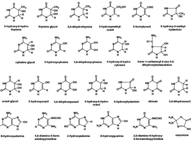
One of the most common reactive oxygen species, the hydroxyl radical reacts with DNA by addition to double bonds of heterocyclic DNA bases and by abstraction of an H atom from the methyl group of thymine and each of the C-H bonds of 2′-deoxyribose (Teoule 1987). In case of purines, hydroxyl radical can be added to the C4, C5, and C8 positions, generating OH adduct radicals. Depending on their redox properties, the redox environment and the reaction partners, radicals are reduced or oxidized. Product types and yields depend on absence and presence of oxygen and on other conditions (Dizdaroglu 1992). So far more than 20 base lesions have been identified (Fig. 1.) The consequences of these DNA lesions are diverse: they can cause mutations, conformational changes in DNA, deletions, epigenetic changes among others /reviewed by Cooke at al. in (Cooke, Evans et al. 2003)/.
Figure 1. Oxidized DNA bases
DNA base products of interaction with reactive oxygen and free radical species (Cooke, Evans et al. 2003)
1.1.1 Formation of 8-oxo-7,8-dihydroguanine (8-oxoG)
The most susceptible base among the DNA and RNA bases to oxidative modification is guanine, due to its lowest reduction potential (midpoint potential is −1.29 mV vs. nickel- hydrogen electrode) (Jovanovic and Simic 1986). In vivo, guanine in DNA and RNA can be modified not only by •OH but also by other reactive species, including reactive oxygen (superoxide anion: O2•–), nonradical (ozone: O3; singlet oxygen: 1O2; hydrogen peroxide:
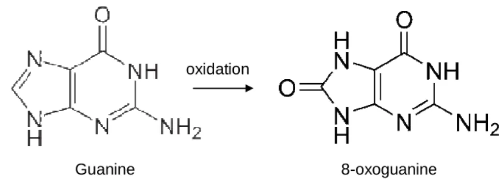
H2O2), and nitrogen species (nitric oxide: NO•; peroxinitrite: ONOO–), as well as nitrosoperoxycarbonate (ONOOCO2–), carbonate anions (CO3–) and the UVA component of solar light (Dizdaroglu, Jaruga et al. 2002; Cadet, Douki et al. 2006). The reaction of •OH with guanine can result in guanine C8-OH-adduct formation (Fig. 2). One-electron oxidation of guanine C8-OH-adduct results in 7,8-dihydro-8-oxoguanine (8-oxoG), while one-electron reduction of the guanine C8-OH-adduct radical leads to a ring opening, resulting in 2,6- diamino-4-hydroxy-5-formamidopyrimidine (FapyG) or its isomer 2,5-diamino-4- hydroxy-6- formamidopyrimidine (Dizdaroglu, Kirkali et al. 2008; Jaruga, Kirkali et al. 2008). The free
8-oxoG base exists in both neutral (N9-H) and anionic (N9:−) forms at physiologic pH. Its presence as a free base in extracellular fluids is one of the most reliable gauges of the oxidative stress load of an organism (Fraga, Shigenaga et al. 1990; Svoboda, Maekawa et al.
2006). Due to its low redox potential, 8-oxoG is more reactive than guanine and serves as a primary target of reactive oxygen species and considered as a protective element in DNA (Sheu and Foote 1995). These observations were supported by findings showing that oligodeoxynucleotide damage and plasmid cleavage by reactive oxygen species (ROS) were inhibited in the presence of 8-oxodG (Kim, Choi et al. 2004).
Figure 2. Guanine and 8-oxoguanine
Estimates show that under physiological conditions, several hundred 8-oxoG lesions could be formed in DNA per eukaryotic cell daily (Lindahl and Barnes 2000). It has been determined that 8-oxoG is one of the most abundant DNA lesions formed in oxidative stress conditions, such as those that exist in diseased and aged cells/tissues (Dizdaroglu 1985;
Dizdaroglu, Jaruga et al. 2001). In mammals, the intra-helical 8-oxoG is recognized by its unique electronic properties (Markus, Daube et al. 2008) and excised by the E. coli Fpg homolog 8-oxoguanine DNA glycosylase 1 (OGG1) from nuclear and mitochondrial genomes during base excision repair (BER) processes (Mitra, Izumi et al. 2002; Dizdaroglu 2005).
Unrepaired 8-oxoG may be paired with adenine during DNA replication, resulting in transversion mutations (Nishimura 2002). During mRNA synthesis, it may serve as a template to transcriptional mutagenesis (Saxowsky, Meadows et al. 2008). Kaneko and his co-workers showed a non-linear accumulation of 8-oxoG in nuclear DNA isolated from brain, heart, liver, and kidneys of rats, detecting a 2-fold increase in 30 month-old tissues compared to 2-24 month-old ones (Kaneko, Tahara et al. 1996). It is believed that this accumulation is caused by higher levels of ROS and/or decreased inactivity of OGG1 during the aging process (Chen, Hsieh et al. 2003).
As RNA molecules are present mostly in single stranded forms without protecting proteins, they are even more prone to oxidative damage (Li, Wu et al. 2006; Kong and Lin 2010). It is estimated that 30-70% of messenger RNA contains 8-oxoG because of the low
Guanine 8-oxoguanine
oxidation
redox potential of guanine and the lack of repair systems (Thorp 2000; Hayakawa, Kuwano et al. 2001; Hayakawa, Uchiumi et al. 2002). Thus, the 8-oxoG level in RNA is estimated ten times higher than in DNA (Shen, Wu et al. 2000; Hofer, Badouard et al. 2005; Hofer, Seo et al. 2006). As the amount of RNA is approximately four times higher than DNA, and both guanine and 8-oxoG are susceptible to further oxidation, an antioxidant protective role has been hypothesized for the RNA pool (Martinet, De Meyer et al. 2005; Kong and Lin 2010).
1.1.2 Defense mechanisms against ROS
Cells have enzymatic and non-enzymatic antioxidants as protection against ROS. Non- enzymatic antioxidants are often reducing agents such as glutathione, ubiquinone, tocopherols (vitamin E), thiols (cysteine), ascorbic acid (vitamin C), beta carotene (precursor to vitamin A) or polyphenols. Hydrophilic antioxidants react with oxidants in the cytosol, while lipophilic antioxidants protect cell membranes from lipid peroxidation (Sies 1997). Many of the non-enzymatic agents are synthesized in the cells, others must be acquired from outer sources (Vertuani, Angusti et al. 2004). Cells also have interacting network of antioxidant enzymes such as glutathione enzymes (glutathione reductase, glutathione peroxidase and glutathione S-transferase), catalases, superoxide dismutases (SOD) and various peroxidases that protect against oxidative stress by metabolizing oxidative intermediates.
Oxidative stress activates the expression of a battery of defensive genes in order to eliminate ROS and to prevent free radical generation and further damage (Dhakshinamoorthy, Long et al. 2000; Jaiswal 2004). The Nrf2 (NF-E2 related factor 2) pathway is regarded as the most important one in the cells to protect against oxidative stress (Nguyen, Huang et al. 2000;
Jaiswal 2004). Nrf2 binds to antioxidant responsive elements (ARE) that regulates the basal and inducible expression of antioxidant genes in response to UV light, xenobiotics, oxidants, heavy metals (Venugopal and Jaiswal 1996; Alam, Stewart et al. 1999; Wild, Moinova et al.
1999; Maher, Dieter et al. 2007; Copple, Goldring et al. 2008). Some of these genes encode enzymes such as γ-glutamylcysteine synthetase, glutathione S-transferases, heme oxygenase 1, quinone oxidoreductases, and ubiquitination enzymes (Dhakshinamoorthy, Long et al.
2000; Kwak, Cho et al. 2007). Other genes encode regulatory proteins with wide variety of cellular activities including signal transduction, proliferation, and immunologic defense reactions. Other factors associated with oxidative stress-induced cellular responses are: NF- κB, heat shock response activator protein 1, p38 kinase, c-jun N-terminal kinases (JNKs) and TP53 (Halliwell and Gutteridge 2007; Wakabayashi, Slocum et al. 2010; Marinho, Real et al.
2014).
ROS molecules can cause DNA damage and start DNA damage response networks.
These DNA damage sensing and signaling pathways enable the cell either to eliminate or cope with the damage or to activate a programmed cell death process, presumably to remove cells with potentially catastrophic mutations (Sancar, Lindsey-Boltz et al. 2004). The DNA damage response networks try to preserve genome integrity and prevent tumor growth while DNA repair mechanisms help to restore the damaged DNA to its original form.
1.2 DNA repair mechanisms
Elevated levels of ROS can generate over a hundred oxidative DNA adducts such as single/double-strand brakes, deoxyribose oxidation, DNA-protein cross-links and base modifications (Cadet, Berger et al. 1997). The majority of DNA damage has endogenous origin (De Bont and van Larebeke 2004) and one of the most common among them is spontaneous hydrolysis of the N-glycosidic bond between the DNA base and the deoxyribose (Lindahl and Nyberg 1972). The nucleobase loss generates an apurinic/apirimidic site (AP site), which is estimated to occur at a rate of ten thousand per cell per day (Lindahl 1993).
Another example of spontaneous hydrolysis is the deamination of DNA bases containing exocyclic amino groups. Uracil from cytosine occurs most frequently (Sugiyama, Fujiwara et al. 1994), but guanine or adenine can also deaminate to form xanthine and hypoxanthine, respectively at a much lower rate (Kow 2002).
Among the ROS generated DNA adducts 8-oxoG is the most extensively studied and generally used as an indicator of DNA damage (Fraga, Shigenaga et al. 1990; Svoboda, Maekawa et al. 2006). Endogenous nitric oxide (NO•) and its derivatives can produce oxidative adducts too (Burney, Caulfield et al. 1999). Lipid peroxides can generate reactive alkylating agents such as methyl radicals, S-adenosylmethionine and nitrosated amines (De Bont and van Larebeke 2004). Nucleobases are being alkylated on the O- and N-atoms primarily. DNA polymerases also can cause endogenous errors by misincorporation of bases or chemically altered nucleotide precursors, such as 8-oxo-dGTP and dUTP (Shimizu, Gruz et al. 2003; McCulloch and Kunkel 2008). Even DNA repair mechanisms may be sources of DNA damage (Bridges 2005).
The environment serves with numerous forms of damaging agents. UV light may induce atypical covalent bond between adjacent pyrimidine bases (Ravanat, Douki et al.
2001). Ionization radiation is another external damaging source, which can be both artificial (X-rays) and natural (gamma radiation). The most harmful damages they induce either indirectly or through generating ROS are double-strand breaks and other DNA lesions (Ward
1988). Chemical agents are very potent at damaging DNA.
are used for treating cancer double strand breaks (Sinha 1995) amines (Sugimura 1997) or N latter also can be found in tobacco
also induce DNA adducts by covalently b produced by Aspergillus flavus
crops.
Figure
The diagram illustrates common DNA damaging agents, examples of DNA lesions caused by these agents, and the relevant DNA repair mechanism responsible for their removal. (DNA Repair of Cancer Stem Cells,
Springer Link, 2013)
Depending on the type o
correct DNA lesions. There are five major DNA repair mechanism
mammalian cells can utilize: mismatch repair (MMR), nucleotide excision re (NER), base excision repair (BER),
homologous end joining repair (NHEJ)
1.2.1 Mismatch Repair (MMR)
The MMR system recognizes and corrects insertion and deletion made by DNA polymerases
Chemical agents are very potent at damaging DNA. Topoisomerase I or II inhibitors used for treating cancer (camptothecin, etoposide, respectively) by inducing single or
(Sinha 1995). Others can be originated from food such as heterocyclic or N-nitrosoamines (Jakszyn and Gonzalez 2006)
can be found in tobacco (Schaal and Chellappan 2014). These type of chemicals induce DNA adducts by covalently binding to DNA bases similarly to aflatoxins
flavus and A. parasiticus (Bedard and Massey 2006)
Figure 3. DNA damage and repair mechanisms
The diagram illustrates common DNA damaging agents, examples of DNA lesions caused by these agents, and the relevant DNA repair mechanism responsible for their removal. (DNA Repair of Cancer Stem Cells,
Depending on the type of damage, organisms developed multiple pathways to There are five major DNA repair mechanism
mammalian cells can utilize: mismatch repair (MMR), nucleotide excision re (NER), base excision repair (BER), homologous recombination (HR) and non homologous end joining repair (NHEJ) (Christmann, Tomicic et al. 2003)
epair (MMR)
recognizes and corrects misincorporated bases, erroneous made by DNA polymerases. Cells lacking MMR have increased Topoisomerase I or II inhibitors (camptothecin, etoposide, respectively) by inducing single or . Others can be originated from food such as heterocyclic (Jakszyn and Gonzalez 2006) from which the . These type of chemicals to DNA bases similarly to aflatoxins (Bedard and Massey 2006) found in various
The diagram illustrates common DNA damaging agents, examples of DNA lesions caused by these agents, and the relevant DNA repair mechanism responsible for their removal. (DNA Repair of Cancer Stem Cells, page 21,
multiple pathways to There are five major DNA repair mechanism (Fig. 3) that mammalian cells can utilize: mismatch repair (MMR), nucleotide excision repair ombination (HR) and non- (Christmann, Tomicic et al. 2003).
misincorporated bases, erroneous . Cells lacking MMR have increased
number of mutations, organisms with defective MMR genes are characterized by variety of cancers including Lynch syndrome or also known as hereditary non- polyposis colon cancer (Peltomaki 2001).
The MMR is a strand specific pathway that remained quite conservative from bacteria to primates. The process consists of three main steps: recognition, excision and repair. In the first step mispaired bases are recognized, in the second one the error containing strand is partially degraded, leaving a gap, and in the third one DNA is synthesized to fill the gap (Fukui 2010). The initiation of the mismatch repair is carried out by two protein complexes: MutS (MutSα and β in humans) and MutL (MutLα, β and γ in humans). MutS is responsible for the mismatch recognition and MutL couples the recognition with downstream events leading to the removal of the error containing strand. Both MutSα and MutSβ are heterodimers (homodimers in E. coli) consisting of a common MSH2 subunit and one MSH6 in MutSα and one MSH3 in MutSβ (Modrich 2006).
The MSH2-MSH6 heterodimer represents the 80-90% of the cellular MSH2 and recognizes insertion/deletion (ID) mispairs and base-base mismatches (Drummond, Li et al. 1995;
Palombo, Gallinari et al. 1995). MutSβ recognizes larger (2-10 IDs), but no base-base mismatches (Genschel, Littman et al. 1998). After the MutS-DNA complex is formed, a MutL homologue (MLH) heterodimers are recruited. The MutLα (MLH1-PMS2 heterodimer) carries out 90 % of the MutL activities, and supports the repair initiated by either MutSα or MutSβ. The other two MutL homologues MutLβ (MLH2-PMS2) and MutLγ (MLH1-MLH3) may have not known or minor roles in MMR (Raschle, Marra et al. 1999; Cannavo, Marra et al. 2005).
The assemblage of ATP-driven MutS-MutL-DNA ternary complex activates the exonuclease 1 (ExoI) and degrades the error containing DNA strand (Galio, Bouquet et al.
1999). ExoI has a 5’→3’ exonucleotic activity and required for repair the base-base and single nucleotide ID mismatches (Tran, Erdeniz et al. 2004). The incision needed for ExoI is made by PCNA/replication factor C (proliferating cell nuclear antigen/RFC)-dependent activity of MutLα (Kadyrov, Dzantiev et al. 2006). DNA polymerase δ accompanied by PCNA and replication protein A (RPA) fills the gap left by ExoI and the repair is completed by DNA ligase I sealing the nick.
1.2.2 Nucleotide Excision Repair (NER)
NER machinery recognizes the bulky distortions of the double helix. Such DNA distorting lesions are cisplatin-DNA intrastrand crosslinks, pyrimidine dimers and 6-4 photoproducts caused by UV light. The process consists of the same biochemical steps both in prokaryotes and in eukaryotes: damage recognition, verification, dual incisions, excision, repair synthesis and ligation (Costa, Chigancas et al. 2003; Gillet and Scharer 2006). While the NER in prokaryotes takes only six proteins, in eukaryotes more than thirty proteins are involved. The process mediated by the sequential assembly of repair proteins and the correct positioning at the site of the DNA lesion. Defects in NER lead to severe diseases including xeroderma pigmetosum, Cockayne syndrome and trichothiodystrophy, caused by genetic mutations of NER proteins. All of them characterized by extreme sun sensitivity and predisposition to cancer, neurodegeneration, immunological defects and premature aging (Nouspikel 2008; Cleaver, Lam et al. 2009). The NER system contain two subpathways: global genome repair (GG-NER) and transcription-coupled repair (TC- NER). The two differ in the damage recognition step and while GG-NER eliminates lesions from the whole genome, TC-NER initiated by the stalling of the RNA polymerase on the coding strand of DNA being transcribed. In GG-NER the damage recognition carried out by XPC/HR23B/CEN2 (XP complementation group C/Rad23 homolog B/Centrin-2) protein complex (Sugasawa, Ng et al. 1998) and in some cases by the UV-damaged DNA binding complex (UV-DDB1, 2) (Sugasawa 2006). The UV- DBB binding to the damaged DNA increases the distortion of the helix and helps the recruitment of the XPC complex to the lesion site (Sugasawa 2010).
TC-NER damage recognition initiated when RNA polymerase II (RNAPII) stalls at the site of the DNA damage (Fousteri and Mullenders 2008). Cockayne syndrome A (CSA) and B (CSB) recruited to the site displacing RNAPII and allow NER proteins to continue with the repair progress (Tornaletti 2009). Following initial damage recognition the two subpathways proceed through the same NER reactions recruiting the ten subunit containing transcription factor TFIIH to the site of damage. With the help of two ATP-dependent helicases (XPB and XPD) TFIIH unwind the DNA helix to form a ~30 nucleotide bubble exposing the lesion. The unwinding allows another protein, XPA access the damaged region and a second level damage recognition (Schaeffer, Roy et al. 1993; Evans, Moggs et al. 1997). The binding of XPA recruits replication protein A (RPA), which helps to stabilize the pre-incision complex. The
lesion is excised by the endonucleases ERCC1-XPF and XPG at positions 3’ and 5’
relative to the damage, respectively (O'Donovan, Davies et al. 1994). Finally DNA polymerase δ or ε resynthesize the gap using the undamaged strand as a template.
The nick is sealed by DNA ligase I or XRCC1-DNA ligase IIIα, completing the NER process (Moser, Kool et al. 2007).
1.2.3 Double-Strand Break Repair
DNA double strand breaks (DSBs) are amongst the threats that endanger genome stability and cell viability. They can be generated naturally during programmed genome rearrangement by nucleases (Paques and Haber 1999), V(D)J recombination (Franco, Alt et al. 2006) and from damaging agents, including ionizing radiation (Khanna and Jackson 2001), UV lights (Limoli, Giedzinski et al. 2002) and chemicals (Bosco, Mayhew et al. 2004). Failure to repair them can cause chromosomal aberrations, deletions leading to genomic instability or development of cancer (Khanna and Jackson 2001). Organisms use two pathways to repair DBSs: homologous recombination (HR) and non-homologous end-joining (NHEJ).
1.2.3.1 Homologous recombination (HR)
HR pathway utilizes the undamaged sister chromatid as template (Li and Heyer 2008) and restricted to the late S and G2 phases of the cell cycle (Morrison, Sonoda et al. 2000). The process starts with generation of 3’-single-stranded tails by the MRN complex (Mre11-Rad50- Nbs1) together with CtIP (RBBP8) at the DNA ends of the DSB (Sartori, Lukas et al. 2007).
Next, BLM helicase (Bloom syndrome, RecQ helicase-like) and Exo1 exonuclease continue the 5’ to 3’ resection (Nimonkar, Ozsoy et al. 2008), which enables RPA to bind to the single stranded tails and prepare the environment for Rad51 recombinase and several other mediator protein such as Rad52, BRCA2 and Rad51 paralogs (Rad51B,C,D, XRCC2,3) (Forget and Kowalczykowski 2010). Then the single-stranded DNA tail coated Rad51 searches for the homologue DNA sequence and once it has been identified Rad51 starts the DNA strand invasion. During this process, the damaged DNA strand invades the template DNA (sister chromatids) and DNA polymerase η starts synthesizing DNA from the 3’-end of the invading strand followed by DNA ligase I, creating a four-way junction intermediate structure (McIlwraith, Vaisman et al. 2005). This so called “Holliday junction” is cleaved by either Gen1/Yen1 (symmetrically) or Slx1/Slx4 (asymmetrically) or dissolved by the BLM-TopIIα complex (Seki, Nakagawa et al. 2006; Ip, Rass et al. 2008) finishing the correction of DSB.
1.2.3.2 Non-Homologous End-Joining (NHEJ)
This DSB repair pathway was named "non-homologous" because the break ends are directly ligated without the need for a homologous template. NHEJ considered to be an error-prone repair, which operates in all phases of the cell cycle (Sonoda, Hochegger et al. 2006). The repair process starts with the recognition and binding of Ku70/80 heterodimer (Ku) to the DSB (Mari, Florea et al. 2006). Ku produces a ring-shaped structure that encircles the DNA helix by binding to the sugar backbone allowing the heterodimer to be sequence independent (Walker, Corpina et al. 2001). Once Ku is bound to the DNA, the heterodimer-DNA complex recruits the catalytic subunit of DNA-dependent protein kinase (DNA-PKcs) to create the DNA-PK holoenzyme. The binding of the DNA-PKcs on opposing ends of DSBs makes a synapsis of the two DNA molecules and results in an autophosphorilaton of the DNA-PKcs promoting an accessible DNA termini (DeFazio, Stansel et al. 2002). In case the DNA termini have single-stranded overhangs, DNA polymerase µ or λ can resynthesize the missing strand or Artemis, a NHEJ-specific nuclease can excise the overhangs (Jeggo and O'Neill 2002;
Lieber, Lu et al. 2008). Other options for make the overhangs ligatable are the lesion-specific base excision repair (BER) enzymes, such as Tdp1, PNKP and APE1 (Chappell, Hanakahi et al. 2002), or exonucleases ExoI and WRN (Bahmed, Seth et al. 2011). The final step is the ligation of the DNA ends by DNA ligase IV/XRCC4 complex with the help of an additional factor called XLF (XRCC4-like factor) (Ahnesorg, Smith et al. 2006).
1.2.4 Base Excision Repair (BER)
Base excision repair pathway removes small, non-helix distorting lesions from the DNA. The process initiated by DNA glycosylases which excise mismatched (uracil) or damaged bases derived from alkylation (3-methyladenine), deamination (hypoxanthine) or oxidation (8- oxoguanine) (David, O'Shea et al. 2007; Zharkov 2008). There are at least twelve DNA glycosylases with very narrow substrate specificity (Jacobs and Schar 2012). They all use a common “flipping” mechanism by which the damaged base is flipped to an extrahelical position for excision (Hitomi, Iwai et al. 2007). DNA glycosylases cleave the N-glycosidic bond between the base and its deoxyribose leaving an abasic (AP) site. These AP sites then processed by apurinic/apyrimidic endonuclease 1 (APE1) which hydrolyzes the phosphodiester backbone 5’ to the AP site, generating a single-strand break bordered by 3’- OH and 5’-deoxyribose phosphate (5’-dRP) termini (Abbotts and Madhusudan 2010). The resulting single-strand break can be further processed by either short patch repair with a single nucleotide replacement or long patch repair where 2-10 nucleotides are replaced (Matsumoto,
Kim et al. 1999; Pascucci, Stucki et al. 1999). Some of the DNA glycosylases (i.e. NEIL1 and 2) have AP endonuclease activity too, and can cleave the AP site via β elimination reaction resulting s 3’-phospho-α, β-unsaturated aldehyde and 5’-phosphate at the ends of the break.
This break contains 3’- and 5’- blocking lesions, which first must be changed to 3’-OH and 5’-phosphate in order to be processed by the subsequent DNA polymerase, then DNA ligase reactions. APE1 also has an intrinsic 3’-phosphodiesterase activity which enables it to restore 3’-OH from 3’-phospho-α,β-unsaturated aldehyde. The 3’-phosphate products generated by the bifunctional glycosylases, are converted to 3’-OH by the 3’-phosphatase activity of PNKP (polynucleotide kinase 3’-phosphatase). The 5’-dRP removal is primarily executed by DNA polymerase β (Polβ) which has an intrinsic dRP lyase activity (Loeb and Monnat 2008).
Besides Polβ, DNA polymerase λ (Polλ) and DNA polymerase ι (Polι) are also capable of removing 5’-dRP terminal groups (Bebenek, Tissier et al. 2001; Garcia-Diaz, Bebenek et al.
2001). Because of the many types of termini, DNA end-processing is a very diverse enzymatic step of BER. Besides the classic end-processing enzymes mentioned before, there are specific ones for the removal of “non-scheduled" single-strand 3’- and/or 5’- blocking lesions, such as tyrosyl-DNA phosphodiesterase 1 (Tdp1) (Interthal, Chen et al. 2005) and aprataxin (APTX) (Ahel, Rass et al. 2006).
After the hydroxyl group at the 3’-, and the phosphate group at the 5’ ends are restored, the Polβ synthesizes the missing base for the short patch, while Polλ and Polε in conjunction with proliferating cell nuclear antigen (PCNA) are believed to be responsible for the DNA synthesis of the long patch pathway (Robertson, Klungland et al. 2009). The Polλ and Polε perform a strand displacement synthesis where the downstream 5’ DNA end is displaced to create a flap intermediate. The displaced DNA strand is removed primarily by flap endonuclease 1 (FEN1) leaving a ligatable site (Storici, Henneke et al. 2002) for DNA ligase I to seal the nick.
There are additional proteins facilitating the BER process. For example X-ray repair cross-complementing protein 1 (XRCC1) functions as a scaffold that coordinates the assembly of BER protein including DNA glycosylases, DNA polymerase β, APE1, APTX, PNKP, Tdp1, and ligase III (Caldecott 2003). Another example is poly (ADP-ribose) polymerase 1 (PARP1), which functions as a sensor of DNA breaks and catalyzes the ADP-rybosilation of itself and other proteins enabling the recruitment of repair proteins (Malanga and Althaus 2005).
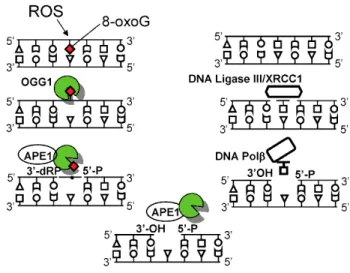
1.2.5 OGG1, a versatile DNA repair enzyme
OGG1 is the dedicated enzyme to excise the 8-oxoG during the DNA base excision repair process. OGG1 is a bifunctional glycosylase, it is able to both cleave the glycosidic bond of the mutagenic lesion and the phosphodiester bonds (3’ and 5’) causing a strand break in the DNA backbone (Chung, Kasai et al. 1991; Chung, Kim et al. 1991). OGG1-initiated BER encompasses four key steps (Fig. 4), including damaged base recognition and excision, 3’deoxyribose phosphate end-processing by AP endonuclease 1 (APE1), filling in the nucleotide gap by DNA polymerase β, and nick-sealing by DNA ligase (Mitra 2001). OGG1’s repair activity is modulated by post-translational modifications, including phosphorylation (Dantzer, Luna et al. 2002), acetylation (Bhakat, Mokkapati et al. 2006), and by interactions with canonical repair and non-repair proteins (Hegde, Hegde et al. 2011). Studies have also unveiled a redox-dependent mechanism for the regulation of OGG1 activity (Bravard, Vacher et al. 2006; David, O'Shea et al. 2007).
Figure 4. Graphical illustration of 8-oxoguanine DNA glycosylase-1 (OGG1)-initiated genome damage repair (Ba, Aguilera-Aguirre et al. 2014)
Depending on the last exon sequence of the C-terminal region of the OGG gene there are two major splice variants of OGG: nuclear (type 1 with 3 isoforms) and mitochondrial (type 2 with 5 isoforms) (Aburatani, Hippo et al. 1997; Nishioka, Ohtsubo et al. 1999). All variants have the N-terminal region in common. In eukaryotes, the N-terminus of this gene contains a mitochondrial targeting signal, essential for mitochondrial localization (Nishioka,
Ohtsubo et al. 1999). A conserved N-terminal domain contributes residues to the 8- oxoguanine binding pocket (van der Kemp, Charbonnier et al. 2004).
Accumulation of 8-oxoG in DNA has conventionally been associated with various diseases, accelerated telomere shortening, inflammatory and aging processes (Markesbery and Lovell 2006; Radak, Bori et al. 2011). In addition, unrepaired 8-oxoG lesion is potentially one of the most mutagenic lesions among oxidatively modified DNA bases, because its pairing with A will cause a GC→AT transition. Unexpectedly, OGG1 knock out (OGG1–/–) mice have an unaltered lifespan, and show only moderate increases in tumor formation. In these animals, no organ failure can be observed despite the supraphysiological levels of 8-oxoG in their DNA (Klungland, Rosewell et al. 1999; Minowa, Arai et al. 2000). Furthermore, OGG1–
/– mice showed an increased tolerance to chronic oxidative stress (induced by KBrO3 treatment), while 8-oxoG levels in the DNA increased by 250- to 500-fold compared to the wild type. Interestingly, lack of OGG1 activity protected mice from the trinucleotide repeat expansions underlying Huntington's disease (Kovtun, Liu et al. 2007).
Mabley and co-workers studied the role of OGG1 in inflammatory processes. They used three models of inflammation: endotoxic shock, diabetes, and contact hypersensitivity.
According their results the OGG1 knockout mice are extremely resistant to most of the lipopolysaccharide-induced effects: LPS-induced organ dysfunction, neutrophil infiltration and oxidative stress, when compared to wild-type (OGG1+/+) controls. Furthermore, OGG1−/−
mice had decreased serum cytokine and chemokine levels and prolonged survival after LPS treatment. In case of multiple low-dose streptozotocin-induced type I diabetes, OGG1−/− mice were found to have significantly lower blood glucose and higher insulin levels followed by fewer incidence of diabetes as compared with wild type mice. These knockout mice also have higher levels of protective Th2 cytokines (IL-4, IL10) while lower levels of chemokine MIP- 1α and Th1 cytokines (IL-12, TNF-α) compared to the levels measured in OGG+/+ controls. In a model of oxazolone-induced contact hypersensitivity, results showed reduced neutrophil accumulation, chemokine (MIP-1, MIP-2), Th1 (IL-1, TNF-α) and Th2 cytokine levels (IL-4) in the ear tissue of OGG1−/− mice. Their results suggest that OGG1 may primarily regulate Th1 cytokine levels rather than Th2 (Mabley, Pacher et al. 2005). On the other hand, mice lacking OGG1 have been shown to be susceptible to high-fat diet induced insulin resistance and obesity as well (Sampath, Vartanian et al. 2012). Others demonstrated that under chronic inflammatory conditions, cytokine-induced nitric oxide inhibits the activity DNA repair enzymes, including OGG1 (Jaiswal, LaRusso et al. 2000; Jaiswal, LaRusso et al. 2001). It has been hypothesized that DNA-dependent kinases recognize the single strand gaps made by
OGG1 and trigger inflammation. In this point of view, it looks more advantageous to down- regulate OGG1 and leave the 8-oxoG in the DNA (Radak and Boldogh 2010). This hypothesis also can explain why OGG1−/− mice with significantly fewer DNA nicks are less prone to inflammation. In support, OGG1 expression was increased in islet cells of type 2 diabetes patients compared to healthy controls (Tyrberg, Anachkov et al. 2002). Although oxidative stress increases 8-oxoG in the DNA, but observations showed a decreased OGG1 activity until normal redox status returned (Bravard, Vacher et al. 2006).
Besides redox modulation, OGG1 activity can also be altered by acetylation/deacetylation. OGG1 activity can be increased by acetylation by transcriptional coactivator p300 in the presence of APE1 (Bhakat, Mokkapati et al. 2006). Recent publications suggest that sirtuins, a group of regulatory proteins with NAD+-dependent deacetylase (or mono-ADP-ribosyltransferase) activity, have important role modifying OGG1’s glycosylase activity. Cheng and colleagues showed a regulatory role of Sirt3 in the maintenance of mitochondrial DNA and turnover of OGG1. They found that by deacetylation, Sirt3 modified OGG1 incision activity and prevented its degradation leading to cell survival under oxidative stress. (Cheng, Ren et al. 2013). Another paper revealed an inverse correlation between Sirt1 and OGG1 in animal studies. They found that rats with higher aerobic capacity had increased Sirt1 and lower acetylated OGG1 levels (lower repair capacity), when compared to low aerobic capacity rats (Sarga, Hart et al. 2013).
OGG1 has been associated with aging because of the accumulation of 8-oxoG in DNA (Dherin, Radicella et al. 1999; Cortopassi 2002). Szczesny and co-workers showed a difference in efficiency of import of OGG1 (and APE1) into nuclear and mitochondrial compartments of young and aged cells. They hypothesized this phenomenon as reason for age-related DNA damage (Szczesny, Hazra et al. 2003). Physical exercise can upregulate OGG1 activity in the liver of old (21 month) rats and reduce the 8-oxoG content of both nuclear and mitochondrial DNA to the level of young rats (11 month) after 2 months regular exercise (Nakamoto, Kaneko et al. 2007).
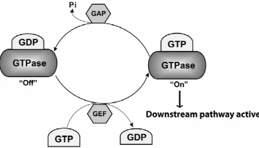
1.3 Small GTPases
Small GTPases are a type of G-proteins found in the cytosol that are homologous to the alpha subunit of heterotrimeric G-proteins (large GTPases). They can hydrolyze guanosine triphosphate (GTP) to form guanosine diphosphate (GDP) (Bourne, Sanders et al. 1990). The GDP-bound form is their inactive state while the GTP-bound form is their active form (Fig. 5) in which they can activate downstream pathways by binding to effectors. Small GTPases
work like molecular switches with the help of
that facilitate GDP dissociation and of GTPase activating proteins (GAPs) that stimulate GTP hydrolysis (Bos, Rehmann et al. 2007)
GTPases cycle between the inactive ‘off’ GDP
state occurs by stimulation of intrinsic GTPase hydrolysis activity by GAPs. Activation is facilitated by GEFs to load GTP and dissociate GDP, allowi
downstream signaling pathways. (Original
1.3.1 Ras
The name “Ras” is an abbreviation of “Rat
the protein family were discovered. Ras protein family members belong to (Wennerberg, Rossman et al. 2005)
Ras is encoded by the ras gene and
proteins, which are all related in 3D structure. They regulate diverse cell behaviors and are involved in transmitting signals
members share a set of conserved G box GDP/GTP N-terminus (Bourne, Sanders et al
domain (Ras residues 5-166) that has a conserved structure and biochemistry shared Ras superfamily proteins. Ras (p21) protein
helices (Vetter and Wittinghofer 2001)
olecular switches with the help of guanine nucleotide exchange factors (GEFs) that facilitate GDP dissociation and of GTPase activating proteins (GAPs) that stimulate GTP
(Bos, Rehmann et al. 2007).
Figure 5. The GTPase activity cycle
GTPases cycle between the inactive ‘off’ GDP-bound state and the active ‘on’ GTP-bound state. The inactive state occurs by stimulation of intrinsic GTPase hydrolysis activity by GAPs. Activation is facilitated by GEFs to load GTP and dissociate GDP, allowing interaction with downstream effectors and in turn activation of
Original picture from Biochemical Society Transactions)
is an abbreviation of “Rat sarcoma” referring the way the first members of the protein family were discovered. Ras protein family members belong to
(Wennerberg, Rossman et al. 2005), as they can bind and hydrolyze guanosine triphosphate gene and the prototypical member of the Ras superfamily proteins, which are all related in 3D structure. They regulate diverse cell behaviors and are involved in transmitting signals (Fig. 6) within cells (Bourne, Sanders et al. 1990)
share a set of conserved G box GDP/GTP-binding motif elements beginning at t (Bourne, Sanders et al. 1991). Together, these elements make up a
166) that has a conserved structure and biochemistry shared
Ras (p21) protein contains a six-stranded beta sheet and 5 alpha (Vetter and Wittinghofer 2001).
guanine nucleotide exchange factors (GEFs) that facilitate GDP dissociation and of GTPase activating proteins (GAPs) that stimulate GTP
bound state. The inactive state occurs by stimulation of intrinsic GTPase hydrolysis activity by GAPs. Activation is facilitated by GEFs to ng interaction with downstream effectors and in turn activation of
picture from Biochemical Society Transactions).
referring the way the first members of the protein family were discovered. Ras protein family members belong to small GTPases nosine triphosphate.
Ras superfamily of proteins, which are all related in 3D structure. They regulate diverse cell behaviors and are (Bourne, Sanders et al. 1990). The family nts beginning at the ether, these elements make up a ∼20 kDa G 166) that has a conserved structure and biochemistry shared by all stranded beta sheet and 5 alpha
Figure 6
Activation of Ras GTPases involves the displacement of GDP with GTP, a process mediated by GEFs (Bourne, Sanders et al. 1990)
dissociate GDP at an increased rate, and then the bound GTP exchange factor leaving the GTPase in an active form
Sjodin, Margarit et al. 1998) Karlovich et al. 1991) and cdc25
domain of the Raf1 serine/threonine kinase
phosphorylation is essential, but not sufficient, for mediating Raf1’s mitogen kinase (MAPK) activity, as phosphorylated Raf1
membrane-lipid interactions (Kyriakis, App et al. 1
downstream and results in the transcription of genes involved in cell growth and division.
While RasGEFs catalyze a "push and pull" reaction which releases GDP from Ras, RasGAPs enhance the catalytic machinery of
slow. Thus, GAPs accelerate Ras
molecule is now bound to a GDP which turns “off” further signaling Because intracellular concentratio
GTP predominantly re-enters the nucleotide binding pocket of Ras. The balance between GEF and GAP activity determines the guanine nucleotide status of Ras, thereby regulating Ras
Figure 6. Ras effectors and downstream pathways
Activation of Ras GTPases involves the displacement of GDP with GTP, a process (Bourne, Sanders et al. 1990). GEFs first interact with GTPase and dissociate GDP at an increased rate, and then the bound GTP promotes the release of exchange factor leaving the GTPase in an active form (Bourne, Sanders et al. 1990; Boriack Sjodin, Margarit et al. 1998). Well known GEFs are Son of Sevenless (Sos)
and cdc25 (Sadhu, Reed et al. 1990). Ras-GTP binds to the RBD 1 serine/threonine kinase (Block, Janknecht et al. 1996), and its subsequent phosphorylation is essential, but not sufficient, for mediating Raf1’s mitogen
kinase (MAPK) activity, as phosphorylated Raf1 requires additional protein
(Kyriakis, App et al. 1992). The MAPK cascade transmits signals downstream and results in the transcription of genes involved in cell growth and division.
While RasGEFs catalyze a "push and pull" reaction which releases GDP from Ras, RasGAPs enhance the catalytic machinery of Ras, since the protein intrinsic GTPase activity is very slow. Thus, GAPs accelerate Ras inactivation. An inorganic phosphate is released and the Ras molecule is now bound to a GDP which turns “off” further signaling (McCormick 1989) Because intracellular concentration of GTP is approximately 10-fold higher than that of
enters the nucleotide binding pocket of Ras. The balance between GEF and GAP activity determines the guanine nucleotide status of Ras, thereby regulating Ras Activation of Ras GTPases involves the displacement of GDP with GTP, a process GEFs first interact with GTPase and promotes the release of rne, Sanders et al. 1990; Boriack- . Well known GEFs are Son of Sevenless (Sos) (Rogge,
GTP binds to the RBD , and its subsequent phosphorylation is essential, but not sufficient, for mediating Raf1’s mitogen-activated protein additional protein-protein and . The MAPK cascade transmits signals downstream and results in the transcription of genes involved in cell growth and division.
While RasGEFs catalyze a "push and pull" reaction which releases GDP from Ras, RasGAPs Ras, since the protein intrinsic GTPase activity is very An inorganic phosphate is released and the Ras (McCormick 1989).
higher than that of GDP, enters the nucleotide binding pocket of Ras. The balance between GEF and GAP activity determines the guanine nucleotide status of Ras, thereby regulating Ras
activity. Another protein may augment the activity of Ras is GDI (GDP Dissociation Inhibitor). This protein functions by slowing the exchange of GDP for GTP and thus, prolonging the inactive state of Ras (Boguski and McCormick 1993).
Ras-regulated signal pathways control cell growth, migration (Lee, Feig et al. 1996), differentiation (Crespo and Leon 2000), actin cytoskeletal integrity, cell adhesion (Chambers, Hota et al. 1993), apoptosis (Kauffmann-Zeh, Rodriguez-Viciana et al. 1997), proliferation and survival (Bonni, Brunet et al. 1999). The clinically most notable members of the Ras subfamily are H-RAS, K-RAS and N-RAS, mainly for being implicated in many types of cancer (Bos 1989). Ras proteins are attached to the cell membrane because of their prenylation and palmitoylation (H-RAS and N-RAS) or prenylation and a polybasic sequence adjacent to the prenylation site (K-RAS). Depalmitoylation releases the proteins from the membrane, and allows them to enter another cycle of palmitoylation and depalmitoylation. It is assumed that this cycle prevents the N-RAS and H-RAS to attach to other membranes over time and to maintain their localization along the Golgi apparatus, secretory pathway, plasma membrane and inter-linked endocytosis pathway (Rocks, Peyker et al. 2006). Ras inhibitor trans-farnesylthiosalicylic acid (FTS) disrupts the membrane attachment of Ras, thus works as an anti-oncogenic drug in many cancer cell lines (Blum, Jacob-Hirsch et al. 2005; Rotblat, Ehrlich et al. 2008). Ras and Ras-related proteins are often deregulated in cancers, leading to increased invasion and metastasis, and decreased apoptosis. Mutations in ras genes can result the production of continuously active Ras proteins. As a result, the signaling pathway(s) remain “switched-on” and the overactive signaling can lead to uncontrolled cell growth and cancer (Goodsell 1999). Ras mutations are found in 20-25% of all human tumors and up to 90% in certain types of cancer (e.g. pancreatic cancer) (Downward 2003).
H-Ras is involved in regulating cell division in response to growth factor stimulation.
Growth factors act by binding cell surface receptors that span the cell’s plasma membrane.
Once activated, receptor stimulate signal transduction events in the cytoplasm, a process by which proteins and second messengers relay signals from outside the cell to the cell nucleus and instructs the cell to grow or divide. Once it is turned on, K-Ras recruits and activates proteins necessary for the propagation of growth factor and other receptors' signal, such c-Raf and PI 3-kinase (Castellano and Downward 2011).
1.3.2 Rac
Rac1, also known as Ras-related C3 botulinum toxin substrate 1 is a protein ubiquitously expressed and involved in signal pathways (Fig. 7) that regulate mobility and other processes
related to membrane trafficking and cell morphology (Ridley 2001; Vega and Ridley 2008). It is encoded by the rac1 gene (Didsbury, Weber et al. 1989; Jordan, Brazao et al. 1999). The Rac protein belongs to the Rho GTPase family. The classical members of the four subfamilies of Rho are: Rac, Cdc42, Rho and Rif. Similarly to other GTPases, the classical Rho GTPases cycle between active GTP-bound forms and inactive GDP-bound forms (Symons and Settleman 2000). Their cycle is also controlled by three types of regulatory proteins: guanine nucleotide exchange factors (GEFs), GTPase-activating proteins (GAPs) and guanine nucleotide dissociation inhibitors (GDIs) (Boguski and McCormick 1993). Rac subfamily has three Rac isoforms: Rac1, Rac2 and Rac3. Rac1 protein is widely expressed in different tissues, whereas as Rac2 expression is restricted to cells of hematopoietic origin. Rac3 is predominantly found in the central nervous system (Bedard and Krause 2007).
Rac can regulate cell survival and proliferation via cell cycle progression by activating p42/p44 and p38 MAPK, JNK, and Akt kinases (Yang, Atkinson et al. 2001; Gu, Filippi et al.
2003; Cancelas, Lee et al. 2005; Carstanjen, Yamauchi et al. 2005). Rac1 is a well characterized member of the Rac subfamily. Previous studies found that Rac1 regulates a diverse array of cellular events, including the formation of lamellipodia and membrane ruffles, cell cycle, cell adhesion and mobility (Ridley 2006). In addition, Rac1 regulates endocytic and exocytic trafficking pathways. Rac1 has a characterized role in clathrin- dependent endocytosis (Lamaze, Chuang et al. 1996). Other physiological functions of the Rac1 GTPase includes: modulation of the cellular redox state, regulation of cell movements, cellular signaling, gene expression, and cell differentiation (Bedard and Krause 2007). In most types of cells, Rac is connected to the production of low levels of ROS which have an implicated role in growth, differentiation, migration, and angiogenesis, as well as in inflammation (Sulciner, Irani et al. 1996; Sundaresan, Yu et al. 1996; Irani, Xia et al. 1997;
van Wetering, van Buul et al. 2002). Rac1 is ubiquitously expressed in nonphagocytic cells, including lung epithelial cells and fibroblasts. Rac1 is one of the three Rac family molecules that control NADPH oxidase (NOX) activity (NOX1, NOX2, and NOX4 as well as NOX3) both in phagocytes and in nonphagocytic cells (Hordijk 2006; Lambeth, Kawahara et al.
2007).
Figure 7. Rac1 related pathways (Original picture from SABiosciences)
Rac2 is expressed primarily in myeloid cells (e.g. human neutrophils) (Hordijk 2006).
Rac2 have a crucial role generating high levels of ROS in neutrophils upon cell stimulation and phagocytosis, and these ROS aid in the killing of ingested pathogens. To assemble a fully active oxidase complex, Rac2 with three other cytosolic proteins - p40phox, p47phox, p67phox - translocate to the plasma membrane and the active complex uses electrons from NADPH to reduce oxygen to form superoxide (Babior, Lambeth et al. 2002). Abnormal activation of Rac has been shown in a number of acute and chronic leukemias, including chronic myelogenous leukemia, chronic lymphocytic leukemias and acute myeloid leukemias
(Wertheimer, Gutierrez-Uzquiza et al. 2012). Rac1 is thought to play a significant role in the development of various cancers, including melanoma (Bauer, Chen et al. 2007; Krauthammer, Kong et al. 2012) and non-small cell lung cancer (McAllister 2012; Stallings-Mann, Waldmann et al. 2012). Another example that Rac1 overexpression may play a role in tumors was shown in pancreatic cancer progression (Wertheimer and Kazanietz 2011). Rac2 has been linked to leukemias (Muller, Schore et al. 2008) and mutations in the Rac2 gene have been found in human brain tumors (Hwang, Lieu et al. 2005).
Rac3 was originally identified from a chronic myelogenous leukemia cell line and has been implicated in human breast cancer (Mira, Benard et al. 2000; Morris, Haataja et al. 2000;
Baugher, Krishnamoorthy et al. 2005), ovarian cancer, cellular transformation (Keller, Gable et al. 2005) and tumor invasion (Chan, Coniglio et al. 2005).
1.4. DNA repair-independent functions of OGG1
Previous studies have implied roles for OGG1 in multiple cellular processes in addition to that of being a canonical DNA BER protein (Mitra 2001; Dizdaroglu, Kirkali et al. 2008). For example, it has been shown that OGG1 colocalizes with centrioles (microtubule organizing center), microtubule networks, and mitotic chromosomes (Dantzer, Luna et al. 2002;
Szczesny, Hazra et al. 2003; Conlon, Zharkov et al. 2004). These data show the implication of OGG1 in chromatin remodeling and transcriptional initiation. In mitochondria, aconitase enzyme has been suggested as a redox sensor because of its vulnerability to oxidative stress leading to inactivation or disassembly (Bulteau, Ikeda-Saito et al. 2003). OGG1 was reported to act as a chaperon for aconitase and to prevent mitochondrial dysfunction and apoptosis by interacting with it. They found that OGG1 silencing augmented oxidant induced caspase-9 activation (Panduri, Liu et al. 2009; Kim, Cheresh et al. 2014).
As described above, OGG1−/− mice have increased resistance to inflammation (Mabley, Pacher et al. 2005). This observation raises the possibility that it may not be the genomic level of 8-oxoG but the free 8-oxoG generated by BER that provides the linkage to disease/aging processes. While several aspects of the involvement of OGG1 in DNA repair- independent cellular functions have already been revealed, the role of OGG1 and free 8-oxoG in the activation of canonical small GTPase-mediated pathways has not been investigated so far.
2. Objectives of the study
Previous studies on OGG1−/− mice suggest that a lack of OGG1 activity is accompanied by dysfunction of signaling pathway(s) linking oxidative stress to cellular responses. These observations raise the possibility that the 8-oxoG base released from the genome by OGG1 could have physiological/pathophysiological relevance. The aim of this study was to reveal whether 8-oxoG and/or OGG1 are able to activate small GTPase-related signaling pathways.
Our hypotheses were the following:
- 8-oxoG has a biological role after being excised and not just a neutral byproduct of DNA repair,
- both exogenously added and endogenously excised 8-oxoG induces cellular responses, - free 8-oxoG base can bind to OGG1,
- 8-oxoG/OGG1 complex can activate small GTPases, Ras and Rac1,
- 8-oxoG/OGG1 complex can induce ROS production via NADPH oxidases by activating Rac1.
3. Materials and methods 3.1 Reagents
8-OxoG was from Cayman Chemicals (Ann Arbor, MI); 7,8-dihydro-8-oxoadenine (8-oxoA) was from Axxora, LLC/BioLog Life Science Institute (San Diego, CA); 8-aminoguanine was purchased from Carbosynth Inc, (Berkshire, UK). Guanine, 8-oxo-deoxyguanosine (8- oxodG), maleic acid diethyl ester (DEM), N-acetyl-L-cysteine (NAC), L-glutathione reduced (GSH); β-nicotinamide adenine dinucleotide 2’-phosphate reduced tetrasodium salt (β- NADPH); 2’-deoxyguanosine, adenine, guanosine, were from Sigma-Aldrich (St. Louis, MO).
8-Oxo-7,8-dihydroadenine (8-OH-Ade); Biolog Life Science Institute, Bremen, Germany);
2,6-diamino-4-hydroxy-5-formo-midopyrimidine (FapyG) was a kind gift of Dr. Miral Dizdaroglu (National Institute of Standards and Technology, Gaithersburg, MD). Rac1 antibody (Thermo Fisher Scientific, Rockford, IL); Rac2 and Rac3, NADPH oxidase subunit Abs (Epitomics, Burlingame, CA); recombinant human Rac1 protein (Cytoskeleton, Denver, CO); recombinant human OGG1, H-Ras, N-Ras, and K-Ras proteins (Novus Biological, Littleton, CO); OGG1 Ab (Abcam, Cambridge, MA). GTP, GDP, and GTPγS were from Cytoskeleton (Denver, CO); Pan-Ras antibody was from Millipore (Darmstadt, Germany);
nickel-nitrilotriacetic acid-agarose beads were from Qiagen (Valencia, CA); K-Ras and N-Ras antibodies were from Santa Cruz Biotechnology (Santa Cruz, CA); antibodies to ERK1/2, MEK1/2, phospo- ERK1/2, -MEK1/2 were from Cell Signaling (Danvers, MA); and FITC- and Alexa Fluor 488-conjugated antibodies were from Invitrogen (Carlsbad, CA). HRP- conjugated anti-rabbit Ab (Southern Biotech, Birmingham, AL), anti-mouse IgG, GE Healthcare UK Ltd, (Pittsburgh, PA). Active Ras and Rac pull-down assay kit was from Pierce Biotechnology (Thermo Fisher Scientific, Waltham, MA); and siRNAs for Ras, Rac1, NOX4, and OGG1 depletion were from Dharmacon (Thermo Fisher Scientific Inc. Waltham, MA). Diphenyleneiodonium chloride (DPI),; 3,3′,5,5′-Tetramethylbenzidine (TMB) substrate (eBioscience, San Diego, CA); H2SO4 (Fisher Scientific, Fair Lawn, NJ); (Mant)-GTP (2′-(or- 3′)-O-(N-methylanthraniloyl)guanosine 5′-triphosphate, trisodium salt, MantGTP) and (Mant)- GDP (2′-(or-3′)-O-(N-methylanthraniloyl)guanosine 5′-diphosphate, disodium salt, and
MantGDP) (Invitrogen, Carlsbad, CA). CD11a, CD11c, CD16, CD34, CD58, CD64, HLA-DR purchased from Immunotech (Commerce, CA), CD11b, CD32, CD36, CD45RA, CD45RO, CD95 (Fas), IL-3Rα, αvβ3, CD206 obtained from BD Pharmingen (San Diego, CA), CCR5, CCR6, CCR7, GITR, E-cadherin purchased from R&D Systems (Minneapolis, MN), αvβ5 (Chemicon, Temecula, CA). The MDR-specific monoclonal antibody was a generous gift from Gabor Szabo, (Department of Biophysics and Cell Biology, University of Debrecen).
Isotype-matched antibodies labeled with the same fluorochrome (all from BD Pharmingen).
pHyPer-Cyto, pHyPer-dMito and pHyPer-Nuc were acquired from Evrogen (Moscow, Russia).
3.2 Cell cultures
MRC-5, a human diploid lung fibroblast (ATCC# CCL-171) and human cervix carcinoma (HeLaS, ATCC# CCL-2.2) cells were maintained in Earle’s minimum essential and Dulbecco’s modified Eagle’s low glucose medium, respectively. A549 type II alveolar epithelial cells (ATCC # CCL-185) were cultured in Ham’s F12 (GIBCO-BRL), U937, a human monocytic cell line (ATCC# CRL-1593.2), were grown in and RPMI-1640. The human myelomonocytic KG-1 (ATCC# CCL-246) cells were grown in Iscove's Modified Dulbecco's Medium. All media were supplemented with 10% fetal bovine serum, glutamine, penicillin, and streptomycin; cells were grown at 37 °C in a 5% CO2. KG1 cells were stimulated with 10 ng/ml PMA (Sigma-Aldrich, Steinheim, Germany) and 100 ng/ml ionomycin (Sigma-Aldrich) for 4 days as described previously (St Louis, Woodcock et al.
1999).
Monocyte-derived DCs were developed as described previously (Thurner, Roder et al.
1999). Briefly, mononuclear cells were isolated from Buffy Coat by Ficoll-Pacque (Amersham Biosciences, Uppsala, Sweden) gradient centrifugation and monocytes were isolated by magnetic cell separation using positive selection with anti-CD14-coated beads (Miltenyi Biotech, Bergish Gladbach, Germany). Purified monocytes were plated at 2×10⁶ cells/ml concentration and cultured in serum-free AIMV medium (Gibco) in the presence of 100 ng/ml IL-4 and 75 ng/ml GM-CSF (Peprotech EC, London, UK) given on days 0 and 2.
Cells were differentiated for 5 days and immature DC were characterized by flow cytometry using anti-CD1a fluorescent antibody (Immunotech, Marseille, France). Activation of immature DC was induced by an inflammatory cocktail containing 10 ng/ml TNF-α, 5 ng/ml IL-1β, 20 ng/ml IL-6, 75 ng/ml GMCSF and 1 µg/ml PGE2 (Sigma-Aldrich). Mature DC were identified by anti-CD83 mAb (Immunotech).
3.3 Animals and treatments
Animal experiments were performed according to the National Institutes of Health Guidelines for Use of Experimental Animals and approved by the University of Texas Animal Care and Use Committee (Protocol number: 0807044A). Eight-week-old female BALB/c mice (The Jackson Laboratory) were challenged intranasally with 60 µl of 8-oxoG (1 µM) in saline (or