Is extravascular and intravascular calcification connected in patients with atherosclerosis?
PhD Thesis Dr. Mátyás Fehérvári
Semmelweis University Doctoral School of Clinical Medicine
Consultant:
Dr. Zoltán Szeberin, Ph.D.
Official reviewers:
Dr. Pál Soltész, Ds.C.
Dr. Marcell A. Szász, Ph.D.
Head of The Final examination comittee:
Professor Lajos Szollár, Ds.C.
Members of the Final exam committee:
Dr. György Wéber, Ds.C.
Dr. Zsolt Pécsvárady, PhD.
Budapest 2017
CONTENT
I. INTRODUCTION 12
I.1. Atherosclerosis 13
I.1.1. The pathophysiology of atherogenesis 13
I.1.1.1. Introduction 13
I.1.1.2. The role of lipoproteins 13
I.1.1.3. Inflammation 14
I.1.1.4. Other important factors 14
I.1.2. Risk factors of atherosclerosis 16
I.1.2.1. Framingham Heart Study 16
I.1.2.2 New risk factors/markers of atherosclerosis 16
I.1.3. The progression of atherosclerosis 17
I.2. Vascular Calcification 18
I.2.1. Anatomical variation of calcification 18
I.2.2. Development of calcification 19
I.2.2.1. Introduction 19
I.2.2.2. Vascular smooth muscle cells 20
I.2.2.3. Vitamin D 20
I.2.2.4. Further pathophysiology of calcification 21 I.2.3. The role of complements in progression of calcification 21 I.2.4. The role of Fetuin-a in the progression of calcification 22 I.2.5. Other factors contributing to plaque development 23
I.3. Bone formation and Osteoporosis 24
I.3.1. Osteogenesis 24
I.3.2. Osteoporosis 24
I.3.2.1. Osteoporosis at a glance 24
I.3.2.2. Risk stratification and markers in osteoporosis 25 I.4. The connection between vascular calcification, bone formation and
osteoporosis 25
I.4.1. History 25
I.4.2. Bone cells in the arterial wall 26
I.4.2.1. Aids of calcification 27
I.4.2.2. Inhibition of calcification 27
I.4.3. Clinical associations 27
I.4.4. About the theories explaining the connection 28 I.4.4.1. Reduced blood flow – less nutrition 28
I.4.4.2. Dyslipidaemia 28
I.4.4.3. Vitamin D 29
II. AIMS 30
II.1. Osteoporosis and vascular calcification – Prevalence, connection, prognosis 30 II.2. The relation of Complement complements and fetuin-A to vascular calcification and their role in the progression of lower limb ischemia 30 II.3. The role of complement component 3 and Fetuin-A in the
progression of lower limb ischemia 31
III. METHODS 32
III.1. Clinical evaluation 33
III.2. Assessment of atherosclerosis and calcification 35
III.2.1. Imaging modalities 35
III.2.2. Laboratory measurements 36
III.3. The evaluation of osteoporosis amongst patient with Peripheral Artery
Disease 37
III.3.1. Biochemical parameters 37
III.3.2. Dual-energy X-ray absorptiometry 37
III.3.3. Angiography, Bollinger score 38
III.3.4 Site specific assessment 39
III.4. The role of Complement component 3 and fetuin A in the development and
progression of vascular calcification 41
III.4.1. Association of the extent of PAD to complement
component 3 and 4 41
III.4.2. Laboratory measurements 41
III.5. The role of complement component 3, 4 and fetuin-A in the progression of
atherosclerosis 41
III.6. Statistical analysis 42
IV. RESULTS 43
IV.1. Osteoporosis in the Hungarian population of patients with atherosclerosis 43
IV.1.1. Bone Mineral Density 44
IV.1.2. The risk factors of osteoporosis and atherosclerosis 46 IV.1.2.1. Prevalence and gender specific comparison 46
IV.1.2.2. Age 47
IV.1.2.3. Body Mass Index 48
IV.1.2.4. Smoking 49
IV.1.3. Bone turnover markers 51
IV.2. Connection between osteoporosis and atherosclerosis 51 IV.2.1. Bone mineral density is associated to the severity of
atherosclerosis if the site of the lesion is considered 52 IV.2.2. BMD is not related to the severity of atherosclerosis in
all patients 53
IV.2.3. The role of Vitamin-D 54
IV.2.4. The role of Dyslipidaemia 55
IV.3. The role of Complement component 3 and 4 in vascular calcification 55 IV.3.1. C3 is significantly higher in atherosclerosis than in healthy
controls 57
IV.3.2. Clinical parameters and complements 58
IV.3.3. Calcification and complements 60
IV.3.4. Other findings 60
IV.4. The role of complement component 3, 4 and fetuin-A in the progression of
atherosclerosis 61
IV.4.1. Complement 3 and 4 and future cardiovascular complication 61
IV.4.2. Fetuin-A and other markers 63
IV.4.3. Regression analysis 63
V. DISCUSSION 65
V.1.Osteoporosis and atherosclerosis 65
V.1.1. General clinical and laboratory results 65
V.1.2. The prevalence of osteoporosis 65
V.1.3. BMD and the mutual risk factors of bone disease and
atherosclerosis 66
V.1.4. Bone turnover markers and atherosclerosis 67 V.2.What is the origin of the connection of atherosclerosis and low bone density? 68 V.2.1. About the blood supply of the sites of the BMD measurements 68
V.2.2. Site specific comparison 68
V.2.3. General findings 69
V.2.4. The role of Vitamin D 70
V.2.5. Dyslipidaemia 70
V.2.6. Bone turnover markers 71
V.3.The role of complement component 3 and 4 in the progression
of atherosclerosis 71
V.3.1. Patient characteristics 71
V.3.2. Level of Complements in patients and controls 72 V.3.3. Complement components and the clinical severity of
atherosclerosis 72
V.3.3.1. ABI and Bollinger score associated to the level of
complements 72
V.3.3.2. Complements and the Fontaine stadiums 73
V.3.4. Calcification and the complements 72
V.4. Complements and the progression of atherosclerosis 74
V.4.1. Baseline C3 predicts future MI 74
V.4.2. Complements, mortality and morbidity 75
VI. CONCLUSION 76
VII. SUMMARY 77
VIII. ÖSSZEFOGLALÁS 78
IX. BIBLIOGRAPHY 79
X. LIST OF AUTHOR'S PUBLICATIONS 99
Köszönetnyilvánítás 101
Figures and Graphs
Figure 1. The different types of calcification
Figure 2. Summary of factors influencing vascular calcification
Figure 3. Assessment of carotid calcification by ultrasound for calcification score Figure 4. DEXA scan
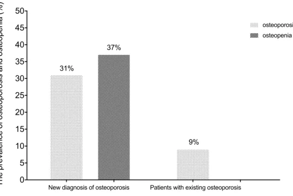
Figure 5. The prevalence of osteoporosis before and after DEXA scan in patients with Peripheral Artery Disease
Figure 6. The prevalence of osteoporosis in PAD patients with different sex Figure 7. The prevalence of osteoporosis in the different age groups
Figure 8. The distribution of osteoporosis across patients with different body habitus Figure 9. Bone disease in smokers, ex smokers and non-smokers
Figure 10. Scatter plot diagram displays the connection between complement component 3 (C3) and heathy control. Mann Whitney U test.
Figure 11. Scatter plot diagram displays the connection between complement component 4 (C4) and heathy control. Mann Whitney U test.
Figure 12. Scatter plot diagram displays the connection between complement
component 3 (C3), complement component 4 (C4), and ankle-brachial Doppler index (ABI). Spearmen rank correlation
Figure 13. Scatter plot diagram displays the association between complement component 3 (C3) and Bollinger angiographic score. Spearmen rank correlation, (p=0.028, r=-0.357)
Figure 14. ROC analysis of C3
Figure 15. Kaplan Meier survival analysis of patients with high and low C3
List of Tables
Table 1. Summary of different study groups, their inclusion and exclusion criteria.
Table 2. Patient Questionnaire Table 3. Patient characteristics
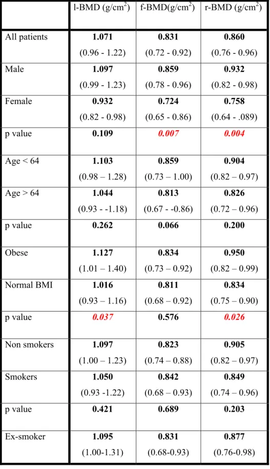
Table 4. Bone mineral density in patients with different risk factors of atherosclerosis and osteoporosis
Table 5. The median (IQR) of bone turnover markers in different patient groups Table 6. Patient characteristics of the different subgroups
Table 7. There is no association between BMD and BS across all patients
Table 8. Burn turnover markers and their relations to BMD in different patient groups Table 9. Clinical characteristics of patients and controls
Table 10. Multivariable prediction analysis using baseline variables
List of Abbreviations
ABI – ankle brachial doppler index
AHSG - alfa-2 Heremans Schmid glycoprotein ALP – alkaline phosphatase
BAP - bone specific alkaline phosphatase BGLAP – osteocalcin
bCTx - beta-crosslaps
BMD – bone mineral density BMP - bone morphogenetic protein BS – Bollinger score
CES – cardio embolic stroke CS - calcification score CI - confidence interval CKD – chronic kidney disease CRP - C-reactive protein CVC - calcifying vascular cell ECM - extracellular matrix
EDRF - endothelial-derived relaxing factor ENPP1 - ekto-nucleotide pyrophosphates -1 DEXA: Dual-energy X-ray absorptiometry f-BMD: femoral head bone mineral density GFR - glomerulus filtration rate
HbA1c - haemoglobin A1c HDL – high density lipoprotein HR - Hazard ratio
IMT – intima media thickness IQR - interquartile range CS – calcification score GP - General Practitioner
l-BMD - lumbar vertebral bone mineral density LDL - low density lipoprotein
MGP - matrix glutamic acid protein MI- myocardial infarction
MMP - matrix metalloproteinase
NPP1 - nucleotide pyrophosphatase/phosphodiesterase NVE – novell vascular event
OPN - osteopontin OR - odds ratio
ox-LDL - oxidised LDL
PAD - peripheral artery disease PTH - parathormon
r-BMD: radial head bone mineral density ROC - reciever operating characteristics ROS- reactive oxygen species
SNP - single-nucleotide polymorphism TGF-β - transforming growth factor-β TNF-α - tumour necrosis factor-α
VEGF - vascular endothelial growth factor VSMC – vascular smooth muscle cell
Summary of investigations - about the findings that this study will present
The connection between intra and extravascular calcification has been evaluated.
Initially, we determined the prevalence of osteoporosis amongst patients with lower limb atherosclerosis followed by the comparison of bone mineral density and some other parameters of osteoporosis to the severity of atherosclerosis. There are 3 main hypotheses explaining the connection between low bone mineral density and atherosclerosis in the normal population. The first one suggests reduced blood flow to the bones as a result of generalised atherosclerosis, the second dyslipidaemia and a third low vitamin D3 as responsible causes. All of the 3 parameters were noted and their affect on bone mineral density has been evaluated in our atherosclerotic patient group.
In a different patient group, the association of complement components to the clinical parameters of chronic lower limb ischemia have been evaluated. In a follow up study we determined the effect of baseline complement component 3 and fetuin-A on future cardiovascular morbidity.
I. INTRODUCTION
Atherosclerosis and subsequent cardiovascular disease are a leading cause of death in the Hungarian population. The morbidity and mortality related to myocardial ischemic events have improved dramatically over the last few years in Hungary. Unfortunately, the outcomes for patients with peripheral arterial disease have not improved and indeed in some aspects have worsened [1]. Consequently, there is an urgent need for research into this field. Calcification is an often investigated process, and several recent findings suggest a connection between the vascular (ectopic) and the extravascular form.
Furthermore, population based clinical studies described an association between atherosclerosis, osteoporosis and bone mineral density. Their effect on each other, however, is not clear. It is well known that beside the poor outcomes and low life expectancy, the quality of life of vascular patients is often impaired by severe comorbidities, such as diabetes, obesity or renal disease. The appropriate management of these comorbidities and to slow down the progression of atherosclerosis are essential in order to improve these patients’ lives. Based on recent research of calcification, osteoporosis can be one of these so far less known comorbidities.
The understanding of atherosclerosis has much improved over the decades since the first population based study, the Framingham study, was launched. The identification and understanding of the risk factors of the disease is an ongoing process. The development of vascular calcification from the atheroma is a highly investigated mechanism and is one of the most clinically significant processes in the progression of the disease. Several markers, such as Complement components or fetuin-A have been associated with vascular disease and calcification. However, less is known about their role in the progression of atherosclerosis and the way they affect cardiovascular outcome. The purpose of this research is to investigate the connection between intra- and extravascular calcification through bone mineral density and other calcifying parameters in patients with atherosclerosis.
I.1. ATHEROSCLEROSIS
I.1.1. The pathophysiology of atherogenesis I.1.1.1 Introduction
Many aspects in the development of atherosclerosis have been thoroughly investigated and fully understood. The existing knowledge of the disease however, expands and changes rapidly. Current research shows that the initial step in atherogenesis is endothelial injury and consequent dysfunction accompanied by extracellular lipid accumulation in the vessel wall [2]. The important role of mechanical factors in atherosclerosis has been highlighted back in 1957 [3, 4]. One of the most important risk factors, hypertension, is a clinical manifestation of increased shearing forces to the wall of the arteries [5, 6]. Many patients with essential hypertension have been diagnosed with endothelial dysfunction and it has been highlighted that these patients have an increased risk to developing further organ damage[7]. Endothelial dysfunction alters the permeability to certain molecules such as low density lipoprotein (LDL), results in vasoconstriction, leukocyte adhesion, thrombosis, and proliferation [8] and it could explain the higher incidence of atherosclerosis in patients with chronic inflammatory disease [9].
I.1.1.2 The role of lipoproteins
Following the injury of the innermost layer of the vessel wall the LDL settles in the sub endothelial layer. Endothelial damage will facilitate the aggregation of platelets which will synthetize vaso-active substances such as serotonin and histamine. The aggregated platelets will also induce smooth muscle proliferation [10]. These muscle cells will migrate towards the endothelial layer and secrete autocrine inflammatory proteins such as cytokines and interleukins [11]. The uptake of different molecular weight lipoproteins, especially the LDL, is regulated by the scavenger receptors[12]. Styrene based lipoproteins innate an inflammatory-immune reaction and macrophages will engulf and digest them and transform to foam cells [13]. These cells are dividing rapidly
forming a lesion called fatty streak, which contains monocyte-derived macrophages, macrophage-derived foam cells, and T lymphocytes. These cells are all filled with lipids.
I.1.1.3 Inflammation
As atherosclerosis progresses, chronic inflammatory processes will mediate the process, which will eventually lead to the development of a complex lesion. The importance of the inflammatory cells has been demonstrated first on intercellular adhesion molecule (ICAM)-1 and P-selectin deficient mice. In these subjects virtually no atherosclerosis can be detected [14]. The raise in adhesional molecules such selectins, integrins and immunoglobulins will help inflammatory cells to attach to the activated endothelium [15, 16]. These cells will release reactive oxygen species (ROS) which will then oxidase LDL in the intimal layer and facilitate further lipid accumulation. Furthermore, it has also been described that inflammation itself increases the permeability of the vessel wall for lipoproteins [17]. The continuous retention of lipids will result in endoplasmic reticulum stress, hence, apoptosis [18]. A central region with increased inflammation and apoptosis will develop in the atherosclerotic plaque. Unlike acute inflammation where pro-inflammatory molecules are followed by anti-inflammatory ones, this chronic process will remain unresolved. This failed resolution is a new field of research for therapeutic agents [19]. The ongoing inflammation results in the vulnerability of the plaque accompanied by further apoptosis and smooth muscle cell death induced by inflammatory cytokines. This will eventually lead to fibrosis and formation of a fibrous cap [20] .
I.1.1.4. Other important factors
In the growth of the fatty streak the matrix metalloproteinases (MMP) play an important role [21]. These enzymes are responsible for the degradation of extracellular matrix.
However, they also play an important role in the smooth muscle proliferation and in the formation of neointima following endothelial injury [21, 22]. Furthermore, they are reported to be responsible for plaque instability [23]. They have many effects on
atherosclerosis, including regulating endothelial cell invasion, migration and lumen formation (angiogenesis) [24].
In response to tissue hypoxia, neovascularisation starts from the vasa vasorum of the adventitial layer towards the innermost intimal layer[25]. This is followed by increased angiogenesis promoting factors. The increased cell wall activity disrupts the homeostasis, leading to necrosis and inflammation. Consequently, this is associated with advanced atherosclerotic lesion and increased plaque vulnerability [26].
Vascular Endothelial Growth Factor (VEGF) affects both hypoxia and inflammation, and also induces neovascularisation in the vessel’s wall. The administration of anti VEGF neutralising antibody in rats resulted in the stop in growth of coronary collateral circulation[27]. Further animal studies were able to demonstrate that the inhibition of VEGF receptors, for example by vaccination, was able to reduce the size of atherosclerotic plaques and the microvascular density [28, 29]. Some clinical data also confirms the important role of VEGF in atherosclerosis. Increased VEGF activity has been shown in relation to interplaque haemorrhage, a sign and cause of plaque instability in carotid endarterectomy specimens in symptomatic patients. The same activity could not be detected in patients without symptoms[30].
Additionally, abnormal vaso activity is shown in response to the injury and endothelia dysfunction. In healthy individuals the endothelial secretes Nitrogen oxide that acts as a vasodilatator. In atherosclerosis the secretion of NO is decreased resulting in vasoconstriction[31]. Furthermore, the bioavailability of NO is also decreased due to the excess of reactive oxygen species (ROS)[32]. This deteriorates to tissue hypoxia.
These processes over many years will result in a complicated lesion, which then will narrow the vessel lumen worsening tissue hypoxia and resulting in organ damage. The complicated lesion can transform to a vulnerable plaque, which is then most likely to be ruptured and result in acute thrombo-embolic event. Several attempts have been made recently to detect vulnerable plaques in order to prevent further clinical progression of the disease [33, 34].
I.1.2. Risk factors of atherosclerosis I.1.2.1 Framingham Heart Study
Simultaneously to the studies on the pathophysiology of atherosclerosis, population based longitudinal prospective studies focused on identifying risk factors for atherosclerosis. Many years have passed since the first study, the Framingham Heart Study, was launched[35]. This was followed by many others focusing on the risk factors of atherosclerosis. The classic Framingham risk factors of atherosclerosis are:
increasing age, hyper or dyslipidaemia, smoking, hypertension, diabetes, lack of physical activity, obesity, male gender and mental stress [35]. Over the years many other factors were investigated and some added to this list if they were found independently predictive.
I.1.2.2 New risk factors/markers of atherosclerosis
Several studies reported the connection between atherosclerosis and osteoporosis.
Systemic vascular calcification is associated with low Bone Mineral Density [36].
Patients with osteoporosis are more likely to suffer from vascular calcification and peripheral vascular disease[37, 38]. Higher cardiovascular mortality also appears to be related to osteoporosis [39]. In our research, we thoroughly investigated this connection and we will present our findings below.
As described in the previous chapter, inflammation is a key element of the atherogenesis. Therefore, the effect of pro-inflammatory cytokines, immune molecules and acute phase proteins on atherosclerosis have been thoroughly investigated.
Numerous studies described a strong association amongst C reactive protein and atherosclerosis [40]. However, the nature of this association is not fully understood.
Recent randomised studies could not find a causative connection amongst them [41].
Beside CRP, the complement system [42], interleukins [43, 44], , tumour necrosis factor-α TNF- α [45] and many more inflammatory agents have been identified playing a role in atherosclerosis. However, an ongoing issue is that the acute-phase proteins and
other inflammatory markers can only be considered as markers and not mediators of the disease [46].
Hyperhomocysteinaemia is one of the few risk factors that have been added to the classical risk factors of Framingham. Homocysteinylated lipoproteins with microorganisms obstruct the vasa vasorum and form vulnerable plaques[47]. It also induces endothelial dysfunction by damaging the endothelial cells[48].
The Human Genome Project made it possible to analyse some of the traits that lead to this multifactorial disease. Mainly single nucleotide polymorphisms have been identified as potential variants. The understanding of these genetic variants helps to better describe inflammation [49]. There is a long way to go in the research of the genetic background of atherosclerosis.
I.1.3 The progression of atherosclerosis
The progression of atherosclerosis has a major effect on a patient’s life. The risk factors for atherosclerosis and peripheral artery disease (PAD) are well known, however they cannot be used to assess the progression of the disease in symptomatic patients [50, 51].
Identifying the risk for rapid worsening of cardiovascular disease in patients with PAD and adjusting their therapy could help improve their quality of life, morbidity and mortality. Clinically, calcification appears to be a very important factor in the progression of the disease. The severity of calcification is strongly associated with cardiovascular mortality. Plaque fissures can be responsible for at least half of all cardiovascular morbidity, although morphologically, vulnerable plaques has a low positive predictive value for major cardiovascular events [52, 53]. The extent of calcification is linked to the instability of the plaques [54]. Therefore, there is a need to better judge the effect of extra and intravascular calcification parameters on the progression of the disease.
I.2. VASCULAR CALCIFICATION
Vascular calcification is an important feature of atherosclerosis as the degree of calcification is strongly associated to cardiovascular mortality. Most recent research suggested that -despite the historic view- vascular calcification is reversible [55]. This makes the understanding of the progression of calcification crucial to prevent the narrowing and obstruction of arteries at an early stage.
I.2.1. Anatomical variation of calcification
Historically, cardio-vascular calcification has been treated as the same disease, however it is fully understood now that the manifestation of it at different anatomical sites develop and behave differently. Calcification most commonly affect elastic type arteries in the systemic circulation. The subtype of this concludes intimal or tunica media calcification and porcelain aorta, which is limited to the ascending part of this vessel. Pulmonary arteries are less likely to be affected by calcification and are usually associated with pulmonary hypertension. Arterioles can also be affected by calcification, usually in chronic kidney disease (CKD). This type of calcification is referred to as calciphylaxis. Cardiac valves, predominantly the aortic valve, can also calcify. This type of calcification shows many similarities to arterial calcification, however at the present time, it is considered a different disease. Finally, myocardial calcification has also been described. The pathogenesis of this disease is currently poorly understood[56]. These anatomical variations are further demonstrated in Figure 1.
Figure 1. The different types of calcification. Figure by Bostrom et al. [56]
“Schematic drawing of different types of vascular calcification affecting elastic arteries in the systemic circulation, including atherosclerotic lesion calcification, calcification of the internal elastic lamina (IEL), coral reef aorta, media sclerosis (Mönckeberg's disease), and porcelain aorta”.
I.2.2. Development of calcification I.2.2.1. Introduction
According to Murshed et al. [57] the development of tissue bone mineralization requires two conditions: a fibrillar collagen rich matrix as a scaffold, and the expression of alkaline phosphatase. These two factors could be found in almost all tissue, including vessels. Different types of calcification have been identified in the adventitial and in the tunica media layers. The deposition of lipoproteins seems to be playing an important role in adventitial calcification but not in medial calcification [58]. The changes in the medial layer, also described as Möckenberg sclerosis, are associated with age, diabetes
and chronic kidney disease. This type of calcification is also visible on plain radiographs[59].
I.2.2.2. Vascular smooth muscle cells
Both layers, but especially the tunica media, are built up from collagen rich matrix and contain vascular smooth muscle cells (VSMC). These cells do not express alkaline phosphatase under healthy conditions, whilst under calcifying conditions alkaline phosphatase activity was measured [60]. Their migration is stimulated by VEGF molecules [61]. The importance of these VSM cells is the potential to transfer to osteoblasts. This could be facilitated by several factors. Intimal calcification appears to be mediated beside oxidative stress by bone morphogenic proteins (BMP) and the level of pyrophosphates. BMPs are inducing inflammation in the plaque facilitating intimal calcification [62].Several types of BMPs appear to be affecting calcification, but BMP-2 appears to be having the most important role as its level is increased in calcified plaques [63].
I.2.2.3. Vitamin D
Vitamin D plays a controversial role in vascular calcification and its effect is most likely be dose dependent [64]. Vitamin D excess leads to calcification in subjects with renal disease. At the same time its level inversely correlates to coronary calcification [65]. Vitamin D receptors have been identified in many cells including VSMC. A high dose of vitamin D induces osteoblastic phenotype of VSMC, and therefore, calcification. The effect of it is so strong that it has been used to induce calcification in animal experiments [66]. Klotho is a gene responsible for premature ageing and it inhibits vascular calcification in mice. The loss of this gene leads to ectopic calcification, but this can be reduced by genetically inhibiting the production of the active form of Vitamin D 1,25(OH)2D at the same time [67]. There are several ongoing clinical studies with Vitamin D supplementation in cardiovascular disease but the benefits of the routine use of Vitamin D have not been confirmed yet [68].
I.2.2.4. Further pathophysiology of calcification
Oxidative stress in relation with inflammation may also induce the process. The mechanism is not completely understood, but atherosclerosis is an inflammatory disease and vascular calcification is a big part of it. Additionally, many inflammatory cytokines have been identified being pro-calcific[69].
The regulation of phosphate levels is also an important feature in calcification.
Nucleotide pyrophosphatase/phosphodiesterase 1 (NPP1) is responsible for the production of extracellular inorganic phosphate. The mutation of the NPP1 gene leads to general arterial calcification of infancy [70]. Renal failure leads to hyperphsophateamia, which consequently triggers vascular calcification [71]
Matrix Gla proteins (MGP) have an important role in the inhibition of vascular mineralisation [72]. They affect the pathway in two different ways. They can directly block sclerotic crystal growth by interacting to hydroxyapatite and calcium ions [73]
and they also regulate BMP-2 expression [74]. Furthermore, in animal experiments it has been shown that if this pathway is inhibited (in MGP -/- mice) the extent of calcification is less severe [75]
I.2.3. The role of complements in progression of calcification
The Complement System is a part of the innate immune system and it activates in 3 main pathways: the classical, alternative and the mannose binding lectin pathway. The activation of the classical pathway requires Complement 3 (C3) while the activation of lectin pathway requires Complement 4 (C4). Inflammatory mediators, such as C- reactive protein (CRP) or the components of complement are present in all stages of atherosclerosis[76]. The third component of complement (C3) is strongly associated to the severity of atherosclerosis [77, 78] and to cardiovascular risk factors in elderly individuals [79] (Please see Figure 2.). C3 increase vascular stiffness by binding to collagen and elastin fibres in the vessel wall [80]. During premature enchondreal bone formation increased activity of C3 was found[81] and its role in the post traumatic cartilage healing[82] was also verified. In female patients the level of C3 is associated
to the progression of plaques and intimal media thickness (IMT) [83] , and also to arterial stiffness[84].
Furthermore, recent evidence suggests that the complement cascade is activated within the atherosclerotic plaque and they may play a role in plaque destabilization [85]. It has also been described that inflammatory changes precede plaque instability [86]. In patients with carotid artery disease, radiological unstable plaques had very low prediction for post interventional embolization, whilst inflammatory status was found to be a predictor of this [87]. Hence, inflammatory agents including C3, seem to be a possible factor for novel vascular events.
Clinical data also suggests an important role for complements in the progression of atherosclerosis. In healthy subjects non independent association was found between higher level of C3 and myocardial infarction[88]. Higher C3 level have been associated with cardiovascular risk in patients with psoriasis[89] and has been identified as a risk for coronary artery calcification in women with systemic lupus erythematosus [90].
This evidence suggests a connection between vascular calcification, bone formation and the serum level of third part and fourth part of complements.
I.2.4 The role of Fetuin-A in the progression of calcification
Fetuin-A also known as Hereman Schmidt glycoprotein is an important factor in the inhibition of calcification, thus on the overall progress of atherosclerotic disease. In the last decade, researches have focused more on regulatory factors associated to the severity of vascular calcification (Please see Figure 2.). These researches first focused on patients with chronic kidney disease (CKD). Among these patients the serum level of Fetuin-A[91] and pyrophosphates[92] are inversely correlated with the severity of calcification. Recent studies focused on patients without renal disease. These studies suggest that Fetuin-A has an effect on calcification among diabetic and non-diabetic patients with peripheral artery disease[93, 94], coronary disease[95] or aortic aneurism[96]. Additionally, they suggest that Fetuin-A has a major role in the inhibition of calcification.
I.2.5 Other factors contributing to plaque development
Several other factors have been highlighted as potential markers of the progression of atherosclerosis. For example, VSMC cells have been identified as contributors to plaque instability[97] or arterial stiffness could also be a prognostic factor for cardiovascular mortality in patients with PAD[98]. VEGF over expression also has an important role in the progression of the disease by inducing plaque instability [30]. Local increase of elastase and consequent decrease in arterial stiffness is related to the production of elastin-derived peptides (EDP). The elimination of the effect of EDP appears to be a promising therapy, however further research is required [99]. In our study, we focused primarily on potential factors contributing to the calcification and development of the plaques such as complement protein or marker related to calcification (Please see Figure 2.).
Figure 2. Summary of factors influencing vascular calcification. Original Figure by Giachelli et al.[55]
I.3. BONE FORMATION AND OSTEOPOROSIS
I.3.1. Osteogenesis
Bone is derived from para axial mesoderm. There are 2 types of bone formation.
Enchondreal ossification begins with formation of chondrocytes from mesenchymal cells and subsequent cartilogenesis. This is followed by the transformation of the cartilage to bone. The importance of this type of ossification is in the growth in length of the bones. Intramembranous bone formation is initiated from the neural crest originated mesenchymal cells. These cells develop as compound nodules and differentiate in to different cells such as capillaries or osteoblasts committed to bone formation. The most important role is in the development of the bones of the cranium and its role in bone healing [100].
I.3.2. Osteoporosis
I.3.2.1 Osteoporosis at a glance
The human bone is a constantly changing tissue. By aging, some of our bone cells dissolve, however simultaneously new bone forms. The remodelling of the bone this way has been first described in 1963 [101]. Up to 10% of the whole bone mass can undergo remodelling at a time. The responsible cells for bone degradation are the osteoclasts, whilst the osteoblasts are rebuilding the bone matrix. If the difference of bone formation and resorption turns negative and more bone dissolves than new forms, the bone mass starts to decrease. This leads to lower bone density and an increased risk of fractures [102]. Approximately 15% of the Caucasian population is affected by osteoporosis around the age of 50 and about 70% over the age of 80. It is more common in women than men [103]. The diagnosis of osteoporosis is based on the measurement of Bone Mineral Density, usually by Dual Energy X-Ray Absorptiometry [104].
I.3.2.2 Risk stratification and markers in osteoporosis
There are several factors highlighted to have an important role in the evaluation of osteoporosis [105] . The most important is to estimate and eventually decrease the risk of osteoporotic fractures. The suggested tool for the assessment of fracture risk by the WHO is the FRAX tool [106]. This calculation encounters the most important risk factors and BMD. These are age, sex, BMI, previous fracture, parent fractured hip, current smoking, glucocorticoids, rheumatoid arthritis, more than 3 units of alcohol per day and secondary osteoporosis (insulin-dependent diabetes, osteogenesis imperfecta in adults, untreated long-standing hyperthyroidism, hypogonadism, premature menopause, chronic malnutrition, and chronic liver disease). In addition to this, neuromuscular disorders, long-term immobilization and low dietary calcium intake have also be identified as potential risk factors[107]. The biochemical markers can be divided in to two subgroups based on bone absorption or formation. The most important markers of bone formation are total and bone specific alkaline phosphatase and osteocalcin.
Resorption is best reflected in B crosslinks and hydroxiplorin. Parathyroid hormone and Vitamin D3 also reflects on bone homeostasis. Some of these risk factors are common for atherosclerosis, vascular calcification and osteoporosis.
I.4. THE CONNECTION BETWEEN VASCULAR CALCIFICATION, BONE
FORMATION AND OSTEOPOROSIS
I.4.1. History
Atherosclerosis has been identified as a calcifying disease in 1983. However, the first studies investigating the connection between osteoporosis and vascular calcification were published 10 years later, at the same time when atherosclerosis was suggested to be an inflammatory disease. The ongoing Framingham Heart Study suggested a connection between metacarpal relative cortical area and abdominal aortic calcification suggesting a connection between bone density and vascular calcification [108]. The classical definition of Calcific diseases describes that Ca uptake is increased,
calcification is significantly related to dysfunction, and the control of calcification may improve the outcome of the disease [109].
I.4.2 Bone cells in the arterial wall
O’Brien et al.[110] were able to demonstrate that vascular calcification is not a result of a degenerative process in the atherosclerotic plaque, but rather an actively regulated process. They were able to isolate osteopontin, a bone matrix protein from cardiac valvular and vascular calcification. This protein was found around calcium deposits and adjacent macrophages. They proved by in situ hybridisation that the osteopontin is secreted by these macrophages, but not by distant ones. Osteopontin is mainly secreted by osteoclasts and to some degree, by osteoblasts. Osteoblasts are single nucleated cells responsible for the synthesis of bone. Osteoclasts are multinucleated cells that develop from the same precursors as macrophages. Their importance is in the absorption of the bone. Under normal circumstances, their importance is in forming the bone marrow canal. The high number of osteoclasts leads to osteoporosis while low number results in osteopetrosis. Further evaluation of calcification demonstrated that the development of vascular calcification shows similarities to bone formation[111]. Several more bone matrix protein have been isolated from vascular calcification[112] suggesting ongoing chondrogenesis and osteogenesis.
As described earlier, vascular calcification can occur in several distinct layers of the vessel wall and they develop in alternate ways. Different types of bone formation have been identified in the intimal layer of the arteries and in the tunica media. Intimal calcification follows the sequence of enchondreal ossification while Möckenberg sclerosis resemble to intramembranous bone formation. The presence of bone morphogenic proteins (BMP), however, is not sufficient to describe the process and understand the link between ectopic and physiological calcification – bone formation.
The question also remains as to where do osteoblasts derive from in ectopic places such as the vessel wall. The most likely explanation is that calcifying mesenchymal vascular cells are able to differentiate in to osteoblasts [113], but it has also been suggested that circulating mesenchymal precursors could be a source of them [114]. Another theory suggests migration of adventitial myofibroblasts, which then mineralises the cells[115].
Either way, vascular smooth muscle cells (VSMC) seem to be playing the most important role in the mineralisation and ossification of the vessel wall[116].
I.4.2.1. Aids of calcification
It has been shown that high calcium and phosphate level induces the differentiation of VSMC to osteoblasts facilitating calcification [117].
Angiogenesis is an important feature of cardio vascular calcification. It has been shown that Vascular Endothelial Growth Factor (VEGF) is a key regulator of vessel and bone formation during enchondreal osteogenesis [118]. It has also been demonstrated that it affects intramembranous bone formation and increases bone mineralisation [119].
I.4.2.2. Inhibition of calcification
The inhibition of ectopic calcification can occur through alkaline phosphatase binding with inorganic pyrophosphate. Pyrophosphate affects hydroxyapatite, which is a potent inhibitor of the development of calcium deposits in the extra cellular matrix. The level of pyrophosphate is controlled by ekto-nucleotide pyrophosphate phosphodiesterase (ENPP1). The lack of this gene will induce arterial calcification in children and severe calcification has been found in ENPP1 mice [120]. Parathyroid hormone (PTH) plays an important role in bone and calcium homeostasis. It has been shown PTH is able to reduce the calcification on VSCMs by inhibiting ALP in the extracellular matrix [121].
I.4.3. Clinical associations
The molecular pathways of ossification-calcification in the vessel wall has been thoroughly studied and many aspects have been clearly identified. However, less has been found about the clinical relation of the different types of calcification. Cross sectional studies and population based research demonstrated the clinical association between the prevalence [37] and the severity [36] of the two diseases. Osteoporosis in patients with vascular calcification worsens the outcome of vascular diseases [39].
Comparison of patients suffering of osteoporosis with or without vascular calcification
demonstrated that the outcome of osteoporosis is worse and the number of fractures are higher in the atherosclerotic group. Ankle brachial index (ABI) is a measure of the severity of atherosclerosis. It has been shown that the ABI is associated to bone mineral density (BMD) [122]. These associations represent a strong connection between these conditions but the exact origin of this relation remains unknown.
I.4.4. About the theories explaining the connection I.4.4.1. Reduced blood flow – less nutrition
Many hypotheses have been proposed over the years to answer the questions about the origin of the connection between bone disease and vascular calcification.
Atherosclerosis and consequent calcification often affect the abdominal aorta and the ilio-femoral arteries. These vessels are important blood supply of the lumbar spine and the femoral head respectively. These bones are the most common sites of BMD measurements beside the radial heads. The reduced blood flow in these bones result in insufficient nutrition impaired bone repair mechanisms[37]. Some authors were reporting site specific association and suggesting that osteoporosis is a result of reduced blood flow due to vascular calcification [123]. A post mortem angiography based study demonstrated the blood supply of the lumbar spine, with the lumbar and medial sacral arteries being more likely to be occluded on angiography in subjects with a history of chronic back pain. This study also precisely describes the blood supply of the lumbar vertebras [124].
I.4.4.2. Dyslipidaemia
Another possible explanation for the described relation suggests an important role for dyslipidaemia in both type of calcification. Serum cholesterol level is an important risk factor of atherosclerosis [35]. Laboratory studies suggested that HMG-co reductase inhibitors, also known as “statins”, are able to improve bone mass and slow down bone turnover. In vitro animal studies suggested that this could be achieved by increasing the production of BMP-2 [125]. The importance of BMP in calcification has been
previously explained. Furthermore, simvastatin was found to have a positive effect on BMD in postmenopausal patients with high cholesterol [126]. However, this association was not confirmed in later studies [127]. Overall, the role of dyslipidaemia in osteoporosis is controversial [128].
I.4.4.3. Vitamin D
As described above, Vitamin D3 appears to be having an important role in vascular calcification and its positive effect on BMD is well known [129]. Many recent data suggest that vitamin D can be the link between the 2 diseases. Vitamin D receptors are located in many different tissue including endothelial cells and smooth muscle cells in the vessels wall. These cells produce the active form, dihydroxycholecalciferol in the kidneys. Vitamin – D deficiency has been associated in clinical and experimental studies with several worsening effects on vascular and extra vascular calcification [130]. Cardiovascular mortality [131], low BMD and increased osteoporotic fractures has all been linked to low vitamin-D levels.
II. AIMS
II.1. Osteoporosis and atherosclerosis – Prevalence, connection, prognosis
According to our knowledge the prevalence, the severity and the treatment of osteoporosis has never been evaluated in the Hungarian population of patients with peripheral vascular disease. It is not known how many of these patients are affected by osteoporosis or how many of them have already been diagnosed and treated. The evaluation of BMD in relation to atherosclerosis is also a remaining question. This is of high importance in order to improve the outcome and the quality of life of these patients. To initiate treatment for osteoporosis appears to be necessary as ectopic calcification negatively influences bone mineralisation [132]. There is a need to further analyse the means of their connection too. The understanding of the nature of this can further aid novel therapies.
We evaluated the extent of osteoporosis and osteopenia based on huge variety biochemical markers (vitamin D3, PTH-, osteocalcin- (BGLAP-), bone specific alkaline phosphatase (BAP). beta-crosslaps- (bCTx-), and bone mineral density (BMD) in patients with severe chronic lower limb ischemia. We recruited patients with symptoms of lower limb ischemia in order to compare site specific bone density (lumbar, femoral and radial) to the site of the vascular lesion (aorto-iliac, femoro-distal). The risk factors for both diseases have also been noted.
II.2. The relation of Complement complements to the clinical parameters of lower limb atherosclerosis
It is known that the level of Complement 3 and 4 is associated with atherosclerosis and vascular calcification, however its relation to the clinical severity of atherosclerosis in patients with lower limb ischemia remains unknown [77]. The progression of atherosclerosis has a major effect on a patient’s life. The risk factors for atherosclerosis and peripheral artery disease are well known, however they cannot be used to assess the progression of the disease in symptomatic patients [50, 51].
We evaluated the connection of C3 and C4 to the severity of atherosclerosis and vascular calcification by using a wild variety of methods.
II.3. The role of complement component 3 and Fetuin-A in the progression of lower limb ischemia
Complements have been associated with the worsening of atherosclerosis and vascular calcification. The role of fetuin-A in calcification has been previously described by our research group, however its role in the progression of the disease has not been published yet. Baseline C3 and Fetuin-A levels in a follow up study have been compared to middle term novel cardiovascular events such as stroke, myocardial infarction and further vascular operative intervention.
III. METHODS
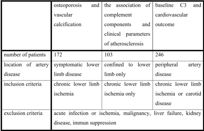
Consecutive patients with peripheral artery disease have been recruited at the Outpatient Department of Semmelweis University Department of Vascular Surgery in 2009 for the purpose of this study. Our inclusion criteria were: patients with present symptoms of atherosclerotic chronic lower limb ischemia or carotid disease who gave written informed consent. We excluded all patients with acute onset of ischemia, clinical or laboratory signs of acute infection, malignant tumour, hepatic disease, end stage renal disease (dialysis), immune suppression, severe medical or surgical conditions (myocardial infarction, stroke, trauma, surgical procedure) in the last 6 months. Patients with serum creatinine level > 100 µmol/l or estimated glomerular filtration (eGFR) < 60 ml/min were also excluded from the study. Please find a summary of the the inclusion and exclusion criteria and the number of patients recruited to each study in Table 1.
Table 1. Summary of different study groups, their inclusion and exclusion criteria.
osteoporosis and vascular
calcification
the association of complement
components and clinical parameters of atherosclerosis
baseline C3 and cardiovascular outcome
number of patients 172 103 246
location of artery disease
symptomatic lower limb disease
confined to lower limb only
peripheral artery disease
inclusion criteria chronic lower limb ischemia
chronic lower limb ischemia only
chronic lower limb ischemia or carotid disease
exclusion criteria acute infection or ischemia, malignancy, liver failure, kidney disease, immun suppression
III.1. Clinical evaluation
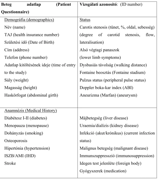
The initial study questioner is presented in original language Hungarian and English translation in brackets in Table 1. Age, sex and past medical history have been noted on the day of presentation to the outpatient clinic or on the day of consequent hospital admission. Past medical history focused on diabetes, ischemic heart disease, cerebrovascular event, liver and kidney disease as well as metal work or other device previously implanted in the patient. Body mass index (BMI) was calculated as weight (kg) / height2 (m). Metabolic syndrome was identified by the presence of three or more risk factors (abdominal obesity, high triglycerides and high density lipoprotein (HDL), elevated blood pressure or treated hypertension, history of diabetes or elevated fasting blood sugar defined by the guidelines of the International Diabetes Federation [133].
Past and present smoking habits and alcohol consumption has been recorded.
Furthermore, history of osteoporosis or osteopenia and treatment received has been noted. Current medical therapy, especially statins, anticoagulants/ anti thrombocyte aggregating agents were also asked in the study questionnaire. Our subjects were asked about their exercise tolerance and their walking distance. The traditional Fontaine classification was used to assess the clinical severity of the chronic lower extremity atherosclerotic disease (groups I, II/a, II/b, III, IV). Group II was separated to “a” and
“b” subgroups at a walking distance of 200 meters.
Table 2: Patient Questioner in original language Hungarian and English translation in brackets
Beteg adatlap (Patient Questionnaire)
Vizsgálati azonosító: (ID number)
Demográfia (demographics) Név (name)
TAJ (health insurance number) Születési idő (Date of Birth) Cím (address)
Telefon (phone number)
Adatlap kitöltésének ideje (time of entry to the study)
Súly (weight) Magasság (height)
Haskörfogat (abdominal girth)
Status
Carotis stenosis (tünet, %, oldal, sebesség) (degree of carotid stenosis, flow, lateralisation)
Alsó végtagi panaszok (lower limb symptoms)
Dysbasiás távolság (walking distance) Fontaine beosztás (Fontaine stadium) Pulzus status (peripheral pulse status) Doppler boka-kar index (ABI)
Aneurizma (Marfan) (aneurysm) Anamnézis (Medical History)
Diabétesz I-II (diabetes) Menopauza (menopause) Dohányzás (smoking) Osteoporosis
Hipertónia (hypertension) ISZB/AMI (IHD)
Stroke
Májbetegség (liver disease) Uraemia/dialízis (kidney disease)
Infekció (akut/krónikus) (current infection status)
Malignus betegség (malignant disease) Immunszuppresszió (immunosuppression) Idegen test jelenléte (foreign body)
Gyógyszerek (medication)
III.2. Assessment of atherosclerosis and calcification
The methods used for the assessment of calcification have been previously described by our research group on several places [78, 94]. For the purpose of the thesis we provide a brief summary of the methods please read the above cited articles for full details.
An experienced vascular surgeon performed physical examination and ankle-brachial index (ABI) measurement with Doppler ultrasound probe. The patients laid in a supine position after resting for pressure measurements over the posterior tibial and dorsal pedal arteries. ABI was calculated as the lowest pressure of the ankles divided by the higher of the left and right arm pressures[134, 135].
III.2.1. Imaging modalities
The extent of calcification was assessed by evaluating the carotid intima media thickness (IMT) and a General Calcification Score which were determined by a single experienced radiologist who was blind to the patients’ clinical information. IMT was measured on a plaque free area at three points of the dorsal wall of the common carotid arteries, using a linear (7.5-11MHz) and convex (3.5-5MHz) transducer of a Toshiba Aplio SSA-770 ultrasound system. The mean value and the maximum IMT was used for calculations[136]. During the same examination carotid stenosis was also determined. Please also see Figure 3. To assess the overall extent of systemic atherosclerosis a calcification score (CS) was calculated after examining the vascular system at seven sites: both carotid bifurcations, the infrarenal aorta, both common femoral arteries, aortic and mitral valves by B-mode ultrasound (see technical details above at carotid IMT measurements). If calcification was noted, the spot was rated as 1.
Sites with no calcification received 0. As we evaluated calcification at 7 sites the calcification range was 0-7 [137-139]. Transthoracic echocardiograms were performed by one experienced cardiologist blind to other study information. Examinations were performed, including Doppler images in all standard views using phased array transducers (2.5-4.5MHz). Mitral and aortic valve calcification was determined if echodense structures were noted at the appropriate views [140].
Figure 3. Assessment of carotid calcification by ultrasound for calcification score (investigations performed by Dr Endre Rimely at the Hear and Vascular Centre of Semmelweis University)
III.2.2. Laboratory measurements
Blood samples were collected after a minimum 6 hours of starvation. These samples were used to evaluate laboratory characteristics of our study cohort. Conventional standardized methods were performed in the core laboratory of Semmelweis University.
On admission Urea and Electrolyte, Full Blood Count, Clothing and Liver Function Test, C-Reactive Protein, HemogblobinA1c, Protein C, Lipid Profile were measured.
We used the Cockcroft-Gault formula for the calculation of glomerular filtration rate.
III.3. The evaluation of osteoporosis amongst patient with Peripheral Artery Disease III.3.1 Biochemical parameters
According to our knowledge, the prevalence of osteoporosis and osteopenia has never been investigated in the Hungarian population of patients with clinically manifest vascular disease. For the purpose of this part of the study, we investigated the BMD of our 172 patients with PAD. In addition to the baseline laboratory measurements we evaluated the level of vitamin D3, beta crosslaps (bCTx), bone alkaline phosphatase (BAP), osteocalcin (BGLAP) and parathyroid hormone (PTH). For the purposes of investigations regarding the role of Vitamin-D we divided our study cohort into high and low Vitamin-D subgroups. According to Holick [141], low level of Vitamin-D was noted if the patient had 20 mg/mL or lower serum concentration. Dyslipidaemia has been diagnosed for the purpose of clarifying its role in the connection of the diseases.
For this purpose, either previous diagnosis of dyslipidaemia or according to Nataraja et al. [142] total cholesterol/ high Density Lipoprotein ratio has been used.
III.3.2 Dual-energy X-ray absorptiometry
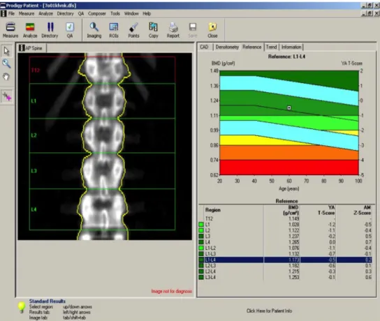
The recommended method by the WHO for the assessment of bone health is Dual- energy X-ray absorptiometry (DEXA) scan. The principal of the scan is based on the simultaneous use of two X-ray beams with different energy. By subtracting the soft tissue absorption from the images the difference between the penetration of the beams to the bone will determine the bone mineral density. The definition of BMD consists of the mineral content of a defined area of the bone surface. Please see also Figure 4. The BMD is measured on 3 different bones. The femoral (f-BMD) and radial (r-BMD) head, and the lumbar spine (l-BMD). The T-score has been calculated based on the different density to healthy bone. According to the WHO guidelines, patients with more than - 2.5 SD to their age matched peak bone density were treated as osteoporotic. If the SD of the T-score was between -1 and -2.5 these patients were considered as having decreased BMD, hence osteopenia[143].
Figure 4. An example of a DEXA scan demonstrating how calcification appear and what type of results does the scan provide. (source: www.orthospinelab.com [144] )
III.3.3. Angiography, Bollinger score
These methods have previously been described in Fehérvári et al [145].
Lower limb angiography was taken by using standard methods. The original pictures were analysed by an experienced radiologist, who was blind to other parts of the study.
According to the clinical symptoms and to the angiographic images the site of the atherosclerotic lesion was also noted.
Angiography based scoring systems are not the most popular way of classifying atherosclerosis, mostly due their complexity. However, for the purpose of this study we have chosen an angiography based score system created by Bollinger et al. [23], also called the Bollinger score (BS). This allowed us to precisely demonstrate the lack of perfusion in the exact anatomical region. Two experienced radiologists -who were blind
from each other’s results- analysed the pictures. An additive score was calculated in order to assess the extent of arteriosclerosis of the infrarenal aorta, iliac, femoral, popliteal and crural arteries on each side. Stenotic lesions and occlusions were noted in each arterial segment on both sides. Four categories of occlusive lesions were defined in descending order of severity: complete occlusion, stenosis narrowing the lumen by more than 50 %, between 25 and 50% and less than 25% (Lower score values were assigned to less severe stenosis.) If the stenotic area exceeded more than half of the length of the vessel, higher values were given and occlusion received the highest scores, especially if it was observed in the full length of the artery.
This score system is particularly suitable to assess systemic atherosclerosis, because it is able to judge stenoses and occlusions in a long segment of the vascular system. It is also particular suited for this study as it takes the anatomical segment in to account. The site of the atherosclerotic lesion was noted and compared to the BMD measurements, which were also site specific.
III.3.4. Site specific assessment
To enable site specific comparison amongst the vascular lesion and the BMD measurement the anatomical site of the vascular lesion has been noted. As described in the introduction there are several explanations behind the connection of low bone mass and vascular disease. One of them suggest that the negative remodelling of the bone is induced by the decreased blood flow and the consequent lack of nutrients. BMD is usually measured in 3 different anatomical regions, the radial and femoral head and the lumbar spine. In our study cohort, patients presenting with lower limb symptoms were arranged into 2 groups based on the anatomical site of the arterial stenosis. Patients with aorto-iliac disease were assigned to one group and patients with infra inguinal disease to another. The blood supply of the lumbar spine and the femoral head derives from the aorto and iliac vessels.
III.4. The role of Complement component 3 and fetuin-A in the development and progression of vascular calcification.
III.4.1. Association of the extent of PAD to complement component 3 and 4
In a cross sectional study design, 103 patients with lower limb atherosclerosis have been recruited. The previously described study questionnaire, laboratory measurements, evaluation of atherosclerosis and calcification have all been registered for these patients.
III.4.2. Laboratory measurements
In addition to the standard laboratory parameters, serum concentrations of C3, C4 were measured by radial immunodiffusion method [146]. The following reagents were used:
Anti-human C3 Complement IgG Fraction, Anti-human C4 Complement IgG Fraction (DiaSorin Inc. Stillwater, MN, USA) for measuring the serum levels of C3, C4, respectively. For calculating the standard values Human Serum Protein Calibrator (DAKO A/S, Glostrup, Denmark) was used referring to C3, C4.
III.5. The role of complement component 3, 4 and fetuin-A in the progression of atherosclerosis
In this case control study 246 consecutive patients with severe peripheral artery disease were recruited at the Department of Vascular Surgery of Semmelweis University, Budapest in 2009. Follow up for them was organized 3 years after their visit to the Outpatient Department.
Serum fetuin-A was determined by the standard radial immunodiffusion method previously described in our group. Five microliters of patient's serum diluted to 1:4 was applied in 11.5 ml of Litex agarose gel (Sigma). Serum samples (1:4 dilution) with known concentrations of fetuin-A served as standards. The incubation was done at room temperature for 48 h. We used two types of antibodies against fetuin-A as the protein is synthesized as a single chain and is rapidly converted to a dipeptide form following the cleavage of a connecting peptide. The commercially available product (anti-fetuin-A,
IgG fraction, Incstar, Cat. No. 81931, 13.7 mg/ml, in a final concentration of 84 µl/11.5 ml gel) recognizes the dipeptide form. The other type of antibody binding to the newly synthesized single chain form of fetuin-A, was raised by immunizing a rabbit with recombinant human protein (final concentration of 568 µl/11.5 ml gel) [140].
Follow up for our patients was organised after 3 years to the original visit of the Outpatient Department. Another patient questionnaire was completed again. Patients were asked prior to their appointment to bring their medical documentation from the past 3 years and medical history was thoroughly revised. All cardiovascular events and their nature were noted. Patients were considered having myocardial infarction with noted rise and/or fall of cardiac biomarkers, with at least one of the values being elevated and symptoms of myocardial ischemia, or new (or presumably new) significant ST-segment/T-wave changes, or left bundle branch block, or development of pathological Q waves on ECG, or new loss of viable myocardium, or regional wall motion abnormality by imaging, or identification of intracoronary thrombus by angiography or autopsy [147]. Any episode of neurological dysfunction caused by focal cerebral, spinal, or retinal infarction that persisted beyond 24 hours or resulting in death within 24 hours [148] was considered as stroke or stroke-related death. All strokes were confirmed with magnetic resonance imaging or computed tomography. Any patient undergoing further vascular intervention was including open surgery and endovascular repair considered as a novel vascular event (NVE).
III.6. Statistical analysis
Statistical analysis was performed with Prism for Windows 5.01 (GraphPad Software, San Diego, CA) and SPSS for Windows 15.0.1 (SPSS Inc., Chicago, IL) statistical software products. As many of the variables had non-Gaussian distributions we used nonparametric tests in the analysis. We used the Mann-Whitney’s U test to compare two independent groups, Kruskal–Wallis to compare multiple groups and Spearman’s rho to calculate correlations. Multiple logistic regression analysis was also performed. All statistical analyses were performed two-tailed and p<0.05 was considered as significant.
Values presented in the text are medians (interquartile ranges, /IQR/), unless otherwise
stated. For the univariate regression models we used Cox regression analysis or logistic regression
We were using the definition for Peripheral arterial disease (PAD) described by the National Institute of Health of the United States of America. We included all patients with clinically significant atherosclerosis on any site excluding the heart.
The Semmelweis University Regional and Institutional Committee of Sciences and Research Ethics approved the study protocol.
IV. RESULTS
IV.1. Osteoporosis in the Hungarian population of patients with atherosclerosis
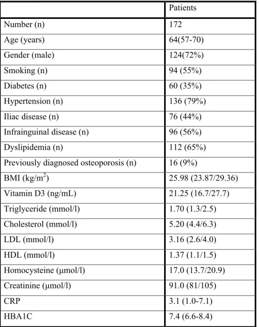
According to our knowledge, this is the first study investigating osteoporosis amongst patients with lower limb atherosclerosis. Table 3. presents the general clinical and biochemical characteristics of our 172 patients: 48 females and 124 males.
Table 3. Patient characteristics. Values are in median and interquartile range. Modified after Fehervari et al. [145]
Patients
Number (n) 172
Age (years) 64(57-70)
Gender (male) 124(72%)
Smoking (n) 94 (55%)
Diabetes (n) 60 (35%)
Hypertension (n) 136 (79%)
Iliac disease (n) 76 (44%)
Infrainguinal disease (n) 96 (56%)
Dyslipidemia (n) 112 (65%)
Previously diagnosed osteoporosis (n) 16 (9%)
BMI (kg/m2) 25.98 (23.87/29.36)
Vitamin D3 (ng/mL) 21.25 (16.7/27.7)
Triglyceride (mmol/l) 1.70 (1.3/2.5)
Cholesterol (mmol/l) 5.20 (4.4/6.3)
LDL (mmol/l) 3.16 (2.6/4.0)
HDL (mmol/l) 1.37 (1.1/1.5)
Homocysteine (µmol/l) 17.0 (13.7/20.9)
Creatinine (µmol/l) 91.0 (81/105)
CRP 3.1 (1.0-7.1)
HBA1C 7.4 (6.6-8.4)
IV.1.1. Bone Mineral Density
As previously described, BMD was measured at 3 anatomical sites with DEXA scan.
The median of the radial BMD was 0.86 (0.7-0.4) the femoral BMD was 0.83 (0.7-0.9) and the lumbar BMD 1 (0.9-1.2). We are displaying the median BMD, Z and T score values in different patient groups in Table 3. As described earlier, we identified important patient groups based on the risk factors of both conditions. The value of BMD on different anatomical sites for patients with different sex, age, BMI, smoking habits, are presented in Table 4. We compared these groups to each other, the level of significances also presented in the same Table.
![Figure 1. The different types of calcification. Figure by Bostrom et al. [56]](https://thumb-eu.123doks.com/thumbv2/9dokorg/1370880.112299/19.892.131.764.124.568/figure-different-types-calcification-figure-bostrom-et-al.webp)
![Figure 2. Summary of factors influencing vascular calcification. Original Figure by Giachelli et al.[55]](https://thumb-eu.123doks.com/thumbv2/9dokorg/1370880.112299/23.892.143.744.563.908/figure-summary-factors-influencing-vascular-calcification-original-giachelli.webp)