DOI 10.2478/pjvs-2013-0001
Original article
Bone mineral density (BMD) and computer tomographic measurements of the equine proximal phalanx in correlation with breaking
strength
P. Tóth
1, C. Horva ´th
2, V. Ferencz
2, B. Tóth
3, A. Va ´radi
4, O. Szenci
1, G. Bodó
51Clinic for Large Animals, Faculty of Veterinary Science, Szent Istva´n University, 2225 U
¨
llo¨, Dóra major, Hungary2First Department of Medicine, Semmelweis University of Medicine, Budapest, Hungary
3Department of Veterinary Clinical Sciences, Purdue University, West Lafayette, IN, USA
4Faculty of Veterinary Science, Szent Istva´n University, Budapest, Hungary, senior student
5University of Bern, Department of Clinical Veterinary Science, Equine Clinic, Switzerland
Abstract
Despite the fact that bone mineral density (BMD) is an important fracture risk predictor in human medicine, studies in equine orthopedic research are still lacking. We hypothesized that BMD correlates with bone failure and fatigue fractures of this bone. Thus, the objectives of this study were to measure the structural and mechanical properties of the proximal phalanx with dual energy X-ray absorptiometry (DXA), to correlate the data obtained from DXA and computer tomography (CT) measurements to those obtained by loading pressure examination and to establish representative region of interest (ROI) for in vitro BMD measurements of the equine proximal phalanx for predic- ting bone failure force.
DXA was used to measure the whole bone BMD and additional three ROI sites in 14 equine proximal phalanges. Following evaluation of the bone density, whole bone, cortical width and area in the mid-diaphyseal plane were measured on CT images. Bones were broken using a manually control- led universal bone crusher to measure bone failure force and reevaluated for the site of fractures on follow-up CT images. Compressive load was applied at a constant displacement rate of 2 mm/min until failure, defined as the first clear drop in the load measurement.
The lowest BMD was measured at the trabecular region (mean±SD: 1.52±0.12 g/cm2; median:
1.48 g/cm2; range: 1.38-1.83 g/cm2). There was a significant positive linear correlation between trabel- cular BMD and the breaking strength (P=0.023, r=0.62). The trabecular region of the proximal phalanx appears to be the only significant indicator of failure of strengthin vitro. This finding should be reassessed to further reveal the prognostic value of trabecular BMD in anin vivofracture risk model.
Key words:bone mineral density, DXA, CT, breaking strength, first phalanx, horse
Correspondence to: P. Tóth, email: Toth.Peter@aotk.szie.hu, tel.: +36 306 070 537
Introduction
Stress fractures are considered as one of the most significant causes of economic losses in race horse industry. According to large retrospective studies, 80% of musculoskeletal injuries in the US (Johnson et al. 1994) and 60% in the UK (Vaughan et al. 1975) are fatal in racehorses. Fractures of the proximal phalanx are one of the most common incidences dur- ing training (Vaughan et al. 1975) and during the daily fracture repair at equine clinics as well (Smith 2010). The most severe longitudinal and comminuted fractures are also observed in the first phalanx (Rooney 1969). Most fractures appear “sponta- neous” despite that they are the summation of com- plex processes with numerous factors involved (Bax- ter and Turner 2002).
The density of bony structures correlate to skel- etal strength and stiffness, which can be estimated by DXA (dual energy X-ray absorptiometry) in people (Genant et al. 1994, Genant et al. 1996, Grier et al.
1996). Further, as a quantitative method, DXA also has the ability to reveal and monitor changes in bone structural properties in humans and in variousin vivo animal models (Griffin et al. 1993, Turner et al.
1995, Grier et al. 1996). Previous research in horses focused on the structural and mechanical properties of the third metacarpal bone (Bynum et al. 1971, El Shorafa et al. 1979, Nunamaker et al. 1989) and the proximal sesamoid bones (Young et al. 1991), al- though little is known about the biomechanical prop- erties of the first phalanx (Thompson et al. 1996, Dzierzęcka and Charuta 2012).
Thus, the objectives of this study were to corre- late the data obtained from DXA and CT (computer tomography) measurements to those obtained by loading pressure examination. Further, this experi- ment aimed to establish representative ROI (region of interest) of the equine proximal phalanx for pre- dicting bone failure force. We hypothesized that bone failure force correlates to bone mineral density (whole bone; medial, lateral and trabecular region) and to the morphometric parameters of the proximal phalanx (cortical width, cortical area, total bone width).
Materials and Methods Samples
Proximal phalanges of the frontlimbs were in- volved in the study. The horses were euthanised un- related to muscolosceletal injuries at Szent Istva´n University, Faculty of Veterinary Science, Clinic for
Large Animals, U
¨
llo¨, Hungary. After dissection and manual removal of all soft tissue, the bones were stored in ethyl-alcohol at room temperature until measurements as previously validated (Beaupied et al.2006). The horses were: 5±3 years old (mean±SD, median: 5.29 years). There were 4 Lipizzaners, 1 Hun- garian half-blood, 1 Hungarian sporthorse, 1 Arabian.
Gender distribution was the following: 5 mares, 1 stal- lion and 1 gelding. Horses were used for either pleas- ure (n=3. 42%), carriage-driving (n=2. 29%) or breeding (n=2. 29%) purposes.
Bone mineral density (BMD) measurement
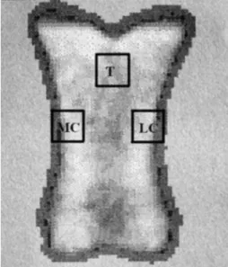
Bones were examined post mortem with a den- sitometer (Norland XR-26a) at the First Department of Medicine, Semmelweis University of Medicine, Budapest, Hungary. Imitation of soft tissue around the bone was required for the software algorithm used to measure the BMD. Therefore, a 20 mm plexiglass was used as a substitution for soft tissue as described in detail elsewhere (Tóth et al. 2010). Bones were measured once from dorsopalmar direction (Tóth et al. 2010). As the first step, the BMD of the whole bone was measured followed by selection of three 1x1 cm ROIs. The ROIs were the entire bone; the medial cortex (CM), the lateral cortex (CL) at the level of the horizontal axis of the mid-third of the bones and the trabecular region of the sagittal plane of the proximal third, 3 mm under the level of the deepest point of the metacarpophalangeal joint surface of the proximal phalanges (T) as shown in Fig. 1.
Fig. 1. DXA image of a proximal phalanx from dorsopalmar direction.Region of interests (ROIs) of the first phalanx.
T: trabecular or cancellous bone, MC: medial cortex, LC:
lateral cortex.
Computer tomographic (CT) measurements
Bones were scanned with a commercially available computer tomography unit (Siemens Somaton Emo- tion 6 Multislice CTb) using the following settings: 130 kV, 20 mAs, slides: 2 mm; at Kaposva´r University, Institute of Diagnostic Imaging and Radiation Oncol- ogy, Kaposva´r, Hungary. Morphometric parameters of the proximal phalanx were taken at mid-diaphyseal plane using a Siemens SIENETb software. Cortex width was measured from dorsal, lateral, medial and palmar sides, then the bone total width in either dor- sopalmar and lateromedial directions (Fig. 2). Corti- cal area was calculated from these data using an equa- tion described by Sherman et al (1995). CT images were repeated after loading test to reveal the fracture sites.
Fig. 2. Transverse CT image at the level of the mid-dia- physeal region of a proximal phalanx. DT: diameter trans- versa; DS: diameter saggittalis; CM: cortex palmaris;
CL: cortex lateralis; CD: cortex dorsalis; CM: cortex me- dialis.
Loading test
Biomechanical properties of proximal phalanges were assessed at Budapest University of Technology and Economics, Laboratory of Biomechanical Re- search. For loading pressure examination a universal bone crusher (ZD-20cuniversal testing machine) was used. Due to the lack of previousex vivo data regard- ing the first phalanx’s breaking force measurements, the machine was arbitrarly set for human lumbar spi- nal preset due to the similar cuboid shape. In order to position the bone properly during measurements, the proximal and distal articular rimms of the P1 bones were removed at the level of the deepest point of the metacarpophalangeal (proximal) and proximal inter-
phalangeal joint (distal) articular surfaces of the proximal phalanges by a bandsaw (Fig. 3). The bones were subjected to loading pressure from a proximodis- tal direction, due to the convex shape of the latero-medial site, which would have prevented accu- rate pressure loading. Bone failure strength was cal- culated from loading pressure values.
Fig. 3. ZD-20 universal testing machine. The bones were subjected to loading pressure from a proximodistal direc- tion. The proximal and distal articular rimms of the P1 bones were removed at the level of the deepest point of the metacarpophalangeal (proximal) and proximal interphalan- geal joint (distal) articular surfaces of the proximal phalanges by a bandsaw.
Statistical Analysis
Statistical analysis of the data was performed with a commercially available program (Minitab 16d).
Mean, standard deviation, median and range were calculated for bone mineral density, cortical area, cor- tex width, bone width, loading pressure, and breaking force. Distribution of the data was assesed with the Shapiro-Wilk test. Pearson’s linear correlation was used to reveal any association between bone mineral density values (whole bone BMD, traecular BMD, lat- eral cortical BMD, medial, cortical BMD), cortical area, cortical width (lateral, medial, dorsal, and pal- mar regions), bone width (saggittal, transversal) and bone breaking strength and loading pressure. Statisti- cal significance was set at P«0.05 and an adequate linear correlation was assumed if regression coeffi- cient was greater than 0.6 (r>0.6).
Results Samples
14 proximal phalanges from 7 cadavers were used in the study. 1 phalanx was excluded from the statisti- cal evaluation because following loading test examin- ations the fracture site could not be located on the CT images.
Bone mineral density (BMD) measurement
Total bone BMD (g/cm2), lateral cortex BMD, medial cortex BMD, trabecular BMD were as shown in Table 1.
Table 1. Descriptive statistical data of the variables meas- ured across the 13 proximal phalanges in this study.
Variable N Mean SD Median Range Whole BMD (g/cm2) 13 1.91 0.14 1.96 1.68-2.13 CL BMD (g/cm2) 13 2.19 0.15 2.2 1.95-2.51 CM BMD (g/cm2) 13 2.1 0.13 2.05 1.93-2.35 T BMD (g/cm2) 13 1.52 0.12 1.48 1.38-1.73 Compression strength
(MPa) 13 74 15.56 72.56 49.96-100
Breaking force (kN) 13 72 13.36 74 45-92
CM (cm) 13 0.97 0.47 1.09 0.22-1.44
CL (cm) 13 0.83 0.45 1 0.15-1.34
CD (cm) 13 0.39 0.13 0.43 0.15-0.59
CP (cm) 13 0.53 0.23 0.63 0.09-0.8
DT (cm) 13 4.66 0.299 4.75 4.17-5.18
DS (cm) 13 2.8 0.55 2.6 2.28-3.78
Area (cm2) 13 5.4 1.90 6.06 2.47-8.1 BMD – bone mineral density, DT – diameter transversa, DS – diameter saggittalis, CM – cortex palmaris, CL – cortex lateralis, CD – cortex dorsalis, CM – cortex medialis, T – trabecular or cancellous bone, SD – standard deviation).
Computer tomographic morphometric measurements
Data are summarized in Table 1 as means of three consecutive measurements of each parameter. Frac- ture lines were identified in the sagittal plane in 13 (92.86%) of 14 specimens. In 5 of 13 (38.46%) cases the fracture lines were located in the proximal and mid-third, in 6 of 13 (46.15%) cases the fracture lines were located in the mid and distal-third and in 2 of 13 (15.38%) cases fracture lines were located in the mid-third in the sagittal plane.
Loading test
Values of compression force and failure force are summarized in Table 1.
Correlations
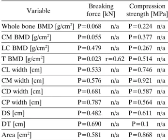
Significant positive linear correlation was found between trabelcular BMD and the breaking force (P=0.023, r=0.62). Other parameters did not signifi- cantly correlate with the breaking force or compres- sion force as shown in Table 2.
Table 2. Pearson’s linear correlations.
Breaking Compression force [kN] strength [MPa]
Variable
Whole bone BMD [g/cm2] P=0.068 n/a P=0.224 n/a CM BMD [g/cm2] P=0.055 n/a P=0.377 n/a LC BMD [g/cm2] P=0.479 n/a P=0.267 n/a T BMD [g/cm2] P=0.023 r=0.62 P=0.514 n/a CL width [cm] P=0.533 n/a P=0.746 n/a CM width [cm] P=0.576 n/a P=0.921 n/a CD width [cm] P=0.681 n/a P=0.587 n/a CP width [cm] P=0.787 n/a P=0.564 n/a
DS [cm] P=0.482 n/a P=0.611 n/a
DT [cm] P=0.690 n/a P=0.1 n/a
Area [cm2] P=0.581 n/a P=0.868 n/a r value is only indicated if P<0.05).
BMD – bone mineral density, DT – diameter transversa, DS – diameter saggittalis, CM – cortex palmaris, CL – cortex lateralis, CD – cortex dorsalis, CM – cortex medialis, T – trabecular or cancellous bone, n/a – not applicable).
Discussion
The lowest BMD was observed in the trabecular ROI. Both the anatomy and physiology of the trabecular region differs from cortical region (Good- ship and Smith 2004). It is composed of small trabeculae with a lower mineral content. Unlike the cortical region, the trabecular region is not only under the regulation of vitamin D, calcitonin or parathyroid hormone, but local forces can also induce marked re- modeling (Lawrence 2005) enabling this region to re- spond to mechanical forces directly. Loss of trabecu- lar bone induced by immobilization has been well documented in people (Kazarian and von Gierke 1969) and is characterized by a complete loss or thinn- ing of trabeculae (Ijiri et al. 1995).
This in vitro study revealed a significant positive linear correlation between the trabecular region BMD and failure strength. This result coincides with the cli-
nical evidence that fracture of the trabecular region is the most common in the proximal phalanx (Nixon 2012). In this study post-fracture CT images revealed that most fracture lines were located in the sagittal plane at the proximal or distal trabecular regions, similarly to an earlier retrospective study (Ellis et al.
1987). Thus, it would be important to focus on this region in further studies, as the trabecular region ap- pears to be the key component in sagittal proximal phalanx fractures. Proximal phalanx fractures are cat- egorized as sagittal or transverse, the latter extends across the bone and does not involve articular surfaces (Ellis et al. 1987). Sagittal proximal phalanx fractures are categorized as incomplete or complete. Although, the most common fracture of the proximal phalanx is the sagittal fracture, breed and usage are important predisposing factors. Fractures of the palmar/plantar processes can occur in Standardbreds (Ruggles 2003), while dorso-frontal fractures can occur both in Stan- dardbreds (Ruggles 2003) and Throroughbreds (Stashak 2002) raced on hard surface. Despite the co- inciding results, it must be emphasized that sagittal trabecular fractures are currently attributed to the mechanical effect of the saggital ridge of the third metacarpal bone (Nixon 2012), which was not part of our in vitro model. Davies performed proximodistal loading of equine metacarpal bones with both ends embedded in fibreglass-impregnated resin (Davies 2009), which also did not account for the mechanical load of the proximal articular surface. Since neither Davies’ nor our in vitro approaches are exact models of in vivo strains, the relationship between the trabecular region of the proximal phalanx and the sag- ittal ridge of the metacarpus, and to evaluate the charectiristics and effects of the subchondral bone un- der the proximal articular surface of the bone war- rants further investigation.
During training, microcracks may occur without evidence of lameness and with complete recovery to normal function after a short period of rest (Baxter and Turner 2002). The bone may withstand a particu- lar strain until remodels or, less frequently, the micro- cracks progress to macroscopic stress fractures (Nunamaker et al. 1990). Light work (compared to regular training load) or immobilization (e.g. longer peroid of stall rest) will decrease BMD in horses, while intense training without transition increases the incidence of bone fractures (Nunamaker et al. 1990).
The bone has a dynamic structure and remodels with exercise (Riggs 2002). BMD has been shown to de- crease with inactivity or increase with high level of training (Sherman et al. 1995). Due to the insidious nature of stress fractures, there would be a need of an in vitro diagnostic tool that correlates or predicts fracure risk in performance horses. Since our results indicate that there is a significant correlation between
failure of strength and trabecular BMD, this hypoth- esis could be tested in vivo as well.
Correct positioning of the patient/subject is a criti- cal part of the densitometry. DXA machine converts a three-dimensional structure into a two-dimensional image. Elliptical bones may have different BMD value depending on the position of the bone (Rozenberg et al. 1995). In this in vitro experiment bones were meas- ured once from dorsopalmar direction, as described previously (Tóth et al. 2010). Nevertheless, in vivo measurements would require careful limb positioning and deep sedation.
In conclusion, this study suggests that the trabecular region is the sole ROI in predicting failure of strength of the proximal phalanx. In future studies identification of the fracture lines and measurement of the BMD of those particular areas should also be investigated. Further, in vivo experi- ments are also warranted to reveal whether or not the trabecular BMD data are useful indicator of frac- ture risk evaluation of the proximal phalanx in horses.
Acknowledgement
The authors would like to acknowledge the Sur- gery Department at the Large Animal Clinic, the densitometry laboratory of the First Department of Medicine at the Semmelweis University, the Labora- tory of Biomechanical Research of the Budapest University of Technology and Economics and the In- stitute of Diagnostic Imaging and Radiation Oncol- ogy of the Kaposvhr University for contributing to this study with their specialized equipment and ex- pertise.
Abbreviations
DXA – dual energy x-ray absorptiometry CT – computer tomograph
BMD – bone mineral density P1 – proximal phalanx ROI – region of interest
Manufacturers’ adresses
a– Norland XR-26, Norland Corporation, Fort Atkin- son, WI, USA
b– Siemens AG, Erlangen, Germany
c– ZD-20 Universal Testing Machine, Jyoti Ltd, East Germany & FIB make (India)
d– Minitab Inc., PA, USA
References
Baxter GM, Turner AS (2002) Diseases of bone and related structures. In: Stashak TS (ed) Adam’s lameness in horses. 5th ed., Philadelphia, Lippincott Williams and Wilkins, pp 401-457.
Beaupied H, Dupuis A, Arlettaz A, Brunet-Imbault B, Bon- net N, Jaffre´ C, Benhamou CL, Courteix D (2006) The mode of conservation does not affect the architecture and the tensile properties of rat femurs. Biomed Mater Eng 16: 253-259.
Bynum D Jr, Ledbetter WB, Boyd CL, Ray DR (1971) Flexural properties of equine metacarpus. J Biomed Ma- ter Res 5: 63-79.
Carter DR, Hayes WC (1977) The compressive behavior of bone as a two-phase porous structure. J Bone Joint Surg Am 59: 954-962.
Davies HM (2009)Ex vivocalibration and validation of in vivo equine bone strain measures. Equine Vet J 41: 225-228.
Dzierzęcka M, Charuta A (2012) Bone minearl density and bone mineral content of the bilateral first phalanges of the thoracic limbs in horses. Pol J Vet Sci 15: 159-161.
El Shorafa WM, Feaster JP, Ott EA (1979) Horse metacar- pal bone: age, ash content, cortical area and failure stress interrelationships. J Anim Sci 49: 979- 982.
Ellis DR, Simpson DJ, Greenwood RE, Crowhurst JS (1987) Observations and management of fractures of the proximal phalanx on young thoroughbreds. Equine Vet J 19: 43-49.
Genant HK, Grampp S, Gluer CC, Faulkner KG, Jergas M, Engelke K, Hagiwara S, Van Kuijk C (1994) Universal standardization for dual x-ray absorptiometry: patient and phantom cross-calibration results. J Bone Miner Res 9: 1503-1514.
Genant HK, Engelke K, Fuerst T, Gluer CC, Grampp S, Harris ST, Jergas M, Lang T, Lu Y, Majumdar S, Mathur A, Takada M (1996) Noninvasive assessment of bone mineral and structure: state of the art. J Bone Miner Res 11: 707-730.
Goodship AE, Smith RK (2004) Skeletal physiology: Re- sponsive to exercise and training. In: Hinchcliff KW, Kaneps AJ, Geor RJ (eds) Equine sports medicine and surgery. 1st ed., Philadelphia, Saunders, pp 111-129.
Grier SJ, Turner AS, Alvis MR (1996) The use of dual-en- ergy x-ray absorptiometry in animals. Invest Radiol 31: 50-62.
Griffin MG, Kimble R, Hopfer W, Pacifici R (1993) Dual-energy x-ray absorptiometry of the rat: accuracy, precision, and measurement of bone loss. J Bone Miner Res 8: 795-800.
Ijiri K, Jee WS, Ma YF, Yuan Z (1995) Remobilization par- tially restored the bone mass in the non-growing cancel- lous bone site following long term immobilization. Bone 17: 213S-217S.
Johnson BJ, Ardans AA, Daft BM (1994) California racehorse postmortem programme: a 4-year overview.
Proc Am Ass Equi Pract 40: 167-169.
Kazarian LE, von Gierke HE (1965) Bone loss as a result of immobilization and chelation. Preliminary results in Macaca mulatta. Clin Orthop 65: 67-75.
Lawrence LA (2005) Effects of exercise and training on skel- etal development in horses. In: Pagan JD (ed) Advances in equine nutrition. 3rd ed., Nottingham, Nottingham University Press, pp 219-226.
Nixon AJ (2012) Phalanges and the metacarpophalangeal and metatarsophalangeal joints. In: Auer JA, Stick JA (eds) Equine surgery. 4th ed., St. Louis, Saunders Else- vier, pp 1300-1325.
Nunamaker DM, Butterweck DM, Provost MT (1989) Some geometric properties of the third metacarpal bone:
a comparison between the thoroughbred and standar- dbred racehorse. J Biomech 22: 129-134.
Nunamaker DM, Butterweck DM, Provost MT (1990) Fa- tigue fractures in thoroughbred racehorses: relationships with age, peak bone strain, and training. J Orthop Res 8: 604-611.
Riggs CM (2002) Fractures-a preventable hazard of racing thoroughbreds? Vet J 163: 19-29.
Rooney JR (1969) Biomechanics of lameness in horses. Bal- timore, Williams and Wilkins, pp 164-167.
Rozenberg S, Vandromme J, Neve J, Aguilera A, Muregan- curo A, Peretz A, Kinthaert J, Ham H (1995) Precision and accuracy of in vivo bone mineral measurement in rats using dual-energy x-ray absorptiometry. Osteoporos Int 5: 47-53.
Ruggles AJ (2003) The proximal and middle phalanges and proximal interphalangeal joint. In: Dyson SJ, Ross MW (eds) Diagnosis ans management of lameness in the horse. 1st ed., St. Louis, Elsevier, pp 343-344.
Sherman KM, Miller GJ, Wronski TJ, Colahan PT, Brown M, Wilson W (1995) The effect of training on equine metacarpal bone breaking strength. Equine Vet J 27: 135-139.
Smith H (2010) Comparison of fracture incidence, type and the associated pain between working equids in Egypt and horses in a UK based referral hospital. Taws Overseas Travel Grant Project Report, pp. 1-17.
Stashak TS (2002) Fractures of the proximal phalanx. In:
Stashak TS (ed) Adam’s lameness in horses. 5th ed., Philadelphia, Lippincott Williams and Wilkins, pp 755-764.
Thompson KN, Cheung TK, Putnam M (1996) Com- puterized bone density analysis of the proximal phalanx of the horse. Equi Pract 18: 26-29.
Tóth P, Horva´th C, Ferencz V, Nagy K, Gligor N, Szenci O, Bodó G (2010) Assessment of the mineral density and mineral content of the equine third metacarpal and first phalanx bone by dual energy x-ray absorptiometry. Acta Vet Hung 58: 317-329.
Turner AS, Mallinckrodt CH, Alvis MR, Bryant HU (1995) Dual-energy X-ray absorptiometry in sheep: experiences with in vivo and ex vivo studies. Bone 17: 381S-387S.
Vaughan LC, Mason BJE (1976) A clinico-pathological study of racing accidents in horses. A report of a study on equine fatal accidents on racecourses.Horserace Betting Levy Board, London, United Kingdom, pp 3-88.
Young DR, Nunamaker DM, Markel MD (1991) Quantitat- ive evaluation of the remodeling response of the proximal sesamoid bones training-related stimuli in thorough- breds. Am J Vet Res 52: 1350-1356.


