BoneAlbumin filling decreases donor site morbidity and augments bone formation after anterior cruciate
ligament reconstruction with patellar autografts
PhD thesis
Károly Schandl
Doctoral School of Clinical Medicine Semmelweis University
Supervisor: Zsombor Lacza, M.D., D.Sc.
Official reviewers: István Flóris, M.D., Ph.D.
Áron Lazáry, M.D., Ph.D.
Head of the Final Examination Committee: György Szőke, M.D., D.Sc., Members of the Final Examination Committee: Zoltán Bejek, M.D., Ph.D.
István Kádas, M.D., Ph.D.
Budapest
2018
1. Introduction
Anterior cruciate ligament (ACL) reconstruction is one of the most common knee interventions, and multiple studies to investigate grafting techniques that result in optimal clinical outcomes.
So far, most data suggest that graft type has limited influence on clinical outcome, and bone- patellar tendon-bone (BPTB) or hamstring tendon can provide stable knee function. As a result, both methods are treatment strategies on the clinical palette, differing in their indications.
However, donor site pain remains a problematic side effect after BPTB reconstruction, affecting 32-43% of the patients. The reason behind this phenomenon is the slow or inadequate bone buildup in the tibial and patellar harvest sites. Leftover autologous spongious bone can be used to fill the defects, but the volume of autologous bone is usually insufficient to fill both defects.
Bone substitution is still the aim of numerous scientific investigations, and there exists a wide range of potential materials suitable for different bone replacement situations. In case of BPTB reconstruction, where the goal is to reduce donor site pain, a fully rebuilding bone substitute is the material of choice. Autologous bone tissue is the best material for bone replacement therapies because it is osteoconductive, osteoinductive and contains osteogenic cells. However, its available volume in this specific indication is low, thus creating the need for other suitable substitute materials. Allogenic bone substitutes are generally used because they are completely biocompatible materials with good osteoconductive properties. The osteoinductivity of these bone substitutes is less favorable because of the harsh physicochemical procedures that are needed to remove immunogenic contents for safety reasons. To reestablish osteoinductivity, allogenic materials can be augmented with different agents, for example proteins, growth factors, or even stem cells. Although these complex, fully loaded implants can achieve very good bone remodeling in experimental studies, their clinical usefulness is rather limited especially in less serious indications such as BPTB surgery. Moreover, application of bone morphogenetic proteins is declining due to the high cost and complication rate, and osteogenic cells pose severe regulatory hurdles. Thus, there is a pressing clinical need for a bone substitute with improved efficacy and minimal cost and complexity.
In our previous experiments, we showed that bone allografts with a lyophilized serum albumin coating (BoneAlbumin) led to the regaining of some lost functions. Mesenchymal stem cells attach and proliferate on the albumin-coated surface readily, and by activating stem cells, BoneAlbumin induces faster ossification and reduces healing time in different animal models.
This was also supported by a first-in-human study, where BoneAlbumin, in the form of large
structural allografts, was used in aseptic prosthesis revisions. In this study, not only were good clinical outcomes achieved, but implanted allografts also showed intensive osteoblast function even after one year, which further supports the idea of endogenous stem cell recruitment.
2. Aims
In the present dissertation, we used BoneAlbumin chips to reduce the size of the bone defects after harvesting patellar tendon, and compared the functional and morphologic differences between donor sites. We hypothesized that donor site morbidity with BoneAlbumin would be less prominent, thereby eliminating a major side effect of BPTB reconstruction and improving overall clinical outcome.
1. Are the BoneAlbumin chips technically feasible to fill the donor site bone defects after harvesting the patellar tendon in anterior cruciate reconstruction? Are there any side effects or complications related to the BoneAlbumin filling?
2. Can the advantageous effects of Bone Albumin on bone regeneration be shown with morphometric measurements using computed tomography?
3. Does BoneAlbumin filling decrease donor site complains after anterior cruciate ligament reconstruction?
3. Methods
Between 2013 and 2014, male and female patients undergoing ACL reconstruction were asked to participate in our prospective, randomized, double-blinded study. Forty patients between the age of 18 and 50 were enrolled with the inclusion criteria of high level sports activity prior to injury. Exclusion criteria were associated collateral ligament damage, immune-rheumatologic pathologies, chondropathies higher than grade II (Outerbridge), preexisting anterior knee pain and previous surgery on the same knee. ACL reconstruction was performed with BTB autografts. Patients were randomly assigned into two groups: 20 patients in the control group (ACL reconstruction with BPTB), and 20 patients in the BoneAlbumin treated group (ACL reconstruction with BPTB plus serum albumin coated allograft implanted into the donor- sites). All patients received detailed information about the risks and benefits of the procedure prior to granting informed consent and undergoing surgery. All patients underwent the same
rehabilitation protocol. The study was performed under the approval of the local ethical committee (ALLO-2013-02).
BoneAlbumin - Serum albumin coated bone chips Bone pieces were harvested from cadavers under the operational license of West-Hungarian Tissue Bank and processed immediately to produce autolysed, antigen extracted allogenic bone according to Urist's method. The preservation method was freeze-drying under aseptic conditions followed by EtOH sterilisation. After freeze- drying, the bone grafts were submerged in sterile 10% human serum albumin solution for 1 minute under aseptic conditions (low-salt-content Biotest human albumin infusion, Biotest Pharma GmbH, Dreieich, Germany) and a second freeze-drying was performed with the same parameters to obtain BoneAlbumin.
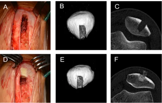
Patients enrolled in the control group were treated according to our clinical practice. BPTB autografts were used to reconstruct the ACL. Autologous spongious bone was used to repair the tibial donor site, while the patellar donor site was filled with blood clot. In the treatment group BoneAlbumin mixed with autologous spongious bone was used to fill the tibial donor site, while due to the limited amount of autologous bone, only BoneAlbumin was used to fill the patellar defect. Approximately 3-3 cm3 grafts were used at each donor site (Fig. 1-2.).
Fig. 1. Patella donor site filling techniques. Panels A, B, and C show the control technique in an intraoperative picture and 1 week post-operative CT images. Panels D, E, and F show the BoneAlbumin filling technique. Note that BoneAlbumin filling was complete in each case intraoperatively, however, small bone graft fragments were visible in the early post-op CT in the pre-
patellar soft tissue space.
Fig. 2. Tibia donor site filling techniques. Panels A, B, and C show the control technique, in an intraoperative picture and 1 week post-operative CT images. Panels D, E, and F show the
BoneAlbumin filling technique.
Patients underwent physical examination and VISA, VAS, Lysholm and IKDC subjective questionnaires. Scores were collected on the postoperative 6th week and 6th month. The VISA score is designed specifically to quantify knee function in subjects with patellar tendinopathy regarding different activities involving knee movement. The score range is 0 to 100. Visual Analog Scale (VAS) ranging from 0 (no pain) to 10 (maximum pain) evaluates the average knee pain during normal daily activities. Patients were asked to point to the value corresponding to their pain on the numeric scale during kneeling, squatting and average daily movement or exercise. Activity level was recorded with the use of International Knee Documentation Committee (IKDC) and Lysholm scores postoperatively.
Computed tomography (CT) of the knee was also performed after the 6th month with a Phillips Brilliance 64 machine. Axial slices were obtained with 120 Kv and 300 mA (slice thickness:
0.9mm, increment 0.45mm, collimation 64 X 0.625mm, pitch 0.579, rotation time 0.75 sec).
Remaining bone defect volume was measured with interpolated images on axial slices. The density (HU) of the regenerating defect was measured in a 20 mm2 circular region of interest placed approximately 2 mm below the cortex level. In a separate group of patients CT was performed at 1 week after surgery in order to illustrate the early positioning of the bone fillers (n=2 per treatment group).
All of the values are reported as the means ± SEM. The statistical analysis was performed using Student's t test using GraphPad Prism statistical software. p values < 0.05 were considered significant.
4. Results
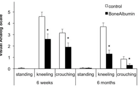
At the examination date 6 weeks postoperatively, both treatment groups were still under recovery from the operation itself, and overall knee function was compromised as evidenced by the IKDC and VISA scores, less so in the Lysholm activity scale (Fig. 3.). However, both the IKDC and VISA scores were significantly lower in the control than in the BoneAlbumin group (Fig. 3.). Pain at the donor site was significantly lower in the treatment group both at kneeling and crouching, while standing pain was negligible in both groups (Fig. 4.).
At six months, when the tendon is considered healed and the patients generally return to their
previous sport activity levels, the knee functional scores were close to normal (IKDC control:
85±2 vs treated: 91±2, Lysholm control: 96±1 vs. treated: 97±1, VISA control: 94±1 vs. treated:
96±2) with a slight but statistically significant advantage for BoneAlbumin in the IKDC score (Fig. 3.). At this time point, donor site pain generally starts to take precedence over other symptoms. Indeed, although standing and crouching pain subsided in both groups, kneeling pain was prominent in the controls but significantly lower in the treatment group (VAS control:
3.70±0.33 vs. treated: 1.30±0.34, p< 0.01) (Fig. 4.).
Fig. 3. Functional knee scores. Panel A shows IKDC, panel B shows VISA and panel C shows Lysholm activity score results at 6 weeks and 6 months post operatively, each with a potential
maximum value of 100.
Fig. 4. Knee pain at 6 weeks and 6 month after BTB surgery. Visual analog scale of knee pain on a 0- 10 scale in standing, kneeling and crouching positions. * represents p < 0.05 between the two
treatment groups.
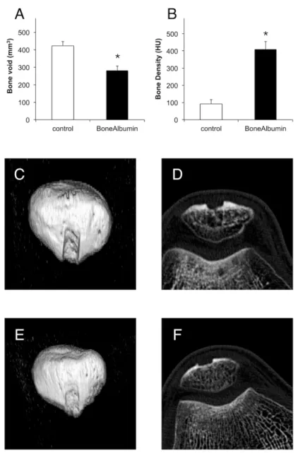
Bone density and the remaining bone defect were measured with computed tomography. In the patella, defects filled with BoneAlbumin had a significantly smaller bone void and higher density values than the controls, which were filled with blood clot only (Fig. 5.) The tibia was either filled with the available autograft only or autograft mixed with BoneAlbumin. Since the volume of the available autograft bone was variable, the completeness of the filling was scored by the operator on a scale of unfilled, moderately, well, or completely filled defects. At 6-month follow-up, the completeness of the filling was not correlated with the bone density in the control group (Table 1.). In case of BoneAlbumin filling, the level was always 'complete'.
BoneAlbumin augmentation of the autograft resulted in a significantly denser new bone than that filled with autograft only (605 ± 41 vs. 407 ± 48 HU, p< 0.05, respectively).
Fig. 5. Morphometric measurements of the patellar site. Panel A shows remaining bone void, while Panel B shows density of the newly formed bone at 6 months post-operatively. Panels C-F show representative CT images from the patella. Panel C shows 3D reconstruction and panel D shows an axial slice of the control group. Panel E shows 3D reconstruction and panel F shows an axial slice of
the BoneAlbumin treated group. There is a significantly lower remaining defect size with a significantly denser remodeled bone in the BoneAlbumin group. indicating a nearly complete tissue
healing. * represents p < 0.05 between the two treatment groups.
Table 1. Tibial site filling in the control group. As the avialable allograft bone was not always sufficient to fill the tibia site, the approximate adequacy of filling was noted by the operating surgeon on a subjective scale of no/noderate/substantial/complete filling. Bone density values are measured in
the remodeled bone stock at 6 months on CT. Number of cases is shown in each category.
5. Conclusions
1. BoneAlbumin chips are feasible bone substitute to fill precisely fill the bony donor sites after anterior cruciate ligaments reconstruction. Throughout the 6 months experiment no side effects or complications related to the bone substitution was observed.
2. According to the size of the remaining bone defects and the density of the newly regenerated bone tissue, pure BoneAlbumin successfully supported regeneration at the patellar donors site after anterior cruciate ligament reconstruction. BoneAlbumin mixed with autologous spongious bone also augmented the new bone formation at the tibial donor site.
3. BoneAlbumin filling of the bone defects after anterior cruciate ligaments reconstruction significantly decreased donor site complains in kneeling and crouching.
6. Summary
In order to prevent the knee joint from degeneration, surgical reconstruction is needed after anterior knee ligament rupture. According to literature data, there is no significant difference in postoperative knee stability between the two common, world wide used techniques.
Additionally, BPTB technique enables early and more invasive rehabilitation, which results in faster return to sport activities. After using the BPTB graft, on the other hand, donor site discomfort appears in significant number of the cases, which decreases the wider application of the method. The donor site morbidity is influenced by the elongated regeneration of the bony defects, which we aimed to decrease by filling the them with BoneAlbumin. According to our results, donor site pain decreased by 44% after six weeks and 75% after six months with BoneAlbumin augmentation, while the surgical technique is not modified significantly and the number of complication remains the same. According to the radio morphologic studies, BoneAlbumin filling increased new bone quantity and quality, without ectopic bone formation.
These results prove that the filling of the bone defects with albumin supports the regeneration of the area and decreases donor site pain, which consequently lead to better clinical results. Our study shows that BoneAlbumin is safe and effective bone substitute, for this reason we are planning to investigate further indications, focusing on cases in which controlled and more complete bone regeneration is needed.
List of publications
Publications of current dissertation:
Schandl K, Horváthy DB, Doros A, Majzik E, Schwarz CM, Csönge L, Abkarovits G, Bucsi L, Lacza Z. Bone-Albumin filling decreases donor site morbidity and enhances bone formation after anterior cruciate ligament reconstruction with bone-patellar tendon-bone autografts. Int Orthop. 2016 Oct; 40(10):2097-2104. IF: 2,520
Skaliczki G, Schandl K, Weszl M, Major T, Kovács M, Skaliczki J, Szendrői M, Dobó-Nagy C, Lacza Z. Serum albumin enhances bone healing in a nonunion femoral defect model in rats:
a computer tomography micromorphometry study. Int Orthop. 2013 Apr;37(4):741-5. IF: 2,019 Skaliczki G, Weszl M, Schandl K, Major T, Kovács M, Skaliczki J, Redl H, Szendrői M, Szigeti K, Máté D, Dobó-Nagy C, Lacza Z. Compromised bone healing following spacer removal in a rat femoral defect model. Acta Physiol Hung. 2012 Jun;99(2):223-32. IF: 0,882 Baló E., Schandl K., Hangody Gy., Abkarovits G., Bodó L., Hangody L. Izolált elülső keresztszalag sérülés ellátásánál felhasznált különböző autológ graft típusok összehasonlító vizsgálata. Magyar Traumatológia, Ortopédia, Kézsebészet, Plasztikai sebészet (2010) IF: 0 Weszl M, Skaliczki G, Cselenyák A, Kiss L, Major T, Schandl K, Bognár E, Stadler G, Peterbauer A, Csönge L, Lacza Z. Freeze-dried human serum albumin improves the adherence and proliferation of mesenchymal stem cells on mineralized human bone allografts. J Orthop Res. 2012 Mar;30(3):489-96 IF: 2,875
Skaliczki G., Weszl M., Schandl K., Major T., Kovács M., Skaliczki J., Szendrői M., Dobó- Nagy Cs, Lacza Zs. Új típusú interpozíciós csontdefektus modell. Magyar Traumatológia, Ortopédia, Kézsebészet, Plasztikai sebészet (2012) IF: 0
Independent publications:
Bikov A, Lazar Z, Schandl K, Antus BM, Losonczy G, Horvath I. Exercise changes volatiles in exhaled breath assessed by an electronic nose. Acta Physiol Hung. 2011 Sep;98(3):321-8.
IF: 0,821
D. B. Horvathy, P. P. Nardai, T. Major, K. Schandl, A. Cselenyak, G. Vacz, L. Kiss, M.
Szendroi, Z. Lacza: Muscle regeneration is undisturbed by repeated polytraumatic injury, Accepted for publication in Eur J Trauma Emerg Surg on May 31, 2010 IF: 0,328